HAL Id: hal-02928164
https://hal.archives-ouvertes.fr/hal-02928164
Submitted on 18 Jan 2021
HAL is a multi-disciplinary open access
archive for the deposit and dissemination of
sci-entific research documents, whether they are
pub-lished or not. The documents may come from
teaching and research institutions in France or
abroad, or from public or private research centers.
L’archive ouverte pluridisciplinaire HAL, est
destinée au dépôt et à la diffusion de documents
scientifiques de niveau recherche, publiés ou non,
émanant des établissements d’enseignement et de
recherche français ou étrangers, des laboratoires
publics ou privés.
Cellulose Secretion Regulator BcsE
Samira Zouhir, Wiem Abidi, Meryem Caleechurn, Petya Violinova Krasteva
To cite this version:
Samira Zouhir, Wiem Abidi, Meryem Caleechurn, Petya Violinova Krasteva. Structure and
Mul-titasking of the c-di-GMP-Sensing Cellulose Secretion Regulator BcsE. mBio, American Society for
Microbiology, 2020, 11 (4), pp.e01303-20. �10.1128/mBio.01303-20�. �hal-02928164�
Structure and Multitasking of the c-di-GMP-Sensing Cellulose
Secretion Regulator BcsE
Samira Zouhir,
a,b,cWiem Abidi,
a,b,cMeryem Caleechurn,
aPetya Violinova Krasteva
a,b,caStructural Biology of Biofilms Group, Institute for Integrative Biology of the Cell (I2BC), CEA, CNRS, Paris-Sud University, Gif-sur-Yvette, France bStructural Biology of Biofilms Group, European Institute of Chemistry and Biology (IECB), Pessac, France
cCBMN UMR 5248 CNRS, University of Bordeaux, Pessac, France
Samira Zouhir and Wiem Abidi contributed equally to this work. Author order was determined alphabetically on first-name basis.
ABSTRACT
Most bacteria respond to surfaces by biogenesis of intracellular
c-di-GMP, which inhibits motility and induces secretion of biofilm-promoting adherence
factors. Bacterial cellulose is a widespread biofilm component whose secretion in
Gram-negative species requires an inner membrane, c-di-GMP-dependent synthase
tandem (BcsAB), an outer membrane porin (BcsC), and various accessory subunits
that regulate synthase assembly and function as well as the exopolysaccharide’s
chemical composition and mechanical properties. We recently showed that in
Esche-richia coli, most Bcs proteins form a megadalton-sized secretory nanomachine, but
the role and structure of individual regulatory components remained enigmatic.
Here, we demonstrate that essential-for-secretion BcsR and BcsQ regulate each
oth-er’s folding and stability and are recruited to the inner membrane via
c-di-GMP-sensing BcsE and its intraoperon partner BcsF. Crystallographic and solution-based
data show that BcsE’s predicted GIL domain is a degenerate receiver-GGDEF domain
tandem (BcsE
REC*
-GGDEF*), where the divergent diguanylate cyclase module binds
both dimeric c-di-GMP and BcsQ through mutually independent interfaces. In
addi-tion, we reveal that a third N-terminal domain (BcsE
NTD) determines the protein’s
homooligomerization and targeting of BcsERQ to the membrane as well as
previ-ously unreported interactions with transcription antitermination complex
compo-nents. Together, the data suggest that BcsE acts on multiple levels to fine-tune
bac-terial cellulose secretion, from the early stages of secretion system assembly to the
maintenance of a membrane-proximal pool of dimeric c-di-GMP for processive
syn-thase activation.
IMPORTANCE
Bacterial cellulose is a widespread biofilm component that can
modu-late microbial fitness and virulence both in the environment and infected hosts.
Whereas its secretion generally involves an inner membrane c-di-GMP-dependent
synthase tandem (BcsAB) across the bacterial domain of life, enterobacteria feature
sophisticated Escherichia coli-like Bcs secretion systems, where multiple additional
subunits are either required for secretion or contribute to the maximal production of
the polysaccharide in vivo. Here, we demonstrate that essential-for-secretion BcsR
and BcsQ regulate each other’s folding and stability and are recruited to the inner
membrane via c-di-GMP-sensing BcsE and its intraoperon partner, BcsF.
Crystallo-graphic and functional data reveal that BcsE features unexpected domain
architec-ture and likely acts on multiple levels to fine-tune bacterial cellulose production,
from the early stages of secretion system assembly to the maintenence of a
membrane-proximal pool of dimeric c-di-GMP for processive synthase activation.
KEYWORDS
biofilm formation, c-di-GMP signaling, cellulose secretion, structural
biology
Citation Zouhir S, Abidi W, Caleechurn M,
Krasteva PV. 2020. Structure and multitasking of the c-di-GMP-sensing cellulose secretion regulator BcsE. mBio 11:e01303-20.https://doi .org/10.1128/mBio.01303-20.
Editor Caroline S. Harwood, University of
Washington
Copyright © 2020 Zouhir et al. This is an
open-access article distributed under the terms of theCreative Commons Attribution 4.0 International license.
Address correspondence to Petya Violinova Krasteva, [email protected]. Received 15 May 2020 Accepted 9 July 2020 Published
crossm
® 11 August 2020on January 4, 2021 by guest
http://mbio.asm.org/
Downloaded from
B
acterial biofilm formation is a ubiquitous adaptational strategy that provides fitness
and resistance advantages to both free-living and clinically important species (1). In
most motile bacteria, the switch from planktonic to biofilm life styles is orchestrated by
an intracellular second messenger, c-di-GMP, that acts at the transcriptional,
transla-tional and posttranslatransla-tional levels to inhibit flagellar motility and induce the secretion
of extracellular matrix components (2, 3). Bacterial cellulose is a widespread biofilm
exopolysaccharide that typically requires an inner membrane, c-di-GMP-dependent
synthase tandem for glucose polymerization and inner membrane transport (BcsAB),
and in Gram-negative species, an outer membrane porin with peptidoglycan-binding
scaffolding motifs (BcsC) (4). Depending on the type of core and accessory subunits,
four major types of cellulose secretion systems are generally recognized among
bac-teria (5). Many Betaproteobacbac-teria and Gammaproteobacbac-teria feature sophisticated
Escherichia coli-like systems for cellulose biogenesis, where multiple additional subunits
are either essential for secretion or contribute to the maximal production of the
polysaccharide in vivo (5, 6).
In particular, the E. coli bcsEFG and bcsRQABZC operons encode a total of nine
subunits that span from the cytosol to the surface of the cell (5, 6) (Fig. 1). The
processive glucose polymerization reaction is carried out by a glycosyl transferase
domain on BcsA (BcsA
GT), whose active site is made accessible by the recurrent binding
of dimeric intercalated c-di-GMP to an adjacent PilZ !-barrel domain on the protein
(BcsA
PilZ) and the displacement of a so-called gating loop capping the
substrate-binding pocket (7, 8). Transport is coupled to polymerization, and the nascent
poly-saccharide chain is extruded, one molecule at a time, through the inner membrane
transport domain of BcsA completed by the C-terminal tail-anchor of the cocatalytic
subunit BcsB (BcsB
TA) (4, 7). We showed earlier that in E. coli, most of the inner
membrane and cytosolic Bcs components interact stably to form a megadalton-sized
Bcs macrocomplex with a seashell-like, layered, and asymmetric architecture (Fig. 1) (6).
In it, multiple copies of BcsB arrange in a fan-like assembly, or “crown,” in the periplasm,
which is proposed to lead the outcoming cellulose toward the outer membrane
secretory component BcsC (6). En route, the synthesized cellulose can undergo
enzy-matic modifications through the addition of phosphoethanolamine residues by BcsG or
limited hydrolysis by BcsZ (4, 5, 9).
Interestingly, E. coli-like cellulose secretion in vivo is absolutely dependent on the
expression of two small cytosolic proteins, BcsR and BcsQ (6), whose genes precede
those for the membrane-embedded secretory components in their respective bcs
operon. We showed earlier that deletion of the BcsB periplasmic modules (BcsB
peri) did
not abolish Bcs macrocomplex assembly (6), indicating that membrane targeting of the
cytosolic components likely precedes the multimerization of BcsB protomers in the
crown. BcsR is a short 7-kDa polypeptide with unknown structure and function,
whereas BcsQ is predicted to belong to the ancient SIMIBI (signal recognition particle,
MinD and BioD) superfamily of NTPases (10, 11). Members of the latter are key to a large
variety of cellular processes, including bacterial flagellar secretion (FlhG and FlhF) and
membrane protein sorting in both prokaryotes and higher organisms (SRP54-SR and
Get3) (10, 11). This, together with our earlier observations that BcsQ affects detection
of the downstream BcsA synthase in the membrane (6), suggests that BcsQ might play
a role in the early stages of cellulose secretion system assembly. A third cytosolic
protein, BcsE, has been shown to significantly boost cellulose secretion in vivo and to
present a second c-di-GMP binding module in addition to the BcsA
PilZdomain (6, 12).
Previous work has defined BcsE as a GGDEF-I-site-like (GIL) domain-containing protein
due to c-di-GMP recognition by a conserved RXXD sequence, which, when found on
diguanylate cyclases, can serve as a product-sensing regulatory motif called “I-site” (12).
Finally, the Bcs secretion system is completed by a short membrane-embedded
poly-peptide, BcsF, that is also necessary for maximal cellulose production in vivo through an
as-yet-unknown mechanism (6).
We showed earlier that both BcsF and the cytosolic BcsERQ components assemble
stably with the inner membrane BcsAB biosynthetic platform to form the seashell-like
on January 4, 2021 by guest
http://mbio.asm.org/
Bcs macrocomplex visualized by single-particle electron microscopy (Fig. 1) (6). The low
resolution of these structural data, however, precluded us from gaining specific insights
into individual regulatory components or the interdependence of Bcs subunit
interac-tions. Here, we determine that essential-for-secretion BcsR and BcsQ determine each
other’s folding and stability and that their membrane targeting is facilitated by
high-affinity interactions with the c-di-GMP sensor BcsE. To unravel the latter’s structure and
function, we solved the crystal structure of a stable, N-terminally truncated BcsE variant
(BcsE
217!523) and reveal that the previously postulated GIL domain is in fact a
degen-erate receiver-GGDEF domain tandem (REC*-GGDEF*). We further show that the
cata-lytically incompetent diguanylate cyclase module senses through separate interfaces
both BcsQ and c-di-GMP and that the dinucleotide likely adopts a dimeric conformation
in solution, such as the one necessary for processive BcsA gating loop displacement
and glucose polymerization (8). We also present evidence that although BcsQ is
recruited by the C-terminal BcsE
GGDEF* domain, efficient BcsERQ membrane targeting
requires the remaining N-terminal module (BcsE
NTD) and that membrane partitioning is
largely triggered by inner membrane BcsF. Finally, we determine that BcsE further uses
its N-terminal domain to both homooligomerize and interact with transcription
anti-termination complex (TAC) components and discuss a putative physiological role for
these unexpected interactions. Together, the data presented here suggest that BcsE
and BcsF proteins might have evolved in E. coli-like cellulose secretion systems to boost
exopolysaccharide production through actions at multiple levels: from high-affinity
sequestration and membrane targeting of essential-for-secretion components to the
maintenance of a membrane-proximal pool of dimeric c-di-GMP for processive
syn-thase activation.
RESULTS AND DISCUSSION
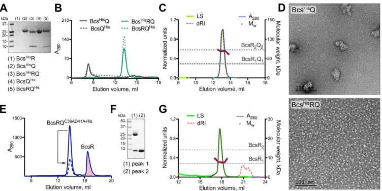
BcsR and BcsQ interdependence and heterocomplex formation. In
cellulose-producing enterobacteria, bcs genes are typically arranged in two separate operons,
FIG 1 E. coli-like cellulose secretion systems. (Left) Thumbnail representation and proposed topology ofthe nine Bcs proteins. (Right) Electron microscopy-based three-dimensional reconstruction of the Bcs macrocomplex, encompassing most of the inner membrane and cytosolic subunits. Known and pro-posed roles for the different subunits and/or protein domains are color coded and annotated at the bottom. GT, glycosyl transferase domain; TA, tail-anchor; CBD, carbohydrate-binding domains; SIMIBI, signal recognition particle, MinD and BioD superfamily; TMD, transmembrane domain(s); IM, inner membrane; OM, outer membrane; NTD, N-terminal domain; CTD, C-terminal domain; TPR, tetratricopep-tide repeats; pEtN, phosphoethanolamine.
on January 4, 2021 by guest
http://mbio.asm.org/
with hallmark bcsRQ and bcsE genes featuring promoter-proximal positions in each (5,
6). Different efforts to purify BcsR and BcsQ constructs on their own were not successful,
with the proteins failing to express stably (Bcs
HisR) or aggregating upon purification
(Bcs
HisQ and BcsQ
His) (Fig. 2; see also Table S1 in the supplemental material).
Coex-pression of the two subunits, however, led to the stable exCoex-pression and purification of
a homogeneous heterotetrameric BcsRQ complex with apparent 2:2 stoichiometry in
solution, where BcsR-dependent BcsQ stabilization appeared independent of the
pres-ence or position of epitope tags on the subunits (Bcs
HisRQ, BcsRQ
His, and BcsRQ)
(Fig. 2A to D). Interestingly, while individual expression of BcsR did not yield detectable
levels of purified protein, we identified empirically BcsQ variants that, when coexpressed
under the same promoter with BcsR (BcsRQ
C39AD41A-Hisand BcsRQ
C39AD41AL43D-His), yielded
an excess of purified BcsR protein, which remained relatively stable in monomeric form
in solution (Fig. 2E to G). These data indicate that the two proteins likely exhibit
chaperone-like functions toward each other, where BcsR stabilizes BcsQ to form
mono-disperse heterotetramers in solution, while BcsQ itself might play a role in the folding
and subsequent stability of BcsR.
Although bacterial operons have now been described for more than half a century
(13), only recently have mechanistic insights into the role of operon organization begun
to emerge. In particular, not only are proteins that function together through the
assembly of heteromeric complexes likely to be encoded by genes in the same or
adjacent operons, but operon gene order has also been reported as generally
opti-mized for the order of protein complex assembly itself (14). This appears to be
especially true for low-copy systems, as are typically the energetically costly secretion
systems, where expression-coupled protein-protein interactions would minimize the
stochasticity of heterocomplex formation (14). Nevertheless, protein folding in the
context of multiprotein assemblies, as well as intraoperon partners remains enigmatic.
FIG 2 BcsR and BcsQ interdependence and complex formation. (A) IMAC elution fractions upon expression of BcsHisR (pProExHTB-BcsHisR), BcsHisQ (pProExHTB-BcsHisQ), BcsHisRQ (pProExHTB-BcsHisRQ), BcsQHis(pET21b-BcsQHis), and BcsRQHis(pET21b-BcsRQHis). (B) Size exclusion chromatography (SEC) profiles of the purified proteins from panel A (2 to 5) using a Superdex 200 Increase 10/30 GL column. (C) SEC-coupled multiangle light scattering (SEC-MALS) of purified tag-free BcsRQ complex. Normalized experimental traces for the light scattering (LS), differential refractive index (dRI), UV absorbance at 280 nm (A280) and calculated molecular weight (Mw) are annotated at the top, theoretical molecular weights for the BcsRQ complex at 1:1 and 2:2 stoichiometries are shown as dashed lines. (D) Electron micrographs in negative stain of the purified BcsHisQ (top) and BcsHisRQ complex (bottom). (E) Purification of folded noncomplexed BcsR upon coexpression with the BcsQC39AD41A-Hismutant following IMAC and SEC. The BcsR peak is colored in pink. A dashed line shows the SEC profile of the mutant BcsRQC39AD41A-Hiscomplex upon reinjection. (F) SDS-PAGE analysis of fractions corresponding to the two peaks in panel E. (G) SEC-MALS of the purified BcsR protein with experimental and theoretical traces as described above.
on January 4, 2021 by guest
http://mbio.asm.org/
Studies on native and engineered proteins have shown that charged or intrinsically
disordered N-terminal domains and protein tails can act as so-called “entropic bristles”
with protein folding helper effects that stabilize fused downstream modules by
mini-mizing their intrinsic aggregation propensity (15–17). We propose here that BcsRQ
complex formation represents a paradigm of similar folding helper effects at the
intraoperon level where upstream expression of an initially disordered BcsR minimizes
the aggregation of its intraoperon partner BcsQ. The sequential expression of the two
proteins could therefore not only limit the stochasticity of complex assembly within a
low-copy cellulose secretion system but also couple the inhibition of intermolecular
BcsQ aggregation with the intramolecular folding of BcsR to secure a stable
stoichio-metric assembly. Moreover, maintenance of separate polypeptides versus the evolution
of genetically fused modules could present further advantages of operon organization,
such as additional regulatory inputs or possible stoichiometry and symmetry variations
upon secretion system assembly and function. In support for this model, recent work
from our group has revealed that even in the context of the stable BcsR
2Q
2hetero-complex, BcsR features a highly flexible and partly disordered N-terminal region that
can partake in nonsymmetric protein-protein interactions, whereas the C-terminal
domain adopts an "-helical fold at the interface of two BcsQ protomers (W. Abidi, S.
Zouhir, M. Caleechurn, S. Roche, and P. V. Krasteva, unpublished).
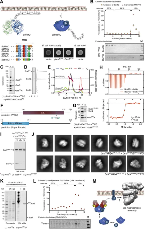
BcsERQ complex formation and membrane targeting. We previously showed
that although predicted as hydrophilic cytosolic proteins, BcsRQ associate stably with
pelleted membranes in cell fractionation experiments and subsequently copurify with
the detergent-extracted Bcs macrocomplex (6). Based on sequence conservation and
putative fold recognition, BcsQ belongs to the ancient family of SIMIBI NTPases, many
of which are involved in membrane-mediated processes such as division septum
inhibition (MinD), flagellar assembly (FlhG and FlhF), protein secretion, and membrane
protein sorting (Srp54-SR and Get3), among others (10, 11). While some of these
proteins are targeted to the membrane via specific protein-protein interactions, others,
such as MinD and FlhG homologs, have intrinsic membrane-targeting sequences (MTS)
that are proposed to adopt an amphipathic "-helical fold upon contact with membrane
lipids (Fig. 3A) (18–20). Comparative sequence analysis shows that a conserved basic
residue midway in the MTSs of MinD and FlhG homologs is replaced by a proline in the
corresponding 10-residue-long C-terminal tail of BcsQ (BcsQ
C10) (Fig. 3A). Although
proline is generally a potent breaker of both "-helical and !-strand secondary
struc-tures in aqueous environments, it is often found in putative transmembrane protein
helices and has been shown to protect "-helical conformations in hydrophobic milieus
(21). To determine the potential role of BcsQ
C10in membrane targeting, we performed
cell-based phenotypic and in vitro lipid-binding assays. Interestingly, deletion of the
BcsQ
C10region had no significant effect on cellulose secretion in a functional
comple-mentation assay in vivo (Fig. 3A), and purified BcsRQ failed to partition with the
lipid-enriched fractions in liposome flotation experiments in vitro (Fig. 3B). As these data
favor protein-based membrane targeting of the essential-for-secretion BcsRQ complex,
we proceeded to determine the nature and sequence of downstream BcsRQ
interac-tions. We started by probing putative interactions with the third cytosolic component,
BcsE, and after testing different strategies for BcsERQ recombinant coexpression (see
Materials and Methods), we were able to purify a stable BcsERQ heterocomplex with
equimolar 2:2:2 stoichiometry in solution (Fig. 3C to E).
BcsE occupies a leader position in its respective bcsEFG operon, which is consistent
with a role in the early stages of Bcs macrocomplex assembly (14). Residues 164 to 474
of the protein were previously defined as a conserved GGDEF I-site-like (GIL) domain
based on the identification of a c-di-GMP binding RXXD motif, similar to the I-site
regulatory sequence often found on diguanylate cyclases (12) (Fig. 3F). Interestingly,
fold recognition programs predict that the N-terminal BcsE region, which features
significantly lower overall sequence conservation (see Fig. S1A), adopts a RecA-like
ATPase fold whose boundaries significantly overlap those of the postulated C-terminal
on January 4, 2021 by guest
http://mbio.asm.org/
FIG 3 BcsRQ membrane targeting and BcsERQ complex formation. (A) Testing the presence and role of a putative
membrane targeting sequence (MTS). (Top left) crystal structure and MTS sequence of the E. coli MinD protein. (Bottom left) MTS conservation among representative SIMIBI proteins and comparison with the corresponding BcsQ C-terminal tail (C10). (Top right) Thumbnail representation of the BcsRQ complex and C10 sequence. (Bottom right) Calcofluor binding assay for
(Continued on next page)
on January 4, 2021 by guest
http://mbio.asm.org/
GIL domain of the protein (Fig. 3F) (22, 23). To identify stable BcsE modules and
examine their role in secretion system assembly, we used both sequence conservation
criteria and predicted three-dimensional fold models to create a series of N- and
C-terminally truncated BcsE variants for recombinant coexpression. From these, we
identified a construct, BcsE
217!523, that copurifies with BcsRQ similarly as full-length
BcsE (Fig. 3G). When individually expressed, the truncated variant featured higher
purity, stability, and protein yields than BcsE
FL, which allowed us to obtain a
thermo-dynamic profile of the BcsRQ ¡ BcsE interaction and reveal a dissociation constant in
the low nanomolar range (K
d"
16 nM) (Fig. 3H). Considering that the binding affinity
is likely even higher in the crowded high-viscosity environment of the bacterial cytosol
and that the typical volume of an E. coli cell is in the low femtoliter range (24), these
data indicate that as soon as the first copies of folded BcsRQ heterocomplex are formed,
they will be bound and sequestered by their BcsE partners in vivo.
Interestingly, biochemical and electron microscopy data show that Bcs complexes
purified via a C-terminal FLAG tag on BcsA fail to efficiently incorporate cytosolic Bcs
components upon deletion of either the N-terminal BcsE
1!217or the C-terminal
BcsE
217!523regions (Fig. 3I and J). While the latter can be explained by disrupted
BcsERQ complex formation through deletion of the BcsRQ binding module, the effects
of BcsE
1!217deletion indicate that this N-terminal domain remains virtually
indispens-able for BcsERQ membrane targeting and its stindispens-able incorporation into the native Bcs
macrocomplex.
We previously showed that deletion of BcsE intraoperon partners BcsF and BcsG
have similar effects of incomplete macrocomplex assembly as the deletion of BcsE
1!217shown here (Fig. 3J) (6). Though both BcsF and BcsG are inner membrane proteins, BcsG
is involved in covalent modifications of the secreted cellulose in the periplasm and does
not purify stably with the assembled Bcs macrocomplex (6, 9). We therefore
hypothe-sized that of the two, BcsF is more likely to act at the early stages of Bcs macrocomplex
assembly as a membrane triggering factor. To test this, we examined BcsE membrane
partitioning in the presence or absence of BcsF (Bcs
HisE
FLversus Bcs
HisE
FLF expression).
Indeed, BcsEF coexpression led to enrichment of BcsE in the pelleted total membrane
fraction (Fig. 3K), and the protein partitioned with membrane-derived proteoliposomes
upon flotation, effectively ruling out potential aggregation in the coexpression context
(Fig. 3L). These data provide further support for coordinated subunit expression and
protein complex assembly, where BcsRQ-bound BcsE is subsequently recruited to the
inner membrane by its immediate downstream operon neighbor, BcsF.
Interestingly, in Pseudomonas putida, the BcsF gene is preceded by two putative
open reading frames (PP_2629 and PP_2630), each of which shares conservation with
FIG 3 Legend (Continued)
cellulose secretion in wild-type (positive control) and mutant (∆bcsQ) E. coli 1094 upon complementation with wild-type or C10-truncated BcsQ. Transformation with an empty pAM238 vector in the ∆bcsQ background was used as a negative control. (B) Liposome flotation assay of potential BcsRQ-lipid interactions. (Top) Relative fluorescence of sucrose gradient fractions after NBD-PE-labeled liposome flotation in the presence of MinHisD (positive control) or BcsRQ. (Bottom) Representative SDS-PAGE analysis of protein distribution along the gradient fractions. Migrated proteins were stained with Coomassie. (C) SDS-PAGE analysis of the IMAC elution fraction upon coexpression of a BcsHisR, BcsQ, and BcsEFLcomplex (pProExHTB-BcsHisRQ plus pRSFDuet1*-BcsEFLcoexpression strategy). (D) Calculated protein ratio in the purified tag-free BcsERQFLcomplex based on densitometric analysis of the SDS-PAGE migrated bands. (E) SEC-MALS of the purified BcsERQFL complex. Experimental and theoretical traces (as described above) are shown for both protein and detergent micelle (Cymal-6m) peaks. (F) Conserved domain detection using sequence alignment and fold prediction tools. (G) SDS-PAGE analysis of the IMAC elution fraction of upon coexpression of a BcsHisR, BcsQ, and BcsE217!523complex (pProExHTB-BcsHisRQ plus pRSFDuet1*-BcsE217-523coexpression strategy). (H) Isothermal titration calorimetry (ITC) profile of the BcsE217!523¡ BcsRQ interaction. (I) Western blot analysis of the BcsHAQ integration into anti-FLAG tag-purified Bcs macrocomplex
(BcsHisRHAQAHA-FLAGB-STREPEFG coexpression) in the context of BcsEFL, BcsE1!217, and BcsE217!523. (J) Representative views
(class averages) of Bcs macrocomplex carrying BcsE217!523(top) versus BcsEFL(bottom, control). (K) BcsEFLmembrane targeting in the context of BcsF coexpression (pProExHTB-BcsHisEFLversus pProExHTB-BcsHisEFLF expression strategies). (Left) SDS-PAGE analysis of the total membrane fractions; (right) Western blot detection of BcsHisEFLin the corresponding fractions. (L) Liposome flotation experiments using NBD-PE-labeled total membrane proteoliposomes from cells coexpress-ing BcsHisEFLand BcsF (pProExHTB-BcsHisEFLF coexpression). (Top) Relative fluorescence of the gradient fractions indicating proteoliposome distribution; (bottom) SDS-PAGE analysis of BcsHisEFL distribution across the corresponding gradient fractions. (M) Results summary showing proposed membrane targeting and macrocomplex integration of the essential for secretion subunits BscR and BcsQ via cytosolic BcsE and membrane-embedded BcsF.
on January 4, 2021 by guest
http://mbio.asm.org/
the BcsE
1!217or Bcs
217!523fragments empirically characterized here (see Fig. S2A and
B). This, together with the significant difference in sequence conservation between the
two BcsE fragments, points toward the evolution of multidomain enterobacterial BcsE
from the genetic fusion of smaller protein subunits to secure not only c-di-GMP
recognition as known for the so-called I-site RXXD motif but also efficient BcsRQ
complexation and subsequent delivery to the inner membrane biosynthetic platform
via high-affinity BcsE-BcsF interactions (Fig. 3M).
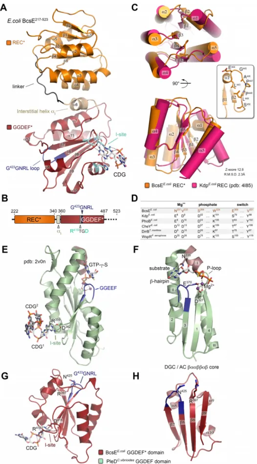
Crystal structure of BcsE
217!523. To gain further insights into BcsE structure and
function, we pursued crystallization of the stable C-terminal BcsE
217!523construct,
which encompasses most of the postulated GIL domain module. Purified untagged
BcsE
217!523crystallized in the presence of c-di-GMP, and its structure was determined
to 2.2 Å using single-wavelength anomalous dispersion (SAD) phasing on crystals
grown from selenomethionine-derivatized protein (Table S2). The protein packed in the
P4
12
12 space group with two BcsE
217!523molecules per asymmetric unit adopting
virtually identical conformations with root mean square deviation (RMSD) of 0.835 Å
over all atoms. A single c-di-GMP is found splayed in a symmetrical conformation
between the two protomers, and nucleotide recognition involves four residues of each
subunit, namely, R
415and D
418from the conserved I site-like motif as well as the side
chain of H
445and the peptide carbonyl of S
432(Fig. 4 and 5). Unexpectedly, the
construct adopts a dual-domain fold with an apparent N-proximal module connecting
via an interstitial helix ("
6) to a C-terminal domain, in which the last #40 residues
(C-terminal tail) remain unresolved in the structure (Fig. 4A and B).
A search for three-dimensional (3D) structural homologs using the fold recognition
server DALI (25) revealed that the N-proximal domain adopts a receiver (REC)
domain-like (!")
5fold (26), where the central five-stranded parallel !-sheet is flanked by 4
"-helices, while the canonical "
1is mostly unfolded in an extended conformation by a
stretch of proline and other small uncharged amino acids (Fig. 4C). Canonical REC
domains are typically found in tandem with DNA-binding modules in response
regu-lator proteins, which use phosphoryl transfer from upstream kinases as input signals for
transcription regulation (26). Structural and sequence alignments of BcsE
REC* with
phosphorylation-competent receiver domains, however, show significant deviation
from the amino acid consensus of key functional motifs, indicating that the module is
unlikely to function in phosphotransfer-dependent signal transduction (Fig. 4D).
Similar DALI search using the resolved C-proximal domain as an input revealed the
closest structural homolog as the cytosolic C-terminal domain of Pseudomonas
aerugi-nosa PelD. Interestingly, the latter is itself a c-di-GMP-binding protein responsible for
the activation of synthase-dependent exopolysaccharide secretion, the Pel system in
pseudomonads, and has been characterized as a degenerate GGDEF
domain-containing protein where c-di-GMP sensing is carried out by the conserved I-site motif
(27–29). Indeed, both BcsE
GGDEF* and PelD
GGDEF* show severe degeneration of the
consensus !""!!"! catalytic core shared by diguanylate and adenylate cyclases (30),
with the substrate-coordinating P-loop and "
1-helix completely missing and catalytic
residues, including those from the signature GGDEF motif, showing significant
diver-gence (Fig. 4E to H; see also Fig. S3). Nevertheless, I-site-dependent c-di-GMP
compl-exation remains virtually unchanged from that of active diguanylate cyclases, with the
dinucleotide participating in both polar and #-stacking interactions with the side
chains of the conserved arginine (R
415) and aspartate (D
418) residues (Fig. 4E to H and
5A; Fig. S3). Taken together, these results classify BcsE as a member of a growing
superfamily of c-di-GMP-sensing proteins, in which canonical signaling (REC, PAS, etc.)
or enzymatic (GGDEF, EAL, etc.) modules have been repurposed to serve
c-di-GMP-dependent signal transduction (2, 3).
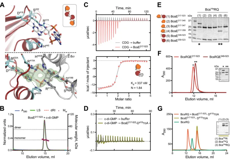
C-di-GMP and BcsQ binding by the BcsE
GGDEF* domain. Although the primary
sequence of the resolved BcsE
REC*
-GGDEF* modules is overall highly conserved, surface
mapping of the amino acid conservation reveals distinct conserved residue clusters on
both the degenerate receiver and diguanylate cyclase modules, which might be
on January 4, 2021 by guest
http://mbio.asm.org/
FIG 4 BcsE217!523crystal structure and domain organization. (A) Crystal structure of BcsE217!523. The degen-erate receiver (REC*) and diguanylate cyclase (GGDEF*) domains are colored orange and red, respectively, and
(Continued on next page)
on January 4, 2021 by guest
http://mbio.asm.org/
indicative of oligomerization or protein-protein interaction interfaces (Fig. S1). To
determine the protein’s homooligomerization propensity in solution, we performed
solution-based light-scattering experiments and determined that the BcsE
217!523con-struct remains monomeric even in the presence of saturating c-di-GMP (Fig. 5B). This is
particularly surprising considering the intrinsic dimerization propensity of REC domains
in general (26), the binding stoichiometry of the BcsE
217!523RQ complex in solution
(N " 0.95, consistent with 2:2:2 binding) (Fig. 3H), and the symmetrical c-di-GMP
conformation in the crystal structure (Fig. 5A, bottom and inset; Fig. S3A and B), where
FIG 4 Legend (Continued)
key motifs are highlighted. Secondary structure elements are numbered without accounting for the missing N-terminal domain. (B) Summary of the resolved domain architecture for the previously predicted GIL domain. (C) Overlay of the E. coli BcsEREC* domain and a canonical receiver domain (E. coli KdpREC) in two different views. Inset, unfolding of the canonical "1 helix into a P-rich loop. Structural alignment scores are calculated in DALI. (D) Comparison of key conserved residues in phosphotransfer-competent response regulators with correspond-ing residues in BcsEREC*. (E) GGDEF domain, I-site-mediated c-di-GMP binding, and substrate homologue coordination of the catalytically active diguanylate cyclase PleDC. vibiroides. (F) Conserved !""!!"! catalytic core shared among adenylate and diguanylate cyclases. Key residues involved in substrate and Mg2$coordination are shown as sticks. Crystal structures of the corresponding BcsEGGDEF* domain (G) and "/! core (H).
FIG 5 c-di-GMP binding and BcsE-BcsQ interactions. (A) c-di-GMP binding to the conserved I-site. (Top) Stick representation of c-di-GMP and the coordinating
residues with only one protein molecule shown. (Inset) Thumbnail representation of the 2:1 protein-to-dinucleotide complexation observed in the crystals. (Bottom) An (|Fo|-|Fc|) partial electron density map calculated from a model prior to inclusion of the dinucleotide and contoured at 2.6$ with both coordinating protomers shown in red/cyan and gray. (B) SEC-MALS of BcsE217!523in the presence of excess c-di-GMP with experimental and theoretical traces as described above. (C) ITC profile of the c-di-GMP ¡ BcsE217!523interaction and thumbnail representation of the calculated binding stoichiometry (1:2, protein to dinucleotide). (D) Control ITC titration of c-di-GMP to the I-site-defective BcsE217!523-D415TGA mutant. (E) SDS-PAGE analysis of IMAC elution fractions testing BcsERQ complex formation upon BcsHisRQ coexpression with various truncated BcsE variants (pProExHTB-BcsHisRQ plus pRSFDuet1*-BcsEtrunccoexpression). (F) SEC profiles of the purified BcsERQGGDEF* complex and BcsERQ217!523. (G) SEC profile of purified BcsRQ preincubated with excess BcsE217!523-D415TGA compared to profiles for separate injections of the individual components. (Inset) SDS-PAGE analysis of IMAC elution fractions assaying BcsE217!523-D415TGA copurification upon coexpression with BcsHisR, BcsHisQ, or BcsHisRQ.
on January 4, 2021 by guest
http://mbio.asm.org/
the dinucleotide bridges two separate BcsE protomers by identical interactions.
Fur-thermore, thermodynamic characterization of the c-di-GMP ¡ BcsE
217!523interaction
reveals a binding stoichiometry consistent with two c-di-GMP molecules binding to a
single BcsE I-site rather than the apparent inverse stoichiometry observed in the crystals
(Fig. 5C versus Fig. 5A; Fig. S3A and B). These results are consistent with both the
propensity of c-di-GMP to adopt diverse conformations, including intercalated dimers
in solution (2, 3), and the capability of GGDEF I-sites to coordinate both monomeric and
dimeric ligands as shown for P. aeruginosa PelD (28, 29) (Fig. S3C and D). Importantly,
the dimeric c-di-GMP conformation derived from the solution-based data is also
consistent with the reported c-di-GMP conformation necessary for BcsA
PilZdomain
binding and gating loop displacement during each step of UDP-glucose coordination
and cellulose incorporation of the sugar moiety (Fig. S3E and F) (8). We therefore
propose that Bcs macrocomplex-bound BcsE could secure the maintenance of a
secretion system-proximal pool of c-di-GMP in a BcsA-activating conformation, thus
limiting dinucleotide diffusion and boosting processive glucose polymerization.
In line with the monomeric state of BcsE
REC*
-GGDEF* in solution, equimolar 2:2:2
BcsE
217!523RQ heterocomplex assembly appears to be driven by the BcsRQ interactions
rather than the BcsE variant itself. To determine how BcsE binds each half of the BcsRQ
complex, we designed a series of shorter BcsE variants for copurification assays and
identified a GGDEF* domain construct, BcsE
349!523, covering the interstitial helix "
i
, the
GGDEF* domain, and the unstructured C-terminal tail that partakes in stable equimolar
interactions with BcsRQ (Fig. 5E and F). We also found that BcsRQ binding is
indepen-dent of c-di-GMP complexation, as BcsERQ complex reconstitution can be carried out
in the absence of dinucleotide and with an I-site mutant incapable of c-di-GMP
complexation (R
415TGD ¡ D
415TGA). Finally, we show that BcsE
GGDEF* interacts with
BcsQ rather than BcsR, as shown in copurification experiments using individual Bcs
HisR
or Bcs
HisQ proteins as baits (Fig. 5F and G). Indeed, a stretch of highly conserved
residues distinct from the c-di-GMP binding I-site is found on one side of the BcsE
GGDEF*
module that could have evolved for high-affinity BcsQ binding (Fig. S1B). Importantly,
nonoverlapping sites for c-di-GMP and BcsQ complexation would allow both stable
assembly of BcsRQ within the Bcs macrocomplex and the possibility of c-di-GMP to
migrate in and out of the I-site in a model where the dinucleotide processively switches
between the BcsE
GGDEF* module and BcsA’s PilZ domain for cocatalytic synthase
regulation.
BcsE
NTD-dependent homooligomerization and binding of conserved Nus
anti-termination complex components. As mentioned above, the N-terminal region of
BcsE (BcsE
1!217) is predicted to adopt a conserved RecA-like ATPase fold (Fig. 6A).
RecA-like motor ATPases are a large family of proteins that use the energy of nucleotide
binding and hydrolysis to oligomerize and perform mechanical work in a variety of
cellular functions, such as the transport or hydrolysis of proteins (e.g., ABC transporters
and proteases) or the binding and remodeling of nucleic acid substrates (e.g., helicases
and recombinases) (31).
Structural and sequence alignments of BcsE
NTDwith catalytically active RecA-like
ATPases show severe divergence of key functional motifs (e.g., the ATP/Mg
2$-coordinating Walker A motif) (Fig. 6A), indicating that BcsE is likely incapable of ATP
binding and hydrolysis. Nevertheless, bacterial two-hybrid assays based on split
ade-nylate cyclase (AC) functional reconstitution (32) suggest that BcsE
FLis prone to
oligomerization and, consistent with the monomeric state of the BcsE
REC*
-GGDEF*
tandem described above (Fig. 5B), that these interactions are BcsE
NTDdependent
(Fig. 6B). Interestingly, blue colony growth indicative of BcsE
FLand BcsE
NTDhomooli-gomerization was only observed in cases when coexpressed AC fragments were fused
to different BcsE termini, regardless of their specific type (T25 or T18) (32) or location
(N or C terminus) in the fusion constructs. These data suggest that the homotypic BcsE
interactions involve different surface regions among the interacting BcsE protomers
and are thus consistent with head-to-tail oligomerization mechanisms that are
fre-quently observed in biologically active RecA-like ATPases (Fig. 6B). However, whether
on January 4, 2021 by guest
http://mbio.asm.org/
BcsE
NTDhomooligomerization plays a functional role in the above-described targeting
of the BcsERQ complex to the inner membrane (Fig. 3) or is involved in additional
regulatory processes (see below), remains to be further examined.
Attempts to recombinantly purify the BcsE
1!217construct consistently led to the
copurification of a second protein species, even in elevated imidazole and salt
con-centrations in the immobilized-metal affinity chromatography (IMAC) purification
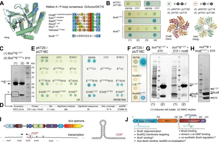
buf-fer (Fig. 6C). Mass spectrometric analyses identified the copurifying species as small
ribosomal protein S10, also known as NusE or RpsJ (Fig. 6D). Interestingly, apart from
associating with the small ribosomal subunit during protein translation, S10 is also
known to moonlight as a key component of the Nus transcription antitermination
complex. Nus factors NusA, NusB, S10/NusE, NusG, and SuhB are mostly essential,
highly conserved bacterial proteins that are known to associate with and reprogram the
transcription apparatus in order to overcome elongation complex dissociation at
certain intrinsic and Rho-dependent transcription terminators (33, 34). Although the
best-studied examples include N protein-dependent antitermination at early % phage
FIG 6 BcsE oligomerization and binding of conserved Nus antitermination complex components. (A) Predicted BcsE1!217RecA-like ATPase fold. (Left) overlay of the prediction model onto Synechococcus elongatus KaiC. (Right) Walker A conservation shown in representative RecA-like ATPases and compared to the corresponding region of BcsE1!217. (B) BcsE1!217and BcsEFLhomooligomerization. (Left and top right) Bacterial two-hybrid assays using different coexpression strategies for the BcsE-AC fragment fusions. (Bottom right) Overlay of modeled BcsE1-217copies (in gray) with head-to-tail oligomers of RecA-like ATPases in superhelical (DnaB) or ring (KaiC) oligomeric states. (C) IMAC elution fractions of BcsHisE1!217when expressed individually or coexpressed with E. coli NusE/S10 protein. (D) Mass spectrometry-based protein identification of a consistently copurifying low-molecular-weight band from an individually expressed, purified, and SDS-PAGE-migrated BcsE1!217. (E) Bacterial two-hybrid assay of interactions between BcsE domains and S10/NusE based on plasmid-based adenylate cyclase functional reconstitution in a cya-defective E. coli strain (BTH101). The positive zip/zip control is based on coexpressed adenylate cyclase fragments each fused to a homodimerizing leucine zipper region of the yeast protein GCN4. Interactions were evaluated by the growth of blue colonies on X-Gal-supplemented LB (left) or M63BI (right) agar plates. (F) Bacterial two-hybrid assays of BcsE1!217-NusB interactions. (G) SDS-PAGE analysis of induced cell lysates, IMAC elution fractions, and S10 copurification upon BcsHisE1!217and S10 coexpression in the absence (left) or presence (right) of NusB. (H) IMAC elution fraction upon NusHisB,
BcsE1!217, and S10 coexpression. (I) Organization of the two bcs operons and schematic representation of bioinformatically detected potential intrinsic
terminators. (Right) Representation of a putative intrinsic terminator in bcsR. (J) Results summary showing BcsE domain architecture and proposed functional roles for the identified structural modules.
on January 4, 2021 by guest
http://mbio.asm.org/
genes and the regulation of bacterial ribosomal (rrn) gene expression (34, 35), a recent
study identified conserved NusB-S10 binding sites upstream of additional genes in
diverse bacterial species (36). Nevertheless, characterized S10 interactions at the
protein-protein level have remained limited to the context of assembled ribosomes or
extensively studied transcription antitermination subcomplexes (e.g., see references 35
and 37).
Intrigued by this, we proceeded to assay the putative BcsE-S10 interaction by
recombinant coexpression/copurification and cell-based bacterial two-hybrid
experi-ments. We observed that BcsE
NTDcopurified at an equimolar ratio with overexpressed
S10 (pProExHTB-bcsE
NTDplus pRSFDuet1*-s10 coexpression) and that BcsE likely
inter-acts with S10 in cellulo as observed by blue colony growth in the context of both the
truncated BcsE
1!217construct (BcsE
NTD) and the full-length BcsE protein (BcsE
FL)
(Fig. 6C and E). We further assayed putative interactions of BcsE
NTDwith a second Nus
factor, NusB, known to interact directly with S10 in the early steps of TAC assembly onto
the target mRNA. Whereas bacterial two-hybrid experiments were indicative of weak
BcsE
NTD-NusB interactions in cellulo, recombinant coexpression of Bcs
HisE
NTDwith
tag-free S10 and NusB led to the purification of excess Bcs
HisE
NTDand only trace
amounts of copurifying Nus factors, even if S10 expression levels appeared virtually
unchanged (Fig. 6F and G). Conversely, recombinant coexpression of Nus
HisB with
tag-free S10 and BcsE
NTDled to the purification of an equimolar amount of NusB-S10
complex and trace amounts of a third species, whose molecular weight corresponds to
that of BcsE
NTD(Fig. 6H). Together, these data indicate that S10 likely uses similar
surface regions to interact with its BcsE and NusB partners, whereas higher-affinity NusB
complexation could cause competitive remodeling of the equimolar BcsE
NTD-S10
as-semblies and subsequent release of free BcsE.
Although the physiological significance of the observed BcsE-Nus factor interactions
remains enigmatic, our findings suggest a possibly broader role for the conserved Nus
antitermination machinery than in the well-studied examples of ribosomal or viral gene
expression. Interestingly, in silico prediction tools (38, 39) detect putative intrinsic
terminators within both the bcsR and bcsQ coding regions. If these potential regulatory
elements indeed function as predicted, then the protein-protein interactions observed
here could serve to target (via S10 complexation upon exiting the ribosome) and
subsequently release (via downstream recruitment of NusB upon antitermination
com-plex assembly) BcsE at the site of bcsRQ expression, prior to binding the newly
synthesized BcsRQ complex directly and delivering it to the inner membrane for
downstream Bcs macrocomplex assembly. Such a hypothesis, however, remains to be
experimentally tested.
Concluding remarks. Bacteria have evolved complex secretion machineries to
deliver large molecules to the cell envelope, external milieu, or host cell targets.
Although these systems are typically not essential for bacterial physiology per se, they
could often provide significant advantages in interspecies competition or be key to a
pathogen’s infection cycle. Bacterial exopolysaccharide secretion shares many
similar-ities with the various types of protein secretion systems in that it typically involves
intricate signal transduction events to induce the expression and assembly of multiple
subunits in order to provide the biosynthetic activities, physical conduit, and energetics
for biopolymer extrusion through the complex bacterial envelope (40).
Often, secretion systems are viewed as such at the level of assembled
macrocom-plexes and substrate extrusion, whereas the initial steps of subunit expression and
sequential protein-protein interactions remain largely overlooked. Here, we present the
E. coli-like Bcs system as a new candidate paradigm for concerted secretion system
assembly and function. We demonstrate that essential-for-secretion BcsR and BcsQ
regulate each other’s folding and stability, whereas BcsE packs a subtle but diverse
toolkit to fine-tune enterobacterial cellulose production (Fig. 6J). We provide structural
and functional data that reveal the protein’s multidomain evolution, fold conservation,
and complexation of synthase-activating intercalated c-di-GMP on one hand, together
on January 4, 2021 by guest
http://mbio.asm.org/
with high-affinity BcsRQ recruitment and facilitated membrane targeting through BcsF
interactions on the other. Although more research is needed to uncover physiological
roles for the observed BcsE-Nus factor interactions or how the essential BcsRQ subunits
control assembly and function of the inner membrane biosynthetic platform, this work
lays an important milestone toward more comprehensive models of operon-encoded
synthase-dependent polysaccharide secretion in bacterial biofilms.
MATERIALS AND METHODS
The experiments were not randomized, and the investigators were not blinded during experimental design, execution, or outcome assessment. However, most experiments were reproduced independently by different investigators, including crystallographic, biochemical, biophysical, and phenotypic functional assays.
Bacterial strains. Plasmids for recombinant protein expression (see below) were propagated in and
isolated from E. coli DH5" cells. All recombinant protein expression for structural and in vitro biochemical studies was carried out in BL21(DE3) Star cells, including the expression of selenomethionine-derivatized protein. An E. coli 1094 ∆bcsQ strain was used for the complementation phenotypic assays with BcsQ variants expressed from a low-copy-number isopropyl-!-D-thiogalactopyranoside (IPTG)-inducible vector (pAM238; see below). Finally, bacterial two-hybrid experiments were performed using chemically com-petent BTH101 cells and the IPTG-inducible pKT(N)25 and pUT18(C) expression plasmids with custom-modified multiple cloning sites (see below). All bacterial strains and plasmids used in this study are available upon request.
Recombinant DNA techniques. DNA manipulations were carried out using standard protocols for
PCR, molecular cloning, transformation, and DNA analyses. Coding regions for BcsR, BcsQ, BcsRQ, BcsE, MinDE, S10, and NusB variants were amplified using E. coli 1094 genomic DNA as a template and a high-fidelity DNA polymerase (Phusion; New England BioLabs) and inserted via digestion/ligation cloning into IPTG-inducible expression vectors with custom-modified multiple-cloning sites (MCS). Point muta-tions, insertion of stop codons, MCS modificamuta-tions, and domain deletions within previously reported and newly generated expression constructs were performed using inverse PCR-based protocols and mutation-specific oligonucleotides as primers. All recombinant vectors and introduced mutations were verified by DNA sequencing and, where applicable, IPTG-inducible protein expression.
Protein expression and purification. All pProExHTB-encoded constructs (BcsHisR, BcsHisQ, BcsHisRQ, BcsHisENTD, NusHisB, and MinHisDE) were expressed as IPTG-inducible variants carrying N-terminal hexa-histidine tags cleavable by the human rhinovirus (HRV) 3c protease. BcsQ was also cloned in a standard pET21b vector yielding a C-terminally hexahistidine-tagged protein. As all BcsR (BcsHisR) and BcsQ (BcsHisQ and BcsQHis) constructs failed to yield stable proteins, the coding region corresponding to the BcsRQ tandem was subsequently amplified and cloned into both the pProExHTB and pET21b expression vectors, adding a cleavable N-terminal or noncleavable C-terminal hexahistidine tag to BcsR (pProExHTB-HisRQ) or BcsQ (pET21b-RQHis), respectively. For coexpression studies, the coding region corresponding to full-length tag-free BcsE was cloned into custom-modified pRSFDuet1* expression vector under the control of the first T7 promoter (pRSFDuet1*-BcsEFL) (see Table S1 in the supplemental material). Full-length BcsE was also cloned in pProExHTB and pET-HisSUMO (see below) vectors for standalone expression, but the purified proteins were judged insufficiently stable or pure for structural studies. Based on sequence conservation (PFAM [41]) and predicted tertiary structure (Phyre2, Robetta [22, 23]), several N- and C-terminal deletions of the pRSFDuet1*-BcsEFLconstruct were tested for expression and copu-rification with BcsRQ. The coding region for the interacting BcsE217!523construct was subsequently cloned for standalone expression into a modified pET-HisSUMO plasmid, yielding a hexahistidine-tagged Ulp1-cleavable SUMO moiety fused to the N terminus of the protein of interest (pET-His
SUMO-BcsE217!523). Based on the resulting crystal structure of the BcsE217!523construct, an additional construct
corresponding to the C-terminal GGDEF* domain was designed (BcsE349!523), and its coding sequence was cloned into the pRSFDuet1* and pET-HisSUMO expression vectors as described above. The BcsE1!217 construct corresponding to the protein’s N-terminal domain was cloned into both pProExHTB and pRSFDuet1* (site 1) vectors. For coexpression of BcsE1!217(in pProExHTB) with S10 and NusB, the coding sequences for the latter were cloned in the first and second sites, respectively, of custom-modified pRSFDuet1* vectors (pProExHTB-BcsHisE1!217plus pRSFDuet1*-S10(site 1)-NusB(site 2) coexpression; the pRSFDuet1* vector was further modified at the second promoter to introduce unique XhoI and HindIII restriction sites) (Table S1). In addition, a pProExHTB-NusHisB plus pRSFDuet1*-BcsE1-217(site 1)-S10(site 2)was also employed. For control liposome flotation studies, the coding region for the MinDE tandem was PCR-amplified and cloned into a pProExHTB vector, and MinD was purified as a partner-free protein (MinHisD) from the clarified cytosolic fraction. Finally, BcsHisEFLand BcsHisEFLF cloned into pProExHTB vectors were used for examining the membrane-targeting role of BcsF. Protein constructs used in the bacterial two-hybrid studies are described separately.
For protein purification, all expression vectors were (co)transformed into chemically competent E. coli BL21(DE3) Star cells. For the expression of native proteins, cells were grown at 37°C under aerobic conditions in terrific broth (TB) medium supplemented with appropriate antibiotics (100 &g/ml ampicil-lin, 40 &g/ml kanamycin, or a combination of 70 &g/ml ampicillin plus 30 &g/ml kanamycin for coex-pressed vectors). At a cell optical density corresponding to an optical density at 600 nm (OD600) of 0.8 to 1.0, the cells were moved to 17°C, and overnight protein expression was induced by the addition of IPTG at a final concentration of 0.7 mM. For the expression of selenomethionine-derivatized proteins, 4 liters of cells was initially grown at 37°C in LB medium to an OD600of 0.5 to 0.6. Cells were then pelleted by
on January 4, 2021 by guest
http://mbio.asm.org/
centrifugation (4,000 % g, 15 min, 20°C), gently washed with 200 ml 1% SelenoMet medium base (Molecular Dimensions), collected again, and resuspended in 1 liter complete SelenoMet medium (Molecular Dimensions) supplemented with 40 mg/literL-selenomethionine and the appropriate
antibi-otic. Cells were then grown for an additional 1 h at 37°C, transferred to 17°C, and induced with IPTG as described above.
After 16 h, cells were harvested by centrifugation, resuspended in lysis buffer, and flash-frozen in liquid nitrogen. The composition of the lysis buffer was 20 mM HEPES (pH 8.0), 120 mM NaCl, 19 mM imidazole (pH 8.0,) 2 mM !-mercaptoethanol, and 1 tablet/50 ml cOmplete protease inhibitors (Roche) for the BcsHisR, BcsHisQ, BcsQHis, BcsHisRQ, MinHisDE, and BcsHisE217!523constructs. For the BcsHisRQ-BcsEFL, BcsHisRQ-BcsE217!523, and BcsHisRQ-BcsE349!523complexes, the IMAC buffer was also supplemented with 0.5 &M c-di-GMP (Jena Bioscience or Sigma-Aldrich), 2 &M AppCp (Jena Bioscience), 5 mM MgCl2, and 10% glycerol. For the expression of BcsHisE1!217and the BcsHisE1!217-S10, BcsHisE1!217-S10-NusB, and NusHisB-S10-BcsE1!217complexes, the concentration of salt in the lysis buffer was increased to 750 mM NaCl.
For all cytosolic protein purifications, cells were thawed and lysed using an Emulsiflex-C3 high-pressure homogenizer (Avestin). Cell debris was removed by centrifugation (1 h at 50,000 % g and 4°C), and the cleared lysates were loaded onto buffer-washed Talon Superflow resin (GE Healthcare) at approximately 0.5 to 1 ml of resin per liter of culture. The resin was subsequently washed with more than 20 volumes of IMAC buffer A (protease inhibitor-free lysis buffer as described above), and bound proteins were eluted in a single step with IMAC buffer A supplemented with 200 mM imidazole (pH 8.0) (IMAC buffer B).
For purification of tag-free BcsRQ, eluted protein BcsHisRQ protein was supplemented with 15 mM EDTA (pH 8.0) and homemade HRV3c protease at 4°C, concentrated to 2.5 ml using an Amicon Ultra centrifugal filter (30-kDa cutoff; Millipore), desalted using a disposable PD-10 desalting column (GE Healthcare), and incubated overnight for tag removal. The cleaved tag and protease were removed by inverse IMAC, concentrated, and subjected to size exclusion chromatography on a Superdex 200 Increase 10/300 GL column (GE Healthcare) equilibrated in gel filtration buffer (20 mM HEPES [pH 8.0], 120 mM NaCl, and 2 mM dithiothreitol [DTT]). Collected protein fractions were analyzed for purity by SDS-PAGE, pooled, concentrated, flash-frozen in liquid nitrogen, and stored at !80°C.
For purification of tag-free BcsE217!523, the elutedHisSUMO-fused protein was mixed with homemade yeast protease Ulp1, concentrated to 2.5 ml, desalted on a disposable PD-10 column, and incubated for HisSUMO cleavage at 4°C overnight. Cleaved protein was collected in the flowthrough fraction during reverse IMAC on the following day, concentrated, and subjected to size exclusion chromatography on a Superdex 200 Increase 10/300 GL column equilibrated with gel filtration buffer (20 mM HEPES [pH 8.0], 100 mM NaCl, and 2 mM DTT). Collected protein fractions were analyzed for purity, concentrated, aliquoted, and flash frozen for storage at !80°C.
Complexes BcsRQ-BcsE217!523and BcsRQ-BcsE349!523were purified in a similar 2-step IMAC proce-dure. Eluted proteins were incubated with the viral HRV3c protease for cleavage of the N-terminal hexahistidine tag on BcsR. Imidazole concentrations were lowered via desalting on a disposable PD-10 column, and after overnight incubation at 4°C, the proteins were subjected to size exclusion chroma-tography using a Superdex 200 Increase 10/300 GL column and gel filtration buffer composed of 20 mM HEPES (pH 8.0), 120 mM NaCl, 5 mM MgCl2, 0.5 &M c-di-GMP, 2 &M AppCp, 2 mM DTT, and 10% glycerol. Collected protein fractions were analyzed for purity and stoichiometric complex assembly, concentrated, and flash frozen in liquid nitrogen for storage at !80°C.
To characterize the complex formation and stoichiometry of interaction between BcsRQ and BcsEFL, a ternary complex was coexpressed and purified using a similar protocol. However, as the complex appeared to be stabilized by the presence of detergents, after cell lysis, the cell debris was pelleted by slower centrifugation (12,000 % g, 15 min, 4°C), and the remaining supernatant was incubated with 0.25% n-dodecyl-!-D-maltopyranoside (!-DDM; Anatrace) for 1 h at 4°C. The lysates were then cleared by
high-speed centrifugation, and the ternary BcsRQ-BcsEFLcomplex was purified as the rest of the BcsERQ complexes while keeping a low concentration of detergent (0.06% Cymal-6; Anatrace) in all buffers. For size exclusion chromatography, the Superdex 200 Increase 10/300 GL column was replaced by a Superose 6 Increase 10/300 GL column.
MinD was purified from clarified cytosolic fraction using a single-step metal-affinity purification (IMAC buffer A with 20 mM HEPES [pH 8.0], 120 mM NaCl, and 19 mM imidazole), followed by size exclusion chromatography on a Superdex 200 Increase 10/300 GL column equilibrated with gel filtration buffer (20 mM HEPES [pH 8.0], 100 mM NaCl, and 2 mM DTT). Clean protein fractions were concentrated and flash-frozen for storage at !80°C.
Cells expressing BcsHisE1!217, BcsHisE1!217-S10, BcsHisE1!217-S10-NusB, and NusHisB-S10-BcsE1!217 were resuspended in high-salt lysis buffer (same as described above but with 750 mM NaCl), and the proteins were purified by a single-step IMAC. The high-salt conditions (750 mM NaCl) were maintained in all buffers.
Finally, expression and purification of the Bcs macrocomplex (pCDFDuet1-BcsHisRQAHA-FLAGB plus pRSFDuet1*-StrepEFG) with various BcsE (BcsStrepEFL, BcsStrepE1!217, or BcsStrepE217!523) and BcsQ (BcsQ or BcsHAQ) variants were performed as reported previously (6). pRSFDuet1*-BcsStrepE1!217FG was generated from pRSFDuet1*-BcsStrepEFLFG via inverse PCR using two different strategies which yielded consistent results: (i) an insertion of a 4-letter STOP codon following BcsE residue A217(TAAT in DNA) and (ii) a deletion of the REC*-GGDEF* tandem while preserving the ribosome-binding site for bcsF to avoid polar effects). pRSFDuet1*-BcsStrepE217!523FG was generated by standard restriction/ligase subcloning. Inser-tion of a hemagglutinin (HA) tag at the N terminus of BcsQ was also conducted by inverse PCR. After
on January 4, 2021 by guest
http://mbio.asm.org/
expression vector cotransformation, culture growth, and overnight expression induction, cells were pelleted by centrifugation and resuspended in ice-cold lysis buffer containing 20 mM HEPES (pH 8.0), 120 mM NaCl, 10% glycerol, 5 mM MgCl2, 10 &M AppCp, 2 &M c-di-GMP, 250 &M cellobiose, 0.5 mg/ml Aspergillus niger cellulase (Sigma-Aldrich), 100 &g/ml lysozyme, and 1 tablet/50 ml cOmplete EDTA-free protease inhibitors (Roche). After lysis (Emulsiflex-C3), cell debris was removed by low-speed centrifu-gation (12,000 % g, 15 min, 4°C), and the membranes were pelleted by ultracentrifucentrifu-gation using an SW 28 Ti Beckman rotor (26,500 rpm, or up to 126,000 % g, for 1 h at 4°C). After removal of the supernatant, the membrane fraction was resuspended in solubilization buffer containing all lysis buffer components except lysozyme and cellulase, as well as a mix of detergents at the following final concentrations: 0.4% (wt/vol) digitonin (Sigma-Aldrich), 0.4% (wt/vol) n-dodecyl-!-D-maltopyranoside (anagrade !-DDM; Ana-trace), 0.4% (wt/vol) decyl maltose neopentyl glycol (DM-NPG; AnaAna-trace), and 0.2% lauryl maltose neopentyl glycol (LM-NPG; Anatrace). After a 60- to 90-min-long incubation at 20°C and under mild agitation, the solubilized membrane fraction was cleared by a second high-speed centrifugation step as described above. The supernatant was then incubated with anti-FLAG M2 affinity gel (50 &l packed resin per liter of induced culture; Sigma-Aldrich) with mild agitation at 4°C for 1 h. After gravity elution of the nonbound fraction, the resin was washed extensively (&30 column bed volumes) with binding buffer containing all lysis buffer components except lysozyme and cellulase, as well as 0.008% (wt/vol) LM-NPG. The bound complexes were then eluted using 4 column bed volumes of elution buffer (affinity buffer supplemented with 3% FLAG peptide at 100 &g/ml) and concentrated on a 100-kDa cutoff Amicon Ultra (MerckMillipore) centrifugal filter.
SDS-PAGE and Western blot analyses. Protein fractions were analyzed by standard denaturing
SDS-PAGE using 4% to 20% gradient mini-gels (Bio-Rad), Expedeon InstantBlue Coomassie stain, and a Li-Cor Odyssey Fc system for Coomassie visualization (700-nm channel). For Western blot analyses, SDS-PAGE-migrated proteins were directly transferred using a standard mini-gel transfer protocol, 0.2-&m polyvinylidene difluoride (PVDF) membranes, and a Trans-blot Turbo transfer system (Bio-Rad). Blocking and antibody incubations were in the presence of 5% skim milk in Tris-phosphate-buffered saline (TPBS); all washes between and after antibody incubations were with 1% TPBS buffer. Rabbit anti-His6 (dilution 1:1,000, ab200537; Abcam) and mouse anti-HA (dilution 1:1,000, number 26183; Thermo Fisher Scientific) antibodies were used as primary antibodies; Alexa Fluor 680-conjugated goat ant-rabbit (dilution 1:10,000, ab175773; Abcam) and donkey anti-mouse (dilution 1:10,000, ab175774; Abcam) were used as secondary antibodies. The Alexa Fluor 680 signal was detected using a Li-Cor Odyssey Fc system in the 700-nm channel.
Crystallization, data collection, and structure determination. Crystals were obtained by sitting or
hanging-drop vapor diffusion by mixing equal volumes of protein (1.5 to 6 mg/ml) and reservoir solution followed by incubation at 4°C. BcsE217!523crystals also appeared within 3 to 14 days under multiple conditions, with diffracting data sets collected on crystals grown in 100 mM morpholineethanesulfonic acid (MES; pH 6.0), 4% polyethylene glycol 4000 (PEG 4000), 200 mM MgCl2, 5% glycerol, and 50 &M c-di-GMP. For cryoprotection, crystals were soaked in reservoir solution supplemented with 25% to 30% glycerol, 1 mM DTT, and 50 &M c-di-GMP. Cryopreserved crystals were flash frozen and stored in liquid nitrogen. Data were collected on frozen crystals at 100 K using synchrotron radiation at beamlines PX1 and PX2 at the Soleil synchrotron.
Data reduction was carried out with the software package XDS (42). Experimental phases were obtained by single-wavelength anomalous diffraction (SAD) experiments on crystals grown from selenomethionine-derivatized protein and with wavelengths corresponding to the experimentally de-termined selenium K-edge. Initial BcsE217!523 models were obtained using the automated model building tools of PHENIX and Buccaneer (43, 44). Reiterative refinements in PHENIX, COOT, and BUSTER yielded the final refined model (43, 45, 46). Data collection and refinement statistics are summarized in Table S2. For illustration purposes, all crystal structures were displayed with the PyMol Molecular Graphics System (Schrödinger, LLC) or UCSF Chimera (47). The latter was also used for displaying the 3D reconstructions of the assembled Bcs macrocomplex.
Single-particle electron microscopy. Negative-stain single-particle electron microscopy was used
for visualization of various Bcs proteins and protein complexes. Briefly, 5 &l of eluted samples (concen-tration, #0.01 to 0.05 mg/ml) were spotted on glow-discharged carbon-coated copper grids (Agar Scientific). After a 1-min incubation, the extra liquid was blotted off, and the grids were passed sequentially through three drops of 2% (wt/vol) uranyl acetate solution, with a second incubation in the last drop before blotting and air drying. Micrographs were taken on a Thermo Fisher Scientific T12 Tecnai electron microscope operated at 100 kV accelerating voltage and equipped with a LaB6 filament and a K2 Base direct electron detector. For the protein complexes purified from cells expressing BcsHisRQAHA-FLAGB plus
BcsStrepEFLFG or BcsHisRQAHA-FLAGB plus BcsStrepE217!523FG, particles were autopicked in EMAN2 (48),
saved as .box coordinates, and converted into a .star particle stack in Relion2 (49). Micrograph contrast transfer function (CTF) correction and two-dimensional (2D) classification were performed in cryoSPARC v2 after particle reextraction using the Relion2-generated .star file as metadata input (50). A total of 3,810 particles were classified for the Bcs macrocomplex carrying full-length BcsE (control) and 6,242 particles for the complex purified from cells expressing the BcsE217!523truncated variant.
Protein identification by mass spectrometry. Coomassie-stained gel bands were excised and
subjected to in-gel enzymatic digestion. Briefly, the bands were extensively washed with acetonitrile and 100 mM NH4HCO3, dried, and treated with 10 mM DTT at 56°C for 30 min. After DTT removal, cysteine carbamidomethylation was performed at room temperature for 30 min by the addition of 55 mM iodoacetamide. The washing procedure was then repeated, the gel slices were dried, and the proteins were digested overnight at room temperature by the addition of 20 &l/band of 10 ng/&l Porcine Gold