HAL Id: tel-02900414
https://tel.archives-ouvertes.fr/tel-02900414
Submitted on 16 Jul 2020HAL is a multi-disciplinary open access archive for the deposit and dissemination of sci-entific research documents, whether they are pub-lished or not. The documents may come from teaching and research institutions in France or abroad, or from public or private research centers.
L’archive ouverte pluridisciplinaire HAL, est destinée au dépôt et à la diffusion de documents scientifiques de niveau recherche, publiés ou non, émanant des établissements d’enseignement et de recherche français ou étrangers, des laboratoires publics ou privés.
Functional multisensory integration and plasticity of
vestibular reflexes
Olga Kuldavletova
To cite this version:
Olga Kuldavletova. Functional multisensory integration and plasticity of vestibular reflexes. Human health and pathology. Normandie Université, 2020. English. �NNT : 2020NORMC405�. �tel-02900414�
THÈSE
Pour obtenir le diplôme de doctorat
Spécialité PHYSIOLOGIE ET BIOLOGIE DES ORGANISMES POPULATIONS
-INTERACTIONS
Préparée au sein de l'Université de Caen Normandie
L'intégratiοn multisensοrielle fοnctiοnnelle et la plasticité des
réflexes vestibulaires
Présentée et soutenue par
Olga KULDAVLETOVA
Thèse soutenue publiquement le 21/02/2020
devant le jury composé de
M. GILLES CLEMENT Directeur de recherche, Université Lyon 1 ClaudeBernard Rapporteur du jury M. JOHN GOLDING Professeur, University of Westminster Rapporteur du jury Mme LILIANE BOREL Directeur de recherche, Aix-Marseille Université Président du jury Mme JOCELYNE VENTRE-DOMINEY Chargé de recherche, Université de Bourgogne Membre du jury Mme GAELLE QUARCK Maître de conférences HDR, Université CaenNormandie Directeur de thèse M. PIERRE DENISE Professeur des universités, Université Caen Normandie Co-directeur de thèse
Thèse dirigée par GAELLE QUARCK et PIERRE DENISE, Mobilités : vieillissement,
pathologie, santé - COMETE
Functional multisensory
integration and plasticity
of vestibular reflexes
3
Acknowledgements
First, I would like to thank the members of my thesis committee: Dr. Gilles Clement, Pr. John Golding, Dr. Liliane Borel and Dr. Jocelyne Ventre-Dominey for accepting to evaluate this thesis, for their professional opinion, their insightful comments and encouragement, as well as for being ready to make a long (and even very long!) way to Caen.
If several years ago someone told me, an engineer student passionate about art, from a small Russian city, that soon I will move alone to France, write a thesis in Neuroscience and give courses at the university in France, I would hardly believe that. However, what has happened was like jumping from a cliff, being such an abrupt change, and such a long, difficult and happy way. Happy about all the new things I have learnt, all the people I have met. I want to thank everyone who made it possible for me to be here. Thank you. Merci. Спасибо. I feel lucky to be surrounded by generous, attentive, professional and caring people.
Thanks to Damien Davenne and Thomas Freret for welcoming me in the laboratory COMETE, for giving me the possibility to publish my work in international peer-reviewed journals and communicate it at international conferences.
My sincere gratitude I would like to express to Pierre Denise. Without you, I would have not been here. Thank you for giving me a chance to work in the laboratory, and also for your help to orient and organize myself in when I just came to France. Thank you for being there, with a good solution and a good advice, despite your busy schedule. Thanks to all the Denise family, especially to Isabelle and Gonzague (twin!) for welcoming me in their house at the very beginning, for introducing me to this beautiful country and language.
Thanks to Gaëlle Quarck, who took me under her wing, when I needed it. Thanks for being the most positive and optimistic director! Thank you for your professional impact, your friendly and caring attitude and your confidence. I have not lost any moment spent with you, whether at work or at personal communication. I keep a lot of great memories, such as our trip to Chicago or music festivals. Thank you for your excellent taste in music, as well as for our philosophic conversations in Franglish J.
4
Thanks to Herve Normand for always being there to help, to give a professional advice, to explain, for being always benevolent and positive, for the smiles and laughs that always accompany you.
Olivier Etard, “-do as I do”, well, I will! I will definitely try to get at least a part of your professionalism mixed with caring nature. Thank you for your invaluable help.
Antoine Gauthier, thanks a lot for giving me a possibility to teach in the UFR STAPS, for your support and attention to people.
Great thanks to Alexander Meigal for having made it possible for me to find this thesis, for spending your time to introduce me to a new challenging topic of research.
My lab friends, I thank you so much! Florane, who has always been by my side, having so much benevolence. Thank you for your company and your support, thank you for your patience when I was just learning speaking French! Houda, thank you for all the crazy laughs we had and for understanding, sharing the same difficulties to be a stranger – alone – in a new country. Daniel, never enough to thank you for your technical help in the experiments, it was so important and also fun! Thanks for all the communications and coffees we’ve had. Nicolas Lefèvre, big thanks for the technical help. I thank all the doctorants and staff of the laboratory: Tristan, Emma, Antoine Jr, Marion, Eva, Solenn, Marc, Elpidio, Valentin, Candice, Eve and Yannick, for the time spent together, for our cosy open-space office with sweets, for all the liters of coffee drank together!
Thanks to my dear friend Catharine Mason for the support and for all the inspiring debates, that could last for hours and hours, without noticing the time passing. Thank you for all the fun and work we have had together. And we will certainly have more!
My great friend Valentine, thank you for all the sketches, drawings, paintings, etchings we have done side by side, for all the hours spent in the theater, as well as in the forest, picking mushrooms. Thank you for the warmest evenings in your house filled with art, animals, good music and smells of your increadible cuisine. Pierre BDL, thak you for the encouragement and your capacity to listen and to share, for your great artistic talent that inspires me to try new and do better. Also
5
thanks for your help with a couple of illustrations for this thesis (they are Figures 5 and 7 within the text, to be precise).
I am grateful to all of my friends that have been near, or far, but always in my heart. My lovely magical Lida, my brave and gifted Katya, my dear cousine Alla, thank you for visiting me in here! I miss you all. Misha, my dearest person. Спасибо. My adored Mom and Dad, thank you for all the support and love you give to me, with no conditions, with any distance. I miss you. (Спасибо любимым маме и папе за поддержку и любовь, на любом расстоянии и в любом состоянии. Я скучаю!)
6
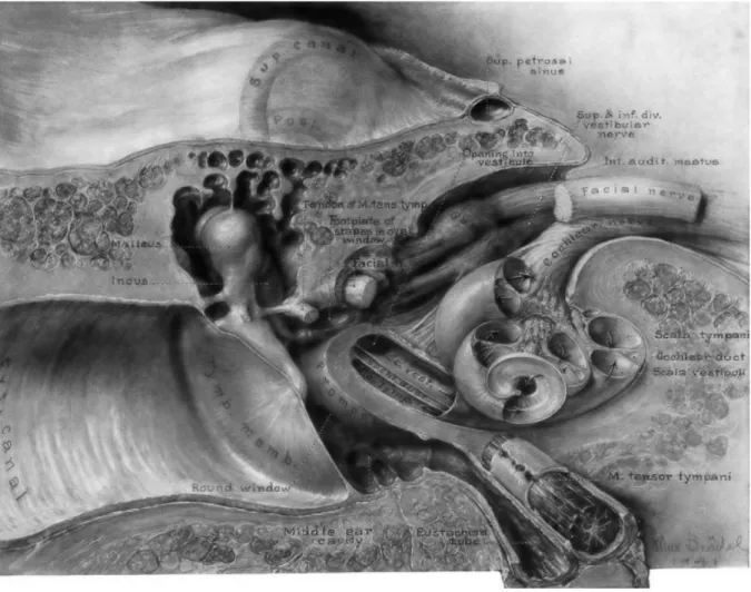
Illustration by Max Brödel, 1941. A drawing of the temporal bone showing the
relationships between the external ear canal with tympanic membrane, middle ear with ossicular chain, the cochlea and the internal auditory canal with vestibular, cochlear and facial nerves.
7
Contents
Abbreviations ... 11Introduction
... 13Part I: Background
... 15 Vestibular system ... 17 Introduction ... 17Anatomy of the vestibular apparatus ... 19
Hair cells and the transduction of a signal ... 21
Otolithic system ... 24
Semicircular canals ... 27
Central vestibular system ... 29
a. Vestibular nerve ... 31
b. Vestibular nuclei ... 32
c. Multisensory integration within VN ... 36
d. Vestibular cerebellum ... 36
e. Multisensory integration within cerebellum ... 38
f. Vestibular thalamus and cortex ... 39
g. Thalamus ... 39
h. Multisensory integration within thalamus ... 43
i. Vestibular cortex ... 43
j. Multisensory integration in cortex ... 46
8
Vestibulo-spinal and vestibulo-collic reflexes ... 49
Vestibulo-ocular reflexes ... 53
a. Canal-Ocular Reflex ... 53
b. Otolith-Ocular Reflex ... 58
c. VOR interactions ... 60
Vestibular effect on autonomic functions ... 60
a. Pathways ... 61
b. Vestibular effect on the cardiovascular control ... 64
c. Vestibular effect on respiration ... 68
Estimation of gravity: tilt-translation ambiguity ... 69
Subjective vertical ... 75
Limitations of the vestibular system ... 77
Sensory Ambiguity ... 77
Motion sickness ... 78
Vestibular plasticity ... 85
Habituation, adaptation and compensation ... 85
a. Habituation ... 85
b. Adaptation ... 88
c. Compensation ... 89
Natural methods used to stimulate the vestibular system in human ... 95
EVAR ... 96
OVAR ... 98
Linear sled ... 100
9
HDNF ... 102
Head Drop ... 103
General notice ... 104
Objectives of the thesis ... 105
Part II: Experiments
... 107Study I: Vestibulo-sympathetic reflex in patients with bilateral vestibular loss ... 109
Background ... 109
Materials and methods ... 111
Results ... 114
Discussion ... 117
Summary ... 121
Study II: Influence of graviceptors stimulation initiated by off-vertical axis rotation on ventilation ... 123
Background ... 123
Materials and Methods ... 125
Results ... 130
Discussion ... 137
Summary ... 141
Study III: Effect of self-motion perception on autonomic control ... 143
Background ... 143
10
Preliminary results ... 154
Discussion ... 158
Study IV: Vestibulo-ocular responses, visual field dependence and motion sickness in aerobatic pilots ... 161
Background ... 161
Materials and Methods ... 165
Results ... 168
Discussion ... 171
Summary ... 174
Part III: General discussion
... 175The overall view ... 177
Our studies ... 180
Our studies within the overall context ... 181
Perspectives ... 183
References
... 18711
Abbreviations
Anatomy
Amb – nucleus ambiguous
[brainstem]
AOS – accessory optic system [brainstem]
CNS – central nervous system CVLM – caudal ventrolateral medulla
DRG – dorsal respiratory group [brainstem]
DVN – descending vestibular nucleus = IVN
IL – intralaminar nuclei
[thalamus]
IO – inferior olive [brainstem] IVN – inferior vestibular nucleus [brainstem]
LC – locus coeruleus [brainstem] LD – lateral dorsal nucleus [thalamus]
LGN – lateral geniculate nucleus [thalamus]
LP – lateral posterior nucleus [thalamus]
LVN – lateral vestibular nucleus [brainstem]
MGN – medial geniculate nucleus [brainstem]
MIP – medial intraparietal area [cortex]
MLF – medial longitudinal fasciculus [brainstem]
MST – medial superior temporal area [cortex]
MVN – medial vestibular nucleus [brainstem]
NOT – nucleus of optic tract [midbrain/brainstem]
NTS – nucleus tractus solitarius [brainstem]
NPH – nucleus prepositus
hypoglossi [brainstem]
NRTP – nucleus reticularis tegmenti pontis [brainstem]
OMN – oculomotor nuclei
[brainstem]
PIVC – parieto-insular vestibular cortex [cortex]
PNS – parasympathetic nervous system
Psol – parasolitary nucleus [brainstem]
RFN – rostral fastigial nuclei [cerebellum] RVLM – rostral ventrolateral medulla [brainstem] SCC – semicircular canals SNS – sympathetic nervous system
SVN – superior vestibular nucleus [brainstem]
VA – ventroanterior nucleus [thalamus]
VI – ventral intermediate [thalamus]
Vim – nucleus ventralis
intermedius [thalamus]
VIP – ventral intaparietal area [cortex]
VL – ventrolateral nucleus [thalamus]
VN – vestibular nuclei
[brainstem]
VNC – vestibular nuclear complex [brainstem]
VP – ventral posterior nucleus [thalamus]
VPI – ventral posterior inferior nucleus [thalamus]
VPL – ventral posterior lateral nucleus [thalamus]
12 VPM – ventral posterior medial nucleus [thalamus]
VRG – ventral respiratory group [brainstem]
Methods, materials,
protocols, variables
ANOVA – analysis of variance AP – arterial pressure BF – breathing frequency BVL – bilateral vestibular loss CBF – calf blood flow
CCW – counter-clockwise CO – cardiac output COR – canal-ocular reflex
cVEMP – cervical vestibular evoked myogenic potential
CVR – calf vascular resistance CW – clockwise
DAP – diastolic arterial pressure ECG – electrocardiography / electrocardiogram
EtCO2 – end-tidal volume (of CO2) [respiration]
EVAR – Earth-vertical axis rotation FRC – functional residual capacity [respiration]
GIA – gravito-inertial acceleration GVS – galvanic vestibular stimulation
HD – head-down
HDNE – head-down neck extension HDNF – head-down neck flexion HPF – high-pass filter
HR – heart rate
HTOT – head-turn-on-trunk HU – head-up
I-E (transition) – Inspiration - Expiration transition [respiration] LED – left ear down
LP – lateral position LPF – low-pass filter lVOR – linear VOR
MAP – mean arterial pressure MS – motion sickness
MSNA – muscle sympathetic nerve activity
MSSQ – motion sickness
susceptibility questionnaire ND – nose-down
NU – nose-up
OOR – otolith-ocular reflex OVAR – off-vertical axis rotation RC – rotational comparator RED – right ear down
RFT – rod-and-frame test
RNI – rotational neural integrator SD – standard deviation
SEM – standard error of the mean SPV – slow phase velocity
SSNA – skin sympathetic nerve activity
SV – stroke volume TC – time constant Ti – inspiratory time tVOR – translational VOR
UVL – unilateral vestibular loss VCR – vestibulo-collic reflex VHIT – video head impulse test VOR – vestibulo-ocular reflex VP – ventral position VR – virtual reality
VSR – vestibulo-spinal reflex Vti – inspiratory tidal volume
13
Introduction
The text of this thesis contains a large view on the problematic of vestibular-driven responses. A functional approach is used in the studies of the thesis, trying to reveal the underlying mechanisms of processing of vestibular information within a “black box” that receives sensory inputs and generates responses for outputs. Nevertheless, the underlying neural structures are reviewed in the Background section in order to illustrate a highly complex, distributed and multisensory character of the network transmitting and processing vestibular afference. Rather than considering just the peripheral vestibular apparatus and its functions in isolation, the vestibular function is viewed as the overall structure, which includes multiple sensory sources and their integrations. We will show that this circuitry is multisensory, individually tuned and plastic.
In the Background part of the text, we will describe the anatomy and physiology of the vestibular apparatus and the central organization of the processing of vestibular signals and their integration with other sensory signals at every step of processing. Then we describe the main responses driven by the vestibular system, focusing first on the stabilization reflexes, the Vestibulo-Ocular, Vestibulo -Spinal and -Collic and Vestibulo-Sympathetic Reflexes, and after on some higher functions such as estimation of the gravity vector. Then we describe what reactions might occur when the vestibular stimulus is out of the natural range and causes the conflict in the sensory processing. These include perceptual illusions and motion sickness. The conflict, occurring persistently, can be a signal for the brain to review the previous sensory processing strategies, so in the next part we are focusing on the plasticity of vestibular responses. Finally, the last part of the Background section would be a brief list of the natural (e.g. induced by motion or orientation) and non-invasive methods used in the research for vestibular stimulation in Human.
In the Experimental part of the text, four experimental studies of the thesis are presented. Three studies are presented as texts of papers, published in peer-reviewed journals. One study that has not yet been published is described with the preliminary, though incomplete results. Study I (published) contains the publication on the vestibulo-sympathetic reflex in patients with bilateral vestibular
14
loss, assessed with the help of the Head-Down-Neck-Flexion (HDNF) protocol. The results indicate that BVL patients present the same cardio-vascular response to changes in tead position with respect to gravity as normal subjests. Two possible expanations of this finding are discussed. Additionally, the significance of the vestibulo-sympathetic reflex in Human is questioned. Study II (published) assesses the influence of graviceptors stimulation initiated by off-vertical axis rotation on ventilation. In this study, the effect of trunk graviceptors and otolithic graviceptors is switched in phase with the Head-Turn-On-Trunk (HTOT) protocol. The phases of the respiratory synchronization to Off-Vertical-Axis-Rotation (OVAR) were analysed in order to find a dominant source of graviceptor information for respiratory synchronsation. The portions of the vestibular and graviceptor impact were found to be individually weighted. Study III uses the OVAR stimulation combined with Virtual Reality visual stimulation to assess the effect of visual stimulation, driving self-motion perception, on autonomic control. The mean levels and modulation of the cardio-vascular and respiratory responses are analysed through different stimulation combinations. The significant effect of OVAR in comparison to non-OVAR (visual-only) stimulation was found for the mean values of cardio-vascular and respiratory parameters. Also, the sustainable and variable between individuals neural effect on the modulation of arterial pressure is observed. Further analysis is required for this study. Study IV (accepted for publication) assesses the vestibulo-ocular responses, visual field dependence and motion sickness in a population submitted to vestibular stimulation out of the normal range (aerobatic pilots) in comparison to control subjects. Contrary to what was expected, the aerobatic pilots did not present clear vestibular habituation. The mechanisms of plastic changes are discussed in attempt to find a possible explanation of the finding.
Finally, in the General Discussion part, the results of all the four studies of the thesis are discussed within a general context.
Part I: Background
17
Vestibular system
The vestibular system is one of the earliest sensory systems developed in living organisms during evolution. However, it is still present and important in almost any life form.
In humans, the sense of balance is represented primarily, but not solely by the vestibular system. Anatomically simple and small, a few millimeters in size, the vestibular organ is linked in human with a complex neural interactions network, that has been drawing attention of researchers for many decades. Indeed, the vestibular system has been shown to be involved in control of numerous motor, autonomic and cognitive functions, and the importance of its understanding is hard to overestimate, especially in the era of the intensive development of transport systems.
Introduction
The vestibular apparatus is an important part of spatial orientation system. This small and simple organ detects accelerations of one’s head. Linear acceleration and angular velocity are coded by the sensory organs of the vestibular system and transmitted by the vestibular nerve to the vestibular nuclei in the brainstem. By multiple pathways and in different combinations the vestibular information is redistributed into numerous brain zones to affect multiple functions, from maintaining clear vision during movement to cardiovascular control during postural changes.
Just as this sense is involved into a variety of operations, many other inputs are involved into processing of vestibular information. Unlike other sensory systems that have major pathways and cortical zones, the vestibular system forms a fine neural web that distributes and integrates sensory information at all levels from brainstem to cortex. This integrative and distributed character makes the vestibular research challenging. We can close our eyes, we can plug ears, but we cannot naturally abolish the vestibular sensory signal. All the possible natural
Vestibular system Introduction
18
stimulations delivered with movement or the orientation changes would inevitably affect other sensors in addition to the vestibular ones. Somatosensation, vision, cognition add to the vestibular sense. Therefore, the choice of an adequate stimulation and examination technique is crucial.
Basically, vestibular function is often reduced to the reflexive control of posture and gaze. However, the spectrum of vestibular activity is much larger.
Here, we describe the main pathways that transfer the vestibular information in different brain zones. Vestibular information does not travel alone, so we describe the sensory integration that happens at every step of processing.
Then, we present different functions, in which the vestibular information is involved. We describe the reflexes, such as spinal and the vestibulo-ocular reflexes. We demonstrate that the sense of gravity direction requires a complex processing and estimation. We also present the link existing between the vestibular inputs and autonomic control.
All these functions require integration with other sensory inputs and internal estimation of the postural and orientation changes. Under unnatural conditions, the estimates can be erroneous and conflicting. We describe the consequences of such conflicts that lead to perceptual illusions or motion sickness.
According to the dominant hypothesis, the same error in the estimate can lead to the neural rearrangement of the sensory processing. We describe findings related to plastic changes of vestibular processing in response to persistent changes in the vestibular or visuo-vestibular inputs. Those can be produced by laboratory experiment protocols (e.g. repetitive trials of rotation, wearing of magnifying glasses etc.), by a specific activity (e.g. gymnastics or piloting), or by deficits in the vestibular function.
Finally, we briefly introduce natural techniques that are used to stimulate the vestibular system. Some techniques would simultaneously stimulate the angular and linear acceleration sensors, some would act differentially on one receptor type. Angular acceleration is easier to induce in laboratory conditions, so the majority of the methods would be related to the stimulation of the receptors detecting linear acceleration and gravity.
Part I: Background
19
Anatomy of the vestibular apparatus
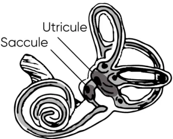
Vestibular signals originate in the labyrinths of the inner ear. The organ is called the vestibular apparatus (Figure 1) and is composed of a system of bony tubes and chambers engraved in the bone of the petrous portion of the temporal bone (Figure 2), symmetrically on both sides. Lying within a bony structure, the membranous labyrinth contains the receptor organs for hearing (the cochlea) and equilibrium (five vestibular sensors) on both sides. The vestibular part includes three semicircular canals oriented orthogonally to each other and detecting angular velocity, and two chambers containing maculae: the utricle and the saccule that are usually referred to as the otolithic system.
Figure 1 – Anatomy of the vestibular apparatus, membranous labyrinth.
Vestibular system
Anatomy of the vestibular apparatus
20
The membranous labyrinth is filled with endolymph, a sodium (Na+) -poor, potassium (K+) -rich fluid, whose composition is maintained by the action of ion pumps in specialized cells. Surrounding the membranous labyrinth, in the space between the membranous labyrinth and the wall of the bony labyrinth, another fluid – perilymph is contained. Perilymph is a high-sodium (Na+) and low-potassium (K+) fluid similar to cerebrospinal fluid, with which it communicates through the cochlear aqueduct. The endolymph and perilymph are kept separate (Herdman, 2007).
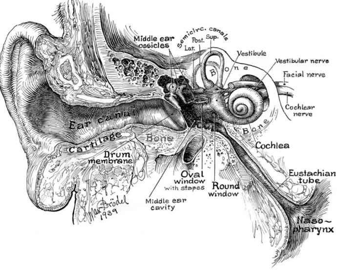
Figure 2 – The inner ear and the location of the vestibular apparatus.
Part I: Background
21
Hair cells and the transduction of a signal
The vestibular sensory neuroepithelium forms either macula for the otolithic system or crista ampullaris for the semicircular ducts. Each of the five receptor organs has a cluster of hair cells responsible for transducing head accelerations into vestibular signals. Vestibular signals are then carried from the hair cells to the brainstem by branches of the vestibulocochlear nerve (cranial nerve VIII).
Each hair cell is embedded in a membrane of neuroepithelium and has a bundle of cilia called the stereocilia and one large cilium called the kinocilium (Figure 3). The stereocilia are grouped in a way that the lengths of the stereocilia increase gradually towards kinocilium.
Figure 3 – The cilia of the hair cell. A high power scanning electron micrograph,
demonstrating a bundle of cilia, gradually increasing in length. Image by the laboratory of David P. Corey, Howard Hughes Medical Institute.
Vestibular system
Hair cells and the transduction of a signal
22
Movements of the head lead to a deflection of the cilia in hair calls. The cilia are tied with each other by a protein-chain connection. When the otoconia weight or the fluid flow in the canals cause the cilia to bend, the cilia pull each other, opening or closing the ion channels leading to the hyperpolarization or depolarization of the cell. The membrane potential of a receptor cell depends on the direction in which the cilia bundle is bent. Deflection of stereocilia towards the kinocilium causes potassium channels in the apical portions of the stereocilia to open. Potassium flows into the cell from the endolymph, causing the cell membrane to depolarize and the rate of firing in the afferent fibers increases. Bending away from the kinocilium causes the cell to hyperpolarize, thus decreasing the afferent firing rate (Figure 4).
Figure 4 – Hair cells receive mechanical stimuli and transduce it into neural signals. Bending of the cilia towards kinocilium excites the cell, increasing the
neural firing, while bending away from cilia decreases firing and inhibits the cell.
There are two structural types of hair cells in the inner ear (Figure 5). Type I cells are more round in shape and are inserted in a nerve calyx of the afferent nerve. These cells have irregular firing and produce variable interspike intervals. Type II cells represent the majority of hair cells of the inner ear. These cells have a normal synaptic connection to their afferent fibers. They create a resting discharge with rather low variability. The vestibular nuclei send efferent signals to the sensory hair cells to modulate their sensitivity (Khan & Chang, 2013).
Part I: Background
23
Vestibular system Otolithic system
24
Otolithic system
Each inner ear has two chambers called the utricle and the saccule (Figure 6). Each of these contains a sensory organ about two millimeters in diameter called macula. The macula of the utricle lies in the horizontal plane on the inferior surface of the utricle. It is oval in shape and connects to the membranous semicircular canals via five openings. It plays an important role in determining the orientation of the head with respect to the direction of linear accelerations including gravity. The macula of the saccule is located on the vertical plane on the medial wall of the saccule. The function of saccular macula is less clear, but it probably participates in the auditory function as well as in the spatial orientation detection, especially when the head is not in a vertical position.
Figure 6 – The maculae. The location of the saccule and utricule.
Each macula is covered by a gelatinous layer composed of acid mucopoly-saccharides. Small calcium carbonate crystals called otoconia inserted on the top of the gelatinous layer (Figure 7). The otoconia range from 0.5 to 30 microns in diameter and are about 3 times heavier than the surrounding tissues. Thousands of sensory hair cells are embedded into the gelatinous layer. The bases and sides of the hair cells synapse with sensory axons of the vestibular nerve and transmit the captured signals to the nervous system. The human utricle contains approximately 30000 hair cells, whereas the saccule contains around 16000 (Goldberg, 2013).
Part I: Background
25
At rest, most of the nerve fibers leading from the hair cells are continuously firing with a frequency averaging at 90impulses per second, for primates. When the head is accelerating or changing its position with respect to gravity, the weight of the otoconia bends the cilia of hair cells. This bending of the cilia towards the kinocilium increases the impulse traffic in its nerve fibers, while bending to the opposite direction inhibits the impulse traffic. Different hair cells are oriented in different directions, so the sensor has a specific signal pattern for every direction of head’s orientation.
Figure 7 – The macular structure illustration. The otoliths are placed on the top
of the gelatinous layer, into which the hair cells are embedded.
The directions of hair cells are separated by a distinctive curved zone running through the center of each macula called the striola (Tascioglu, 2005). The hair cells of a macula on each side of the striola are oriented so that their kinocilia point in opposite directions (Figure 8). In the utricule, the kinocilia face towards the striola and in the saccule they face away from it. As a consequence, displacement of macula’s otolithic membrane in one direction has an opposite physiologic influence on the set of hair cells on each side of the striola. The striola is curved,
Vestibular system Otolithic system
26
which allows hair cells to orient at different angles, detecting multiple directions of displacement.
Figure 8 – Directional preference of the hair cells in maculae of the utricle and saccule. The line separating the maculae in two parts with different directional
preference is called the striola.
Maculae detect the tilt of the head with respect to gravity. In the near-vertical positions, the utricular macula can detect as little as half degree of tilt (Goldberg, 2013). This precision is required to maintain vertical balance and to launch the reflex responses for posture stabilization. As the head leans farther from the vertical, determination of the head orientation becomes poorer (Aubert, 1861). On the other hand, maculae detect inertial linear acceleration. When the head accelerates forward, the otoconia fall backwards due to their inertia and bend the cilia, which send the signal to the brain. As this signal is identical for the inertial acceleration due to motion and a gravitational acceleration due to the head tilt, the
Part I: Background
27
brain uses the information gathered from other sensors, and the characteristics of the signal (e.g. frequency) to discriminate the nature of the signal (Angelaki & Dickman, 2003; Merfeld, Zupan, & Peterka, 1999; Wood, 2002).
Semicircular canals
Each vestibular apparatus contains three membranous tubes with a cross-section of 0.4 milimeters, each forming about two thirds of a circle with a diameter of about 6.5 milimeters, called the semicircular canals. They are arranged strictly orthogonally to each other to represent three planes in space and are referred to as anterior, posterior and horizontal semicircular canals (Figure 9). The horizontal canals are oriented horizontally to the earth when the head is bent approximately 30 degrees forward (Khan & Chang, 2013). In this position, the anterior canals are located in vertical planes, but project forward and 45 degrees outward. The posterior canals are also then in vertical planes and project backward and 45 degrees outward.
Vestibular system Semicircular canals
28
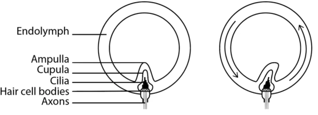
Semicircular canals are filled with endolymph. Each canal has an enlargement at one of its ends called the ampulla. In each ampulla there is a small crest called crista ampullaris with a gelatinous mass similar to the one of the otoliths and called the cupula. Sensory hair cells are embedded into this cupula and connect to the vestibular nerve to transmit the signals of excitation. When the head rotates, the canals with ampullas move, while the endolymph stays still due to inertia. The flow of liquid relatively to the canals bends the ampulla, and excites the sensory organ (Figure 10). As in the otoliths, bending of the cilia to one side, caused by the flow of endolymph inside the canal, stimulates hair cells while bending into the opposite direction inhibits them. However, unlike the hair cells of the otoliths, those of the cupula are all oriented in the same direction.
Figure 10 – Deflection of the ampulla containing the cupula with sensory hair cells by the movement of endolymph.
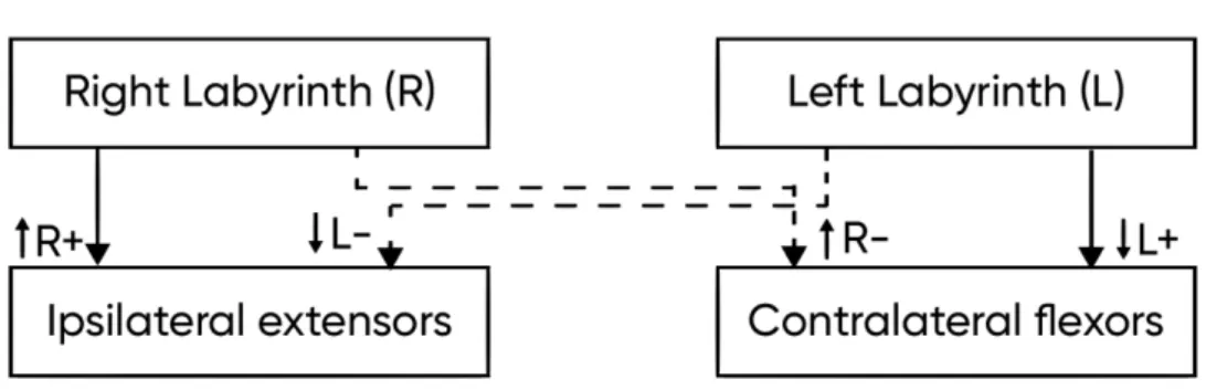
Six semicircular canals from both internal ears make three coplanar pairs: (1) right lateral and left lateral canals, (2) right anterior and left posterior canals and (3) right posterior and left anterior canals. Angular head acceleration causes the endolymph in anterior-posterior coplanar pairs to move in opposite directions, causing one canal to increase the neural firing and the other canal to decrease it. This is often referred to as the push-and-pull organization that helps to provide sensory redundancy. Apart from that, this push-and-pull organization prevents erroneous sensations when the two canals in pair change their firing unidirectionally, which may happen due to internal changes such as in body temperature or chemistry (Herdman, 2007).
Part I: Background
29
When the head is not moving, a spontaneous tonic discharge passes through the vestibular nerves equally on both sides. The balance in firing rates of bilateral discharge indicates the stillness of the head.
When the head rotates with a constant velocity, on the onset of rotation the inert endolymph does not move along with the head, causing the sensory cupula to bend. The inclination angle of the cupula correlates with the sensed angular velocity. After a while (~7 seconds) (Herdman, 2007), the liquid catches up with the movement of the containing structure, releasing the ampulla. At this point, no rotation is mechanically detected anymore. After the head rotation stops, the fluid continues rotating by inertia, while the head is still, causing the cupula to bend in the opposite direction. This might evoke an illusionary sensation or rotation, when no rotation is physically happening, and ocular movements called post-rotatory nystagmus.
Central vestibular system
Vestibular signals require to be processed at different levels, as they drive motor postural and ocular reflexes, add to autonomic control and form the perception (Figure 11). Vestibular neural structure is based on bilaterally afferent and efferent pathways and at least four crossings: three in the brainstem and one in the cortex. The vestibular system can be subdivided into five main components:
● Peripheral receptors placed in the inner ear capturing and transducing head acceleration into neural signals.
● The vestibular nerve that transmits the neural signal.
● Vestibular nuclei in the brainstem that are responsible for receiving, integrating, and distributing information that controls motor activities (such as eye and head movements, postural reflexes, and gravity-dependent autonomic reflexes) and spatial orientation (Jang, Lee, Yeo, & Kwon, 2018; Yakushin, Raphan, & Cohen, 2017).
Vestibular system
Central vestibular system
30
● Cerebellar projections that modulate motor control of the posture, head and eyes, as well as participate in autonomic control (Doba & Reis, 1974; Ito, 1982; Rondi-Reig, Paradis, Lefort, Babayan, & Tobin, 2014; Yates, Holmes, & Jian, 2000).
● The vestibulo-thalamo-cortical network that is responsible for the conscious perception of movement and spatial orientation (Lopez & Blanke, 2011).
Figure 11 – The central vestibular system, its projections and functions.
The main responses initiated by the vestibular system serve to maintain gaze and posture stability and homeostasis during postural changes. They include:
● Vestibulo-ocular reflex (stabilization of the image on the retina of the eye during movement).
Part I: Background
31
● Vestibulo-spinal and vestibulo-collic reflexes (posture stabilization of body and head).
● Vestibulo-sympathetic reflex (autonomic control).
Despite they are mostly called “reflexes” in the literature, those responses often require complex processing, integration and internal models.
a. Vestibular nerve
Neural impulses from otoliths and semicircular canals are carried by the afferent neural fibers that altogether form the vestibular nerve, a branch of the VIII-th cranial nerve (vestibulocochlear nerve). Cell bodies of these fibers form the vestibular ganglion (also referred to as Scarpa’s ganglion). The innervation pattern of vestibular endorgans is specific: the utricle together with horizontal and anterior semicircular canals send their signals through the superior vestibular nerve, while the saccule and the posterior canals are innervated by the inferior vestibular nerve (Kandel, 2013).
The vestibular afferents have two patterns of firing. Regular afferents have little variability of the discharge and a regular tonic rate. Irregular afferents usually do not produce firing at rest, but develop a high discharge variability when stimulated by motion.
Most of the vestibular nerve fibers end in the vestibular nuclei that are located approximately at the junction of the medulla and the pons. However, some fibers pass further to the fastigial nuclei, uvula, and flocculonodular lobes of the cerebellum. Some fibers ending in the vestibular nuclei have synapses with second order neurons projecting into the same areas of the cerebellum as well as to the cortex of other portions of the cerebellum, the vestibulospinal tract, the medial longitudinal fasciculus, and to other areas of the brainstem, particularly the reticular formation.
Vestibular system
Central vestibular system
32
b. Vestibular nuclei
The vestibular nuclear complex consists of four well-described major nuclei and minor nuclei that are less investigated. The whole structure is situated in the pons with extension to the medulla. The mutually inhibitory commissural paths connect the symmetric parts of the vestibular nuclear complex to provide the information exchange between the two sides and implement the push-and-pull pairing of the semicircular canals.
The vestibular nerve projects to the vestibular nuclear complex located on either side of the brainstem and subdivided into four separate anatomical and functional nuclei (Figure 12) (Goldberg et al., 2012):
● The superior (Bechterev’s; SVN) and medial (Schwalbe; MVN) vestibular nuclei receive fibers mainly from the semicircular canals (predominantly horizontal and superior SCC) and project into the medial longitudinal fasciculus (MLF) to trigger the vestibulo-ocular reflex as well as to the medial vestibulospinal tract to cause appropriate movements of the neck. Neurons in the medial vestibular nucleus are mostly excitatory and neurons in the superior vestibular nucleus are mostly inhibitory.
● The lateral vestibular nucleus (the Deiter’s nucleus; LVN) gathers signals primarily from the utricle and possibly saccule and synapses with the neurons going to the spinal cord through the lateral vestibulospinal tract to control postural reflexes.
● The inferior (or descending) vestibular nucleus (IVN or DVN) receives signals predominantly from the otolithic system and from the posterior semicircular canals, and in turn sends signals into both the cerebellum and the reticular formation of the brainstem, as well as to the contralateral nuclei and the spinal cord. This nucleus is involved in integration of vestibular inputs with central motor information.
These four subdivisions are innervated directly by the vestibular nerve and are present in most species.
There are also other minor nuclei distributed in the brainstem and lying close to the four major nuclei that receive primary vestibular inputs and can participate in the processing of the vestibular information. These include parasolitary nucleus
Part I: Background
33
(Psol), y-group, nucleus intercalatus (Barmack, 2003). The y-group consists of dorsal and ventral subdivisions. The dorsal part receives ipsilateral vestibular primary afferents and bilateral secondary vestibular projections. The ventral division projects to the ipsilateral flocculus, nodulus and contralateral oculomotor complex.
Nucleus prepositus hypoglossi (NPH) also receives scattered primary vestibular afferents as well as secondary vestibular projections and projections from the cerebellum (uvula-nodulus and the flocculus). NPH send projections bilaterally to the vestibular nuclei, flocculus and reticular formation. In addition, the neurons in NPH project to the ipsilateral oculomotor nucleus and abducens nucleus. NPH is thought to encode the vestibular and eye position information. The other small nuclei that can be related to vestibular function are the nucleus x and nucleus z.
Figure 12 – The illustration of the bilateral vestibular nuclear complex and their primary vestibular projections from the three semicircular canals, utricle and saccule. SVN, LVN, MVN, DVN – superior, lateral, medial and descending
vestibular nuclei.
While the primary vestibular afferents transduce the information from single vestibular organs, already at the level of secondary vestibular neurons, the
Vestibular system
Central vestibular system
34
convergence of signals appears. It can be the convergent signal from several semicircular canals, or this of canals and otolith organs together. However, some secondary projections relay inputs exclusively from the otolith organs, possibly to help the discrimination between the signals in an ambiguous frequency range (Barmack, 2003).
Figure 13 – Vestibular nuclei, their commissural projections and functions.
Patterned zones localize the majority of neurons responsible for eye movements (small dots), balance and posture (horizontal stripes), cognition (vertical stripes) and autonomic control (big dots). Dashed arrows show commissural homologous connections of the vestibular nuclei. Plain arrows demonstrate non-homologous connections to contralateral nuclei.
Human vestibular nuclei pathway tracing study showed 100% connectivity of vestibular nuclei with cerebellum, thalamus, oculomotor nucleus, trochlear nucleus, abducens nucleus, and reticular formation related to the functions of the VN (equilibrium, control of eye movements, conscious perception of movement, and spatial orientation). High connectivity (over 70%) was seen with the sensory-motor cortex (primary sensory-motor cortex, primary somatosensory cortex, presensory-motor
Part I: Background
35
cortex, and posterior parietal cortex), hypothalamus, and lateral prefrontal cortex (Jang et al., 2018).
Ascending pathways from the vestibular nuclei include principally the projections to thalamus and cortex and cerebellum, in particular ventrobasal thalamus, parietal visual cortex, cerebellar vermis and flocculus.
Figure 14 – Projections of the vestibular nuclei for the motor reflexes. SVN, LVN,
MVN, DVN – superior, lateral, medial and descending vestibular nuclei. MLF – Medial longitudinal fasciculus.
Vestibular system
Central vestibular system
36
Descending pathways to the spinal cord form two principal vestibulospinal tracts: medial and lateral (Figure 14). Axons of the lateral vestibulospinal tract terminate in the ipsilateral lumbosacral region where they connect to motoneurons. The medial vestibulospinal tract terminates bilaterally in the medial part of the cervical ventral horn and drives the vestibulo-collic reflex.
Vestibular nuclei also project to the autonomic control centers. DVN, MVN and Psol project to the Solitary Nucleus (NTS) that receives also autonomic afferents. Fastigial nucleus also receives projections from the vestibular nuclei. It responds to changes in blood pressure, heart rate and respiration, moreover, electrical stimulation of this nucleus can alter these autonomic functions (Barmack, 2003).
c. Multisensory integration within VN
Primarily, neurons in vestibular nuclei integrate signals from otolithic sensors and semicircular canals to model the position and movement in space and distinguish between tilt and linear translation, which are physically indistinguishable (Angelaki & Dickman, 2003; Merfeld, Young, Oman, & Shelhamer, 1993; Merfeld, Zupan, & Gifford, 2001). Also, the integration is happening to differentiate active, voluntary movements from passive ones (Cullen & Roy, 2004).
Vestibular nuclei neurons respond to vestibular, optokinetic (Waespe & Henn, 1978), tactile, proprioceptive (Cullen & Roy, 2004; Tomlinson & Robinson, 1984) inputs.
All the sensory inputs are merged in the vestibular nuclei and the cerebellum, with weighting of every input according to their reliability, leading to the best possible estimate of motion which was demonstrated in monkeys and humans (Fetsch, Turner, DeAngelis, & Angelaki, 2009).
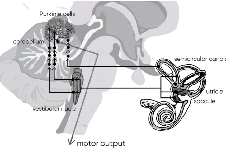
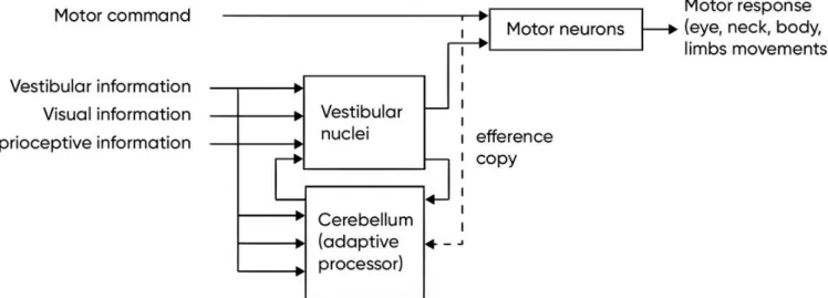
d. Vestibular cerebellum
Vestibular cerebellum acts as an adaptive processor for the vestibular performance (Figure 15). The removal of cerebellar input does not abolish the vestibular reflexes, but makes them imprecise and ineffective (Herdman, 2007).
Part I: Background
37
There are five main regions of the cerebellum that receive either primary (i.e. directly from afferents) or secondary (i.e. from vestibular nuclei) vestibular input, including:
● the nodulus and ventral uvula,
● the flocculus and ventral paraflocculus, ● the oculomotor vermis of posterior lobe, ● lobules I-V of the anterior lobe, and
● the deep cerebellar nuclei (e.g. fastigial nuclei).
The ipsilateral cerebellum has direct efferent connections to ipsilateral vestibular nuclei and to the contralateral vestibular nuclei indirectly through the fastigial nucleus. The latter projects to the contralateral vestibular nuclei via the juxarestiform body (Khan & Chang, 2013). These areas participate in the adjustment of posture- and orientation-related reflexes. In particular, approximately half of the neurons of the rostral fastigial nuclei (RFN) encode the motion of the body in space responding to both vestibular and proprioceptive inputs, while the other half encode the motion of the head in space in a manner similar to neurons in the vestibular nuclei (Brooks & Cullen, 2009). The caudal part of the fastigial nuclei participates in the adjustment and modulation of the saccadic eye movements (Kleine, Guan, & Büttner, 2003). The cerebellar flocculus takes part in the vestibulo-ocular reflex (VOR) and smooth pursuit function by continuously modulating the eye velocity (Ito, 1982). Modulation here is continuous correction of the gain errors and adapting the VOR to different conditions (Miles & Eighmy, 1980). It is essential, for example, for people wearing correction eyeglasses. Lenses change the focal point and so the gain of the VOR must adapt to this change. While the flocculus adapts the VOR gain, cerebellar nodulus adjusts its duration, playing role in its habituation and suppressing (Angelaki & Hess, 1994; Waespe, Cohen, & Raphan, 1985). The lesions of nodulus also affect the processing of otolithic input, causing erroneous eye nystagmus when tilting the head (Herdman, 2007). The nodulus is required for motion sickness (Bard & Woolsey, 1947). Anterior-superior vermis of the cerebellum is involved in postural regulation through the vestibulo-spinal reflex.
Vestibular system
Central vestibular system
38
Figure 15 – Primary and secondary vestibular projections to cerebellum to modulate the motor output.
e. Multisensory integration within cerebellum
The cerebellum is a center for monitoring and modulating motor function. Cerebellum receives multimodal sensory information, which it integrates together with the efference copy of the motor command. Apart from vestibular projections that originate in part directly from the vestibular organs (primary projections to the uvula-nodulus), but mostly from vestibular nuclei (secondary projections to multiple cerebellar lobules), cerebellum receives multiple other sensory projections. Primarily two areas of posterior cerebellar cortex, lobule VII and the dorsal paraflocculus receive visual projections (Kralj-Hans, Baizer, Swales, & Glickstein, 2007; Rondi-Reig et al., 2014). Cerebellum receives also proprioceptive projections (Brooks & Cullen, 2009) from limbs, neck and shoulders, which are
Part I: Background
39
integrated with visuo-vestibular signals for navigation (Brooks & Cullen, 2009; Gdowski & McCrea, 2000). There is evidence that cerebellum participates in processing of tactile information from whiskers in animals (Rondi-Reig et al., 2014). The multiple sensory inputs are converged by cerebellum in the superior colliculus (Rondi-Reig et al., 2014).
f. Vestibular thalamus and cortex
Vestibular signals play a role in many cognitive functions such as navigation in space (Berthoz, Israel, Georges-Francois, Grasso, & Tsuzuku, 1995; Mittelstaedt, 1999), distinguishing self-motion and object motion (McIntyre, Zago, Berthoz, & Lacquaniti, 2001; Straube, Brandt, & Probst, 1987; Werkhoven, Snippe, & Toet, 1992), perceiving the vertical (Mittelstaedt, 1983), bodily self-consciousness (Lopez, Halje, & Blanke, 2008), and even circadian rhythmes (Fuller & Fuller, 2006; Martin et al., 2016) and emotions (Lopez, 2016).
Unlike visual or auditory systems, that have a specific cortical region for processing the sensory information, the vestibular system does not seem to have an assigned cortical zone. Vestibular information is redistributed to multiple cortical areas through the thalamus. Cortical zones receiving the projections from thalamus differ significantly among species that makes it impossible to definitively match primate cortical zones with human ones. Moreover, the methods currently available to study the vestibulothalamocortical connection in human do not permit the profound investigation of this widespread network, including often a small number of neurons per connection.
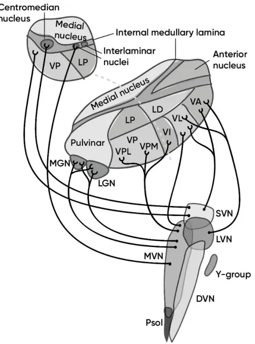
g. Thalamus
As for every other sensory signal processing, the thalamus relays vestibular signals to the cortex (Figure 16). Nevertheless, the vestibular traffic organization differs fundamentally from other sensory systems. Most sensors send projections to a modality-specific thalamic nucleus and after to a respective cortical or subcortical area, creating a sensory map. The vestibular sensory information is converged in the vestibular nuclei, which makes the signals more complex and indirect. The vestibular nuclei send signals to the thalamus, however there is no specifically
Vestibular system
Central vestibular system
40
assigned nucleus relaying vestibular signals. Various thalamic nuclei receive second order neurons projected from the vestibular nuclei to process and redistribute the vestibular signals to various cortical zones. Most of the thalamic nuclei receiving vestibular projections are not specific to the vestibular system, receiving multiple inputs from other sensory systems and cortical zones. This fact accentuates the multisensory and integrative character of the vestibular information.
In animals, multiple thalamic zones have been reported to receive vestibular projections. Ventral posterior lateral nucleus (VPL) and the nucleus ventralis intermedius (Vim) in the ventroposterior complex in rat receive projections primarily from the medial and superior vestibular nuclei (MVN, SVN) (Nagata, 1986) and from descending vestibular nuclei (DVN) (Shiroyama, Kayahara, Yasui, Nomura, & Nakano, 1999). The VPL-Vim complex is considered a major relay of proprioceptive and cutaneous inputs to primary and secondary somatosensory cortex in primates including human (Behrens et al., 2003; Lopez & Blanke, 2011). This thalamic nuclear complex also projects to other multiple cortical zones, such as anterior suprasylvian cortex and posterior cruciate region, superior parietal lobule and intaparietal sulcus (Lopez & Blanke, 2011). The VPL receives efferents from the somatosensory cortex. The ventral posterior thalamus is thought to be the origin of the main thalamocortical pathway to the parieto-insular vestibular cortex (PIVC), which is known to be the principal vestibular cortical zone in non-human primates (Akbarian, Grüsser, & Guldin, 1992). The ventral posterior inferior nucleus (VPI) neurons respond to rotatory vestibular stimulation and are connected with the primary somatosensory cortex (Akbarian et al., 1992). Ventral posterior medial (VPM) nucleus neurons receive inputs primarily from the MVN and SVN and respond to both rotational and translational stimulus, as well as electrical stimulation of the vestibular nerve (Deecke, Schwarz, & Fredrickson, 1977; Matsuo, Hosogai, & Nakao, 1994; Meng et al., 2001). The ventroanterior thalamic nucleus (VA) and ventrolateral thalamic nucleus (VL) receive inputs from the MVN and SVN via the medial longitudinal fasciculus and, in Rhesus monkey, also from the cerebellar nuclei. The VA-VL thalamic nuclear complex relays the major vestibulomotor pathway, connecting vestibular nuclei with the primary motor and premotor cortex. The intralaminar nuclear complex (IL) serve as a major relay of the vestibulo-thalamo-strial pathway, likely involved in the control of body and limb movements. The medial geniculate nucleus (MGN) neurons
Part I: Background
41
receive projections from MVN, SVN and DVN and respond to electrical stimulation of the vestibular nerve. This nucleus is involved in auditory, vestibular and somatosensory integration. The lateral geniculate nucleus (LGN) is known to be the main visual pathway from the retina to the primary visual cortex, however it contains vestibular projections that respond to the horizontal or vertical angular stimulations (Magnin & Putkonen, 1978). Neurons in the pulvinar also respond to vestibular activation.
In human, the methods for tracing vestibular higher pathways are limited. Neuroimaging and clinical neurological data help to describe vestibular processing in the human thalamus. While the thalamic connections for other sensory systems have been successfully traced with neuroimaging, description of the vestibular connections meets strong limitations. Small sizes of the thalamic nuclei, their deep location and moreover, the fact that the vestibular information is redistributed by small patches of neurons to multiple nuclei over thalamus, make the signal harder to detect (Lopez & Blanke, 2011).
Neuroimaging during galvanic vestibular stimulation demonstrated the activation of the paramedian and dorsolateral thalamus (Bense, Stephan, Yousry, Brandt, & Dieterich, 2001). Caloric stimulation activated the posterolateral and posteromedial thalamus (Dieterich, 2003). Nucleus medialis, nucleus habenularis and pulvinar have been reported to participate in vestibular processing in human (Bucher et al., 1998). Neurological data support the participation of posterolateral thalamus in relaying vestibular signals to the cortex (Ceballos-Baumann et al., 2001; Dieterich et al., 2005). The MGN is also suggested to be involved in vestibulothalamic connection in human (Hawrylyshyn, Rubin, Tasker, Organ, & Fredrickson, 1978).
Vestibular system
Central vestibular system
42
Figure 16 – Projections of the vestibular nuclei to the thalamus. Thalamic nuclei
abbreviations: LP – lateral posterior, LD – lateral dorsal, VP – ventral posterior, VPL – ventral posteromedial, VPM – ventral posteromedial, VI – ventral intermediate, VL – ventral lateral, VA – ventral anterior nuclei. SVN, LVN, MVN, DVN – superior, lateral, medial and descending vestibular nuclei.
Part I: Background
43
h. Multisensory integration within thalamus
The neurons in the vestibular thalamus have responses that are very similar to those of vestibular nuclei, they respond to vestibular, proprioceptive, visual and tactile stimuli (Lopez & Blanke, 2011). As has already been described, all thalamic nuclei that use vestibular information, also receive inputs from other sensory systems. Basing on the clinical studies where thalamic lesions or strokes led to the perturbances of visual vertical and postural vertical (Johannsen, Broetz, Naegele, & Karnath, 2006; Karnath, Ferber, & Dichgans, 2000; Masdeu et al., 2005), it has been suggested that the thalamus acts not only as a signal relay, but that thalamic nuclei participate in the complex processing of vestibular information (Wijesinghe, Protti, & Camp, 2015). It was demonstrated that thalamic neurons respond to a combination of visual and vestibular information (Magnin & Putkonen, 1978; Meng, May, Dickman, & Angelaki, 2007). Vestibular neurons in ventral posterior medial, lateral and inferior thalamic nuclei respond to proprioceprive inputs from joints, muscles and skin from limbs, trunk, neck and head (Blum & Gilman, 1979; Deecke et al., 1977). Vestibular nuclei in thalamus were also shown to be responsive to touch in paws in cats (Sans, Raymond, & Marty, 1970). It is possible that thalamus acts as one of the centers of multisensory integration for signals from the labyrinth and other sensory signals (Tyll, Budinger, & Noesselt, 2011).
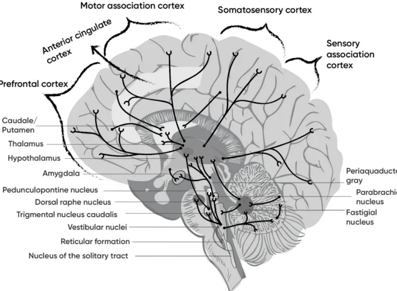
i. Vestibular cortex
Walzl and Mountcastle realized the first description of vestibular projections to the cortex in cats in 1949. Later, a number of studies have been devoted to describe “vestibular cortex” in various species including human (Figure 17).
In animals, the parietal cortex, the area 2v of the primary somatosensory cortex was first shown to receive vestibular inputs (Fredrickson, Figge, Scheid, & Kornhuber, 1966). In cat and monkey the 2v area responded to both otolith and semicircular canals inputs (Büttner & Buettner, 1978; Jijiwa, Kawaguchi, Watanabe, & Miyata, 1991). Also the area 3aHv situated in the somatosensory cortex area representing the hand and arm in squirrel monkey and the forelimb in cat and guinea pig (Odkvist, Liedgren, Larsby, & Jerlvall, 1975; Odkvist, Rubin, Schwarz, & Fredrickson, 1973; Odkvist, Schwarz, Fredrickson, & Hassler, 1974), and the 3aNv zone in the area encoding the neck and trunk representations and
Vestibular system
Central vestibular system
44
stretching till the primary motor cortex (Akbarian, Grüsser, & Guldin, 1994; Guldin & Grüsser, 1998) are responsive to vestibular inputs. The ventral intaparietal area (VIP) and medial intraparietal area (MIP) of the parietal sulcus have also been revealed to receive vestibular neurons (Bremmer, Klam, Duhamel, Ben Hamed, & Graf, 2002; Schlack, 2005; Schlack, Hoffmann, & Bremmer, 2002). The VIP area participates in three-dimensional spatial coding and in visuo-vestibular integration and is reciprocally connected with motor cortex (Schlack, 2005; Schlack et al., 2002). Area 7 has shown vestibular activation in monkeys (Faugier-Grimaud & Ventre, 1989; Ventre & Faugier-(Faugier-Grimaud, 1986), however some researchers tend not to consider the area 7 as a part of the vestibular cortex due to a low percentage of neurons responsive to vestibular inputs (Guldin & Grüsser, 1998). Grüsser and Guldin first described the area called “parieto-insular vestibular cortex” (PIVC) that is considered now the principal vestibular cortical area in non-human primates (Grüsser, Pause, & Schreiter, 1990a, 1990b; Guldin & Grüsser, 1998). However, the PIVC location is variable between species (for example, it is located on the parietal lateral sulcus in macaque and on the temporal lateral sulcus in squirrel monkey). Visual posterior sylvian area (VPS) posteror to PIVC was also found to be responsive to vestibular signals in about 30% of its neurons together with visual information (Guldin & Grüsser, 1998). Some PIVC neurons converge vestibular, somatosensory and visual signals and also respond to muscle pressure, vibrations and rotation of the neck (Grüsser et al., 1990b). In the experiment of Grüsser and collegues (Grüsser et al., 1990b), trunk rotation with the stable head activated the PIVC neurons, which were sensitive to the direction of rotation as well as velocity and acceleration. The vestibular and neck proprioceptive signals interact in the PIVC possibly to differentiate between passive and active movement. Also the PIVC neurons in macaque are reported to respond to both rotational and translational vestibular stimulation (Chen, DeAngelis, & Angelaki, 2010). Areas in primary motor cortex (area 4) and premotor cortex (area 6) that are associated with motor and oculo-motor control also receive vestibular inputs (Ebata, Sugiuchi, Izawa, Shinomiya, & Shinoda, 2004; Fukushima, Akao, Kurkin, Kaneko, & Fukushima, 2006; Fukushima et al., 2010). There is evidence that these areas participate in generation of eye saccades and smooth pursuit eye movements (Fukushima et al., 2006). Cingulate sulcus also has neurons that participate in vestibular processing in monkey, even though they do not receive direct vestibular input (Guldin & Grüsser, 1998). This region
Part I: Background
45
probably participates in integration of visual and self-motion cues for spatial coding and navigation (Cooper, Manka, & Mizumori, 2001). Vestibular projections have been found in primary and secondary visual cortex in cats (areas 17, 18, 21 and 19) (Grüsser & Grüsser-Cornehls, 1972; Vanni-Mercier & Magnin, 1982). Vestibular inputs are found also in medial superior temporal area (MST) in monkey, which represents a major area for visual motion perception, including self-motion derived from visual cues (Bremmer et al., 2002; Bremmer, Kubischik, Pekel, Lappe, & Hoffmann, 1999; Page & Duffy, 2003). Finally, the hippocampus is modulated by vestibular stimulation (Horii, Russell, Smith, Darlington, & Bilkey, 2004). Even though direct vestibular projections have not been found, the firing of “place cells” encoding the location in space and “head direction” cells encoding the face direction, was found to be adjusted by vestibular inputs.
In humans, functional neuroimaging studies revealed the anterior part of the intraparietal sulcus region to respond to vestibular stimulation (Fasold et al., 2002; Lobel, Kleine, Bihan, Leroy-Willig, & Berthoz, 1998), which might be a human analogue of cat’s and monkey’s area 2v (Lopez & Blanke, 2011). Other neuroimaging studies have described the primary somatosensory cortex to be activated following vestibular stimulation, this area could be homologue to the areas 3aHv and 3aNv in monkeys (Emri et al., 2003; Fasold et al., 2002). A human homologue to area 7 in monkey can be the inferior parietal lobule or lateral superior parietal lobule which is activated by head movements and change of gaze direction (Fasold et al., 2002; Lobel et al., 1998; Vitte et al., 1996). Intraparietal sulcus has also demonstrated the vestibular activation both by otolithic and canalar inputs (Miyamoto, Fukushima, Takada, de Waele, & Vidal, 2007; Suzuki et al., 2001). Human cortical zone analogous to PIVC could be in the posterior insula and temporo-parietal junction (Emri et al., 2003; Fasold et al., 2002; Lobel et al., 1998; Miyamoto et al., 2007; Vitte et al., 1996). However, the exact location of the PIVC-homologue area is not yet clear as the difference in its location varies between studies. Anterior insula being an important center of bodily awareness is also involved in the vestibular processing (Bense et al., 2001; Dieterich et al., 2005; Fasold et al., 2002; Trousselard, Barraud, Nougier, Raphel, & Cian, 2004). Several frontal zones are also involved in vestibular processing in human, such as regions in primary motor cortex (precentral gyrus), the premotor cortex, superior, middle and inferior frontal gyri (Bense et al., 2001; Emri et al., 2003; Fasold et al., 2002; Lobel et al., 1998). The frontal gyri neurons are involved in the control of saccadic
Vestibular system
Central vestibular system
46
eye movements, smooth pursuit and nystagmus (Blanke et al., 2000). Anterior and posterior cingulate cortex in human might be homologue to the monkey vestibular cingulate region (Bense et al., 2001; Fasold et al., 2002; Miyamoto et al., 2007; Suzuki et al., 2001). The analogue to the optic-related MST in monkey could be the middle temporal gyrus (area 37) that are activated during galvanic vestibular stimulation (Bense et al., 2001). It was also found that several visual cortex zones are deactivated during vestibular stimulation, while visual fixation was found to be related to a deactivation of the vestibular cortex (Bense et al., 2001; Lobel et al., 1998), which might suggest the existence of reciprocal inhibitory visuo-vestibular interactions (Brandt, 1998). In addition, vestibulo-hippocampal interaction on human is suggested.
j. Multisensory integration in cortex
As it has been described above, there is no vestibular-specific cortical area. Instead, the vestibular signals are redistributed to multiple cortical zones, which are highly multimodal and integrative. The PIVC area in monkeys is connected to all the other vestibular-sensitive regions and thus is considered to be the primary vestibular region, the vestibulo-visuo-somatosensory convergence is happening in this area (Grüsser et al., 1990a, 1990b; Guldin & Grüsser, 1998). Visual-vestibular convergence was also demonstrated in the MST area (Bremmer et al., 1999), and vestibulo-somatosensory convergence was found in the intraparietal sulcus and areas 2v and 3aHv and 3aNv in monkeys and the secondary somatosensory cortex in humans (Bremmer et al., 2002; Fasold et al., 2002; Guldin & Grüsser, 1998).