HAL Id: inserm-01070064
https://www.hal.inserm.fr/inserm-01070064
Submitted on 30 Sep 2014HAL is a multi-disciplinary open access
archive for the deposit and dissemination of sci-entific research documents, whether they are pub-lished or not. The documents may come from teaching and research institutions in France or abroad, or from public or private research centers.
L’archive ouverte pluridisciplinaire HAL, est destinée au dépôt et à la diffusion de documents scientifiques de niveau recherche, publiés ou non, émanant des établissements d’enseignement et de recherche français ou étrangers, des laboratoires publics ou privés.
Biomolecular Recognition: The Current Challenge
Pierre Bongrand
To cite this version:
Pierre Bongrand. Biomolecular Recognition: The Current Challenge. A.R. Bizzari et R Cannistraro. Dynamic force spectroscopy and biomolecular recognition, CRC press - boca Raton, pp.1-50, 2012. �inserm-01070064�
Biomolecular Recognition: The Current Challenge
Pierre Bongrand
Lab. Adhesion and Inflammation, INSERM U600, CNRS U6212
Parc Scientifique de Luminy
Case 937, 13288 Marseille Cedex 09, FRANCE
[email protected], http://lai.sciences.univmed.fr
May 29, 2014
This author-edited text appeared in the book entitled : Dynamic Force Spectroscopy and biomolecular recognition, edited by A.R. Bizzari and R Cannistraro, CRC Press, Boca-Raton, 2012, pp 1-50.
Contents
1 Introduction 2
2 What is the use of biomolecular interactions 4
2.1 Cell structure: statics and dynamics. . . 4
2.2 Cell differentiation . . . 4
2.3 Cell adhesion . . . 5
2.4 Immune recognition . . . 7
2.5 Signal generation . . . 9
3 Brief historical outline of recent investigations made on biomolecule recognition at the single bond level 10 3.1 Studying bond rupture at the single molecule level . . . 10
3.2 Measuring bond formation at the single molecule level . . . 14
4 Which parameters do we need to account for biomolecule recogni-tion ? 15 4.1 The affinity constant . . . 16
4.2 Kinetic constants : the on-rate and the off-rate . . . 17
4.2.1 The force-dependent dissociation rates . . . 17
4.2.2 Distance-dependent association rates . . . 19
4.3 Avidity of biomolecule interactions: an incompletely defined parameter 21 4.4 Specificity of biomolecule interactions: an essential property that is dif-ficult to define accurately . . . 22
4.5 Ligand-receptor interactions are influenced by parameters that are
ex-trinsic to both ligand and receptor molecules . . . 23
5 Relationship between biomolecule structure and recognition events 24 5.1 Intermolecular forces in the biological milieu . . . 25
5.1.1 Electrostatic forces . . . 25
5.1.2 The hydrogen bond . . . 26
5.1.3 Different timescale: electrodynamic interactions . . . 27
5.1.4 Using the formalism of surface physical-chemistry. Hydrophobic bonds. . . 28
5.2 General properties of ligand-receptor association . . . 30
5.2.1 A static view of ligand-receptor complexes . . . 30
5.2.2 Dynamics of ligand-receptor interaction . . . 31
5.2.3 Mechanisms influencing the specificity of biomolecule interaction 33 5.3 Information yielded by computer simulation . . . 35
5.3.1 Limitations and technical advances. . . 37
5.3.2 New information on molecular association may be provided by computer simulations . . . 38
6 Conclusion 39
7 References 39
Abstract
Biomolecules essentially fulfill their function through continual recognition of and binding to other molecules. Biomolecular recognition is therefore a phe-nomenon of prominent importance. When the progress of monoclonal antibody technology and genetic engineering allowed biologists to characterize and iso-late an impressive variety of receptor molecules, it was first felt that affinity constants and kinetic rates provided a satisfactory account of receptor-ligand interactions. However, a number of advances that occurred during the last two decades showed that i) the conventional framework was not sufficient to predict the behaviour of biomolecules in many physiologically relevant situations, ii) a number of techniques allowed investigators to dissect biomolecule interactions at the single bond level and obtain new information on the kinetic and mechanical properties of these interactions, iii) new theoretical techniques and the devel-opment of computer simulation as well as the enormous increase of available structural data provided new avenues to relate structural and functional proper-ties. The aim of this introductory chapter is to present a brief outline of these advances and pending issues.
1
Introduction
Life relies on myriads of interactions between the molecular components of living sys-tems. Proteins are a remarkable example in view of their diversity (the very name of proteins stems from Proteus, a greek god known for his capacity to change shape).
Several decades ago, the author of a well-known treatise on proteins (Creighton 1983) wrote that ... the biological function of proteins almost invariably depends on their direct physical interaction with other molecules. More recently, systematic use of pow-erful techniques such as yeast double hybrid or mass spectrometry was a basis for a large scale attempt to build exhaustive databases of protein interactions, the so-called interactome (Blow 2009). Over 250,000 interactions between about 22,000 proteins were recorded in the Unified Interactome Database on year 2008 (Chaurasia 2008).
Until recently, it seemed that the conventional concepts and methods used to study chemical equilibria provided a suitable framework to deal with biomolecular recogni-tion. As reckoned two decades ago (Williams 1991), the concepts of specificity and affin-ity had seemed sufficient to deal with biological phenomena for many years, and only conventional kinetic constants had to be added to explain some recent findings. How-ever, a number of reports supported the importance of forces in biological interactions (Capo 1978, Jaalouk 2009) and theoretical models of cell functions such as adhesion have included mechanical parameters (Bell 1984, Mege 1987). This was an incentive to devise experimental methods allowing us to study the response of biomolecules to forces with high temporal and spatial resolution, up to the single molecule level. Si-multaneously, continuous progress in molecular dynamics allowed computer scientists to report on simulations of the response of biomolecules to external forces (Grubmuller 1996, Izrailev 1997, PuklinFaucher 2006), thus allowing deeper interpretation of exper-imental results (Florin 1994, Rief 1997). These advances were also facilitated by the tremendous increase of structural data on biomolecules, based on X ray cristallography and nuclear magnetic resonance, and the use of genetic engineering techniques to re-late structural and functional data, as exemplified by alanine-scanning that consists of systematically replacing aminoacids with alanin in protein-protein interaction areas to obtain a direct estimate of their contribution to binding energy (Cunningham 1989). The development of dynamic force spectroscopy is a remarkable example of an innovative approach stemming for a number of different advances and yielding a new kind of information that might shed a new light on important and unresolved issues.
The goal of this chapter is to present as palatably as possible a number of biological processes and recent methodological advances that played an important role in the development of dynamic force spectroscopy and may benefit from this growing domain. The first section includes selected examples of biological situations that are heavily dependent on biomolecular recognition. This will be the basis for defining the questions we need to ask. The next section is a brief outline of recent progress done in the study of molecular interactions, particularly at the single bond level, which shaped the present state-of-the art. The next section is intended to define and analyze the parameters required to provide an adequate account of biomolecule interactions, i.e. to include the pieces of information that are needed to predict the behaviour of a given ligand-receptor couple under physiological conditions. The last section gives a brief description of the application of conventional physical-chemical knowledge and newer computer simulation methods to the study of links between biomolecule structure and association properties. Admittedly, the field of biomolecule interactions is too vast to be exhaustively discussed in the limited space available. Also, it is unavoidable that the topics selected in this chapter should reflect the limitations of the author’s fields of
competence and interest. Therefore, I apologize for the omission of many key references that would certainly have enriched this presentation.
2
What is the use of biomolecular interactions
The goal of this section is to describe several important biological processes in order to illustrate the role of biomolecule interactions and the constraints that must be met.
2.1
Cell structure: statics and dynamics.
Clearly, any living cell or organism would fall into pieces in absence of the molecular interactions linking their components. It is important to emphasize that both quali-tative and quantiquali-tative properties of these interactions are essential. Thus, it is well recognized that cell formation requires an autoorganization capacity of biomolecules that must be able to bind to each other with sufficient specificity to avoid durable presence of potentially harmful molecular interactions (Vavouri 2009). In addition, the rheological properties of cells are considered to be driven by the properties of underlying cytokeletal elements, which are themselves dependent on the kinetic and mechanical properties of intermolecular associations (Wachsstock 1994). These points are important in view of the recently recognized importance of cell mechanics in situ-ations of medical interest such as cancer cell metastasis (Glinsky 2003, Remmerbach 2009) or letal inflammatory processes such as the acute respiratory disease syndrome (Nishino 2005). Cell shape is considered to be highly dependent on the dynamic or-ganization of a network of rodlike structures including actin microfilaments, tubulin microtubules and intermediate filaments. These are highly plastic structures whose growth or retraction is determined by a variety of interaction events, and particularly polymerization/depolymerization as a consequence of tunable kinetics of monomer association or dissociation. Other important events are movements driven by so-called motor molecules such as myosin or kinesin that are able to generate force-dependent displacements. Much effort was recently done to investigate the mechanisms of associ-ation/ dissociation and force generation by these molecules.
2.2
Cell differentiation
A remarkable feature of living cells composing complex organisms is their capacity to acquire different structural and functional capacity whereas they share a common set of genes. While the mechanisms of differentiation are not yet fully understood, a primary process is the selective synthesis of particular proteins as a consequence of gene activation by a combination of over 100 DNA binding proteins with a specificity for a number of regulatory sites on the DNA. Such a complex set of interactions remains incompletely known, but an extensive network of DNA/protein interactions clearly plays an important role in differentiation (Badis 2009).
2.3
Cell adhesion
As previously reviewed (Pierres 2000), cell adhesion is a fundamental process that influences nearly all steps of cell function. Thus, cell survival and proliferation are often dependent on a strong attachment to solid surfaces, a phenomenon known as anchorage dependence (Folkman 1978, Chen 1997). An attractive interpretation of experimental findings was that cell adhesion might be required to induce marked cell flattening and spreading on the surface, and that cell behaviour might be shape-sensitive (Pierres 2002, Neves 2008).
Cell migration on a surface is also highly dependent on the qualitative and quanti-tative properties of cell-surface interactions. It has long been shown that efficient cell migration required that binding strength, i.e. the mechanical force required to de-tach adherent cells, fell within a particular range (Palecek 1997). Too strongly adherent cells are expected to remain stuck on a fixed place (Jay 1995). In contrast, a minimal adhesion efficiency is probably required in order that a lamellipodium sent forward by a motile cell be able to remain stuck on the surface and drag forward the cell body with concomitant detachment of the rear part of the cell (Palecek 1998). More recently, it was reported that moving cells were able to probe the rigidity of underlying surfaces and move towards more rigid regions, a phenomenon called durotaxis (Lo 2000).
Cell differentiation is also strongly influenced by the properties of underlying sur-faces. While this well-known phenomenon has long been interpreted by hypothesizing that cells were essentially sensitive to the biochemical structures of ligands exposed by surrounding surfaces and recognized by their receptors (Kaplan 1982), more recent experiments showed that cell responses were also dependent on the stiffness of these surfaces (Engler 2006). The mechanisms allowing cells to measure surface stiffness re-main poorly understood, but it is likely that this involves the response to forces of surface biomolecules adhering to nearby ligands.
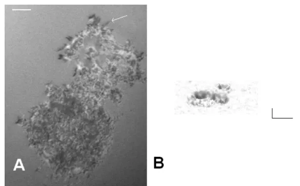
Indeed, cells continually probe their environment to adapt their shape, motion, and other functions such as proliferation or mediator release. Environment sensing may re-sult from the uptake of soluble ligands by membranes. However, a more accurate and less noise-sensitive way of probing cellular environment may result from mechanical exploration through continual formation and retraction of protrusions such as lamel-lipodia (Dobereiner 2006) and finger-like filopodia (Faix 2006) or through transverse membrane undulations (Zidovska 2006, Pierres 2008, thus inducing transient contacts between membrane receptors and fixed ligands, which may provide a powerful way of rapidly gathering information (Pierres 2009). The outcome of interactions is heavily dependent on the kinetics of bond formation between surface-attached ligands and receptors, as well as the strengh of attachements. These phenomena are highly de-pendent on the kinetics and mechanics of receptor-ligand interactions. Arguably, cells use dynamic force spectroscopy to probe their environment (see Figure 1).
Inflammation is an ubiquitous process used by multicellular organisms to cope with various forms of aggression, and particularly infection. A key step is the adhesion of flowing blood leukocytes to the vessel walls, with subsequent transmigration through these walls and entry into tissues containing infectious agents or damaged cells. Unrav-eling the mechanisms of leukocyte interaction with endothelial cells coating the vessel
Figure 1: Cells probe their environment. A. A monocytic THP-1 cell was deposited on a surface coated with fibronectin and examined with interference-reflection contrast microscopy (IRM). Short filopodia (white arrow) appear as black lines. B. The image shows the underside of a lymphocyte falling on an activating surface, and tridimensional shape was derived from IRM images. A dynamic study revealed undulations of a few nm amplitude and Hz frequency. Horizontal bar length is 5 µm and vertical bar length is 100 nm. see (Cretel 2010) for details.
walls was a major task during the last two decades, and this provided a model of prominent biophysical interest (Fig. 2).
It has been known for more than a century that locally activated endothelial cells are able to bind to flowing leukocytes which undergo a nearly hundredfold velocity decrease (typically from 1 mm/s to 10 µm/s). Leukocytes then display a characteristically jerky motion called rolling. During the rolling phase, leukocytes remain sufficiently close to the wall to detect specific molecules with a capacity to activate strong leukocyte at-tachment and arrest. Displacement towards interendothelial junctions and migration to the peripheral tissues then follow. The progress of molecular biology and mono-clonal antibody technology allowed identification of the adhesion molecules involved in leukocyte/endothelial interaction during the eighties, and the overall mechanisms of rolling and firm adhesion were disclosed in 1991 (Lawrence 1991, vonAndrian 1991). Briefly, proper stimulation of endothelial cells was shown to generate rapid expression of so-called selectin molecules on the membranes of endothelial cells. Thus, P-selectin that is stored in specialized granules may be externalized within minutes. P-selectin is a long (about 40 nm) molecule the distal extremity of which bears a binding sites specific for characteristic structures bearing the sialyl − Lewisx tetrasaccharide. This
ligand is exposed on molecules borne by leukocyte membranes such as PSGL-1 (a 40 nm carbohydrate-rich molecule). The jerky rolling motion may be accounted for by a rapid formation and dissociation of P-selectin/PSGL-1 bonds as shown with model
Figure 2: Leukocyte arrest on activated endothelium. Rapidly flowing leukocytes are first tethered by receptors such as P-selectin that appear on the membrane of activated endothelial cells (1). Then they begin rolling with a nearly hundredfold velocity decrease (2), which allows them to detect activating mediators such as chemokines on endothelial surfaces. These molecules activate leukocyte integrins, which results in firm cell adhesion (3). Then cells migrate towards endothelial cell junctions (4), and undergo impressive deformation that allows them to traverse the endothelial junctions. Finally, after crossing the basal membrane, they accede to inflamed tissues (5)
systems (Brunk 1996). During the rolling phase, leukocyte adhesion receptors belong-ing to the integrin family get activated by molecules linked to the endothelial cell pericellular matrix. These integrins then strongly bind to their ligand and induce a durable arrest. As an example, leukocyte integrin LFA-1 (which means lymphocyte function associated-1, also called CD11aCD18) will bind to ICAM-1 (intercellular cell adhesion molecule I, CD54) on endothelial cell surfaces. Flow chambers (see below) were used to study leukocyte arrest on planar surfaces coated with endothelial cell monolayers or molecules. A question that rapidly emerged consisted of understanding why P-selectin/PSGL-1 interaction resulted in rolling, whereas integrin/ligand associ-ation could not occur in absence of rolling, even if leukocyte integrins were activated before the experiment. Since the affinity of P-selectin/PSGL-1 and integrin/ligand interaction fell into the same range, it was soon suggested that P-selectin/PSGL-1 interaction might display peculiar physical properties, with high association and dissociation rates (allowing rapid cell attachment and detachment) and high me-chanical strength to resist hydrodynamic forces (otherwise, these interactions would not generate any detectable cell arrest).
A general conclusion of these studies is that cell function is dependent on precise kinetic and mechanical properties of their adhesion receptors as well as tight regulation of these parameters.
2.4
Immune recognition
The immune system provides particularly important models of biological recognition. The task of immune cells consists of detecting foreign and potentially harmful particles or molecules in order to destroy them. Foreign particles may be pathogens, cancer
cells or damaged cells that may release harmful metabolites. Immune recognition is of utmost importance and failure may entail devastating consequences. Indeed, a marked immune deficiency is known to result in letal infection within a few days or even hours after birth. Conversely, excessive immune activation may result in death as may be observed in allergic conditions or autoimmune diseases. It is probably because of this utmost importance that three complementary recognition mechanisms evolved and re-mained active in higher vertebrates.
Antibodies are protein molecules that may be generated by injecting animals with foreign substances that are consequently called antigens (which means: antibody generators). Antibodies share remarkable structural properties shared by the plasma proteins called immunoglobulins. Each antibody molecules possesses between 2 and 10 identical antigen binding sites called paratopes. There seems to be no limit to the recognition capacity of antibodies: they can specifically bind to proteins, carbohy-drates, lipids, nucleic acids and even totally artificial structures such as dinitrophenol. Further, antibody efficiency is dependent on quantitative properties of binding sites such as affinity constant or association kinetics (Foote 1991, Diz 2008), as explained below. Antibodies bind antigens with an affinity constant that may be as high as 1010 − 1012M−1 and their specificity is illustrated by their capacity to discriminate
between antignenic sites (called epitopes) differing by a single aminoacid. The study of antibodies was long made difficult by the high heterogeneity of antibodies raised after injecting animals with a given antigen. However, monoclonal antibodies provide a highly efficient basis for studying molecular recognition (Murphy 2008).
The specific antigen receptors born by T lymphocytes (T cell receptors, or TCRs) represent a different recognition system. A major task of T lymphocytes consists of detecting cells containing foreign material such as viral proteins. The recognition principle is remarkable: most cells express on their surface on the order of 10,000 oligopeptides of 10-15 aminoacids nearly randomly sampled from the proteins they synthesize. Each oligopeptide appears as a few units bound to specialized membrane molecules encoded by genes belonging to the major histocompatibility complex (MHC) (Murphy 2008) It is remarkable that a T lymphocyte can detect a few or even a single foreign oligopeptide on a cell after scanning its membrane for 5-10 minutes (Bongrand 1998). Another remarkable point is that a number of studies strongly sup-ported the hypothesis that the outcome of the recognition of a foreign oligopeptide by a T lymphocytes is dependent on the physical properties of TCR/ligand interaction. Indeed, the lifetime of individual TCR/ligand bonds might be a key determinant of T lymphocyte activation, since a too short interaction might result in cell paralysis rather than activation of effector functions (McKeithan 1995). Thus, quantifying these interactions between membrane-bound receptors and ligands is a current challenge of prominent importance (Huppa 2010, Huang 2010).
While the aforementioned two recognition mechanisms have been a focus of intense investigation during the last three or for decades, it is well recognized that the immune function also requires a set of so-called innate recognition mechanisms that are able to detect foreign microorganisms or damaged cells. Thus, a variety of receptors such as scavenger receptors (Greaves 2009) or toll-like receptors (Imler 2001) can de-tect remarkable structures such as double stranded RNAs that are not expressed by
eukaryotic cells or denatured proteins and altered lipids that appear in damaged cells. The exquisite specificity of antibodies and TCRs may be responsible for the necessity of an additional recognition mechanism: since a given lymphocyte bears receptors of a single specificity (this is a basic tenet of the so-called clonal theory), due to the high amount of receptor specificities, the probability that an foreign particle entering a multicellular organism be recognized by a lymphocyte it had just encountered is very low. Since immune defences would be uneffective if an excessive amount of time was required to initiate an immune response, there is a need for rapid ways of detecting the presence of foreign material with limited specificity. Understanding the involved recognition mechanisms is a challenge of high current interest.
2.5
Signal generation
A general consequence of biorecognition events is the selective binding of specific molecules by cell membrane receptors and subsequent generation of intracellular sig-nals that drive cell function. It has long been considered that this phenomenon was fully accounted for by the specificity of intermolecular recognition events. Also, sig-nal generation was usually ascribed to two prominent mechanisms: (i) in many cases, ligand-receptor association was found to result in a change of receptor conformation with concomitant acquisition of signaling sites. G protein-coupled receptors are a gen-eral example, and more of 700 of these receptors appear to be encoded in the human genome out of about 25,000 genes (Alberts 2008). (ii) An other general mechanism of signal generation is the surface aggregation of membrane receptors following associa-tion with multivalent ligands. This aggregaassocia-tion may result in conformaassocia-tional changes, or in encounter between enzymes and substrata bound to the intracellular part of re-ceptors. Thus, T lymphocyte activation often involves a clustering of tyrosine kinases such as p56lck, that are constitutively bound to cytoplasmic domains of so-called co-receptors. Co-receptor clustering may thus trigger the phosphorylation of tyrosines borne by the cytoplasmic chains of nearby molecules. These phosphorylated tyrosines will then become ligands for intracellular scaffolding proteins bearing cognate SH2 domains (SmithGarvin 2009). However, while the importance and frequency of afore-mentioned mechanisms are well established, recent reports supported the view that a number of membrane receptors might behave as force sensors and generate signals through different kinds of mechanisms. Binding of surface-attached ligands might re-sult in force generation, thus generating conformational changes and appearance of binding sites that might nucleate signaling scaffold. Thus, recent data suggested that TCR signaling might indeed be influenced by forces (Ma 2008). Also, it was recently reported that a force of only a few piconewtons applied on molecule talin might result in the appearance of new reactive sites (delRio 2009). Thus, the effect of forces on molecules involved in recognition events is of direct functional significance.
In conclusion, most aspects of cell function are dependent on speficic interactions between biomolecules. The outcome of interaction depends on affinity, but also on association and dissociation kinetics and bond sensitivity to disruptive forces. Further, in view of the tremendous number of potential interactions occurring in the biological environment, the specificity of binding molecules is a key properties that needs to be
rigorously evaluated. Finally, biomolecule interactions may involve soluble molecules, but also surface-attached receptors. As a consequence of this situation, it appeared during the last decades that the conventional theoretical framework developed during the last century to account for soluble phase (also called 3D) interactions was insuf-ficient to deal with cell function. This was an incentive to develop new methods of studying interactions between surface-attached molecules (i.e. 2D interactions). These methods gave accurate information on bond formation and dissociation at the single molecule level. This exquisite sensitivity provided investigators with a direct grasp on specific aspects of molecular behaviour such as random thermal fluctuations. Data interpretation thus required a reexamination of older theoretical models. These recent developments will be rapidly sketched in the following section.
3
Brief historical outline of recent investigations
made on biomolecule recognition at the single
bond level
The purpose of this section is to give a brief account of a series of investigations essen-tially performed during the last two decades to analyze interactions between surface-bound molecules at the single bond level. Indeed, the kind of understanding brought by these studies proved highly relevant to biomolecule function, and this was an incentive to reexamine theoretical frameworks elaborated more than a century ago to account for the basic mechanisms of molecule association and separation (Eyring 1935, Kramers 1940, Hanggi 1990). It is hoped that this brief outline will help the reader grasp more easily the rationale of more recent work.
3.1
Studying bond rupture at the single molecule level
A theoretical paper authored by George Bell (Bell 1978) may be considered as a start-ing point to all recent work on the force sensitivity of sstart-ingle bonds. The purpose of Bell’s paper was to find a relationship between the function of cell membrane recep-tors responsible for adhesive phenomena and the properties of soluble forms of these molecules. Two main points of this paper consisted (i) of separating the encounter phase of interaction, that was supposed to be different under 2D and 3D conditions, and the second phase of complex formation that was postulated to be similar in free and surface-anchored molecules, and (ii) of suggesting an simple model to account for the effect of disruptive forces on dissociation rates, leading to the so-called Bell’s law: kof f(F ) = kof f(0)exp(F xβ/kBT ) = kof f(0)exp(F/F0) (1)
where kof f(F ) is the dissociation rate of a bond subjected to force F, as shown on
figure 3, xβ is a parameter with the dimension of a length that was interpreted as
the distance between the equilibrium distance and the transition state of the ligand-receptor complex as observed on a one-dimensional energy landscape, and kB and T
Figure 3: Bells law. Bells interpreted bond rupture as the exit of an energy well on a unidimensional energy landscape. Assuming that the frequency of particle attempts at crossing the barrier was a constant, the probability of success was estimated at exp(Ea/kBT ), where Ea is the activation energy. The effect of a force is to lower the energy curve in proportion to the distance (broken line). (Bell 1978)
are Boltzmann’s constant and the absolute temperature. F is a parameter with the dimension of a force that may be viewed as an indicator of bond mechanical strength. This formula is now denominated as Bell’s law. A theoretical justification based on Smoluchowski’s equation was elaborated a few years later by E. Evans (Evans 1997). Also, while it seemed reasonable to expect that a disrupting force should reduce the lifetime of a bond, rigorous thermodynamic reasoning lead M. Dembo and colleagues (Dembo 1988) to notice that a disrupting force should reduce the affinity of a bond, but since the affinity constant is the ratio between the association and dissociation rates, it was conceivable that a pulling force might somewhat paradoxically increase bond lifetime. The authors dubbed slip bonds “normal” bonds displaying decreased lifetime in presence of forces, and catch bonds “strange” bonds displaying increased lifetime in presence of force.
Remarkably, within a few years, several complementary methods (Bongrand 1994) allowed a number of investigators to test the theoretical predictions that had recently been reported. H. Goldsmith used a moving capillary tube to monitor the rupture of doublets made between osmotically sphered red cells coated with a minimal amount of antibodies (Tha 1986) and subjected to shear flow. The normal force at separation ranged between 60 and 197 pN. Assuming that binding involved a few or even one antibody molecule, this order of magnitude was consistent with Bell’s prediction. A few years later, E. Evans used a dual pipette apparatus to monitor the rupture of at-tachments between red cells bound by a minimal amount of antibodies (Evans 1991). He estimated at a few tens of piconewtons the rupture force and ascribed it to the uprooting of membrane molecules, a possibility already suggested by Bell (Bell 1978).
Soon thereafter, laminar flow chambers (Figure 4) were used to monitor the for-mation and rupture of attachments between moving particles and surfaces coated with receptor and ligand molecules.
Figure 4: Studying molecular interactions with a flow chamber. Optimal information can be obtained by studying the motion of receptor-coated microspheres near ligand-coated surfaces in presence of a wall shear rate of a few s−1. Using microspheres of a few µm diameter, trajectories can be monitored with an accuracy of several tens of nm and time resolution of 20 ms with standard video equipment. The force exerted on a particle of 1.4 µm radius may be a fraction of a piconewton, and the force on the bond may be estimated at a few pN when the wall shear rate is on the order of several s−1, which provides high sensitivity. The possibility to scan extensive contact areas is well suited to the use of low surface density coatings and determination of association rates. The capacity of the flow chamber to measure the kinetic and mechanical properties of weak bonds is described in a recent review (Pierres 2008).
This approach proved a highly sensitive way of observing single bond formation and dissociation, since a cell size sphere subjected to a wall shear rate on the order of a few s−1displays a translational velocity of a few µm/s and is subjected to a distractive force on the order of a piconewton, which is sufficiently low to permit a single weak bond to maintain a particle at rest during a detectable amount of time. The lifetime of single bonds formed between E-selectin molecules and ligands borne by flowing neutrophils was estimated at about 2.4 s (Kaplanski 1993). During the following years, flow cham-bers were used to estimate Bell’s F0 coefficient for the force dependence of dissociation rates, yielding about 90 pN for P-selectin/PSGL-1 couple (Alon 1995). However, it was soon reported that single bond rupture was more complex than predicted with Bell’s law, since ligand-receptor association behaved as a multiphasic reaction (Pierres 1995, Pierres 1996). Another problem that was later emphasized was the difficulty of ensur-ing that sensur-ingle bonds were indeed observed (Zhu 2002). This difficulty may provide an explanation for the discrepancy found between different estimates of parameter F0 (Alon 1995, Evans 2001).
Atomic force microscopy (AFM) provided another way of tackling with single bonds. Initial studies (Florin 1994, Lee 1994) were performed on the avidin/biotin in-teraction, that is known for its high affinity constant on the order of 1015M−1. H.Gaub
deriva-tized with avidin and agarose beads coated with biotin or analogs. The avidin/biotin separation forces appeared as integer multiples of 160 piconewtons, which was inter-preted as the strengh of a single bond (Florin 1994). As previously reviewed (Bongrand 1999), during the following years, different authors used flow chambers, atomic force microscopes, and also optical tweezers (Thoumine 2000) to measure the rupture force of a number of ligand-receptor couples. A major advance came from E. Evans’ laboratory when he markedly enhanced the power of his micromechanical approach by develop-ing the so-called biomembrane force probe (BFP) (Evans 1994, Merkel 1999): He glued a latex microbead on an erythrocyte that was used as a tunable cantilever. He used pipettes mounted on a piezoelectric system allowing computer-controlled dis-placement with high velocity and subnanometer accuracy. Finally, a rapid videocamera allowed excellent time resolution. This device allowed Evans to convince the scientific community that the unbinding forces commonly reported in AFM-based studies were not intrinsic parameters of a given ligand-receptor couple : indeed, even with an interaction as strong as the avidin-biotin bond, spontaneous rupture will occur in absence of force if observation is performed for a sufficiently long time (that may be centuries!). When individual bonds were subjected to a pulling force increasing at con-stant rate (the so-called loading rate, expressed as pN/s, the force at the moment of rupture was linearly dependent on the logarithm of the loading rate. When the loading rate was varied over an impressive range of six orders of magnitude and the rupture force was plotted versus the logarithm of the loading rate, the curve appeared as a sequence of straight lines that could be related to the localization of barriers in the energy landscape (see below). This method dubbed dynamical force spectroscopy (DFS) (Evans 2001) provided a powerful way of analyzing ligand-receptor interac-tions. At this stage, bond rupture might be viewed as the serial passage of a series of barriers in an one-dimensional energy landscape that could be analyzed with DFS (Evans 2001). Each barrier was crossed with a frequency that seemed to increase in presence of forces following Bell’s law. Flow chambers and AFM or BFP appeared as consistent and complementary methods. Thus, while BFP gave accurate informa-tion on a notable part of energy landscapes, flow chambers operated at several wall shear rates allowed direct visualization of the random character of bond rupture (as illustrated by the distribution of bond lifetimes). The multiplicity of bound states, corresponding to the multiplicity of energy barriers, was an early finding (Pierres95). Results obtained on a same molecular model such as homotypic cadherin association with a flow chamber (Perret 2002) and BFP (Perret 2004 appeared fairly consistent. A general finding was that flow chambers were better suited to probe weak interac-tions or to analyze the outer part of energy landscapes of strong interacinterac-tions such as avidin/biotin association (Pierres 2002a), while AFM and BFP provided more infor-mation on the inner part of these energy landscapes. While Bell’s law was considered to account quite satisfactorily for many experimental models (Chen 2001) as a conve-nient zeroth order phenomenological theory (Dudko 2006), experimental results obtained with flow chambers (Thomas 2002, Marshall 2003) and AFM (Marshall 2003) on a bacterial model of lectin-mediated adhesion and the P-selectin/PSGL-1 interac-tion strongly supported the view that the catch bonds fancied by Dembo and collegues actually existed.
This was an incentive to reexamine the theoretical framework used to analyze ex-perimental data. Further details will be provided in the following chapters.
3.2
Measuring bond formation at the single molecule level
As previously emphasized (Pierres 1998), studying bond formation between surface-attached molecules appeared more difficult than aforementioned investigations on bond dissociation for several reasons: First, while it is relatively easy to compare the fre-quencies of bond rupture under 2D and 3D conditions since both are expressed in the same units (i.e. second−1), rates of molecular association are respectively expressed in M−1s−1 and in molecule−1µm2s−1 under 3D and 2D conditions respectively (Pierres
2001, Dustin 2001). Secondly, while it is relatively simple to exert a force on a bond until it breaks, studying bond formation requires to bring two molecules into close con-tact, wait for a given amount of time, then exert a force to determine whether molecules are bound. However, the choice or force or waiting time is quite arbitrary and many combinations must be tried. Thirdly, while bond strengh is mainly dependent on the molecular properties of interaction sites (but see Evans 1999), the properties of linkers between molecules and surfaces may play a dominant role in binding kinetics (Pier-res98a). The earliest determination of biomolecule association rate at the single bond level was performed with atomic force microscopy by P. Hinterdorfer (Hinterdorfer 1996) who studied the interaction between a mica surface coated with bovine serumal-bumin (BSA) and a cantilever tip coated with anti-BSA antibodies connected through a 8 nm long polyethyleneglycol linker. The association rate was derived from the bind-ing frequency, assumbind-ing free motion of the antibody site (paratope) in a half sphere. The association rate kon was estimated at 5 × 10⁴M−1s−1 which was deemed
compara-ble to values reported on several antibody/peptide couples. Soon thereafter, a laminar flow chamber was used to measure the binding frequency of beads and planar surfaces coated with fragments of C-cadherin, an homotypic adhesion molecule (Pierres 1998). The binding frequency was estimated on the basis of computer simulations yielding a quantitative estimate of the actual interaction time between beads and surfaces, as a consequence of vertical brownian motion. The estimate of about 1.2 × 10⁻³s−1 for the binding frequency would yield an association rate of about 0.2M−1s−1 as estimated with a similar reasoning as that suggested by Hintertorfer. This value is much lower than an estimate of cadherin association rate obtained with atomic force microscopy (Baumgartner 2000). On the same year, a clever way of estimating association rates was reported in C. Zhu’s laboratory (Chesla 1998). This consisted of generating numer-ous transient encounters between erythrocytes coated with immunoglobulin G (IgG) and transfected CHO cells expressing IgG receptors. This was achieved with two mi-cropipettes and a piezoelectric system was used to vary the encounter time in the several second range. Binding events were revealed by transient deformation of softer erythrocytes, which allowed exquisitively sensitive count. Concomitant determination of the surface density of ligands and receptors allowed quantitative determination of the product between the conventional association rate kon and contact area which was
estimated at 2.6 × 10⁻⁷µm4s−1. The molecular contact area was estimated at a few
this method by using a more sensitive way of detecting attachments: instead of looking for a deformation of the softer cell membrane, they recorded the thermal fluctuations of the biomembrane force probe (Chen 2008, Chen 2008a). This allowed them to measure bond formation and dissociation with better than 100 ms resolution. During the same period of time, flow chambers went on being used to study bond formation kinetics. Careful analysis of the relationship between contact duration between surfaces and binding probability led to the intriguing finding that the probability of bond forma-tion was not proporforma-tional to encounter duraforma-tion, but rather that a minimum contact time was required to allow binding (Robert 2009). This finding might cast a doubt on the suitability of the association rate parameter to account for different experimental models. Note that this conclusion does not mean that the use of an association rate is incorrect. Only, if a binding reaction is highly multiphasic, i.e. if it involves numer-ous sequential reactions with a number of association rate parameters, it was found that association kinetics might be approximated with a simple law involving a single parameter. This point will be further discussed in the following section.
4
Which parameters do we need to account for
biomolecule recognition ?
The examples provided in section 2 show that a quantitative description of biomolecule recognition is required to understand how these biomolecules fulfill their function. The historical outline given in section 3 shows that a new kind of knowledge is now available concerning biomolecule interaction. On the basis of this progress, it is now warranted to reexamine the suitability of older parameters used to investigate molecular inter-actions. It is important to notice that there is a certain degree of freedom in the choice of basic parameters. As an example, either forces or energies might be chosen as primitive parameters for developing theoretical mechanics. However, it is important to understand that an improper choice might lead to conceptual limitation and seri-ously hamper a quantitative interpretation of experimental data. This point may be illustrated with the following two examples.
(i) When the force-induced rupture of molecular bonds began being studied at the single molecule level, a natural parameter might be the rupture frequency kof f, as was
naturally chosen when laminar flow chambers were used as experimental devices (Ka-planski 1993, Alon 1995, Pierres 1996). However, the unbinding force was chosen by investigators using atomic force microscopy (Florin 1994, Hinterdorfer 1996, Baumgart-ner 2000). Theoretical (Evans 1997) and experimental (Merkel 1999) advances were needed to show that unbinding forces were not intrinsic parameters and were strongly dependent on loading rates. This new understanding may be considered as the starting point for dynamic force spectroscopy (Evans 1997, Merkel 1999).
(ii) A common theoretical procedure initiated by H. Eyring consisted of modeling bond formation and dissociation as consequences of time dependent evolution of a single coordinate in a unidimensional energy landscape, which was interpreted as a valley in a multidimensional hypersurface (Eyring 1935). Deeper analysis was needed to understand that the choice of a reaction coordinate is by no means straightforward,
since an improperly chosen coordinate cannot provide an ”intrinsic” description of a system if it is dependent on the system history (Best 2005).
Thus, a good set of interaction parameters must satisfy the following criteria: (a) it must be sufficiently exhaustive to predict biomolecule behaviour when numerical values of parameters are known, (b) This should be liable to experimental determination, (c) this must be sufficiently “intrinsic” to be independent of a particular experimental setup used for experimental study, (d) it should be feasible to relate each parameter to molecular structure.
Commonly used interaction parameters (or coordinates) will now be rapidly con-sidered.
4.1
The affinity constant
As previously acknowledged (Williams 1991), the concept of affinity still dominated most thinking about complex biological reactions only two decades ago. Starting from the standard equation:
A + B →
←(AB) ; Ka=
[AB]
[A][B] (2)
where A and B are a ligand and a receptor molecule, [A], [B] and [AB] are respectively the molar concentrations of isolated molecules A and B and of the molecular complex AB, and Ka is the affinity constant, we can in principle calculate the amount of
com-plex if we know the total amounts of molecules A and B. Further, determining the affinity constant between soluble receptors and ligands may be easily achieved with powerful and widely available methods such as are based on optical biosensors (Schuck 1997) (some caution is however warranted (Rich 2006). Finally, the thermodynamic relationship :
Ka= exp(−∆G0/RT ) (3)
allows us to relate the affinity constant to the free enthalpy of reaction under standard conditions (see standard treatises or (Bongrand 1999) for more details). However, there are two problems with this formalism:
Firstly, while equation 2 is useful under equilibrium conditions, life works out of equilibrium. As an example, the affinity constant may conveniently account for the amount of occuped receptors on the cell membrane in a stable environment, but it is certainly insufficient to account for the evolution of rapid signaling cascades.
Secondly, while equation 2 can be used to deal with two soluble reactants, or a cell receptor interacting with soluble ligands, it cannot account for interactions between surface-attached molecules. A major problem is related to the reaction entropy. As emphasized by Page and Jencks, the standard free enthalpy ∆G0 is the sum of an
“intrinsic term” that represents the intrinsic binding energy and a connecting term that represents the loss of entropy generated by complex formation (Page 1971, Jencks 1981). The problem is that both terms are of comparable order of magnitude and they may be quite different when interacting molecules are bound to surfaces, which may dramatically restrict their motion and number of degrees of freedom.
Reasoning with kinetic parameters instead of affinity constants may suffice to deal with out-of-equilibrium processes. As was emphasized, dealing with surface- attached
molecules will result in the replacement of two numbers, the reaction on-rate and off rate, with two functions, namely kof f(F ), i.e. the dissociation rate as a function of
applied force, and kon(d), i.e. the association frequency of two molecules maintained at
a fixed distance d (Pierres 1996b). The suitability of these function will be considered below. Unfortunately, this additional complexity remains insufficient to deal with all situations of biological interest.
4.2
Kinetic constants : the on-rate and the off-rate
The kinetic description of molecular interaction may seem more intuitive than the thermodynamic description. It makes use of two parameters as follows:
A + B kon→←
koff AB ; d[AB]/dt = kon[A][B] − kof f[AB] (4)
Accounting for the kinetics of molecular interactions certainly contributed a major advance to the study of many biological phenomena. Thus, kinetics certainly plays a major role in determining the respective role of selectin and integrin adhesion receptors in leukocyte interaction with blood vessels. When a cell briefly encounters a foreign surface, only kinetic information can tell us whether contact will be durable enough to allow bond formation provided suitable receptors and ligands are expressed on surfaces. The remarkable treadmilling phenomenon (Alberts 2008) reported on cell cytoskeletal elements is understandable only on the basis of kinetic data: actin microfilaments are oriented, and while the thermodynamics of monomer association/dissociation are similar on both ends, there is a tenfold difference between kinetic constants. Also, signaling cascades generated by membrane receptors require the rapid formation of multimolecular scaffolds that are strongly influenced by interaction kinetics as well as molecular localization. Finally, recent methodological advances such as the use of surface plasmon resonance technology allowed rapid increase of available data on the kinetics of a number of ligand-receptor couples (Schuck 1997) and experimental progress was an incentive to consider more thoroughly the significance of kinetic rates. For the sake of clarity, bond formation and dissociation will be considered separately. Bond dissociation will be first considered in view of its greater simplicity and historical order.
4.2.1 The force-dependent dissociation rates
Since the principles of bond dissociation with AFM and BFP and theoretical interpre-tations are described with much detail in chapters 2 and 3, only some key points will be mentioned.
- First, dissociation rates are highly relevant to important experimental situations: as mentioned above, the outcome of interactions between a ligand and a receptor is certainly dependent on interaction lifetime. Prominent examples are (i) cell adhesion, since an essential factor of adhesion efficiency is the capacity of a single bond to main-tain a cell in contact with an adhesive surface until a second bond occurred. This is the critical step to the formation of a firm adhesion that will be maintained by hun-dreds or thousands of bonds (Bongrand 1984, Pierres 2000). and (ii) signaling, since
in some cases exemplified by the TCR the duration of interaction will shape the cell response (McKeithan 1995). It is therefore of obvious interest to determine dissociation rates. As illustrated by the many studies on leukocyte-endothelium interaction, the force dependence of interaction plays a dominant role in some situations.
- Second, as described above and in other chapters, some techniques allow experi-mental determination of kof f(F ), i.e. the rupture frequency of a given bond in presence
of a disruptive force F. This may be achieved with a flow chamber which yields direct determination of kof f(F ) (Pierres 2008b) or with AFM or BFP, since theoretical
mod-els allow us to relate constant-force binding frequencies and unbinding forces measured at constant pulling speed (Dudko 2008, Freund 2009). An important point is that the force-free dissociation rate and mechanical resistance may behave as different pa-rameters: thus, when the ligand CD34 of L-selectin was subjected to mild periodate oxidation, the force free dissociation rate kof f(0) was not substantially altered, in
con-trast with dissociation rates measured in presence of disruptive forces, as evidenced with a flow chamber (Puri 98). This example supports the use of considering the force dependence of dissociation rates.
- Third, an important question is to know whether kof f(F ) may be viewed as an
intrinsic property of a given ligand- receptor complex AB. While a positive answer might have seen obvious a few years ago, two recent papers (Pincet 2005, Marshall 2005) reflected the feeling that bond lifetime and dissociation rates were not intrinsic parameters since they depended on the history of studied complexes. This apparent paradox is indeed a consequence of a clear approximation in our language: it is only an approximation to refer to a complex AB, since it is well known that AB may span a number of states that appear as local minima in a muldidimensional energy land-scape or even in a one-dimensional reaction path (Zwanzig 1988, Pierres 1995, Merkel 1999). Therefore, if the amount of time required to reach equilibrium is higher than the period of time between complex formation and dissociation rate determination, mea-sured parameters will depend on thie initial state of the molecular complex and on the time allowed for equilibration between different substates before beginning measure-ments. An additional point is that the dissociation probability of a molecular complex subjected to a time-dependent disruptive force is dependent on the history of force application (Marshall 2005, Walton 2008) and possibly, as suggested by molecular dy-namics simulation, on the precise location of atoms at the moment of force application (Walton 2008).
There are other properties that hamper the universality of function kof f(F ). Firstly,
dissociation may depend not only on the intensity of a disruptive force but also on its direction (Astrof 2006, Zhu 2008). This may be important if free rotation is not allowed between binding molecules and surfaces. Secondly, dissociation is not only dependent on the properties of binding sites but also on linker molecules connecting these sites to surfaces (Evans 1999, Walton 2008).
Fourth, several authors developed theoretical models to relate dissociation frequen-cies under constant load or loading rates to the location and depth of energy landscapes. The next step would be to relate these geometric and energy parameters to structural properties of binding molecules. This point will be rapidly considered in the next section.
In conclusion, while it might appear for some years that equation 1 provided a tractable way of describing the force dependence of molecular bonds (Chen 2001), more recent work showed that i) kof f(F ) was often more complicated than suggested
by Bell’s law due to the existence of multiple barriers and possibly multiple dissociation pathways, as suggested to interpret catch bond behaviour (Pereverzev 2005), and ii) a function such as kof f(F ) may not exist, even with a more complicated form than
equation 1, due to the effect of history and dependence on the properties of linker molecules.
4.2.2 Distance-dependent association rates
The importance and significance of association rates (i.e. kon parameter) will now be
discussed.
First, there are many important examples supporting the prominent biological im-portance of association rates. As indicated above, the efficiency of selectin molecules was ascribed to their capacity to tether rapidly flowing leukocytes to endothelial cells, which required a particularly high association rate. Also, experimental data supported the view that the association rate of antibodies progressively increased during the so-called maturation of immune responses, a finding that was intepreted as a premium on the capacity to bind target rapidly (Foote 1991). Finally, the cell capacity to probe its environment is dependent on the capacity of membrane receptors to bind to their ligand during a transient approach of a receptor-bearing membrane protrusion towards a ligand-bearing surface. In all these case, it seems that the efficiency of bond forma-tion should be calculatable if we knew a funcforma-tion kon(d) defined as the frequency (per
unit of time) of bond formation between a ligand and a receptor molecules located at distance d. Such a function would include sufficient information to account for inter-actions between soluble molecules (i.e. 3D conditions) and surface-attached molecules (i.e. 2D conditions). Unfortunately, the determination and even the very definition of such a function are fraught with difficulties for at least two complementary reasons.
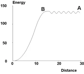
i) if the association between molecules A and B is a multiphasic reaction involv-ing a high number of interaction states, the discrimination between free and bound states may be somewhat arbitrary. Indeed, if bond formations is not an all-or-none phenomenon but requires a progressive strengthening, it it not obvious to chose a threshold to discriminate between free and bound states. Thus, while the streptavidin-biotin interaction might have been considered as strong enough to allow easy detection of bound states, several investigators reported on the time-dependent maturation of this interaction (Pincet 2005) and existence of a number of weak association states (Pierres 2002). Indeed, if the number of intermediate states is high, the concept of association constant becomes meaningless. This point was recently demonstrated in a quantitative study made on the binding efficiency of antibody-coated microspheres encountering antigen-bearing surfaces in a laminar flow chamber (Robert 2009): the probability of bond formation scaled as a power of encounter duration that was sig-nificantly higher than 1, and under a number of conditions this probability varied as erf c[(t/t0)1/2], where t was the contact time and t0 was a constant on the order of
where the reaction landscape was modeled as an unidimensional curve with a rugged segment (Figure 5). It may be useful to emphasize that this problem could not have been detected in studies of 3D interactions, since in this case the encounter time is de-termined by the laws of diffusion and is not expected to display substantial variations between different experimental setups.
Figure 5: Model of bond formation. Bond formation is modeled as a passage through a rough segment of an energy landscape, represented as passing from A to B. This was found to match experimental findings obtained with a flow chamber (Robert09).
ii) Atomic force microscopy and micropipette-based methods, that met with impres-sive success in analyzing bond rupture, may be less well suited to the study of bond formation because they do not allow easy control of contact duration, since contact is usually difficult to detect. Also, the contact area is often difficult to estimate since it is difficult to observe (Chesla 1998) and it may be markedly altered by forces exerted by the apparatus to induce molecular contact (Thoumine 2000). Finally, while hundreds or thousands of approach/retraction cycles can be performed on a given contact area, it is more difficult to sample extensive areas, which may be useful if low ligand and receptor densities are used in order to ensure that binding events are representative of single bond formation and dissociation.
Further, when ligands and receptors are attached to surfaces, association rates are less “intrinsic” parameters than dissociation rates because bond formation is highly de-pendent on the properties of linker molecules (Jeppesen 2001). Indeed, if molecules are rigid, association will be impossible if ligands and receptors are not suitably oriented to allow proper match between interacting areas. In contrast, association will be strongly enhanced if ligands and receptors are forced against each other with binding configu-ration. Also, the microtopology of surfaces bearing ligands and receptors may strongly influence association rates. As an example, the association rate between capsules bear-ing immunoglobulins and immunoglobulin receptors displayed 50-fold decrease when a smooth erythrocyte was replaced with a rough nucleated cell (Williams01).
In conclusion, The new kind of information that was recently obtained on biomolecule interactions by studying single bond formation and dissociation in presence of forces is directly relevant to a number of important biological processes. However, connecting this information to structural data still requires significant theoretical and experimen-tal progress. In addition, accounting for biological processes still requires to consider other less well defined parameters than kof f and kon. Thus, we shall briefly discuss the
frequently used concepts of avidity and specificity.
4.3
Avidity of biomolecule interactions: an incompletely
de-fined parameter
While aforementioned development might convey the view that ligand-receptor inter-action are liable to rigorous quantification, it has long been recognized that the affinity constant or association rates did not fully account for biological phenomena. Antigen recognition by antibodies provides a suitable example in view of the huge diversity of interactions and number of applications in hospital and research laboratory. As writ-ten in a standard treatise several decades ago (Glynn 1977) In the literature, affinity and avidity commonly are used synonymously ... However, it is now accepted that the term affinity is a thermodynamic expression ... Avidity also involves other contribut-ing factors such as antibody valence, antigen valence. A similar opinion remains in use today (Murphy 2008) ... The total binding strength of a molecule with more than one binding site is called the avidity. Thus, although it is accepted that avidity is not defined as accurately as affinity, a general concept is that this may be related to the capacity of forming multivalent associations. Indeed, many situations suggest that a most common way of forming strong associations involves the formation of multiple bonds. The following examples are intended to support the importance of the concept of avidity and the complexity associated to the multivalency of molecular interactions. Many biological interactions need to be multivalent. There are many exam-ples suggesting that a single noncovalent interaction between a ligand and a receptor may be too transient to be significant. Cell adhesion is driven by a number of membrane receptors that often require multivalent interactions. Cadherins, that are thought to play a dominant role in the stability of epithelia, are an important example. The im-portance of lateral clustering was very elegantly demonstrated (Yap 1997) by studying the adhesion of cells expressing engineered cadherins that could be oligomerized at will by bridging the intracellular domains with a drug. Similarly, integrins play a promi-nent role in cell adhesion to extracellular matrix compopromi-nents. It has long been known that cell surface integrins are often in an inactive state and events including clustering or conformational changes are required to enable these integrins to bind their ligands. Some recent examples clearly demonstrated that clustering integrins could directly en-hance the binding to multivalent, not monovalent ligands without any affinity change (Bunch 2010). As another example, ICAM-1, a ligand of integrin LFA-1, was reported to bind to immobilized LFA-1 with high avidity (dissociation constant was 8 nM) after dimerization, while no measurable interaction was observed with monomeric ICAM-1 (Miller 1995). It was further checked that ICAM-1 monomer expressed a complete LFA-1 binding surface (Jun01)
It is difficult to relate the properties of divalent and monovalent interac-tions. As emphasized above, it is because of this difficulty that single molecule studies revolutionized our understanding of biomolecule interactions. This difficulty is due to several reasons. Firstly, the rupture frequency of multivalent attachments may be drastically decreased by the possibility of rebinding events. Indeed, while a monovalent attachment is expected to break spontaneously as a consequence of thermal fluctua-tions, a multivalent attachment may need an external force for rupture if rebinding occurs (Seifert 2001). Also, the force sensitivity of multivalent attachments is strongly dependent on force sharing between different bonds, and unbinding forces may follow a number of different laws depending on forces and bond arrangement (Seifert 2000, Sulchek 2005, Tang 2007).
In conclusion While single molecule studies essentially provided an accurate de-scription of the interaction between binding sites exposed by biomolecules, we need to better understand the requirement for multivalent association. Clearly, multiva-lency is dependent on the topographical relationship between different binding sites and molecular flexibility. When interactions involve surface-attached molecules, other additional factors are important, including static and dynamic length and flexibility of linkers between surfaces and binding sites, as well as rugosity of the surface region surrounding molecules, and lateral mobility of molecules. These points will be briefly listed in a later section.
4.4
Specificity of biomolecule interactions: an essential
prop-erty that is difficult to define accurately
Obviously, biomolecules must bind specifically to adequate targets in order to fulfil their task. Specificity seems easy to define qualitatively : a ligand-receptor interaction is the more specific as the interaction between the same receptor and a “slightly” different ligand is “weaker”. However, there is no general way of defining the similarity of two molecules or the strength of an interaction.
First, two molecules may differ according to their shape (e.g as mentioned above for ortho- or para-dinitrophenol), their electric charge, their hydrogen bonding capacity or their hydrophobicity. As indicated in the next section, all these properties are involved in biomolecule recognition, but their relative importance may be different in varying situations. The similarity (or dissimilarity) between two molecules is not an absolute quantitative concept. This arbitrariness was indeed pointed out many years ago (Janin 1996).
Second, an interaction may be considered as “weaker” than another one if it oc-curs less often under physiological conditions. Thus, the affinity constant may be the dominant parameter if we are interested in the proportion of receptor molecules that are occupied by their ligand at equlilibrium, e.g. the number of insulin receptors at a given moment. However, if we are interested in the detection of an immobilized ligand on a surface dynamically explored by a cell protrusion, the kinetic rate of bond formation may be more important. Finally, if we are interested in the specificity of cell tethering on a surface, the interaction strengh may be the dominant parameter.
Interestingly, these parameters are not necessarily correlated. Thus, when mutant streptavidin molecules were made to bind biotin, the rupture forces were different, but they were correlated to the thermodynamic enthalpy rather than free energy of reac-tion (Chilkoti 1995), which is tightly related to the affinity constant, as recalled in eq. 2. Also, as mentioned above, the zero-force dissociation rate may not be correlated to the force resistance as represented with Bell’s distance parameter (Puri 1998, Pierres 2006)
Third, the difficulty of defining specificity is further illustrated by so-called promis-cuous receptors that may bind specifically a number of very different ligands, while a slight alteration of a given ligand may abolish the interaction. As an example, a mon-oclonal antibody was reported to bind specifically 2,4 dinitrophenol (Kd = 20nM ),
with a negligible affinity for the close analogs 2-nitrophenol and 2-nitro-4-iodophenol (Kd> 100µM ), but which also bound unrelated compounds such as furazolidone with
high affinity (Kd= 1.2µM ) (James03)
In conclusion, while it is recognized that both affinity and specificity are essential properties of ligand-receptor interactions (Janin 1996, Wang 2006), the latter may re-main difficult to define unambiguously. Specificity cannot be considered as an intrinsic parameter. A receptor may be considered as specific for its ligand if it does not interact with other molecules that it is liable to encounter under biologically relevant situations. The significance of interaction specificity will be discussed more precisely in a further section devoted to the structural basis of biomolecule interactions.
4.5
Ligand-receptor interactions are influenced by parameters
that are extrinsic to both ligand and receptor molecules
In addition to the parameters we have just mentioned, it is important to recall that molecular associations occurring in the biological milieu may be deeply influenced by a number of external parameters that may obscure the intrinsic properties of interacting sites. Receptor-mediated cell adhesion provides many examples as shown below. We shall give selected examples to illustrate this point.
Presence of repellers on receptor-bearing surfaces. It is well known that the surfaces of living cells are coated with a carbohydrate-rich layer with a thickness of several tens of nanometers or more, called the glycocalyx or pericellular matrix. Much experimental evidence supports the view that i) The glycocalyx may substantially impair the receptor capacity to bind to ligands, particularly during short encounters as occur in a laminar flow chamber (Sabri 1995, Patel 1995). ii) Under some cir-cumstances, cells may increase their receptor capacity by rapid removal of glycocalyx components, thus increasing the accessibility of membrane receptors (Sabri 2000). This inhibitory effect of the pericellular matrix is an example of so-called steric repulsion (Pierres 2000).
Lateral mobility of binding molecules It seems obvious that the probability of encounter between surface-attached binding molecules may be strongly enhanced if molecules can move freely on surfaces. This has long been demonstrated experimen-tally. As an example, when cells bearing CD2 surface molecules were micromanipulated into contact with surfaces coated with CD58, a ligand of CD2, either in immobilized
form or freely diffusing in a supported lipid bilayer, adhesion efficiency was strongly increased when ligand molecules were mobile, and this effect was more apparent when the ligand density was decreased (Chan91). As another example, the adhesive efficiency of cell surface integrins was reported to increase in parallel with lateral mobility, as measured with enhanced video microscopy and single particle tracking (Kucik96).
Localization of binding molecules on surfaces. As previously indicated, sur-face roughness may strongly decrease the accessibility of sursur-face receptors (Williams 2001). It is understandable that this phenomenon might depend on the localization of binding molecules as suggested by some experimental evidence. Thus, the capacity of selectin molecules to mediate binding of rapidly flowing leukocytes to the vessel walls was found to require the localization of these selectins on the tip of cell surface protru-sions. Indeed, when this localization was prevented by changing the transmembrane domain of adhesion molecules, the dynamic binding capacity was abolished although binding sites were intact (vonAndrian 1995, Buscher 2010).
Interactions between soluble biomolecules are also environment-sensitive. Recently, the kinetics of DNA hybridation was studied in living cells transfected with FRET-labeled double strand DNA (Schoen 2009) : different kinetics were observed within cells and in the extracellular milieu, and differences were dependent on the length of strands. Further, the authors did not observe any direct effect of molecular crowding in vitro. Other authors concluded on the basis of experiments and computer simulation that the molecular crowding observed in the cell interior might change pro-tein conformation (Homouz 2008).
In conclusion, the function of biomolecules involved in recognition events is de-pendent on a wide spectrum of parameters, that are not all determined by the structure of binding sites or event of linker parts of binding molecules. It is certainly warranted to devote much attention to all these parameters in the forthcoming years.
5
Relationship between biomolecule structure and
recognition events
As shown in the previous sections, the efficiency and selectivity of biomolecule interac-tions are dependent on a number of thermodynamic, kinetic and mechanical parameters that can be determined experimentally with exquisite sensitivity, using a number of recently developed methodologies. These advances increase our need for a theoretical framework allowing us to relate these quantitative binding parameters to structural properties. In addition to a mere intellectual appeal, such a framework would be use-ful i) to help us integrate a daunting amount of available data, ii) to take advantage of increasingly available structural data to predict the interaction behaviour of important molecules, and iii) to facilitate the rational design of molecules with desired interaction properties, e.g. to act as drugs. In the present section, three points related to this goal will be considered: i) the main intermolecular forces responsible for biomolecule recognition will be rapidly listed. ii) We shall describe some experiments aimed at determining which forces are involved in the interactions between molecules of known structure. iii) Lastly, we shall rapidly discuss the interest of computer simulations as