Publisher’s version / Version de l'éditeur:
Physical Review A, 67, 6, p. 063404, 2003-06-17
READ THESE TERMS AND CONDITIONS CAREFULLY BEFORE USING THIS WEBSITE. https://nrc-publications.canada.ca/eng/copyright
Vous avez des questions? Nous pouvons vous aider. Pour communiquer directement avec un auteur, consultez la première page de la revue dans laquelle son article a été publié afin de trouver ses coordonnées. Si vous n’arrivez pas à les repérer, communiquez avec nous à [email protected].
Questions? Contact the NRC Publications Archive team at
[email protected]. If you wish to email the authors directly, please see the first page of the publication for their contact information.
NRC Publications Archive
Archives des publications du CNRC
This publication could be one of several versions: author’s original, accepted manuscript or the publisher’s version. / La version de cette publication peut être l’une des suivantes : la version prépublication de l’auteur, la version acceptée du manuscrit ou la version de l’éditeur.
For the publisher’s version, please access the DOI link below./ Pour consulter la version de l’éditeur, utilisez le lien DOI ci-dessous.
https://doi.org/10.1103/PhysRevA.67.063404
Access and use of this website and the material on it are subject to the Terms and Conditions set forth at
Nonsequential double ionization of D2 molecules with intense 20-fs
pulses
Sakai, Hirofumi; Larsen, Jakob Juul; Wendt-Larsen, Ida; Olesen, Johannes;
Corkum, Paul; Stapelfeldt, Henrik
https://publications-cnrc.canada.ca/fra/droits
L’accès à ce site Web et l’utilisation de son contenu sont assujettis aux conditions présentées dans le site LISEZ CES CONDITIONS ATTENTIVEMENT AVANT D’UTILISER CE SITE WEB.
NRC Publications Record / Notice d'Archives des publications de CNRC:
https://nrc-publications.canada.ca/eng/view/object/?id=b4495bb2-d7ee-460c-89d5-03965adbe21c https://publications-cnrc.canada.ca/fra/voir/objet/?id=b4495bb2-d7ee-460c-89d5-03965adbe21cNonsequential double ionization of D
2molecules with intense 20-fs pulses
Hirofumi Sakai,1,*Jakob Juul Larsen,2Ida Wendt-Larsen,3Johannes Olesen,3Paul B. Corkum,4and Henrik Stapelfeldt3 1Department of Physics, Graduate School of Science, The University of Tokyo, 7-3-1, Hongo, Bunkyo-ku, Tokyo 113-0033, Japan
2Institute of Physics and Astronomy, University of A˚ rhus, Ny Munkegade, DK-8000 A˚rhus C, Denmark 3Department of Chemistry, University of A˚ rhus, Langelandsgade 140, DK-8000 A˚rhus C, Denmark
4National Research Council of Canada, 100 Sussex Drive, Ottawa, Ontario, Canada K1A 0R6
~Received 19 April 2002; published 17 June 2003! The kinetic-energy distribution of D1fragments obtained from the ionization of D
2molecules with intense
20-fs pulses includes a high-energy component extending up to ;10 eV. These fragments are only present for linearly, or slightly elliptically, polarized light. Both the maximum kinetic-energy and the ellipticity depen-dence are consistent with nonsequential double ionization caused by recollision.
DOI: 10.1103/PhysRevA.67.063404 PACS number~s!: 33.80.Rv, 33.80.Wz
Many new strong laser field phenomena, such as bond softening @1# and zero photon dissociation @2#, were first identified in H2 ionization experiments. Two recent papers have measured high-kinetic-energy distribution of the frag-ment ions produced when H2 is ionized with a short intense laser pulse @3,4#. The high energy cutoff of the fragment kinetic-energy approaches ~but is a bit less than! the energy expected, if the H2 ground state wave function were trans-ferred instantly to the double ion. At least some of the frag-ments come in correlated pairs @4#.
These new observations are compatible with a number of possible mechanisms.
~1! The molecule might double ionize instantaneously @5# and Coulomb explode. If so, the kinetic-energy distribution provides information about the correlated electron dynamics in H2. It might also be used to measure @6,7# the vibrational wave function of excited H2.
~2! The molecule might ionize sequentially. First H21 is formed. Then those H21 molecules that do not decay by other means ~bond softening @1# or enhanced ionization @8–10#! ionize again in the remaining part of the pulse @3,4#. ~3! The high-kinetic-energy fragments might be produced by recollision. If so, the recollision electrons can serve as probes in pump-probe experiments @11#. We show that the high-kinetic-energy fragments originate from nonsequential double ionization caused by recollision.
Our paper is related to a recent work @12# in which lower intensity laser pulses were used. In that paper, recollision electrons are identified as the source of the high-energy com-ponent in the fragment distribution when H2 is ionized. However, in that experiment the laser intensity was kept very low to discourage sequential processes. Our intensity is about ten times that in Ref. @12#, but similar to that used in Ref. @3# and to one of the intensities used in Ref. @4#. Our results show that recollision also remains the dominant mechanism at high intensities in the order of 1015W/cm2, which is misinterpreted in Ref. @3# and not mentioned in Ref. @4#.
Our paper is also related to recent works that identify recollision excitation and double ionization in more complex
molecules @13,14#. By clearly demonstrating double ioniza-tion in D2, we have identified a simple molecule in which to explore the similarities and differences between double ion-ization in atoms and in molecules. In addition, working at an intensity of 1015 W/cm2 adds an extra degree of simplifica-tion, since any excited state of D21 produced by inelastic scattering will almost immediately ionize.
We identify recollision by studying the number of ener-getic fragments as a function of laser ellipticity. Sensitivity to ellipticity of the ionizing laser light is fundamental to all recollision processes @15,16#. First an electron tunnels through the potential modified by the strong linearly polar-ized laser field, then the electron is driven away from the ion by the field, only to be forced with ;50% probability to return after the field reverses its direction. In the electron-ion recollision that might follow, the remaining electrons are col-lisionally excited or ionized @17,18#. If the light deviates very much from linear when the electron returns to the ion, it is offset in the lateral direction and it can miss.
Our results only include data for D2 molecules. That is because, for H2, we must subtract background signals origi-nating from H1produced by dissociative ionization of con-taminants in the vacuum chamber. However, the observed features were the same for both D2 and H2 molecules. In agreement with previous measurements @3,4#, we observe that the kinetic-energy distribution of D1fragments extends continuously up to ;10 eV.
We now briefly describe the experimental setup. A fuller description is given elsewhere @19,20#. A pulsed supersonic beam, formed by expanding ;600 Torr of D2 gas through a 0.5-mm-diameter nozzle, is crossed at 90° by the focused laser beams. The 800-nm 20-fs pulses were generated using a hollow wave guide for spectral broadening and two prisms for chirp compensation @21#. A 10-cm-focal-length parabolic mirror, placed in the vacuum chamber, focused the laser beam to an intensity ;231015W/cm2. Since the saturation intensity is ;1015W/cm2 @22#, we can assume that most molecules ionize near that intensity. A zero-order quarter-wave plate adjusted the ellipticity.
An electrostatic field accelerated the D1 ions toward an ion detector positioned on-axis with the molecular beam. The ion detector consists of a microsphere plate backed by a *Electronic address: [email protected]
phosphor screen. A charge-coupled device ~CCD! camera re-corded the ion image on the phosphor screen. Fast electronic gating of the CCD camera allows us to record mass and charge selected ion images. We used velocity map imaging @23# to achieve good energy resolution.
Figure 1 shows a typical image of D21 and D1ions ~the image is not Abel inverted @23,24#!. The direction of the laser polarization is shown in the figure. Three distinct compo-nents of ion image are observed.
~1! D21ions appear at the center of the image. These are the molecules that only ionized once, and did not fragment. ~2! A pair of D1 peaks ~kinetic-energy 0.3 and 1 eV per fragment! on each side of the D21 feature forms the next prominent feature. They originate from the dissociation of D21. These are the well-known bond softening peaks @1#.
~3! The broad set of peaks that are the outermost feature in the figure are due to enhanced ionization @8–10#.
As the D21 begins to dissociate, it enters a region where the ionization rate is greatly enhanced. After ionization, D221 Coulomb explodes, yielding pairs of D1 ions with kinetic-energy ;3 eV/fragment. The ion image shows a strong spatial anisotropy along the polarization direction. Coupling between Sgand Su is essential for both bond
soft-ening and enhanced ionization.
Not seen in the figure is a diffuse cloud of very high kinetic-energy fragments. Their angular distribution is about twice as broad as the fragments due to enhanced ionization, but integrated over all angles, they make up a significant fraction of the signal.
Figure 2 shows kinetic-energy distributions of D1 ions obtained by angularly integrating the signals from images such as that in Fig. 1, where the peak intensity was kept constant and the ellipticity « was varied from «50 ~linear polarization! to «51 ~circular polarization!. Data for six dif-ferent ellipticities are included. Concentrating on the linearly
polarized results ~solid curve!, the three peaks that were clearly seen as pairs of peaks ~equal energy but oppositely directed! in Fig. 1 are now single peaks in Fig. 2, since each member of the pair had the same energy.
The kinetic-energy calibration of the fragment ions was performed by simulation software. For the horizontal axis of Fig. 2 ~and Fig. 4 shown below!, we use ‘‘radial kinetic-energy,’’ which corresponds to the projection of the velocity vector onto our two-dimensional ion detector. The real kinetic-energy distribution can only be obtained when the laser polarization is linear, through Abel inversion. For the data recorded with elliptical polarization the lack of cylindri-cal symmetry prevents Abel inversion. Clearly visible in the figure is a high-energy component extending up to ;10 eV. Although our data are uninverted, they agree well with pre-vious observations @3,4#.
Our concern is less with the kinetic-energy distribution than with the physics that produces it. The high-kinetic-energy fragments observed in Fig. 2 and in Refs. @3# and @4# can only originate from D221 molecules exploding at small internuclear distances. They extend into the region of direct Coulomb explosion of D2, although not up to the maximum kinetic energy expected from Coulomb explosion of D2, cor-responding to the Coulomb energy at the equilibrium inter-nuclear distance. We first concentrate on the ellipticity de-pendence seen in Fig. 2, then on the kinetic-energy distribution.
In any recollision event involving such high laser fields, the classical motion of the electron following ionization is important. An electron leaving the molecule with near zero initial energy, moves about a distance s;2qE/mv2(E is the electric field amplitude of the laser pulse at angular fre-quency v, and q and m are the electron’s charge and mass, respectively! ;60 Å in the direction of the field vector be-fore its motion is reversed. Depending on the phase of birth of the electron, it can recollide with its parent ion with the maximum recollision kinetic-energy of 3.17m(qE/2mv)2 ;200 eV—enough to knock the remaining electron from D21 ~ionization potential is ;30 eV). The electron-impact ionization cross section for D21 ion is more than 1.0
FIG. 1. A typical image of D21 and D1 ions. Three different
components of fragment ions are observed: ~1! D21ions appear at
the center of the image; ~2! the inner pairs ~0.3- and 1-eV peaks! of ions originate from bond softening, producing D11D; and ~3! the
outer pair originates from enhanced ionization, producing D1
1D1. The image is not Abel inverted.
FIG. 2. The relative number of ions plotted as a function of the radial kinetic-energy of D1 ions. The peak intensity of 2 31015W/cm2 was kept constant and the ellipticity « was varied from «50 ~linear polarization! to «51 ~circular polarization!.
SAKAI et al. PHYSICAL REVIEW A 67, 063404 ~2003!
310217cm2 for incident electron energies of 45–345 eV @25#.
A slight ellipticity of the laser light affects the recollision process, offsetting the electron from the ion by ;5«qE/mv2, making recollision less probable @16#. Look-ing closely at Fig. 2, we find that the yield of all channels decreases with ellipticity, but the high-energy component falls rapidly with increasing ellipticity and almost disappears at ellipticity «50.5. This is the behavior expected for recol-lision phenomena. To make the ellipticity dependence clearer, we integrated the D1ion signals from 1.6 eV to 5.0 eV and from 5.0 eV to 10 eV and plotted them as a function of ellipticity. The 5-eV limit was determined by referring to the kinetic-energy distributions for linear and circular polar-izations with the E-field magnitude kept constant ~see Fig. 4!. The result is shown in Fig. 3. The yield in each energy region is normalized to ten at «50.
In addition to the sensitivity of recollision to ellipticity, there are three processes that influence the ion yield as a function of ellipticity.
~1! The maximum electric field falls as the ellipticity in-creases. In the tunneling regime, the ion yield follows the maximum electric field. Therefore, the yield should decrease as well. In Fig. 2, the yield of all components decreases with increasing ellipticity. However, the high-energy peak de-creases more quickly, as shown in Fig. 3.
~2! We change the ellipticity by rotating a quarter-wave plate. This rotates the major axis of the ellipse by the same angle that the wave plate rotates. The projection of the kinetic-energy distribution of all fragments is affected. Based on the shifts of the peaks due to enhanced ionization, we can estimate the effect of the rotation of major axis to be ;0.3 eV. Since the high-kinetic-energy component ranges from ;5 eV to ;10 eV and is broader than other compo-nents, it is reasonable to think that it is less affected by the small shift of ;0.3 eV.
~3! It is possible that D2, or more likely D21, aligns to
the laser field at least to some extent. If it does, then the alignment might decrease as the ellipticity increases. If it does not, then those effects that depend on the alignment of the molecule to the field will become more important with circular polarization @26#. By studying yields using linear and circular polarized light @26#, we will show below that neither D2 nor D21aligns.
None of the effects described above can influence the re-sults for small ellipticities since the field strength along the major axis hardly changes. The high-kinetic-energy fragment signal has fallen to less than 40% of the peak value and the cutoff energy is almost unchanged for an ellipticity of 0.25. By ellipticity ;0.5, where the electric field ;89% of its maximum value, the high-energy component is almost unob-servable. The only mechanism compatible with such strong ellipticity dependence is recollision.
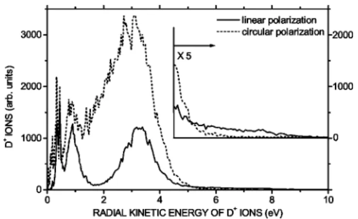
We now confirm this interpretation by comparing the kinetic-energy distributions of D1ions for linear and circular polarizations where the field amplitude is kept constant. Keeping the field constant eliminates the influence of the first two processes mentioned above and allows us to evaluate the third process. Concerning the first two processes one can say the following.
~1! Since the ionization rate depends on the field strength in the tunneling regime @27#, the ionization rate should be essentially the same if the field is the same.
~2! With circular polarization, the polarization becomes parallel to the detector surface twice in an optical period. The cutoff kinetic-energy comes from those molecules that are aligned parallel to the detector plane irrespective of the po-larization.
Combining these two facts, the effect caused by rotation of the major axis in Fig. 2 can be completely eliminated.
The result is shown in Fig. 4, where the peak intensity was 231015 W/cm2 for linear polarization and thus 4 31015 W/cm2 for circular polarization. It can be seen that the enhanced ionization yield ~the number of D1 ions! for circularly polarized light increases by a factor of 3.8 com-pared to that for linearly polarized light. This shows that
FIG. 4. The relative number of ions plotted as a function of the radial kinetic-energy of D1ions for linear polarization ~solid curve! and circular polarization ~dotted curve!. The same laser field was used for these measurements. The peak intensities were 2 31015W/cm2for linear polarization and 431015W/cm2for circu-lar pocircu-larization.
FIG. 3. The relative number of D1 ions in the 1.6–5 eV range ~open circles show enhanced ionization! and 5–10 eV range ~solid circles show recollision! plotted as a function of the laser ellipticity. All the error bars for enhanced ionization and some for recollision are smaller than the size of their symbols. The peak intensity of 2 31015W/cm2was kept constant and the signals were normalized to 10 at «50.
there is little dynamic alignment of D2and/or D21@26#. This conclusion is consistent with our numerical simulations where the dynamic alignment of D2 and D21has been stud-ied by solving the time-dependent Schro¨dinger equation. In contrast to the increased strength of the enhanced ionization peak, the high-kinetic-energy fragments are not observable with the circular polarization used, as expected for any recol-lision process @15#. A recent experimental study, with H21 ions as the target @28,29#, suggests that the neutral parent plays an important precursor role in the laser-induced frag-mentation dynamics of H2, corroborating the present results and considerations.
Before concluding, it is important to show how recollision can be responsible for the high-kinetic-energy fragments that we, and others @3,4# observe. Nonsequential double ioniza-tion is really a ‘‘sub-laser-cycle’’ process. Following single ionization, the newly ionized electron is pulled away from the ion, only to be driven back. The typical time required before the electron makes its first attempt at recollision is ;2/3 of a laser period. During this time, the D21 wave packet is able to move a bit. If the electron recollides with sufficient energy, it can excite the molecule to any of the numerous states, or further ionize the singly charged molecu-lar ion by inelastic scattering. Consistent with our observa-tions, the high-energy cutoff of the fragment kinetic-energy distribution will be slightly lower than that observed for true
Coulomb explosion. We refer the reader to Ref. @12# for a detailed calculation.
In addition, Coulomb focusing and non-hard-sphere colli-sions @17,18# provide the electron wave packet with signifi-cant probability of subsequent recollisions. These occur in the few laser cycles following ionization @17# and will con-tribute lower-energy deuterons to the spectrum. In Ref. @12# this dynamics is measured at low intensity. Our measure-ments of the kinetic-energy spectrum of the D1 ions indi-rectly measure this dynamics at high intensity in the order of 1015 W/cm2.
In summary, the kinetic-energy distributions of D1 frag-ments obtained from the Coulomb explosion of D2 mol-ecules with intense 20-fs pulses included a fractional contri-bution with a high kinetic-energy extending up to ;10 eV. All our results are consistent with the interpretation that the energetic D1 fragments are caused by nonsequential double ionization caused by recollision. We expect similar behavior for any two-electron dimer, for example Na2. Ionization can be controlled to allow only a small range of times for recol-lision @30#. Thus, the recolrecol-lision electron can excite the ion at a well-defined ~short! time after ionization. Coulomb explo-sion then images the nuclear position.
The authors are grateful to Dr. Shinichirou Minemoto ~The University of Tokyo! for his help in the electronic sub-mission of the manuscript.
@1# A. Zavriyev et al., Phys. Rev. A 42, 5500 ~1990!. @2# J.H. Posthumus et al., J. Phys. B 33, L563 ~2000!.
@3# C. Trump, H. Rottke, and W. Sandner, Phys. Rev. A 60, 3924 ~1999!.
@4# A. Staudte et al., Phys. Rev. A 65, 020703~R! ~2002!. @5# D.N. Fittinghoff et al., Phys. Rev. Lett. 69, 2642 ~1992!. @6# H. Stapelfeldt, E. Constant, and P.B. Corkum, Phys. Rev. Lett.
74, 3780 ~1995!.
@7# S. Chelkowski, P.B. Corkum, and A.D. Bandrauk, Phys. Rev. Lett. 82, 3416 ~1999!.
@8# T. Seideman, M.Yu. Ivanov, and P.B. Corkum, Phys. Rev. Lett.
75, 2819 ~1995!.
@9# E. Constant, H. Stapelfeldt, and P.B. Corkum, Phys. Rev. Lett.
76, 4140 ~1996!.
@10# T. Zuo and A.D. Bandrauk, Phys. Rev. A 52, R2511 ~1995!. @11# H. Niikura et al., Nature ~London! 421, 826 ~2003!. @12# H. Niikura et al., Nature ~London! 417, 917 ~2002!. @13# V.R. Bhardwaj et al., Phys. Rev. Lett. 86, 3522 ~2001!. @14# For example, C. Guo and G.N. Gibson, Phys. Rev. A 63,
040701~R! ~2001!, and references therein. @15# P.B. Corkum, Phys. Rev. Lett. 71, 1994 ~1993!. @16# P. Dietrich et al., Phys. Rev. A 50, R3585 ~1994!.
@17# T. Brabec, M.Yu. Ivanov, and P.B. Corkum, Phys. Rev. A 54, R2551 ~1996!.
@18# G.L. Yudin and M.Yu. Ivanov, Phys. Rev. A 63, 033404 ~2001!.
@19# H. Sakai et al., J. Chem. Phys. 110, 10 235 ~1999!. @20# J.J. Larsen et al., J. Chem. Phys. 111, 7774 ~1999!.
@21# M. Nisoli, S. De Silvestri, and O. Svelto, Appl. Phys. Lett. 68, 2793 ~1996!.
@22# T.D.G. Walsh, F.A. Ilkov, and S.L. Chin, J. Phys. B 30, 2167 ~1997!.
@23# A.T.J.B. Eppink and D.H. Parker, Rev. Sci. Instrum. 68, 3477 ~1997!.
@24# C.J. Dash, Appl. Opt. 31, 1146 ~1992!.
@25# Y.-K. Kim, K.K. Irikura, and M.A. Ali, J. Res. Natl. Inst. Stand. Technol. 105, 285 ~2000!.
@26# Ch. Ellert and P.B. Corkum, Phys. Rev. A 59, R3170 ~1999!. @27# M.V. Ammosov, N.B. Delone, and V.P. Krainov, Zh. E´ksp.
Teor. Fiz. 91, 2008 ~1986! @Sov. Phys. JETP 64, 1191 ~1986!#. @28# I.D. Williams et al., J. Phys. B 33, 2743 ~2000!.
@29# K. Sa¨ndig, H. Figger, and T.W. Ha¨nsch, Phys. Rev. Lett. 85, 4876 ~2000!.
@30# M. Ivanov et al., Phys. Rev. Lett. 74, 2933 ~1995!.
SAKAI et al. PHYSICAL REVIEW A 67, 063404 ~2003!