HAL Id: hal-02345781
https://hal.archives-ouvertes.fr/hal-02345781
Submitted on 4 Nov 2019
HAL is a multi-disciplinary open access
archive for the deposit and dissemination of
sci-entific research documents, whether they are
pub-lished or not. The documents may come from
teaching and research institutions in France or
abroad, or from public or private research centers.
L’archive ouverte pluridisciplinaire HAL, est
destinée au dépôt et à la diffusion de documents
scientifiques de niveau recherche, publiés ou non,
émanant des établissements d’enseignement et de
recherche français ou étrangers, des laboratoires
publics ou privés.
A high pressure cell using metallic windows to
investigate the structure of molecular solutions up to
600 MPa by small-angle neutron scattering
Burkhard Annighofer, Arnaud Hélary, Annie Brûlet, Alexandre Colas de la
Noue, Camille Loupiac, Sophie Combet
To cite this version:
Burkhard Annighofer, Arnaud Hélary, Annie Brûlet, Alexandre Colas de la Noue, Camille Loupiac, et
al.. A high pressure cell using metallic windows to investigate the structure of molecular solutions up
to 600 MPa by small-angle neutron scattering. Review of Scientific Instruments, American Institute
of Physics, 2019, 90 (2), pp.025106. �10.1063/1.5051765�. �hal-02345781�
solutions up to 600 MPa by small-angle
neutron scattering
Cite as: Rev. Sci. Instrum. 90, 025106 (2019); https://doi.org/10.1063/1.5051765
Submitted: 12 August 2018 . Accepted: 17 January 2019 . Published Online: 13 February 2019
Burkhard Annighöfer , Arnaud Hélary, Annie Brûlet, Alexandre Colas de la Noue, Camille Loupiac , and Sophie Combet
Review of
Scientific Instruments
ARTICLE scitation.org/journal/rsiA high pressure cell using metallic windows
to investigate the structure of molecular solutions
up to 600 MPa by small-angle neutron scattering
Cite as: Rev. Sci. Instrum. 90, 025106 (2019);doi: 10.1063/1.5051765
Submitted: 12 August 2018 • Accepted: 17 January 2019 • Published Online: 13 February 2019
Burkhard Annighöfer,1 Arnaud Hélary,1Annie Brûlet,1,a)Alexandre Colas de la Noue,1,2Camille Loupiac,1,3 and Sophie Combet1,a)
AFFILIATIONS
1Laboratoire Léon-Brillouin (LLB), UMR 12 CEA-CNRS, Université Paris-Saclay, CEA-Saclay, F-91191 Gif-sur-Yvette CEDEX, France 2Laboratoire CIRAD, UMR QualiSud, F-34398 Montpellier CEDEX 5, France
3Equipe PCAV, UMR PAM, Université de Bourgogne Franche-Comté, AgroSup Dijon, F-21000 Dijon, France
a)Authors to whom correspondence should be addressed:sophie.combet@cea.frandannie.brulet@cea.fr
ABSTRACT
We report on a high pressure (HP) cell designed for the determination of the structure of molecular solutions by small-angle neutron scattering (SANS). The HP cell is fitted up with two thick metallic windows that make the device very resistant under hydrostatic pressures up to 600 MPa (or 6 kbar). The metallic windows are removable, offering the possibility to adapt the HP cell to a given study with the pressure desired on an appropriate spatial range to study the structure of various molecular solutions by SANS. In this context, we report the absorption, transmission, and scattering properties of different metallic windows. Finally, we describe, as a proof of principle, the solution structure changes of myoglobin, a small globular protein.
Published under license by AIP Publishing.https://doi.org/10.1063/1.5051765
I. INTRODUCTION
Pressure is an important thermodynamic variable not only for physics and chemistry but also for biology. Depend-ing on the thermodynamic environment, the solution struc-ture of proteins can fluctuate in multiple conformational sub-states, which differ in their partial molar volumes, ranging from folded to unfolded states.1,2 This equilibrium between different substates contributes to the protein intrinsic flex-ibility. While a number of studies on protein unfolding have been focused on thermal and/or chemical effects, much less has been performed under high pressure (HP), which is how-ever a unique tool.3,4The first observation that high hydro-static pressure is able to unfold proteins was the coagulation of albumin, as shown by Bridgman in 1914.5
Thanks to an appropriate spatial range, small-angle neutron scattering (SANS) upon HP is an invaluable tech-nique to provide direct information on the three-dimensional conformation of various molecular solutions, especially in
biology. Many studies have been published using SANS and HP on different proteins. For instance, measurements performed on various concentrations of azidometmyoglobin (myoglobin being a small and globular protein, whose function is to stock dioxygen in muscles) showed that the compactness of the pro-tein is not altered by HP up to 300 MPa (or 3 kbar),6but some changes have however been observed on the specific vol-ume of this protein as a function of pressure.7 Ortore et al. reported that pressure induces dissociation of β-lactoglobulin in both D2O and a 50% mixture of water and ethylene-glycol, even if the protein shows a higher stability in 50% ethylene-glycol.8Osaka et al. compared the heat- and pressure-induced gelation of β-lactoglobulin aqueous solutions by both SANS and dynamic light scattering.9 The denaturation of calmod-ulin, in contrast to heat denaturation, has been shown to be reversible up to pressures of 300 MPa.10 Structural studies by SANS have also been associated with dynamics studies by quasielastic neutron scattering on the bovine pancreatic trypsin inhibitor11,12 and the human acetylcholinesterase.13
Rev. Sci. Instrum. 90, 025106 (2019); doi: 10.1063/1.5051765 90, 025106-1
in solution does not change with pressure up to 150 MPa but unfolding process of proteins in solution is in general induced by a pressure larger than 200 MPa.13,15For instance, nuclear magnetic resonance (NMR) experiments showed that 500 MPa is the half denaturation pressure value of myoglobin.16,17
However, SANS can exhibit technical limitations that can be exacerbated by HP, due to several issues: (i) the weakness of the SANS signal due to the small size of the proteins, (ii) the huge propensity of protein aggregation when increasing the protein concentration, (iii) the large sample volume and the flat geometry of the HP cell, requiring resistant and thick win-dows, and (iv) in order to access the spatial necessary range, the usual large neutron beam diameter, compared to light or X-rays, requiring a large exit angle and therefore large and thick windows. Pressure values up to 600 MPa have previously been reached by SANS on proteins using cylindrical metallic alloy HP cells.11However, this geometry is not well adapted to SANS measurements7and, instead, a planar geometry is pre-ferred, which was the motivation of the present study. Metal-lic windows have first been proposed in SANS technique.11 However, due to both high neutron scattering and absorption of such windows, a huge protein concentration is necessary, which prevents from measuring the conformation of a single protein in solution. Recent developments have rather focused on HP cells using sapphire windows,7,18–20which, in contrast to metallic windows, have a low noise and are virtually “trans-parent” for neutrons. Sapphire windows have been also pro-posed since they have the advantage of allowing both a visual control of the sample aspect and a combination with in situ light scattering measurements.19However, sapphire is brittle and single crystals used as windows are fragile and can sud-denly break at any pressure value. In conclusion, while very appealing to study the solution structure of proteins, SANS under pressure is not much performed and generally limited to 300 MPa.7,19
In order to reach pressures up to 600 MPa in SANS for molecular solutions, such as biological samples, in a more sys-tematic manner, we studied the neutron scattering properties of various metallic alloys, in order to use them as HP cell windows. In Sec.II, we describe the HP cell device for SANS experiments with removable metallic windows and the experi-mental procedure. In Sec.III, we compare the neutron scatter-ing properties of different materials compatible with the SANS technique. In Sec.IV, we show HP measurements on protein solutions at 20 g/l up to 600 MPa. Finally, in Sec.V, as a proof of principle, we compare the effect of HP on dilute solutions of myoglobin (5 and 7 g/l) in different environments.
II. HIGH PRESSURE CELL FOR SANS EXPERIMENTS We developed a HP cell with removable windows to study the conformation of biological molecules by SANS. This HP cell is available to the scientific community at the LLB neutron facility, but also on other neutron facilities, through neutron beam access and/or collaboration. The pressure cell body and the window abutments are made from a precipitation
hard-(Aubert & Duval, France), which was aged after machining dur-ing 4 hours at 520◦C to reach the desired strength and stress corrosion resistance. This stainless steel gives us the possibil-ity to study the sample under HP combined with a good cor-rosion resistance. The neutron beam size has to be collimated to a maximum diameter of 5.5 mm, centered on the entry of the HP cell. A removable cadmium-diaphragm can be placed in front of the cell for the accurate cell alignment on the incident neutron beam. The beam passes through the sealed metal-lic entry window, interacts with about 3.8 mm-thick sample, and passes through the sealed metallic exit window. The con-tact area between the sample and the metallic windows has been minimized to reduce putative protein modification. This geometry has a wide exit angle (24◦ total conical opening angle) to allow access to high Q-values, up to 0.2 Å−1, where
Q= 4π sin θλ is the momentum transfer (called also the ing vector), λ is the neutron wavelength, and 2θ is the scatter-ing angle. The sample chamber volume is ∼140 µl. Most of sam-ples in biology or physico-chemistry use a temperature-range of 4-90◦C and a pH-range of 2-12. Until now, our HP cell has been used at the calibrated temperature of 20◦C, using a ther-mostatic bath and a copper piece with circulating fluid inside, which can be mounted, either on its top (as shown in the inset of Fig. S1), or on its bottom, or on both. But the temperature-range of 4-90 ◦C should be possible provided that the con-densation at a low temperature can be dried by a continuous air jet. Since molecular solutions are generally prepared in buffers with a pH-range from 2 to 9, between each sample, the cleaning process of the HP cell consisted in rinsing several times with HCl solutions (pH 2) and then with NaOH solutions (pH 13), to eliminate the sample residuals, followed by a final cleaning with distilled water and then with D2O, before dry-ing under vacuum. While keepdry-ing the same HP cell body, the alloy nature and the thickness of the metallic windows can be easily changed to offer the possibility of optimizing both their scattering and absorption, as a function of the sample scatter-ing and the maximum pressure desired.Figure 1shows the HP cell design with thinned (3.95 mm) Ti-alloy windows, but the typical thickness of the windows we used in the present study was about 6.1 mm to perform pressure measurements up to 600 MPa.
About the sealing design, we used inner O-rings, for the low pressure sealing, surrounded by MARVAL X12 anti-extrusion rings using the “knife” form to seal them against the window by creating a local plastification of the window. We avoided to use lead rings to cover the anti-extrusion rings, since lead spread inside the sealing place, gluing everything together. Instead, we tested tin-plated (tin-alloy: S-Sn96Ag-221) pure copper rings. Using this material, it was much easier and faster to change the windows during the SANS experi-ments. However, the highest achievable pressure was limited to 570 MPa, due to the less plasticity of this material (com-pared to pure lead) that did not fill the gaps. Therefore, we finally preferred using an uncoated pure copper ring below the anti-extrusion ring but with a pure lead ring above the rounded edge of the “knife” form. Besides, a short overhang was added to the anti-extrusion ring to remove it with an
Review of
Scientific Instruments
ARTICLE scitation.org/journal/rsiFIG. 1. (Left) 3D design and (right) middle-cut view of the SANS HP cell, shown with thinned windows. The incom-ing neutron beam is indicated by an arrow. The detail A of the cell sealing design is shown inFig. 2.
extractor. Thanks to the present configuration, we were able to both measure pressures up to 600 MPa and reduce the window changing time (Fig. 2).
The overall configuration of the HP device on the SANS instrument is made as follows (Fig. S1 of thesupplementary material): a HP sensor-tipped hand-driven SITEC 700 MPa pressure generator, from where the D2O filled capillary goes to an own-developed Bridgman sealed separator. This sepa-rator leads the sample to a T-fitting, whereby the free exit is used for the pressure measurement with a second sensor, behind the free-floating 4 O-ring sealed separation piston. The capillary then enters into the HP cell body. On the opposite side, a plug is installed for easy cleaning and filling.
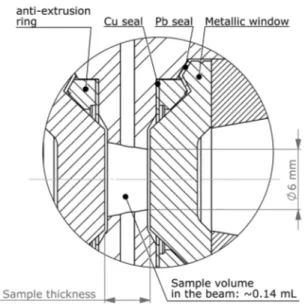
FIG. 2. View of the sealing design (corresponding to the detail A inFig. 1) of the SANS HP cell, showing the copper and lead seals surrounding the anti-extrusion ring. The sample chamber dimensions are indicated.
It is very important to note that the scattering signal from metallic materials changes when mechanical plastifica-tion occurs. This is due to modificaplastifica-tions of dislocaplastifica-tions and grain boundaries and sizes. Therefore, a pressure test before sample measurement at the maximum pressure afforded in the plastification regime is mandatory. This prevents a change in the background signal during the sample measurements under various applied pressures.
III. SCATTERING PROPERTIES OF THE MATERIALS USED AS HP CELL WINDOWS
The scattering intensity of dilute protein solutions is gen-erally weak, about 0.2-1 cm−1, and decreases as a function of Q. Moreover, HP cell windows absorb and scatter neu-trons, which may obscure the sample scattering itself. The choice of the materials for such windows is then crucial. Sapphire has a very high transmission for the wavelengths used in SANS: about 0.94 for 12 mm-thick windows at 6 Å wavelength (see Table S1 of the supplementary material). Besides, the scattering of the windows made of single crys-tals of sapphire, with well-polished surfaces, is weak, at about 3.5 × 10⁻³ cm−1 [see Fig. 3(a)]. With such characteristics, sapphire is a very good choice to study dilute solutions of proteins under pressure. Unfortunately, this material is very brittle. Tensile and stress concentration must be avoided, as well as inner stress due to the growing process of single crys-tals. Moreover, since the material only resists to compression stress, the sealing process has to be very sophisticated. In practice, failures often occur below the theoretical maximum pressure limit without any apparent reason. So, using the HP cell for SANS with sapphire windows is rare. Anyway, owing to the potentialities of sapphire, developments are still ongo-ing to improve the reliability of pressure cells for SANS up to 500 MPa.19
In a previous paper,21 we showed that, by following a method which implies careful and accurate measurements of empty beam, empty cell, electronic background, and transmis-sion, we can in principle recover the signal of a sample placed in a container displaying a high scattering signal. Therefore,
Rev. Sci. Instrum. 90, 025106 (2019); doi: 10.1063/1.5051765 90, 025106-3
FIG. 3. Scattering curves of various compounds normalized to both their sample thickness and transmission. (a) “Low absorption” materials. (b) “High absorption” materials. (c) Ti–Al alloys. Neutron scattering intensities, I(Q), are in absolute units (cm−1). Measurements were performed on PACE and PAXY SANS instruments
at the LLB (Saclay, France) and on D11 SANS instrument at the Institut Laue-Langevin (ILL, Grenoble, France).
despite their possible absorption, absence of transparency, and significant scattering, we chose metallic alloys as win-dow materials because of much better mechanical properties compared to those of sapphire.
Previously, some of these materials had already been used in HP cells for SANS experiments. For instance, CuBe2 and TiZr alloys, the latter one being also called the “null matrix alloy,” display good mechanical properties, but their neutron
rial). That is the reason why HP cell windows for SANS are rather made of niobium or aluminium alloys, exhibiting high transmissions. However, they display smaller maximum pres-sure limits than CuBe2 and TiZr. Actually, it is very difficult to compare the scattering properties of all possible materi-als, since both their transmission and scattering properties are generally not reported in the literature. Therefore, in order to optimize the choice of the materials as a function of the max-imum pressure limit desired and of the sample scattering, we undertook a comparative study of all these materials in the same conditions as our own device.
Table S2 of the supplementary material reports the mechanical parameters of the window materials we tested, i.e., the shear stress factor y (y being an estimation of the ductil-ity, which depends on the crystal structure, heat treatment, plastic deformation, and temperature) and the ultimate ten-sile stress UTS, given by the suppliers. Pressure and shear sections (Spand Sτ) were calculated for the geometry of our HP cell, as described in Sec.II(Fig. 1). The shear strength is equal to Sτ∗y∗UTS. The pressure at shear stress is defined as the ratioPressure sectionShear force . Using these values, we could estimate a maximum pressure limit, which depends on the safety factor against the burst pressure used at the facility, this factor being usually between 1.5 and 2.
Metallic materials can be used with neutron wavelengths between roughly 6 and 10 Å. Indeed, below 5 Å, we observe Bragg diffraction peaks due to the ordered structure of metal-lic alloys. Above 10 Å, we get multiple scattering due to the nanometer-scale grain boundaries of polycrystalline materi-als. Both phenomena are amplified by the large thickness of the windows required for HP cells.
The measured transmission values of various compounds measured at 6 Å are listed in Table S1 of the supplemen-tary material. A transmission value of 0.5 means that half of the neutron beam is “lost,” i.e., it will not be scattered by the sample itself. From these measurements, we estimated the linear absorption coefficient µ of the compound at 6 Å-wavelength using T= exp−µt, from which we can calculate the transmission for any value of thickness. The calculated trans-missions for 10 and 20 mm are reported in Table S1 of the
supplementary material. These calculations assume that no multiple scattering occurs through the pieces. This assump-tion is reasonable for most of the tested compounds: their scattering is not huge and they have reasonable high values of transmissions. We checked the absence of multiple scat-tering, especially for the aluminium alloys, by measuring the scattering and transmission of a second piece of material, but with a double thickness and verifying that scattering curves nicely superimposed. For CuBe2, which has a low transmis-sion for a rather small thickness and scatters a lot, the occur-rence of multiple scattering is probable. This was not checked since the huge scattering signal and the low transmission are two redhibitory reasons to use this material for HP cell win-dows for SANS. In conclusion, we can separate the compounds in two groups: (i) the “low absorption” compounds, includ-ing Si, sapphire, Al alloys, Nb, and ZrNb1, and (ii) the “high absorption” compounds, including TiZr, CuBe2,M30NW steel,
Review of
Scientific Instruments
ARTICLE scitation.org/journal/rsiand TiAl6V4. For the first group, we can afford windows up to 20 mm thickness without much neutron absorption (about 20%-35%), while, for the second group, more than 60% of the neutron beam are absorbed using 10 mm-thick windows. Unfortunately, as seen in Table S2 of thesupplementary mate-rial, the “low absorption” compounds are also the “weakest” materials in terms of pressure resistance. Scattering curves normalized to the thickness and transmission are shown in
Fig. 3. Among the “low absorption” metallic compounds, nio-bium is a good candidate for HP cell windows. At high-Q val-ues, its scattering is especially low, even lower than the one of sapphire, while at low Q-values, it is the second smallest, after sapphire. Among aluminium alloys, the high transmission alloy 7049A (AW-AlZn8MgCu) in the T6 condition, with the high-est achievable strength for conventional Al alloys, scatters a lot [Fig. 3(a)]. This scattering is due to the strengthening pro-cess, which leads to create tangle dislocations. The 2017A Al alloy (AW-AlCu4MgSi) has a lower strength, but its scattering is much lower than the one of 7049A, while the transmis-sions are equivalent. The scattering of ZrNb1 is similar to the one of Al2017A, but its mechanical properties, close to pure Zr ones, are not remarkable and so cannot be used for HP measurements above 200 MPa.
In this compound family, we also tested two kinds of ceramics, zirconia-toughened alumina (ZTA) and alumina-toughened zirconia (ATZ). These materials have indeed been successfully used for neutron diffraction measurements and quasi-elastic measurements under high pressure.22However, their SANS signal (not reported here) is huge, coming from the numerous interfaces between small grains and air. In addition, we also suspected the occurrence of multiple scattering, and, so, the use of these ceramics for a high pressure device for SANS was not possible.
Among the “high absorption” materials, the Aubert & Du-val non-magnetic stainless steel M30NW (X4CrNiMoN21-9-4) and TiZr materials were also tested because of their interest-ing mechanical properties. Their scatterinterest-ing signals were com-parable to the one of Al2017A [Fig. 3(a)and3(b)], but, because of their low transmissions, these two alloys were not a good choice for HP windows. CuBe2 (C17200 alloy in the TF/TH2 condition), in addition to a low transmission, has probably a huge multiple scattering and therefore has to be avoided for SANS experiments. Finally, Fig. 3(c) shows the scattering of a series of Ti-Al alloys. TiAl6V4 displayed a rather low scat-tering at low Q, comparable to the one of niobium [Figs. 3(a)
and 3(c)]. Surprisingly, TiAl6V4 ELI (“Extra-Low-Interstitial” version for lower impurities) scattered more than TiAl6V4, while we expected less scattering due to the presence of less impurities. Multiple scattering cannot explain this dif-ference since their linear absorption coefficients were very similar, while they were deduced from very different sam-ple thicknesses (Table S1 of thesupplementary material). The difference in scattering would be rather due to its crystal structure, depending on heat treatment and plastic defor-mation. Replacing vanadium by niobium in these alloys did not reduce the scattering signal at high Q as expected. On the opposite, the scattering signal of this alloy was much larger than those of the two TiAl6V4 compounds. Finally,
regarding the high strength and reasonably low scattering of TiAl6V4 alloys, we can consider reducing their thickness in order to have a transmission of about 30% for HP cell windows of 10 mm thickness (Table S1 of thesupplementary material).
IV. SANS MEASUREMENTS ON PROTEIN SOLUTIONS USING THE METALLIC WINDOWS
As a proof or principle, we studied the effect of HP on a biological solution, namely, a solution of myoglobin protein. The method would be the same for most of the molecular solutions in biology or physico-chemistry. Protein solutions, generally studied at only a few g/l, give low SANS signals and, therefore, the window material, the neutron beam has to pass through, has to interfere with the sample signal as little as possible. It is also very important to check that no degrada-tion or modificadegrada-tion of the sample occurs due to its contact with the metallic windows. During a SANS experiment, trans-missions have to be carefully measured, as well as the sample thickness at each pressure point, to make a double check of constant sample thickness and absence of leakage of D2O (the pressure transmitter) coming from the separator towards the sample space. The cell was used at ambient temperature, but a copper piece with circulating fluid inside can be mounted, either on its top (see the inset in Fig. S1 of thesupplementary material), or on its bottom, or on both. Since protein solutions are generally prepared in buffers with a pH-range from 2 to 9, between each sample, the cleaning process of the HP cell consisted in rinsing several times with HCl solutions (pH 2) and then with NaOH solutions (pH 13), to eliminate the sample residuals, followed by a final cleaning with distilled water and then with D2O, before drying under vacuum.
We performed several experiments on solutions of myo-globin protein at concentrations of about 20 g/l with the var-ious metallic materials presented in Sec.III. SANS data were recorded on the PACE instrument at the LLB (Saclay, France). We started with 6.1 mm-thick windows of pure niobium. Using this “high transmission and low scattering” material, we could easily recover the sample signal in a wide Q-range, as shown in Fig. 4(a), compared to the signal deduced from the same sample placed in a quartz Hellma cell. However, Nb is a soft material, with a low maximum tensile strength, and therefore can only reach 280 MPa by using 6.1 mm-thickness windows in the HP cell.
Using Al alloys, despite their huge scattering signal, we managed to extract the signal of the same protein solutions [Fig. 4(a)and4(b)]. However, we observed a small decrease in the scattering intensities at low Q-values for Mb as com-pared to the data measured in a quartz Hellma cell or with Nb windows [Fig. 4(a)]. By comparing the raw data, we observed that these deviations occurred when the window scattering became higher than the one of the samples. We used this rough rule to determine the minimum Q-value above which we could confidently extract the data from SANS measure-ments.21 So, depending on the concentration of the studied solution, we had to reduce the thickness of windows and thus reduce the maximum pressure limit. These alloys gave us a
Rev. Sci. Instrum. 90, 025106 (2019); doi: 10.1063/1.5051765 90, 025106-5
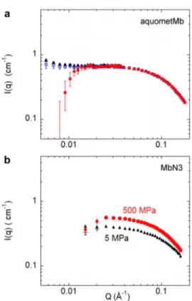
FIG. 4. Scattering intensity of myoglobin solutions at a concentration of about 20 g/l after data corrections.21The incoherent background is not subtracted. (a) Aquometmyoglobin (aquometMb) solution at pD = 6 measured in a quartz Hellma cell (N) or in the HP cell using 2∗6.1 mm-thick windows made of either Nb (◽)
or Al2017A (•), at a pressure of 5 MPa. (b) Azidometmyoglobin (MbN3) at pD = 6
measured in a quartz Hellma cell (N) or in the HP cell with 2∗6.1 mm-thick
win-dows made of Al7049A, at 5 MPa (+) or 500 MPa (•). Note that data from 5 MPa (+) are almost superimposed on those measured at ambient pressure in a quartz cell (N).
maximum pressure of 450 MPa and 500 MPa for Al-2017A and Al-7049A alloys, respectively. The Q-range was limited to val-ues above 0.02 Å−1, which was nevertheless suitable to study protein conformation.
Finally, we tested the “high absorption but rather low scattering” TiAl6V4 ELI compound [TiAl6V4 ELI displays a slightly higher scattering than TiAl6V4, as shown inFig. 3(c)]. Due to the high strength of this alloy, it is possible to reduce the window thickness. Using 4.7 mm-thickness windows of TiAl6V4 ELI allowed us to measure samples up to 600 MPa (see below), whereas a 3 mm-thickness improved the window transmission at possible pressures up to 300 MPa.
V. HIGH PRESSURE EFFECTS ON MYOGLOBIN SOLUTIONS WITH DIFFERENT LIGANDS
SANS experiments under HP were performed on the PACE spectrometer (Fig. S1 of the supplementary material) at the LLB (Saclay, France) at 6 Å neutron wavelength, using 4.7 mm-thick windows of TiAl6V4 ELI alloy. Using these windows,
detector distance of about 1.6 m was chosen in order to reach a minimum Q-value of 0.02 Å−1, that is, the minimum Q limit for which we have previously observed we could correctly recover a weak sample signal, and a maximum Q value of 0.2 Å−1, permitted by the opening angle of the cell. Two solutions of Mb were prepared in the same 100 mM Tris buffer (pD 6): aquometmyoglobin (aquometMb) at 7 g/l and azidometmyo-globin (MbN3) at 5 g/l. These samples were studied by SANS at pressures up to 500 and 600 MPa, respectively. We also mea-sured the buffer (not shown) in order to mimic, as closely as possible, the incoherent signal of both protein solutions and its putative variation according to the pressure up to 600 MPa. For comparison, both buffer and protein samples were also measured in quartz Hellma cells (2 mm-thickness) at atmo-spheric pressure. The buffer signal, while small (around 0.06-0.07 cm−1) at ambient pressure, slightly increases with HP. We fitted this signal to a constant and subtracted this constant as a function of the pressure to the sample scattering data we got at the corresponding pressure.
SANS curves of MbN3 and aquometMb solutions, after incoherent background subtraction, are shown inFig. 5. MbN3 data recorded in a quartz cell at atmospheric pressure and in the HP cell at 5 MPa (this low pressure being considered as almost “no pressure” in the HP cell) superimposed perfectly within the Q-range [Fig. 5(a)]. This demonstrated that, even using a “high absorption and scattering” cell, we were able to recover a weak scattering sample signal.
For MbN3[Fig. 5(b)], the SANS intensity was virtually the same all along the pressure increase, in agreement with the data obtained on this protein in similar conditions, but with pressures limited to 300 MPa, using a HP cell with sapphire windows.5,6Here, we show that the absence of variation was confirmed up to 500 MPa. The behavior of the protein solution under HP was significantly different compared to aquometMb [Fig. 5(c)]. The scattering intensity changed significantly at 300 MPa, compared to the atmospheric pressure, and this variation was more pronounced at 600 MPa. From 7.5 MPa, protein oligomerization and/or aggregation might occur and these processes might be enhanced by increasing pressure. At 600 MPa, a clear aggregation took place, as shown by the huge increase in the scattered intensity at low Q.
Meanwhile, we can already conclude that it is notewor-thy to be able to reach such HP values, which is now possible with this new HP cell device. The comparison between the response of MbN3 and aquometMb under high hydrostatic pressures highlights the role of the ligands on the protein-protein interactions. A further quantitative analysis is out of the scope of the present article.
In conclusion, we developed a new HP cell to study molecular solutions, especially soluble proteins such as myo-globin, by SANS. The metallic alloys used as HP cell windows allowed us to reach or even overcome the maximum pressure limit of usually 300 MPa,6,7 more rarely 500 MPa,19 previ-ously reached using single crystals of sapphire. Among vari-ous compounds, the TiAl6V4 ELI metallic alloy gave us access to higher pressures, up to 600 MPa, while allowing to study dilute (∼5 g/l) solutions of proteins. For smaller maximum
Review of
Scientific Instruments
ARTICLE scitation.org/journal/rsiFIG. 5. Scattering intensities of myoglobin solutions in 100 mM Tris buffer (pD 6), after subtraction of a constant background. (a) Azidometmyoglobin (MbN3) at 5 g/l
in a quartz Hellma cell (N) or in the HP cell using TiAl6V4 ELI windows at 5 MPa (+). (b) MbN3at 5 g/l at 5 MPa (+), 300 MPa (◽), or 500 MPa (•) using the same
HP cell. (c) Aquometmyoglobin (aquometMb) at 7 g/l at 7.5 MPa (+), 300 MPa (◽), or 600 MPa (•) using the same HP cell.
pressures, up to 280 MPa, pure Nb windows would be a good alternative.
SUPPLEMENTARY MATERIAL
Seesupplementary materialfor Fig. S1 (photograph of the overall HP cell device installed on a SANS instrument), Table S1 (transmission of thick pieces of compounds measured at 6 Å wavelength), and Table S2 (mechanical properties of various compounds tested as pressure windows).
ACKNOWLEDGMENTS
A huge part of this device development was supported by the European Commission under the 7th Framework Pro-gram through the “Research Infrastructures” action of the “Capacities” Program, NMI3-II Grant No. 283883. We warmly thank Dr. R. Schweins, responsible for D11 SANS instrument at the Institut Laue-Langevin (ILL, Grenoble, France), for his precious help to test some metallic alloy windows on D11. We also thank the Sample Environment group of the ILL, especially Dr. E. Lelièvre-Berna and J. Gonthier, for valuable discussions.
REFERENCES
1H. Frauenfelder, S. G. Sligar, and P. G. Wolynes, “The energy
landscapes and motions of proteins,” Science 254, 1598–1603
(1991).
2H. Frauenfelder, P. W. Fenimore, and R. D. Young, “Protein dynamics and
function: Insights from the energy landscape and solvent slaving,”IUBMB Life59, 506–512 (2007).
3H. Frauenfelder et al., “Proteins and pressure,”J. Phys. Chem.94, 1024–1037
(1990).
4C. R. Chen and G. I. Makhatadze, “Molecular determinant of the effects
of hydrostatic pressure on protein folding stability,”Nat. Commun.8, 14561 (2017).
5P. W. Bridgman, “The coagulation of albumen by pressure,” J. Biol. Chem.
19, 511–512 (1914).
6C. Loupiac, M. Bonetti, S. Pin, and P. Calmettes, “High-pressure effects on
horse heart metmyoglobin studied by small-angle neutron scattering,”Eur. J. Biochem.269, 4731–4737 (2002).
7M. Bonetti and P. Calmettes, “High-pressure cell for small- and
medium-angle neutron scattering measurements up to 300 MPa,”Rev. Sci. Instrum. 75, 440–444 (2004).
8M. G. Ortore et al., “High pressure small-angle neutron scattering study
of the aggregation state of β-lactoglobulin in water and in water/ethylene-glycol solutions,”Chem. Phys. Lett.418, 342–346 (2006).
9N. Osaka, S. Takata, T. Suzuki, H. Endo, and M. Shibayama, “Comparison of
heat- and pressure-induced gelation of β-lactoglobulin aqueous solutions studied by small-angle neutron and dynamic light scattering,”Polymer49, 2957–2963 (2008).
10G. Gibrat et al., “High-pressure SANS and fluorescence unfolding study of
calmodulin,”Biochim. Biophys. Acta, Proteins Proteomics1844, 1560–1568 (2014).
11M. S. Appavou, G. Gibrat, and M. C. Bellissent-Funel, “Influence of
pres-sure on structure and dynamics of bovine pancreatic trypsin inhibitor (BPTI): Small angle and quasi-elastic neutron scattering studies,”Biochim. Biophys. Acta, Proteins Proteomics1764, 414–423 (2006).
12M. S. Appavou et al., “The influence of a medium pressure on the structure
and dynamics of a bovine pancreatic trypsin inhibitor protein the influence of a medium pressure on the structure,”J. Phys.: Condens. Matter17, S3093– S3099 (2005).
13J. Marion et al., “Pressure-induced molten globule state of human
acetylcholinesterase: Structural and dynamical changes monitored
by neutron scattering,” Phys. Chem. Chem. Phys. 17, 3157–3163
(2015).
14E. Banachowicz, M. Kozak, and A. Patkowski, “High-pressure small-angle
neutron scattering studies of glucose isomerase conformation in solution,” J. Appl. Crystallogr.42, 461–468 (2009).
15S. Kunugi and N. Tanaka, “Cold denaturation of proteins under high
pressure,”Biochim. Biophys. Acta1595, 329–344 (2002).
16V. Le Tilly, O. Sire, B. Alpert, and P. T. T. Wong, “An infrared study of
2H-bond variation in myoglobin revealed by high pressure,”Eur. J. Biochem. 205, 1061–1065 (1992).
Rev. Sci. Instrum. 90, 025106 (2019); doi: 10.1063/1.5051765 90, 025106-7
Biochemistry12, 4217–4228 (1973).
18H. Takeno et al., “High pressure cell for small-angle neutron and light
scattering studies of phase transitions in complex liquids,”Polym. J.29, 931–939 (1997).
19J. Kohlbrecher, A. Bollhalder, R. Vavrin, and G. Meier, “A high pressure
cell for small angle neutron scattering up to 500 MPa in combination with light scattering to investigate liquid samples,”Rev. Sci. Instrum.78, 125101 (2007).
neutron scattering experiments on gas hydrates,”Can. J. Phys.81, 381–385 (2003).
21A. Brûlet, D. Lairez, A. Lapp, and J. Cotton, “Improvement of data
treat-ment in small-angle neutron scattering,”J. Appl. Crystallogr.40, 165–177 (2007).
22K. Komatsu et al., “Crystal structure of magnesium dichloride
decahy-drate determined by x-ray and neutron diffraction under high pressure,” Acta Crystallogr., Sect. B: Struct. Sci., Cryst. Eng. Mater.71, 74–80 (2015).