RESEARCH OUTPUTS / RÉSULTATS DE RECHERCHE
Author(s) - Auteur(s) :
Publication date - Date de publication :
Permanent link - Permalien :
Rights / License - Licence de droit d’auteur :
Institutional Repository - Research Portal
Dépôt Institutionnel - Portail de la Recherche
researchportal.unamur.be
University of Namur
Local mitochondrial-endolysosomal microfusion cleaves voltage- dependent anion
channel 1 to promote survival in hypoxia
Brahimi-Horn, M. Christiane; Lacas-Gervais, Sandra; Adaixo, Ricardo; Ilc, Karine; Rouleau,
Matthieu; Notte, Annick; Dieu, Marc; Michiels, Carine; Voeltzel, Thibault; Maguer-Satta,
Véronique; Pelletier, Joffrey; Ilie, Marius; Hofman, Paul; Manoury, Bénédicte; Schmidt,
Alexander; Hiller, Sebastian; Pouysségur, Jacques; Mazure, Nathalie M.
Published in:
Molecular Cell Biology
DOI:
10.1128/MCB.01402-14
Publication date:
2015
Document Version
Early version, also known as pre-print
Link to publication
Citation for pulished version (HARVARD):
Brahimi-Horn, MC, Lacas-Gervais, S, Adaixo, R, Ilc, K, Rouleau, M, Notte, A, Dieu, M, Michiels, C, Voeltzel, T,
Maguer-Satta, V, Pelletier, J, Ilie, M, Hofman, P, Manoury, B, Schmidt, A, Hiller, S, Pouysségur, J & Mazure, NM
2015, 'Local mitochondrial-endolysosomal microfusion cleaves voltage- dependent anion channel 1 to promote
survival in hypoxia', Molecular Cell Biology, vol. 35, no. 9, pp. 1491-1505. https://doi.org/10.1128/MCB.01402-14
General rights
Copyright and moral rights for the publications made accessible in the public portal are retained by the authors and/or other copyright owners and it is a condition of accessing publications that users recognise and abide by the legal requirements associated with these rights. • Users may download and print one copy of any publication from the public portal for the purpose of private study or research. • You may not further distribute the material or use it for any profit-making activity or commercial gain
• You may freely distribute the URL identifying the publication in the public portal ?
Take down policy
If you believe that this document breaches copyright please contact us providing details, and we will remove access to the work immediately and investigate your claim.
Dependent Anion Channel 1 To Promote Survival in Hypoxia
M. Christiane Brahimi-Horn,aSandra Lacas-Gervais,bRicardo Adaixo,cKarine Ilc,aMatthieu Rouleau,dAnnick Notte,eMarc Dieu,e Carine Michiels,eThibault Voeltzel,fVéronique Maguer-Satta,fJoffrey Pelletier,aMarius Ilie,a,gPaul Hofman,a,gBénédicte Manoury,h Alexander Schmidt,cSebastian Hiller,cJacques Pouysségur,a,iNathalie M. Mazurea
Institute for Research on Cancer and Aging of Nice, CNRS-UMR 7284-INSERM U1081, University of Nice Sophia-Antipolis, Centre Antoine Lacassagne, Nice, Francea
; Centre Commun de Microscopie Appliquée, University of Nice Sophia-Antipolis, Nice, Franceb
; Biozentrum, University of Basel, Basel, Switzerlandc
; Laboratoire de PhysioMédecine Moléculaire, UMR 7370 CNRS, University of Nice Sophia-Antipolis, Faculty of Medicine, Nice, Franced
; URBC-NARILIS, University of Namur, Namur, Belgiume
; Centre de Recherche en Cancérologie de Lyon, INSERM U1052, CNRS U5286, Centre Léon Bérard, Lyon, Francef
; Human Tissue Biobank Unit/CRB and Laboratory of Clinical and Experimental Pathology, Louis Pasteur Hospital, Nice, Franceg
; INSERM U1013, Hôpital Necker, Paris, Franceh
; Centre Scientifique de Monaco (CSM), Monacoi
The oxygen-limiting (hypoxic) microenvironment of tumors induces metabolic reprogramming and cell survival, but the
under-lying mechanisms involving mitochondria remain poorly understood. We previously demonstrated that hypoxia-inducible
fac-tor 1 mediates the hyperfusion of mitochondria by inducing Bcl-2/adenovirus E1B 19-kDa interacting protein 3 and
posttransla-tional truncation of the mitochondrial ATP transporter outer membrane voltage-dependent anion channel 1 in hypoxic cells. In
addition, we showed that truncation is associated with increased resistance to drug-induced apoptosis and is indicative of
in-creased patient chemoresistance. We now show that silencing of the tumor suppressor TP53 decreases truncation and increases
drug-induced apoptosis. We also show that TP53 regulates truncation through induction of the mitochondrial protein Mieap.
While we found that truncation was independent of mitophagy, we observed local microfusion between mitochondria and
en-dolysosomes in hypoxic cells in culture and in patients’ tumor tissues. Since we found that the endolysosomal asparagine
endo-peptidase was responsible for truncation, we propose that it is a readout of mitochondrial-endolysosomal microfusion in
hyp-oxia. These novel findings provide the framework for a better understanding of hypoxic cell metabolism and cell survival
through mitochondrial-endolysosomal microfusion regulated by hypoxia-inducible factor 1 and TP53.
H
ypoxia is a natural occurring stress that results in
compensa-tory changes in metabolism and cell survival during
embry-onic development and tumor growth. Hypoxia stabilizes and
ac-tivates the transcription factor hypoxia-inducible factor (HIF)
through inhibition of oxygen-dependent hydroxylases that
ear-mark the alpha subunit of HIF for proteasomal degradation (
1
).
HIF induces or represses the expression of genes implicated in a
myriad of functions, including those regulating metabolism and
resistance to drug-induced cell death. Genes coding for the
en-zymes of the glycolytic pathway, including hexokinase, are highly
induced by HIF-1, and this is in part responsible for the switch in
metabolism from mitochondrial respiration to glycolysis in
can-cer cells. Considerable studies have pointed to the Warburg effect,
also termed aerobic glycolysis, as the major adaptive response of
cancer cells, but mitochondrial metabolism and mitochondrial
dynamics are also starting to be recognized as important adaptive
strategies of cancer cells (
2
). Mitochondria are critical organelles
that regulate both metabolism and cell death. They are dynamic
organelles that continuously undergo fission and fusion during
cell growth (
3
,
4
). Under stress conditions, such as nutrient
deple-tion or hypoxia, mitochondria either fragment or are degraded by
HIF-dependent mitophagy (mitochondrial removal by
au-tophagy) (
5
) or hyperfuse together to form elongated or rounded
structures that optimize ATP production and promote cell
sur-vival (
6–11
).
We reported previously that certain cell lines exposed to
hyp-oxia contained enlarged mitochondria (
6
). We found that the
mechanism was HIF-1 and Bcl-2/adenovirus E1B 19-kDa
inter-acting protein 3 (BNIP3/BNIP3L) dependent but that it was
inde-pendent of mitophagy. In addition, the hypoxic cells were more
resistant to stimulated cell death than normoxic cells (
12
).
Fur-thermore, we reported that the mitochondrial outer membrane
protein voltage-dependent anion channel 1 (VDAC1) was
post-translationally cleaved at the C terminus in these cells in a
HIF-1-dependent manner and in human lung adenocarcinoma tissue
(
12
). VDAC mediates the transport of ions and small metabolites,
such as ADP/ATP, from and into mitochondria (
13
). Three
mam-malian isoforms of VDAC exist in eukaryotic cells. VDACs bind
hexokinase, the first enzyme of the glycolytic pathway, and in so
doing provide ATP for conversion of glucose to
glucose-6-phos-phate. VDACs also play a key role in apoptosis through Ca
2⫹Received 19 November 2014 Returned for modification 16 December 2014 Accepted 6 February 2015
Accepted manuscript posted online 17 February 2015
Citation Brahimi-Horn MC, Lacas-Gervais S, Adaixo R, Ilc K, Rouleau M, Notte A, Dieu M, Michiels C, Voeltzel T, Maguer-Satta V, Pelletier J, Ilie M, Hofman P, Manoury B, Schmidt A, Hiller S, Pouysségur J, Mazure NM. 2015. Local mitochondrial-endolysosomal microfusion cleaves voltage-dependent anion channel 1 to promote survival in hypoxia. Mol Cell Biol 35:1491–1505.
doi:10.1128/MCB.01402-14.
Address correspondence to Nathalie M. Mazure, Nathalie.Mazure@unice.fr. Supplemental material for this article may be found athttp://dx.doi.org/10.1128 /MCB.01402-14.
Copyright © 2015, American Society for Microbiology. All Rights Reserved.
doi:10.1128/MCB.01402-14
on April 19, 2015 by Xavier De Bolle
http://mcb.asm.org/
regulation of VDAC1 expression and binding of antiapoptotic
proteins of the Bcl-2 family (
14
,
15
).
The TP53 transcription factor plays an important role in the
response to and regulation of metabolic stress in cancer (
16
,
17
). It
is known that a TP53-inducible protein, Mieap (also referred to as
Spata18) (
18
), controls mitochondrial quality through interaction
with the HIF-1-inducible protein BNIP3 (
19
). In addition, Mieap
has been proposed to induce the accumulation of lysosomal
pro-teins within mitochondria by way of repairing damaged
mito-chondria (
20
).
In the present study, we investigated further the mechanism
behind the hypoxic regulation of the truncation of VDAC1. We
propose that enlarged hypoxic mitochondria make fusional
con-tact with late endolysosomes through TP53-induced Mieap in
promoting cell survival. Furthermore, we report that VDAC1 is
cleaved at loop 14 by the endolysosomal protease asparagine
en-dopeptidase (also termed legumain). Intimate contact between
mitochondria and vacuoles has been described only in
Saccharo-myces cerevisiae yeast (
21
,
22
) and in erythroid cells (
23
). This
cross talk between organelles was found to regulate lipid
trans-port, cellular metabolism, and iron transport. We now show that a
spatial and functional interorganellar connection also exists in
eukaryotes, both in cells in culture and in lung adenocarcinomas
of patients. We propose, finally, that VDAC posttranslational
modification marks mitochondria for protection from mitophagy
and reflects a survival strategy of hypoxic cancer cells in vitro and
in vivo in patients.
MATERIALS AND METHODS
Cells and hypoxic conditions. Cells were grown in Dulbecco’s modified
Eagle’s medium (DMEM; Gibco-BRL) supplemented with 5 or 10% in-activated fetal bovine serum, as appropriate. M. van de Wetering provided the LS174 cells. p53⫹/⫹and p53⫺/⫺mouse embryonic fibroblasts (MEFs)
were provided by P. Roux. An Invivo2 200 anaerobic workstation
(Ruskinn Technology Biotrace International Plc) set at 1% O2, 94% N2,
and 5% CO2was used for hypoxic incubation. HeLa cells were incubated
for 48 h in hypoxia, while the other cells, unless otherwise indicated, were incubated for 72 h.
Pharmacological inhibitors and chemicals. Bafilomycin A1 was
pur-chased from Calbiochem. Brefeldin A, chloroquine diphosphate, LY-294002, 3-methyladenine, rapamycin, valinomycin, pepstatin A, E64d, rotenone antimycin, oligomycin, and trifluorocarbonylcyanide phenyl-hydrazone (FCCP) were from Sigma.
Transfection of plasmids and RNA interference. The plasmid
carry-ing yellow fluorescent protein (YFP)-tagged Parkin (YFP-Parkin) was purchased from Addgene (24). The 21-nucleotide RNAs were synthesized by Eurogentec or OriGene. HeLa or LS174 cells were transfected with 40 or 100 nM small interfering RNA (siRNA) twice at an interval of 24 h prior to normoxia or hypoxia, as described previously (6).
siRNA sequences were as follows: siRNA against VDAC1 (forward), 5=-GAUACACUCAGACUCUAAA-3=; siRNA against VDAC1 3= un-translated region (forward), 5=-CUCCAGGUUAAAGUUGAUUCA-3=; siRNA against VDAC2 (forward), 5=-GAAGAUUUGGCCUUAAUAU-3=; siRNA against TP53#1 (forward), 5=-GACUCCAGUGGUAAUC UAC-3=; siRNA against TP53#3 (forward), 5=-UAUGGCGGGAGGUAG ACUG-3=; siRNA against autophagy-related gene 5 (ATG5)/ATG6/ATG7/be-clin, the sequences were described previously (25); siRNA against ATG4B (forward), 5=-CUGAAGAUGACUUCAAUGA-3=; siRNA against Lamp2
(forward), 5=-GAAAAUGCCACUUGCCUUU-3=; siRNA against
Mieap#1 (forward), 5=-GUAGCAGUGACUUAAGGCUAAG-3=; siRNA against Mieap#2 (forward), 5=-GGUGCAGGGACAACUCUUUGGG-3=; siRNA against Mieap#3 (forward), 5=-GAGAUAUGUUGCAUUGCCUU UGC-5=; siRNA against Mieap#4 (forward), 5=-GCACUAUCUGCCUAG
GUAACUGC-5=; siRNA against Lamp1A (forward), 5=-AGAAAUGCAA CACGUUA-5=; siRNA against Lamp2A (forward), 5=-GCUGUGCGGUC UUAUGCAU-5=; siRNA against BNIP3 and B3L, the sequences were described previously (25); and siRNA against mitofusin 1 (Mfn1), the se-quence was previously described (12). Three siRNAs against asparagine en-dopeptidase and a scrambled siRNA were purchased from Origene; the se-quences were not disclosed.
Caspase activation and cell death induced by STS. Quantification of
caspase 3/7 activity was done using a luciferin/luciferase-based assay (Caspase-Glo 3/7 kit; Promega) according to the manufacturer’s instruc-tions. Tests under each condition were performed eight times, and the entire experiment was performed three times. Significant differences (P⬍ 0.005) were based on the Student t test. Staurosporine (STS; 1M) was added 4 h prior to the assay for caspase 3/7 activity. Cell death was also determined by trypan blue exclusion and confirmed with an Adam cell counter.
Transmission electron microscopy. Cells were fixed in situ with 1.6%
glutaraldehyde in 0.1 M phosphate buffer at room temperature for at least 1 h and then conserved at 4°C. Samples were rinsed in the same buffer and then postfixed with 1% osmium tetroxide and 1% potassium ferrocyanide in 0.1 M cacodylate buffer for 1 h at room temperature to enhance the staining of cytoplasmic membranes. Cells were rinsed with distilled water, dehydrated in alcohol, and embedded in epoxy resin. Embedded samples were then processed for ultrathin sectioning and observed with a JEM1400 transmission electron microscope (JEOL, Tokyo, Japan) equipped with a Morada charge-coupled-device camera (Olympus SIS, Rungis, France).
For immunogold staining, cells were fixed with 4% paraformaldehyde and 0.1% glutaraldehyde in 0.1 M phosphate buffer (PB; pH 7.4) for 2 h and were processed for ultracryomicrotomy according to a slightly mod-ified Tokuyasu method. In brief, a cell suspension was spun down in 10% gelatin. After immersion in 2.3 M sucrose (in pH 7.4 0.1 M PB) overnight at 4°C, the samples were rapidly frozen in liquid nitrogen. Ultrathin (70-nm-thick) cryosections were prepared with an ultracryomicrotome (Leica EMFCS, Austria) and mounted on Formvar-coated nickel grids (Electron Microscopy Sciences, Fort Washington, PA). Immunostaining was per-formed with an automated immunogold labeling system (Leica EM IGL) as follows: the grids were incubated successively in phosphate-buffered saline (PBS) containing 50 mM NH4Cl (2 times for 5 min each time), PBS containing 1% bovine serum albumin (BSA; 2 times for 5 min each time), PBS containing the relevant primary antibody (for VDAC1, antibody ab15895; for Lamp1, antibody ab25630; for Lamp2, antibody ab25631 [all antibodies were from Abcam]) in 1% BSA for 1 h, PBS containing 0.1% BSA (3 times for 5 min each time), PBS containing 1% BSA and 15-nm colloidal gold-conjugated protein AG (Cell Microscopy Core, University Medical Center, Utrecht, The Netherlands), PBS containing 0.1% BSA for 5 min, and PBS twice for 5 min each time. Lastly, the samples were fixed for 10 min with 1% glutaraldehyde, rinsed in distilled water, and con-trasted with a mixture of methylcellulose-sucrose and 0.3% uranyl acetate on ice. After drying in air, the sections were examined under a JEOL 1400 transmission electron microscope. For Lamp1 and Lamp2 staining with mouse primary antibodies, an additional step was added before the incu-bation with the colloidal gold protein AG: the grids were incubated with a secondary rabbit antibody raised against mouse IgG (antibody Z0259; Dako).
Respirometry and extracellular acidification. The cellular oxygen
consumption rate (OCR) and extracellular acidification rate (ECAR) were obtained using a Seahorse XF96 extracellular flux analyzer from Seahorse Bioscience (North Billerica, MA). The final concentrations of the agents are given in the appropriate figure legends. The experiments were per-formed according to the manufacturer’s instructions. Protein standard-ization was performed after each experiment, and no noticeable differ-ences in protein concentration and cell phenotype were detected.
Immunoblotting. For immunoblotting, cells were lysed in 1.5⫻ SDS
buffer and the protein concentration was determined using a
on April 19, 2015 by Xavier De Bolle
http://mcb.asm.org/
choninic acid assay. Forty micrograms of protein from whole-cell extracts was resolved by SDS-PAGE and transferred onto a polyvinylidene diflu-oride membrane (Millipore). The membranes were blocked in 5% nonfat milk in TN buffer (50 mM Tris-HCl, pH 7.4, 150 mM NaCl) and incu-bated in the presence of the primary antibody and then secondary anti-bodies in 5% nonfat milk in TN buffer. The rabbit polyclonal antibody to the central regions of VDAC1 was purchased from Abcam (antibody ab15895). Rabbit polyclonal anti-HIF-1␣ antibody (antiserum 2087) was produced and characterized in our laboratory (26). Anti-phospho-TP53 (Ser15) (Cell Signaling Technology, Boston, MA), anti-transactivation TP73 (anti-TA TP73) (catalog number IMG-246; Imgenex), anti-Mieap (Novus Biologicals, Cambridge, United Kingdom), anti-p62 (Sigma-Al-drich, St. Louis, MO), and anti-TP63 and anti-beclin 1 (Novus Biological, Cambridge, United Kingdom) were obtained from the indicated manu-facturers. Mouse anti--tubulin and -actin were from Sigma, and anti-asparaginyl endopeptidase (anti-AEP; EC 3.4.22.34; Sigma Prestige) was from Sigma. Anti-BNIP3 and anti-BNIP3L were described previously (25). After washing in TN buffer containing 1% Triton X-100 and then in TN buffer, immunoreactive bands were visualized with an ECL system (Amersham Biosciences). The enhanced chemiluminescence (ECL) signal for-tubulin or -actin was used as a loading control.
Pulldown with BNIP3 and mass spectrometry. HepG2 cells with
sta-ble expression of the tetracycline repressor (pcDNA6/TR; Life Technolo-gies) were transiently transfected with the pcDNA 4/TO plasmid express-ing the HaloTag-BNIP3 fusion protein usexpress-ing the Lipofectamine reagent. At 24 h after the transfection, the cells were then incubated in the presence of 1g/ml tetracycline to induce HaloTag-BNIP3 expression and incu-bated without or with etoposide 50M in hypoxia (1% O2) for 16 h.
Protein pulldown was performed according to the HaloTag mamma-lian pulldown system protocol (Promega). Tobacco etch virus protease cleavage (30 units of Protev proteases in 50l of Protev cleavage buffer for 1 h at 25°C) was used to isolate the entire complex, including the bait protein BNIP3 fused to the HaloTag. After elution, proteins were boiled for 5 min with 3-[3-(1,1-bisalkyloxyethyl)pyridin-1-yl]propane-1-sulfo-nate (PPS; final concentration, 0.8%; silent surfactant; Protein Discov-ery), reduced for 30 min at 50°C with dithiothreitol (5 mM), and then alkylated for 30 min in the dark using iodoacetamide (15 mM). Samples were then digested overnight at 37°C with trypsin (Trypsin Gold, mass spectrometry [MS] grade; Promega). Prior to MS, samples were acidified with 1l of 12 N HCl, and PPS detergent was hydrolyzed after a 45-min incubation at 37°C, followed by centrifugation (10 min, 16,000⫻ g) at 4°C. Peptides were analyzed using a nano-liquid chromatography (LC)-electrospray ionization-tandem MS maXis Impact ultra-high-resolution time of flight system (Bruker, Bremen, Germany) coupled with a nano-LC UltiMate 3000 system (Thermo). The digests were separated by reverse-phase liquid chromatography using a reverse-reverse-phase Thermo column (75 m by 250 mm; Acclaim PepMap 100 C18) in an Ultimate 3000 liquid
chromatography system. Mobile phase A was 95% water, 5% acetonitrile, and 0.1% formic acid. Mobile phase B was 20% water, 79.9% acetonitrile, and 0.1% formic acid. The digest (8l) was injected, and the organic content of the mobile phase was increased linearly from 5% mobile phase to 40% mobile phase B in 85 min and from 40% mobile phase B to 100% mobile phase B in 10 min. The column effluent was connected to a Captive Spray apparatus (Bruker). In the survey scan, MS spectra were acquired for 0.5 s in the mass-to-charge (m/z) ratio range of from 50 and 2,200. The 15 most intense peptide ions with charges of⫹2 or ⫹3 were sequenced. The collision-induced dissociation (CID) energy was automatically set according to the m/z ratio and charge state of the precursor ion. The maXis and Thermo systems were piloted by Compass HyStar (version 3.2) software (Bruker).
Peak lists were created using the DataAnalysis (version 4.0) program (Bruker) and saved as an MGF file for use with the ProteinScape (version 3.1) program (Bruker) and Mascot (version 2.4) as the search engine (Matrix Science). Enzyme specificity was set to trypsin, and the maximum number of missed cleavages per peptide was set to 1.
Carbamidomethyla-tion, oxidation of methionine, and Gln–pyro-Glu were allowed as variable modifications. The mass tolerance for the monoisotopic peptide window was 7 ppm, and the MS/MS tolerance window was set to 0.05 Da. The peak lists were searched against the NCBInr mammalian database (entries 02122011 and 2075986) with an automatic decoy database search. Scaf-fold (version 4.3) program (Proteome Software) was used to validate the MS/MS-based peptide and protein identifications. Peptide identifications were accepted if they could be established at a greater than 95% probabil-ity by use of the PeptideProphet algorithm (27) with Scaffold delta-iden-tified peptides. Protein identifications were accepted if they could be es-tablished at greater than 5.0% probability to achieve a false discovery rate (FDR) of less than 1.0% and contained at least 2 identified peptides. Pro-tein probabilities were assigned with the ProPro-teinProphet algorithm (28).
Mass spectrometry of VDAC1 and VDAC1-⌬C. Imperial
blue-stained bands excised from SDS-polyacrylamide gels, corresponding to full-length VDAC1 from normoxic and hypoxic cell mitochondria and a
truncated form of VDAC1 (VDAC1-⌬C) from hypoxic HeLa cell
mito-chondria, were digested in gel with trypsin, as recently described (29). For LC-MS analysis, peptides were separated on a reverse-phase LC column (75m by 20 cm) packed in-house with C18resin (Magic C18AQ; particle size, 3m; Michrom BioResources, Auburn, CA) using a linear gradient from 95% solvent A (98% water, 2% acetonitrile, 0.15% formic acid) and 5% solvent B (98% acetonitrile, 2% water, 0.15% formic acid) to 30% solvent B over 40 min at a flow rate of 0.2l/min. Each survey scan acquired in the Orbitrap analyzer at 60,000 full width at half maximum was followed by 20 MS/MS scans of the most intense precursor ions in the linear ion trap with dynamic exclusion enabled for 20 s. Charge state screening was employed to select for ions with at least two charges and rejecting ions with an undetermined charge state. The normalized colli-sion energy was set to 32%, and one microscan was acquired for each spectrum. The ion accumulation time was set to 300 ms (MS) and 50 ms (MS/MS).
For peptide identification, raw files were converted to MGF format using the MassMatrix conversion tool (version 3.9;www.massmatrix.org) and searched against a decoy human Swiss-Prot database consisting of forward and reverse protein sequences (download date, 16 May 2012) containing VDAC and known contaminants, resulting in a total of 41,251 protein sequences using the Mascot (version 2.4) search engine (Matrix Science). The search parameters were set as follows; semitryptic specificity was required, up to two missed cleavages were allowed, carbamidomethyl of cysteine residues was set as fixed, oxidation of methionine residues was set as a variable modification, the precursor mass tolerance was 10 ppm, and the fragment mass tolerance for CID tandem mass spectra was 0.6 Da. After importing the data to Scaffold software (version 4.2.1; Proteome Software), the FDR was set to⬍1% for peptide identifications by the Scaffold local FDR algorithm.
For peptide quantification, raw files were loaded into the Skyline soft-ware tool (version 2.4) to generate extracted ion chromatograms and determine peak abundances for all identified peptide ions generated by the VDAC protein. All integrated peaks/transitions were manually in-spected and validated, if required.
SDS-PAGE of recombinant AEP incubated with recombinant VDAC1 or hypoxic HeLa cell mitochondria. Recombinant VDAC1
ex-pressed in Escherichia coli was reconstituted into either N,N-dimethyldo-decylamine N-oxide (LDAO) micelles or large unilamellar vesicles (LUVs) following published protocols (30,31) and incubated with low-pH-autoactivated (0.1 M citrate buffer, pH 5.5) recombinant AEP (rAEP) (32) for 180 min at room temperature. Samples of inactive or low-pH-autoactivated rAEP and recombinant VDAC1, together with samples of active AEP incubated with VDAC1 LUVs or LDAO micelles, were ana-lyzed by SDS-PAGE. Gels were stained with Coomassie blue. Mitochon-dria from HeLa cells were isolated using a mitochondrion isolation kit (MitoSciences). Mitochondria were resuspended in 0.1 M citrate buffer (pH 5.5), and rAEP was added. AEP-expressing plasmid pcDNA3.1 was used for overexpression of AEP.
on April 19, 2015 by Xavier De Bolle
http://mcb.asm.org/
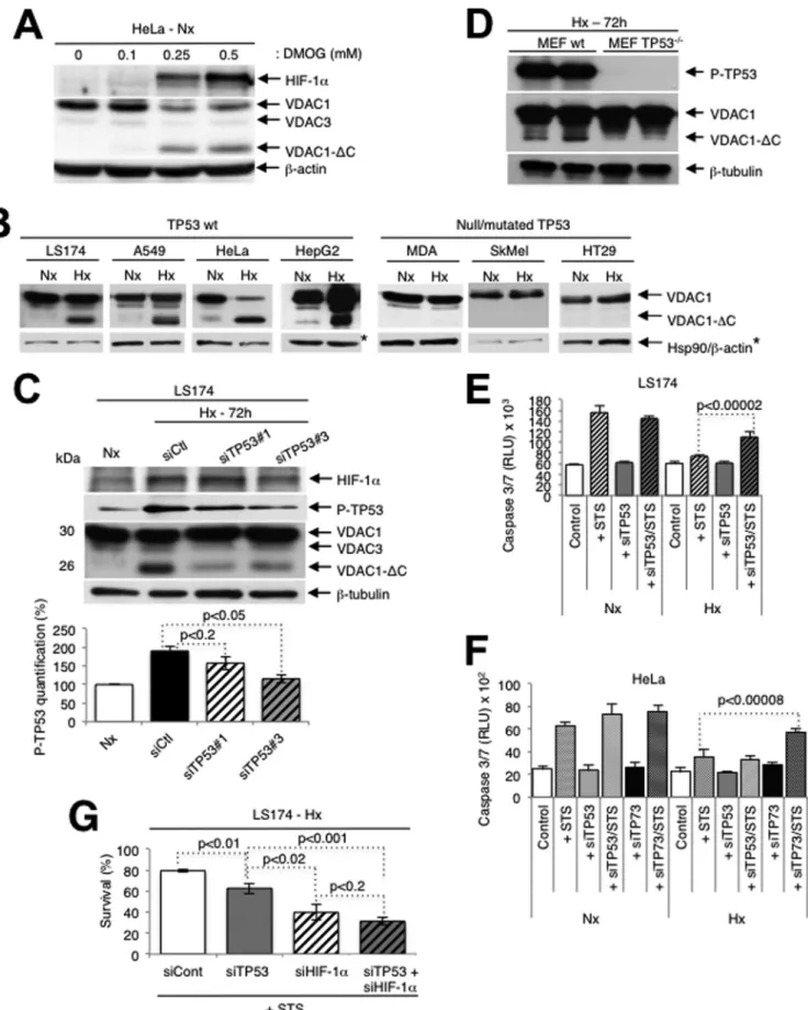
FIG 1 Truncation of VDAC1 requires wild-type TP53. (A) HeLa cells were treated for 24 h with dimethyl-oxalylglycine, and cell lysates were analyzed by
immunoblotting for HIF-1␣, VDAC, or -actin. (B) Wild-type (wt) or TP53 null or mutated cells were analyzed by immunoblotting for VDAC, HSP90, or -actin. (C) (Top) LS174 cells were transfected with control siRNA (siCtl) or TP53-specific siRNA (siTP53; 40 nM) in normoxia (Nx) or hypoxia (Hx), and cell lysates were analyzed by immunoblotting for HIF-1␣, TP53, phospho-TP53 (P-TP53), VDAC, or -tubulin. (Bottom) Quantification of TP53 is shown. (D) wt
on April 19, 2015 by Xavier De Bolle
http://mcb.asm.org/
Patients and tissue sample preparation. Ten patients who underwent
surgery for lung adenocarcinoma between May 2007 and May 2010 at the Pasteur Hospital (Department of Thoracic Surgery, CHU of Nice, Nice, France) were selected. The patients received the necessary information concerning the study, and consent was obtained. The study obtained the approval of the Ethics Committee of the CHU of Nice. The main clinical and histopathological data are summarized in Table S2 in the supplemen-tal material. Morphological classification of the tumors was assigned ac-cording to the WHO criteria (33). The tumors were staged according to the international tumor-node-metastasis system (34). Tumors with at least 80% tumor cells were selected. Protein and microRNA were ex-tracted from the same tissue sample using an AllPrep DNA/RNA/protein kit according to the protocol described by the manufacturer (Qiagen).
Statistics. All values are the means⫾ standard deviations (SDs).
Sig-nificant differences are based on the Student t test, and P values are indi-cated in the figures. All statistical tests were two-sided, and P values of ⬍0.05 indicated statistical significance.
RESULTS
Cleavage of VDAC1 is regulated by TP53. We previously
demon-strated that in hypoxia (1% O
2for 48 to 72 h) certain cell lines (the
CCL39, LS174, A549, 786-O, HeLa, and MEF cell lines) showed
enlarged functional mitochondria and enhanced resistance to
staurosporine (STS)-induced apoptosis, while others (the PC3,
SkMel, MDA-MB, and HT29 cell lines) showed a normal tubular
mitochondrial network and were sensitive to apoptosis (
6
). More
recently, we showed that hypoxic cells with enlarged
mitochon-dria contained a truncated form of VDAC1 (VDAC1-⌬C), the
production of which was HIF-1 dependent (
12
). We now confirm
the HIF-1 dependence of VDAC processing with the
pharmaco-logical inhibitor dimethyl-oxalylglycine (DMOG), which
stabi-lizes HIF-1␣ in normoxia through inhibition of HIF dioxygenases
(
Fig. 1A
). In contrast to the findings in hypoxia, DMOG treatment
led to more rapid VDAC1 processing (24 versus 48 h). However,
unlike the findings in hypoxia, high concentrations of DMOG (1
mM) induced cell death. So, subsequent experiments were done
under the more physiological condition of hypoxia. VDAC1-
⌬C
was not detected or only minimally detected in normoxia in any of
the cell lines tested (
Fig. 1B
). We now note that the cells with
enlarged mitochondria were TP53 wild type, while the other cell
lines were either TP53 null or mutant (
Fig. 1B
). Some of these cell
lines also showed increased expression in hypoxia. We showed
that in LS174 cells transfected with TP53-specific siRNA, the level
of VDAC1-⌬C diminished (
Fig. 1C
). The amount of full-length
VDAC1 was substantially increased, which further confirmed that
VDAC1-⌬C is a product of full-length VDAC1, as we previously
demonstrated (
12
). In addition, we examined mouse embryonic
fibroblasts either wild type or null for TP53 and noted that the
former showed VDAC1-
⌬C in hypoxia, while the latter did not
(
Fig. 1D
). Since we found previously that the formation of
VDAC1-
⌬C is associated with increased resistance to
STS-in-duced apoptosis (
12
), we examined if silencing of TP53 modified
the level of resistance. Indeed, in hypoxia, LS174 cells transfected
with TP53-specific siRNA showed more STS-induced cell death
than control transfected cells challenged with STS (
Fig. 1E
).
Knowing that HeLa cells are wild type for TP53 but that the level of
TP53 is low due to degradation by the human papillomavirus E6
protein (
35
), as confirmed here (see Fig. S1A in the supplemental
material), we questioned further the involvement of TP53. In
mammals, the TP53 family comprises two additional proteins,
TP63 and TP73, that activate TP53 target genes (
36
). So, we
hy-pothesized that in HeLa cells these proteins might substitute for
TP53 and thereby promote VDAC cleavage and protection from
induced apoptosis. However, TP63 was present at only very low
levels (see Fig. S1B in the supplemental material) and TP63 siRNA
(DeltaNp63- and Tap63-specific siRNAs) did not modify the level
of VDAC1-
⌬C (see Fig. S1C in the supplemental material) in
hy-poxic HeLa cells. In contrast, siRNA against TP73 diminished the
level of VDAC1-⌬C (see Fig. S1D in the supplemental material)
and enhanced sensitivity to STS-induced cell death (
Fig. 1F
) in
HeLa cells. So, TP73 was able to substitute for TP53 in HeLa cells.
We also noted that the LS174 cells were mutated in TP73 (
37
),
which further confirmed the involvement of TP53 in truncation,
as described above. Silencing of TP53 or of HIF-1␣ in hypoxic
LS174 cells diminished survival, and silencing of the two together
further diminished survival in the presence of STS (
Fig. 1G
).
TP53 regulation of VDAC1 cleavage occurs through
mito-chondrial Mieap and is dependent on the endolysosomal pH. It
is known that a TP53-inducible protein, Mieap (
18
), controls
mi-tochondrial quality (
19
). Using TP53-specific siRNA, we
con-firmed, as published previously (
19
), that Mieap is TP53 inducible
(
Fig. 2A
). Silencing of Mieap expression (see Fig. S1E in the
sup-plemental material) diminished the truncation of VDAC1 (
Fig.
2B
) and increased STS-induced cell death in hypoxia (
Fig. 2C
).
HeLa cells express only TP73, while LS174 cells express only TP53,
yet a similar level of expression of Mieap was noted (
Fig. 2A
; see
also Fig. S1E in the supplemental material). This suggests
ex-changeable gene activation by TP53/TP73. These results
demon-strate that TP53 regulates VDAC1 truncation via Mieap. In fact,
Mieap has been proposed to induce accumulation of lysosomal
proteins within mitochondria by way of repairing damaged
mito-chondria (
20
). However, we were unable to detect the lysosomal
membrane protein Lamp1 or Lamp2 in mitochondria by
immu-noelectron microscopy (see Fig. S2A to C in the supplemental
material), and siRNA against Lamp1 and/or Lamp2 did not
mod-ify the truncation of VDAC1 (see Fig. S2D in the supplemental
material). Instead, we questioned whether lysosomes could
con-tribute to mitochondrial quality control or metabolism by acting
from the outside of the mitochondria rather than from the inside.
HeLa cells were first incubated in hypoxia in the presence of agents
that increase the lysosomal pH, including bafilomycin A1, an
in-hibitor of lysosomal V-ATPase, as well as chloroquine and NH
4Cl,
which are weak bases. All these compounds efficiently inhibited
the cleavage of VDAC1 to VDAC1-⌬C, confirming an important
lysosomal function in VDAC1 cleavage (
Fig. 2D
; see also Fig. S2E
in the supplemental material). Treatment with bafilomycin A1 or
or TP53 null MEFs in hypoxia were analyzed by immunoblotting. (E) LS174 cells were transfected with either control siRNA or TP53-specific siRNA in normoxia or hypoxia and challenged or not challenged with STS (1 mM) for 4 h or not challenged. Apoptosis was evaluated with caspase 3/7 (three independent experiments were performed in quadruplicate, and values are means⫾ SDs). RLU, relative light units. (F) HeLa cells were transfected with either control siRNA or TP73 siRNA in normoxia or hypoxia and analyzed by immunoblotting. (G) LS174 cells were transfected with control siRNA (siCont) or siRNA against TP53, HIF-1␣ (siHIF-1␣), or both TP53 and HIF-1␣ and incubated in hypoxia. Cells were then challenged or not challenged with STS (1 mM) for 4 h or not challenged. Apoptosis was evaluated with an Adam system (independent experiments were performed in triplicate, and values are means⫾ SDs).
on April 19, 2015 by Xavier De Bolle
http://mcb.asm.org/
chloroquine was also associated with an increase in drug-induced
cell death (
Fig. 2E
).
However, as bafilomycin A1 and chloroquine are also known
to inhibit the maturation and degradation of autophagic vacuoles,
as confirmed here (
Fig. 2F
; see also Fig. S2F in the supplemental
material), and as hypoxia also induces autophagy through HIF-1␣
stabilization, as seen from the enhanced expression of p62/
SQSTM1, the autophagic adaptor protein (see Fig. S3A in the
FIG 2 Truncation of VDAC1 requires TP53-induced Mieap and an increase in endosomal-lysosomal pH-inhibited VDAC1-⌬C formation. (A) LS174 cells were
transfected with either control siRNA (siCtl) or TP53-specific siRNA (siTP53#3; one of three TP53-specific siRNAs; 40 nM), incubated in hypoxia (Hx), and lysed for analysis by immunoblotting. (B) HeLa cells were transfected with either control siRNA or two different Mieap-specific siRNAs (siMieap#1 and siMieap#2; 40 nM), incubated in hypoxia, and lysed for analysis by immunoblotting. (C) HeLa cells were transfected with control siRNA or Mieap-specific siRNA (siMieap; 40 nM), incubated in normoxia (Nx) or hypoxia, and then challenged or not challenged with STS (1 mM) for 4 h. Apoptosis from caspase 3/7 activity was evaluated. Data are from at least three independent experiments performed in quadruplicate, and values are means⫾ SDs. (D) HeLa cells were incubated in normoxia or hypoxia in the absence or presence of bafilomycin A1 (Baf) or chloroquine (CQ) at the indicated concentrations. The formation of VDAC1-⌬C was detected by immunoblotting. (E) HeLa cells were incubated in normoxia or hypoxia without or with bafilomycin A1 (0.03 mM) or chloroquine (17 mM) and then incubated without or with STS (1 mM) for 24 h. Cell death was evaluated by trypan blue exclusion. (F) Electron micrographs of hypoxic HeLa cells incubated without (control) or with bafilomycin A1 (0.03M). AV, autophagic vacuole.
on April 19, 2015 by Xavier De Bolle
http://mcb.asm.org/
supplemental material), and BNIP3/BNIP3L(NIX) expression (
5
,
25
), we investigated the role of autophagy in VDAC1 processing.
However, known inhibitors (LY-294002 and 3-methyladenine) or
activators (rapamycin) of autophagy (
38
) did not modify the
pro-cessing of VDAC1 (see Fig. S3B in the supplemental material). The
inhibitors pepstatin A and E64d (
38
) did not and did inhibit
trun-cation, respectively (see Fig. S3C in the supplemental material). In
addition, siRNA against autophagy-related gene (ATG) proteins,
key components of the autophagic machinery, did not modify
VDAC processing but did lower the expression of p62 (see Fig.
S3D in the supplemental material). Although we have already
demonstrated in HeLa cells that hypoxia did not induce mitophagy,
we examined further the possible implication of mitophagy, using the
model of trifluorocarbonylcyanide phenylhydrazone
(FCCP)-depo-larized mitochondria. Exogenous overexpression of YFP-Parkin in
HeLa cells (see Fig. S3E in the supplemental material) without or with
FIG 3 BNIP3 and mitochondrial fusion are implicated in VDAC1 truncation. (A) HeLa cells were transfected with a control siRNA (siCtl), BNIP3-specific
siRNA (siB3; 40 nM), BNIP3L-specific siRNA (siB3L; 40 nM), or siRNAs against both BNIP3 and BNIP3L (siB3/siB3L; 40 nM each), incubated in hypoxia (Hx), and lysed for analysis by immunoblotting for the indicated proteins. (B) HeLa cells were transfected with a control siRNA or BNIP3- or BNIP3L-specific siRNA, and the oxygen consumption rate (OCR) was evaluated with a Seahorse apparatus. Glucose (Glu.; 10 mM), oligomycin (Oligo.; 1M), FCCP (1 M), and rotenone (Rot.; 1M) plus antimycin (AA; 1 M) were injected at the indicated times. (C) HeLa cells were transfected with control siRNA or two different VDAC1-sprecific siRNAs (siVDAC#1 and siVDAC#2), and the oxygen consumption rate was evaluated with a Seahorse apparatus. Glucose (10 mM), oligomycin (1M), FCCP (1 M), and rotenone (1 M) plus antimycin (1 M) were injected at the indicated times. (D) Valinomycin was added to HeLa cells and then placed in hypoxia, and lysates were analyzed by immunoblotting. (E) HeLa cells were transfected with control siRNA or Mfn1-specific siRNA (siMfn1#1) and incubated in hypoxia, and lysates were analyzed by immunoblotting. (F) HepG2 cells expressing a tetracycline-inducible HaloTag-BNIP3 fusion protein were incubated in hypoxia (16 h) and then incubated without or with etoposide (Eto; 50 mM). Cell lysates were pulled down using the HaloTag mammalian pulldown system protocol, and proteins were identified by mass spectrometry. The major proteins pulled down are presented for each condition. Results are representative of those from three experiments. Details concerning the individual proteins are given inTable 1. (G) HeLa cells were incubated in hypoxia without or with brefeldin A, and cell lysates were analyzed by immunoblotting.
on April 19, 2015 by Xavier De Bolle
http://mcb.asm.org/
FCCP, used as previously described (
24
), did not induce the
trunca-tion of VDAC1 in normoxia (see Fig. S3F in the supplemental
mate-rial). Taken together, these results suggest that lysosomes are
impli-cated in VDAC processing but that the mechanism does not involve
autophagy and/or mitophagy.
Truncation of VDAC1 is dependent on BNIP3 and
mito-chondrial fusion but not on the mitomito-chondrial membrane
po-tential. As Mieap has been reported to interact with BNIP3/
BNIP3L at the mitochondrial outer membrane (
19
), we
investigated the role of BNIP3 and BNIP3L in VDAC1 truncation.
Using siRNA against BNIP3 and BNIP3L (
25
), we found that these
proteins are necessary for processing of VDAC1 into VDAC1-
⌬C
(
Fig. 3A
). In addition, BNIP3L-specific siRNA diminished
oxida-tive phosphorylation (OXPHOS) (
Fig. 3B
) to a level similar to that
obtained with VDAC1-specific siRNA (
Fig. 3C
), whereas
BNIP3-specific siRNA significantly reduced OXPHOS. As we previously
showed that enlarged mitochondria resulted from hyperfusion
through BNIP3 and BNIP3L (
6
), we first tested valinomycin, a K
⫹ionophore that uncouples OXPHOS, as confirmed here (see Fig.
S4 in the supplemental material), and prevents the fusion of
mi-tochondria (
39
). Valinomycin inhibited truncation (
Fig. 3D
),
while neither oligomycin (see Fig. S5A in the supplemental
mate-rial) nor FCCP (see Fig. S5B and C in the supplemental matemate-rial)
affected truncation in hypoxic cells. However,
valinomycin-treated cells partially maintained glycolysis in both normoxia and
hypoxia, thereby allowing cells to survive. These results suggest
that mitochondrial fusion is a prerequisite to VDAC1 truncation.
In addition, siRNA against mitofusin 1 (Mfn1), a protein that we
previously showed is essential for hypoxic mitochondrial
hyper-fusion (
6
), partially silenced Mfn1 and diminished the level of
VDAC1-⌬C (
Fig. 3E
).
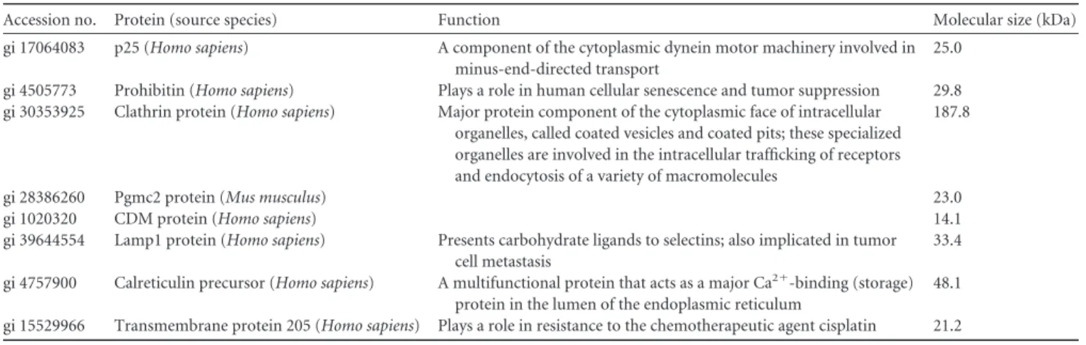
To further examine the role of BNIP3, we used the HaloTag
mammalian pulldown system protocol with BNIP3 as the bait in
HepG2 cells. Indeed, these liver-derived cells, which are enriched
in mitochondria, presented high levels of VDAC1 and
VDAC1-⌬C, which were even upregulated in hypoxia (see Fig. S1A in the
supplemental material). We identified by mass spectrometry the
binding of several proteins, including clathrin, a structural protein
involved in vesicle trafficking (
40
), in hypoxic HepG2 cells. When
apoptosis was induced with etoposide in hypoxic cells, which we
previously found induced the same level of cell death as STS (
12
),
BNIP3 also pulled down the lysosomal protein Lamp1 (
Fig. 3F
;
Table 1
), which further implicates lysosome involvement in
resis-tance to apoptosis in hypoxia. However, Mieap was not pulled
down, suggesting that BNIP3 and Mieap did not bind directly,
which contrasts with the findings of a previous report (
19
). Since
pulldown experiments showed that clathrin bound BNIP3 in
hy-poxic cells and that clathrin is involved in vesicle trafficking, the
effect of brefeldin A, an inhibitor of transport of secretory proteins
from the endoplasmic reticulum to the Golgi apparatus, was
ex-amined. Brefeldin A inhibited VDAC1-
⌬C formation (
Fig. 3G
),
which further suggests that secretory vesicles are involved in
trun-cation.
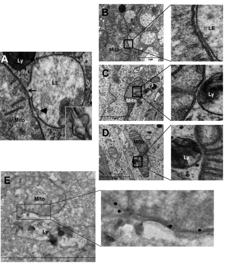
Mitochondria make contact with endolysosomal
compart-ments in hypoxia. We observed on electron micrographs of
hy-poxic LS174 cells that hyperfused mitochondria make contact
with late endosomes and lysosomes (
Fig. 4A
). The membranes of
mitochondria and endolysosomes showed fusional contact, where
some late endosomes puckered up to mitochondria (arrow) or
pinched into mitochondria (arrowhead) (
Fig. 4A
, inset).
Al-though it was difficult to quantify, we frequently noted an
inter-action between these organelles (
Fig. 4B
to
D
) and confirmed the
identity of mitochondria by performing immunogold labeling of
VDAC (
Fig. 4E
). In addition, we did not observe enlarged
mito-chondria in the proximity of autolysosomes. So, these data further
support a process other than mitophagy in the transformation of
mitochondria in hypoxia.
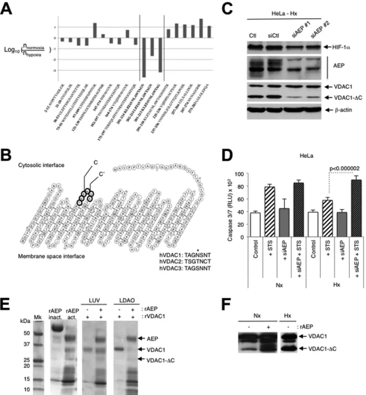
VDAC1 is C-terminally truncated at asparagine 214 by
en-dolysosomal asparagine endopeptidase. Mass spectrometric
analyses showed that the site of cleavage in VDAC1 was at
aspar-agine 214 (major cleavage site) and glycine 213 (minor cleavage
site) (
Fig. 5A
and
B
; see also Fig. S6A in the supplemental
mate-rial). These residues are located in loop 14, which is accessible to
the cytosol. The asparagine residue is not conserved in VDAC2 or
VDAC3, but the glycine is (
Fig. 5B
), and these residues are highly
conserved in VDAC1 proteins ranging from Xenopus to humans
(see Fig. S6B in the supplemental material). Since we suspected the
involvement of late endosomes and lysosomes in truncation, we
screened siRNAs against some endolysosomal enzymes and found
that transfection of asparaginyl endopeptidase (AEP)-specific
siRNA lowered the level of expression of AEP and the amount of
VDAC1-⌬C (
Fig. 5C
). We did not detect any difference in the
TABLE 1 Details of major proteins pulled down with a HaloTag-BNIP3 fusion protein expressed in hypoxic HepG2 cells challenged or not
challenged with etoposidea
Accession no. Protein (source species) Function Molecular size (kDa)
gi 17064083 p25 (Homo sapiens) A component of the cytoplasmic dynein motor machinery involved in
minus-end-directed transport
25.0
gi 4505773 Prohibitin (Homo sapiens) Plays a role in human cellular senescence and tumor suppression 29.8
gi 30353925 Clathrin protein (Homo sapiens) Major protein component of the cytoplasmic face of intracellular
organelles, called coated vesicles and coated pits; these specialized organelles are involved in the intracellular trafficking of receptors and endocytosis of a variety of macromolecules
187.8
gi 28386260 Pgmc2 protein (Mus musculus) 23.0
gi 1020320 CDM protein (Homo sapiens) 14.1
gi 39644554 Lamp1 protein (Homo sapiens) Presents carbohydrate ligands to selectins; also implicated in tumor
cell metastasis
33.4 gi 4757900 Calreticulin precursor (Homo sapiens) A multifunctional protein that acts as a major Ca2⫹-binding (storage)
protein in the lumen of the endoplasmic reticulum
48.1 gi 15529966 Transmembrane protein 205 (Homo sapiens) Plays a role in resistance to the chemotherapeutic agent cisplatin 21.2 a
HepG2 cells were in hypoxia for 16 h, and etoposide was used at 50 mM. Results are representative of those from three experiments.
on April 19, 2015 by Xavier De Bolle
http://mcb.asm.org/
FIG 4 Hypoxic LS147 cells show close interactions between mitochondria and late endosomes and lysosomes. (A) A representative image from an electron
micrograph of an LS174 cell showing an interaction between a mitochondrion (Mito) and a late endosome (LE) showing membrane rupture (arrow) and a late endosome with a lysosome (Ly) showing membrane rupture (arrowhead; see the inset). (Inset) Enlarged view of a late endosome showing blebbing at the mitochondrion-late endosome contact site. (B to D) (Left) Representative electron micrographs of LS174 cells showing the interaction of a mitochondrion with late endosomes and lysosomes. (Right) Enlarged views of the interaction sites. (E) (Left) Representative electron micrograph of LS174 cells showing immuno-gold-stained VDAC, allowing detection of mitochondria making contact with a lysosome. (Right) Enlarged view of the contact site.
on April 19, 2015 by Xavier De Bolle
http://mcb.asm.org/
FIG 5 Mitochondria-endolysosome contact in hypoxia leads to truncation of VDAC1 at asparagine 214 by the endolysosomal asparagine endopeptidase. (A)
Ratios of the abundance of human VDAC1 (hVDAC1) peptides from the normoxic sample to the abundance from the hypoxic sample. Residue numbers are indicated together with the peptide amino acid sequence. The y axis displays the decimal logarithm of the relative abundance of a given peptide in normoxia versus hypoxia. (B) Structure of VDAC1 showing the major cleavage site of VDAC1 C terminal to asparagine 214 (site C) and a minor cleavage site at glycine 213 (site C=). The consensus sequence between human VDAC1, VDAC2, and VDAC3 at the cleavage site is shown by boldface circles. (C) HeLa cells transfected with two different AEP-specific siRNAs (siAEP#1 and siAEP#2) incubated in hypoxia (Hx) and analyzed by immunoblotting (Ctl, control; siCtl, scrambled control siRNA). (D) HeLa cells were transfected with either a control siRNA or asparagine endopeptidase-specific siRNA (siAEP; 100 nM), incubated in normoxia (Nx) or hypoxia, and challenged with STS (1 mM) for 4 h. Apoptosis was evaluated from the level of caspase 3/7. Data from two independent experiments performed in quadruplicate are given as means⫾ SDs. (E) rAEP was activated by low pH and incubated with recombinant VDAC1 (rVDAC1) reconstituted into either large unilamellar vesicles (LUVs) or N,N-dimethyldodecylamine N-oxide (LDAO) micelles for 180 min at room temperature. The samples were analyzed by SDS-PAGE. Mk, molecular mass marker; rAEP inact., inactive rAEP; rAEP act., pH-activated rAEP. (F) Autoactivated rAEP was added to mitochondria isolated from normoxic HeLa cells, and the mixture was incubated for 30 min at 37°C. The samples were analyzed by immunoblotting for VDAC.
on April 19, 2015 by Xavier De Bolle
http://mcb.asm.org/
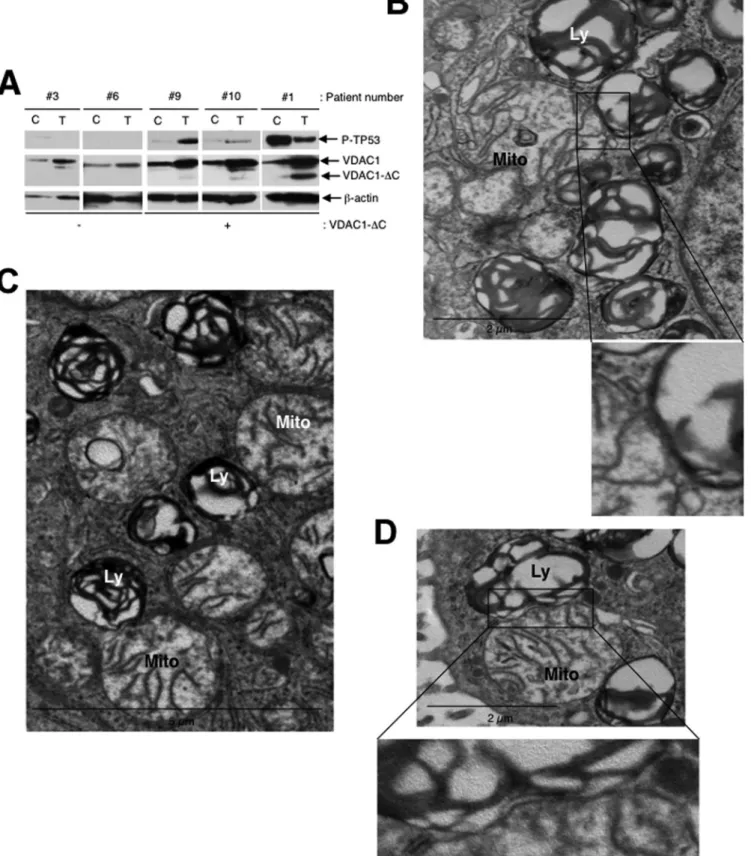
FIG 6 Truncated VDAC is present in lysates of lung adenocarcinoma tissue from patients, and tumor tissue sections show a close interaction between
mitochondria and lysosomes. (A) Representative immunoblots of protein extracts of control tissue (lanes C) and tumor tissue (lanes T) from patients’ lungs. (B) (Top) A representative electron micrograph of a cell from a patient’s lung adenocarcinoma showing the interaction between a mitochondrion (Mito) and lysosomes (Ly); (bottom) enlarged view of the contact site. (C) A representative electron micrograph of tumor tissue showing the interaction of a mitochondrion with lysosomes. (D) (Top) A representative electron micrograph of tumor tissue showing the interaction of a mitochondrion with lysosomes; (bottom) enlarged view of the interaction site.
on April 19, 2015 by Xavier De Bolle
http://mcb.asm.org/
expression levels of the AEP proteins in normoxia compared to
hypoxia by immunoblotting. Drug-induced apoptosis was also
higher in hypoxic cells transfected with AEP-specific siRNA (
Fig.
5D
). Incubation of low-pH-autoactivated recombinant AEP (
32
)
with recombinant VDAC1 in large unilamellar vesicles led to
min-imal degradation; however, in micelles, VDAC1 was degraded, but
degradation was substantial (
Fig. 5E
). Addition of
low-pH-auto-activated recombinant AEP to mitochondria that were isolated
from normoxic cells and that thus contained only full-length
VDAC showed the production of a signal at the level of
VDAC1-
⌬C (
Fig. 5F
). Incubation of hypoxic mitochondria with
recombinant AEP only minimally increased the amount of the
truncated form of VDAC1, VDAC1-⌬C (data not shown). This
may suggest that there is some posttranslational modification to
VDAC1 that is required for processing but that not all of the
full-length form is modified and thus remains insensitive to AEP,
though it may be sensitive to another lysosomal enzyme. Note that
incubation with trypsin did not modify the ECL profile of samples
of normoxic or hypoxic mitochondria (data not shown).
How-ever, overexpression of AEP in HeLa cells did not result in either
the substantial truncation of VDAC1 in normoxia or an increase
in truncation in hypoxia (see Fig. S6C in the supplemental
mate-rial). Since AEP is normally localized in endolysosomes and active
only at low pH, this may suggest that hypoxia-induced local fusion
is a prerequisite to truncation and that the degree of local fusion is
limiting. In addition, the time frame required to obtain VDAC1
processing in hypoxia is relatively long, 48 h in HeLa cells and 72 h
in LS174 cells, which is much longer than the time required to
stabilize HIF-1
␣. We speculate from these results that AEP is
re-sponsible for the major cleavage of VDAC1 during microfusional
contact of intact mitochondria with endolysosomes in hypoxia,
but since we detected two adjacent sites of cleavage, another
en-zyme or partner protein yet to be identified is also required for
efficient truncation.
Truncated VDAC1 is present in tumor tissue from patients
with wild-type TP53 lung adenocarcinomas, and enlarged
mito-chondria made contact with endolysosomes. Using a small
co-hort of 10 patients with lung adenocarcinomas for whom we had
the medical history, we found a correlation between the
expres-sion of phosphorylated-TP53 and cleavage of VDAC1 (
Fig. 6A
).
However, this needs to be confirmed with a larger cohort. This
would also allow correlation between the levels of the activity of
TP53, the amount of VDAC1 processing, and the activation of
HIF-induced targets. Nonetheless, the findings obtained with this
small cohort allow us to propose the basic principle of cross talk
between mitochondria and lysosomes in higher eukaryotes.
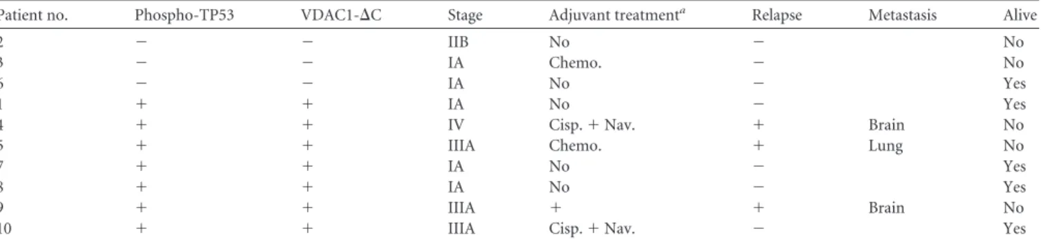
As in our previous study with a cohort of 46 patients with lung
adenocarcinoma (
12
), VDAC1 truncation tended to be associated
with a higher tumor stage and more metastatic relapse in an
inde-pendent population of lung adenocarcinomas (
Tables 2
and
3
).
Enlarged mitochondria in close contact with lysosomes were
de-tected by electron microscopy of tumor sections (
Fig. 6B
to
D
).
This was observed in all the patients showing truncated VDAC1.
Even though it was not a rare event, it was difficult to quantify. We
did not confirm that the regions in the tissue were hypoxic. This
would require doing investigations using light and electron
mi-croscopy. Contact between mitochondria and lysosomes was not
found in patients not showing truncated VDAC1. While these
findings are for a small cohort, they do allow us to propose the
basic principle of cross talk between mitochondria and lysosomes
in higher eukaryotes.
DISCUSSION
The tumor suppressor TP53 has been reported to function not
only as a guardian of the genome but also as a guardian of
mito-chondrial integrity and function (
2
). It regulates both mitophagy
and mitochondrial metabolism through the induction of genes
that regulate, respectively, autophagy and components of the
elec-tron transport chain, glycolysis, and the pentose phosphate
path-way (
41
). In addition, TP53 has been reported to activate necrosis
by opening the mitochondrial permeability transition pore (
42
).
Although our results point to the involvement of TP53-induced
Mieap and BNIP3-mediated regulation of the hypoxic processing
of VDAC1, we do not conclude, as suggested previously (
19
,
20
),
that lysosome-like organelles or lysosomal proteins accumulate in
mitochondria. However, the latter studies examined exogenous
overexpression of adenovirus-infected Mieap in normoxia, which
contrasts with the conditions of the present study, which
exam-ined endogenous proteins in hypoxic cells. We also did not find
evidence for the production of mitochondrion-derived vesicles,
which are involved in a pathway for shuttling cargo from
mito-chondria to lysosomes that is an alternative to mitophagy (
43
,
44
).
However, this pathway was followed after short-term oxidative
stress rather than long-term hypoxia, as in our study. Instead, our
results suggest that after long-term hypoxia, late endosomes and
TABLE 2 Characteristics of patients and their tumors and patient
outcomes
Characteristic Value
No. (%) of patients 10 (100)
No. (%) of patients by gender
Female 2 (20)
Male 8 (80)
Median (range) age (yr) at time of surgery 62.7 (38–74)
No. (%) of patients with tumor size of:
T1 5 (50)
T2 2 (20)
T3 3 (30)
No. (%) of patients with node stage of:
N0 5 (50)
N1 2 (20)
N2 3 (30)
No. (%) of patients with the following stage at diagnosis:
IA 4 (40)
IIB 1 (10)
IIIA 4 (40)
IV 1 (10)
No. (%) of patients with the following histology:
Adenocarcinoma 10 (100)
Relapse (locoregional or metastatic) 3 (30)
No. (%) of patients who died 5 (50)
Median (range) length of follow-up (mo) 57.5 (38.4–72.0)
on April 19, 2015 by Xavier De Bolle
http://mcb.asm.org/
lysosomes dock onto mitochondria and bring the outer
mito-chondrial membrane proteins into contact with their contents.
This is in agreement with the findings of the study by Gomes et al.,
who found that mitochondrial elongation is not dependent on
autophagosome formation (
8
). Our data suggest that mitophagy
is not the basis for the mechanism behind VDAC1 processing.
This finding confirmed data from our previous study, in which we
showed that in hypoxia the area occupied by mitochondria,
deter-mined by electron microscopy, was equivalent to or even larger
than that in normoxia and that the mass of mitochondria
deter-mined by fluorescence-activated cell sorter analysis remained
constant (
6
). So, we propose a novel mechanism in which enlarged
mitochondria escape mitophagy and promote metabolic
effi-ciency. Why some mitochondria undergo mitophagy in hypoxia
(
5
) while others persist is still not clear, but it has been suggested
that it may be simply sterical; that is, elongated mitochondria may
not fit into autophagosomes (
8
). Our results provide an additional
or complementary explanation. We suggest that after long-term
stresses, posttranslational modification of VDAC1 marks
mito-chondria for protection from autophagy and metabolic efficiency.
Under such stress conditions, cells maximize ATP production to
promote cell survival (
6
), as do the stress-induced hyperfused
mi-tochondria of MEFs (
10
), which also rendered them more
resis-tant to cell death. The cleavage of VDAC1 at residues 213 and 214
would result in a protein without
 strands 15 to 19 and probably
a change in conformation, as suggested by our previous results
obtained by immunofluorescence of hypoxic cells with an
anti-VDAC antibody (
12
). This C-terminal segment of the protein
contains the binding site for
-NADH (
30
), which promotes the
closed state of VDAC1. If the residual 14 strands resided in the
membrane and formed a pore, VDAC1-
⌬C would remain
consti-tutively open, allowing optimal efficiency in hypoxia to promote
glycolysis through its binding to hexokinases I and II, which we
previously showed still bind to VDAC1-⌬C (
12
). VDAC1-⌬C
re-constituted into a planar lipid bilayer also interacted with Bcl-x
Land reduced its channel conductance (
12
). At this stage, we do not
know if the C-terminal fragment of VDAC1 is further degraded or
if it plays a functional role in the mitochondrial membrane.
We showed that VDAC1 is cleaved by AEP (EC 3.4.22.34), a
cysteine protease mainly located in endolysosomes. AEP plays
im-portant roles in regulation of the immune system (
45
) and in
cancer (
46
,
47
) and has recently been shown to cleave the protein
tau in Alzheimer’s disease (
48
,
49
). In addition, its expression and
activity are regulated by TP53 (
50
), and it cleaves peptide bonds
carboxy terminal to asparagine (
51
). The asparagine residue of
VDAC1 cleaved by AEP is exposed on the cytoplasmic side of the
mitochondria, so this residue could come into contact with the
contents of lysosomes. We suggest that BNIP3 acts as a docking
site for lysosomes. We also observed that drug-induced apoptosis
in hypoxia involves the interaction of BNIP3 with clathrin and
Lamp1.
Finally, in the present study we have provided experimental
evidence supporting the conclusion that cleavage of VDAC1
re-flects a survival response of hypoxic cells that is the readout of an
interaction between hyperfused mitochondria with
endolyso-somes. We show that this phenomenon exists not only in vitro but
also in vivo in patients with lung adenocarcinomas. This cross talk
between organelles is mediated by TP53-induced Mieap and
bind-ing to hypoxia-induced BNIP3. Further understandbind-ing of the
function of cancer cell mitochondria should stimulate
investiga-tion into pharmacological approaches to modulate mitochondrial
function to design better cancer treatments.
ACKNOWLEDGMENTS
We thank C Pierreux (de Duve Institute, UC Louvain, Louvain, Belgium) for his help with HaloTag protein purification and G. Chinnadurai (Saint Louis University Health Sciences Center, St. Louis, MO) for providing the HA-BNIP3 cDNA-containing plasmid.
This research was supported by grants from the Fondation ARC, Fon-dation de France, ANR, INCA, la Ligue Nationale Contre le Cancer (Équipe Labelisée LNCC), METOXIA and MOMP (FP7-EU programs), MRT, and Canceropôle PACA. The Institute for Research on Cancer and Aging of Nice is funded by the Centre A. Lacassagne, CNRS, and INSERM. The funders had no role in study design, data collection and analysis, the decision to publish, or preparation of the manuscript. We disclose no potential conflicts of interest.
The authors’ contributions are as follows: conception and design, M. C. Brahimi-Horn and N. M. Mazure; development of methodology, M. C. Brahimi-Horn, C. Michiels, S. Hiller, and N. M. Mazure; acquisi-tion of data (acquired and managed patients, provided facilities, etc.), S. Lacas-Gervais, R. Adaixo, K. Ilc, M. Rouleau, A. Notte, M. Dieu, T. Voelt-zel, V. Maguer-Satta, M. Ilie, P. Hofman, and A. Schmidt; analysis and interpretation of data (e.g., statistical analysis, biostatistics, computa-tional analysis), M. C. Brahimi-Horn, S. Lacas-Gervais, A. Schmidt, S. Hiller, and N. M. Mazure; writing, review, and/or revision of the manu-script, M. C. Brahimi-Horn, J. Pouysségur, and N. M. Mazure; technical or material support, J. Pelletier and B. Manoury; and study supervision, N. M. Mazure.
TABLE 3 Phospho-TP53 and VDAC1-⌬C status of 10 patients with lung adenocarcinomas, determined by immunoblotting, and their tumor grade,
adjuvant treatment, relapse, and metastases
Patient no. Phospho-TP53 VDAC1-⌬C Stage Adjuvant treatmenta Relapse Metastasis Alive
2 ⫺ ⫺ IIB No ⫺ No
3 ⫺ ⫺ IA Chemo. ⫺ No
6 ⫺ ⫺ IA No ⫺ Yes
1 ⫹ ⫹ IA No ⫺ Yes
4 ⫹ ⫹ IV Cisp.⫹ Nav. ⫹ Brain No
5 ⫹ ⫹ IIIA Chemo. ⫹ Lung No
7 ⫹ ⫹ IA No ⫺ Yes
8 ⫹ ⫹ IA No ⫺ Yes
9 ⫹ ⫹ IIIA ⫹ ⫹ Brain No
10 ⫹ ⫹ IIIA Cisp.⫹ Nav. ⫺ Yes
a
Chemo, chemotherapy; Cisp, cisplatin; Nav, navelbine.