RESEARCH OUTPUTS / RÉSULTATS DE RECHERCHE
Author(s) - Auteur(s) :
Publication date - Date de publication :
Permanent link - Permalien :
Rights / License - Licence de droit d’auteur :
Institutional Repository - Research Portal
Dépôt Institutionnel - Portail de la Recherche
researchportal.unamur.be
University of Namur
Myeloperoxidase-catalyzed oxidation of cyanide to cyanate
Delporte, Cédric; Boudjeltia, Karim Zouaoui; Furtmüller, Paul G.; Maki, Richard A.; Dieu,
Marc; Noyon, Caroline; Soudi, Monika; Dufour, Damien; Coremans, Catherine; Nuyens,
Vincent; Reye, Florence; Rousseau, Alexandre; Raes, Martine; Moguilevsky, Nicole;
Vanhaeverbeek, Michel; Ducobu, Jean; Nève, Jean; Robaye, Bernard; Vanhamme, Luc;
Reynolds, Wanda F.; Obinger, Christian; Van Antwerpen, Pierre
Published in:
Journal of Biological Chemistry DOI:
10.1074/jbc.M117.801076 Publication date:
2018
Document Version
Publisher's PDF, also known as Version of record Link to publication
Citation for pulished version (HARVARD):
Delporte, C, Boudjeltia, KZ, Furtmüller, PG, Maki, RA, Dieu, M, Noyon, C, Soudi, M, Dufour, D, Coremans, C, Nuyens, V, Reye, F, Rousseau, A, Raes, M, Moguilevsky, N, Vanhaeverbeek, M, Ducobu, J, Nève, J, Robaye, B, Vanhamme, L, Reynolds, WF, Obinger, C & Van Antwerpen, P 2018, 'Myeloperoxidase-catalyzed oxidation of cyanide to cyanate: A potential carbamylation route involved in the formation of atherosclerotic plaques?', Journal of Biological Chemistry, vol. 293, no. 17, pp. 6374-6386. https://doi.org/10.1074/jbc.M117.801076
General rights
Copyright and moral rights for the publications made accessible in the public portal are retained by the authors and/or other copyright owners and it is a condition of accessing publications that users recognise and abide by the legal requirements associated with these rights. • Users may download and print one copy of any publication from the public portal for the purpose of private study or research. • You may not further distribute the material or use it for any profit-making activity or commercial gain
• You may freely distribute the URL identifying the publication in the public portal ? Take down policy
If you believe that this document breaches copyright please contact us providing details, and we will remove access to the work immediately and investigate your claim.
Myeloperoxidase-catalyzed oxidation of cyanide to cyanate:
A potential carbamylation route involved in the formation of
atherosclerotic plaques?
Received for publication, June 8, 2017, and in revised form, February 20, 2018 Published, Papers in Press, March 1, 2018, DOI 10.1074/jbc.M117.801076 X Ce´dric Delportea,b1,2, Karim Zouaoui Boudjeltiac1,X Paul G. Furtmu¨llerd,X Richard A. Makie,f, Marc Dieug,
X Caroline Noyona3, Monika Soudid, Damien Dufoura,b, Catherine Coremansa,b3, Vincent Nuyensc, Florence Reyea, Alexandre Rousseauc, Martine Raesg, Nicole Moguilevskyh, Michel Vanhaeverbeekc, Jean Ducobuc, Jean Ne`vea, Bernard Robayei, Luc Vanhammej4, Wanda F. Reynoldsf,X Christian Obingerd1, andX Pierre Van Antwerpena,b1,5
From theaLaboratory of Pharmaceutical Chemistry andbAnalytical Platform, Faculty of Pharmacy, Universite´ Libre de Bruxelles, 1050 Brussels, Belgium, thecLaboratory of Experimental Medicine, CHU de Charleroi, A. Ve´sale Hospital, Universite´ Libre de
Bruxelles, 6110 Montigny-le-Tilleul, Belgium, thedDepartment of Chemistry, Division of Biochemistry, University of Natural Resources and Life Sciences (BOKU), 1180 Vienna, Austria,eTorrey Pines Pharmaceuticals, Del Mar, California 92014, the fSanford-Burnham-Prebys Medical Discovery Institute, La Jolla, California 92037, thegLaboratory of Cellular Biology and hTechnology Transfer Office, University of Namur, 5000 Namur, Belgium, theiInstitute of Interdisciplinary Research, Institut de
Recherche Interdisciplinaire en Biologie Humaine et Mole´culaire, Faculty of Sciences, Universite´ Libre de Bruxelles, 6041 Gosselies, Belgium, and thejLaboratory of Molecular Parasitology, Institut de Recherche Interdisciplinaire en Biologie Humaine et
Mole´culaire, Faculty of Sciences, Universite´ Libre de Bruxelles, 6041 Gosselies, Belgium
Edited by Ruma Banerjee
Protein carbamylation by cyanate is a post-translational mod-ification associated with several (patho)physiological condi-tions, including cardiovascular disorders. However, the bio-chemical pathways leading to protein carbamylation are incompletely characterized. This work demonstrates that the heme protein myeloperoxidase (MPO), which is secreted at high concentrations at inflammatory sites from stimulated neutro-phils and monocytes, is able to catalyze the two-electron oxida-tion of cyanide to cyanate and promote the carbamylaoxida-tion of taurine, lysine, and low-density lipoproteins. We probed the role of cyanide as both electron donor and low-spin ligand by pre-steady-state and steady-state kinetic analyses and analyzed reaction products by MS. Moreover, we present two further pathways of carbamylation that involve reaction products of MPO, namely oxidation of cyanide by hypochlorous acid and
reaction of thiocyanate with chloramines. Finally, using an in
vivo approach with mice on a high-fat diet and carrying the
human MPO gene, we found that during chronic exposure to cyanide, mimicking exposure to pollution and smoking, MPO promotes protein-bound accumulation of carbamyllysine (homocitrulline) in atheroma plaque, demonstrating a link between cyanide exposure and atheroma. In summary, our find-ings indicate that cyanide is a substrate for MPO and suggest an additional pathway for in vivo cyanate formation and protein carbamylation that involves MPO either directly or via its reac-tion products hypochlorous acid or chloramines. They also sug-gest that chronic cyanide exposure could promote the accumu-lation of carbamylated proteins in atherosclerotic plaques.
Carbamylation is a post-translational modification of biolog-ical molecules bearing an amino group. It occurs in several physio- and/or pathophysiological conditions. For example, it is well known that the⑀-amino group of lysine residues of pro-teins can be converted to carbamyllysine (1–4), also named homocitrulline (Hcit),6 and that chronic renal insufficiency
correlates with enhanced carbamylation mediated by urea-de-rived cyanate (⫺O–C'N). In patients with renal disease, car-bamylation of low-density lipoproteins (LDLs) is considered as a nontraditional risk factor for cardiovascular insufficiency. It manifests all of the biological effects relevant to atherosclerosis, including endothelial cell injuries, expression of adhesion mol-ecules, and vascular smooth muscle cell proliferation (5). Car-bamylation of type I collagen has been shown to induce confor-mational changes and reduce its ability to activate human
This study was supported by Austrian Science Funds Grant FWF P 20664 (to P. G. F.) and doctoral program BioToP Grant W1224 (to M. S.), Belgian National Fund for Scientific Research (FRS) Grants 3.4553.08 and T.0136.13 PDR, Universite´ Libre de Bruxelles Grant FER-207, and Department of Inter-national Relationships of the Universite´ Libre de Bruxelles Grant BRIC SJ/AL/BRIC/130. This study was also supported in part by National Insti-tutes of Health Grants RO1-NS074303 (to W. F. R.) and R43 AG040935 (to R. A. M.). This work was also supported by the Institute of Pathology and Genetics (IRSPG, Loverval, Belgium) and the “CHU of Charleroi.” Additional funds to support this work were provided by Sanford Burnham Prebys Medical Discovery Institute. The authors declare that they have no conflicts of interest with the contents of this article. The content is solely the respon-sibility of the authors and does not necessarily represent the official views of the National Institutes of Health.
This article containsFigs. S1–S5.
1These authors contributed equally to this work.
2Postdoctoral researcher supported by the Belgian Fund for Scientific
Research (FRS-FNRS).
3Research Fellow of FRS-FNRS. 4Research Director of FRS-FNRS.
5To whom correspondence should be addressed: Laboratory of
Pharmaceu-tical Chemistry and AnalyPharmaceu-tical Platform, Faculty of Pharmacy, Universite´ Libre de Bruxelles, Brussels, Belgium. Tel.: 3226505263; Fax: 32265052489; E-mail:pvantwer@ulb.ac.be.
6The abbreviations used are: Hcit, homocitrulline; LDL, low-density
lipopro-tein; MPO, myeloperoxidase; LS, low-spin; EPO, eosinophil peroxidase; LPO, lactoperoxidase; ESI, electrospray ionization; QTOF, quadrupole TOF; LDLR, LDL receptor; HRP, horseradish peroxidase; CE, collision energy.
cro
ARTICLE
6374
J. Biol. Chem. (2018) 293(17) 6374 –6386polymorphonuclear neutrophils involved in the immune response (6).
Recently, myeloperoxidase (MPO) has been demonstrated to be indirectly involved in protein carbamylation (7). This enzyme, a member of the peroxidase-cyclooxygenase super-family (8), is secreted at inflammatory sites from stimulated neutrophils and also from monocytes (9). It catalyzes the two-electron oxidation of thiocyanate (⫺S–C'N), thereby produc-ing hypothiocyanous acid (HO–S–C'N) and cyanate as a minor by-product (7). Protein carbamylation may therefore be driven not only by uremia but also by inflammation. Upon exposure of LDL to either MPO/H2O2/⫺SCN or the potassium
salt of ⫺O–C'N, protein recognition by LDL receptor was attenuated, whereas its interaction with the scavenger receptor SR-A1 was enhanced. A clear relationship between the plasma levels of protein-bound⑀-carbamyllysine (Hcit) and an in-creased risk of cardiovascular disease could be established (7).
We hereafter demonstrate the existence of a new alternative mechanism for cyanate formation and potential protein car-bamylation at sites of inflammation, according to the following reaction, which involves the hydrogen peroxide–mediated oxi-dation of cyanide (⫺C'N) catalyzed by MPO (Reaction 1).
H–O–O–H⫹⫺C⬅N O¡ MPO
⫺O–C⬅N ⫹ H–O–H Reaction 1
Cyanide is well known as a product of organic material com-bustion (10) (atmospheric pollution, tobacco smoking) or from intake of cyanogenic glycosides (almond) (11). In vivo, it is a universal ligand able to bind to and inhibit heme enzymes. Exposure to high concentrations results in death within sec-onds to minutes by impairment of dioxygen (O2) distribution
within the body and inhibition of the respiratory electron trans-port chain in the mitochondria (12). Five-coordinated ferric high-spin heme proteins (e.g. cytochrome c oxidase, methemo-globin, metmyomethemo-globin, catalase, peroxidases, etc.) are its pref-erential targets. Cyanide, which at most biological pH levels is protonated (pKa⫽ 9.14), binds as an anion to Fe(III) and
trans-forms the corresponding heme protein into its six-coordinated low-spin (LS) form, thereby impeding the interaction with nat-ural substrates (e.g. O2or H2O2). The binding rates and
com-plex equilibria involved are very variable. Typically, eukaryotic peroxidases (including MPO) and catalases bind cyanide rap-idly (1.1⫻ 105
M⫺1s⫺1to 1.3⫻ 106M⫺1s⫺1) (13–17), taking up
both the anion and a proton (18). So far, this LS complex for-mation has been thought to effectively block the redox chemis-try of all heme enzymes without any exceptions (KD⫽ 1–10 M). However, the present study unequivocally demonstrates
that cyanide can act both as ligand of MPO and electron donor to compound I of MPO.
Myeloperoxidase is a unique oxidoreductase in human phys-iology. The reconstructed phylogeny of mammalian peroxi-dases indeed demonstrates that MPOs form a well-separated clade within the vertebrate peroxidases (i.e. Family 1 of the per-oxidase-cyclooxygenase superfamily) (8). This superfamily also includes peroxidasins, peroxinectins, cyclooxygenases, and
dual oxidases (8).Fig. 1Adepicts the family of vertebrate per-oxidases, with the clades of myeloperoxidases and eosinophil peroxidases (EPOs) being well separated from the other branches. Segregation of MPO is well reflected by its unique biophysical and biochemical properties (19) that are closely related to its physiological role and the nature of its substrates. It participates in innate immune defense mechanisms through formation of microbicidal-reactive oxidants and diffusible rad-ical species. Its unique substrate is chloride that is oxidized to antimicrobial hypochlorous acid (HOCl). Chloride oxidation is a thermodynamically challenging reaction that requires the for-mation of strong oxidizing redox intermediates (i.e. the so-called “compound I” formed by oxidation of ferric MPO by hydrogen peroxide) (Fig. 2A) (20). The standard reduction potential of the couple compound I/MPO(III) (E°⬘ ⫽ 1.16 V) provides the driving force of this reaction (Fig. 2A). Moreover, as will be demonstrated below, it also uniquely enables MPO to oxidize cyanide to cyanate, thereby contributing to a so far unknown route for protein carbamylation. The presented data also suggest multiple pathways of cyanate formation and pro-tein carbamylation that involve MPO either directly or its reac-tion products hypochlorous acid and/or chloramine. Experi-ments in mouse model demonstrate the relevance of these pathways in vivo as cyanide is directly metabolized and pro-motes accumulation of protein-bound Hcit in atheroma plaque.
Results
Hydrogen peroxide consumption mediated by cyanide
In a preliminary study, the consumption of hydrogen perox-ide by MPO, promoted by cyanperox-ide, was probed by amperomet-ric monitoring.Fig. 1B clearly shows that 0.5MMPO
con-sumes 100MH2O2within 300 s in the presence of 100M
cyanide, whereas no effect is observed under identical condi-tions with related enzymes, such as lactoperoxidase (LPO) and EPO. At lower cyanide concentrations (50 –100M), MPO
rap-idly consumes H2O2, whereas the inhibitory effect is evident at higher concentrations (500 –1000M;Fig. 1B). The slow H2O2
consumption observed in control experiments (absence of cya-nide) is due to the well-known low (pseudo)catalase activity of mammalian peroxidases (20).
Cyanide acts as electron donor for MPO compound I
Myeloperoxidase catalyzes one- and two-electron oxidation reactions (Fig. 2A). The relevant redox intermediate involved in both reactions (compound I; i.e. an oxoiron(IV) porphyrin rad-ical cation) is formed via a two-electron oxidation of the ferric enzyme (PorFe(III)) by H2O2. PorFe(III)⫹ H2O2O¡ k1 ⫹䡠PorFe(IV)⫽O ⫹ H 2O Reaction 2
In two-electron transitions, compound I (about 50% hypo-chromicity of Soret band compared with ferric MPO) is directly reduced to ferric MPO (Soret band at 428 nm), whereas in one-electron transitions, compound II (Soret band at 456 nm) is
formed. These reactions can easily be probed by sequential stopped-flow spectroscopy. Fig. 1C shows a representative series of spectra obtained by mixing 4MMPO compound I
with 100Mcyanide. The reaction is monophasic and linearly
dependent on cyanide concentration. It rapidly forms a MPO species that had its Soret band at 456 nm, which could derive from either compound II or the LS complex between ferric MPO and cyanide (19). The determined apparent bimolecular rate constant is (5.3⫾ 0.5) ⫻ 105
M⫺1s⫺1at pH 7.0 and 25 °C. It
is unlikely that cyanide mediates the reduction of compound I to compound II, because this reaction is thermodynamically unfavorable (see below). Moreover, the reaction of preformed compound II with cyanide probed in the sequential stopped-flow mode clearly demonstrated that compound II does not participate in the turnover of the MPO/H2O2/⫺CN system. By contrast, the observed spectral transition suggests the cyanide-mediated direct reduction of compound I to ferric MPO. The
latter rapidly binds excess cyanide, forming the LS complex with Soret maximum at 456 nm (see alsoFigs. S4 and S5).
For comparison, the reaction of cyanide with preformed compound I of LPO and EPO as well as horseradish peroxidase (a model plant-type peroxidase) was also investigated under identical conditions. With LPO and EPO, compound I is slowly transformed to compound II (2 s⫺1), which is a well-known phenomenon of intramolecular electron transport in the absence of exogenous electron donors (21–23) (seeFig. 1A). Horseradish peroxidase compound I is stable in the presence of cyanide (seeFig. S1B).
MPO-mediated cyanide oxidation generates cyanate
To prove direct reduction of compound I to ferric MPO and concomitant two-electron oxidation of cyanide to ⫺OCN, MPO was incubated with both H2O2 and the isotopomers
Figure 1. A, reconstructed phylogenetic tree showing the family of
verte-brate peroxidases, including the mammalian enzymes MPO, EPO, LPO, and thyroid peroxidase (TPO). B, amperometric measurements (650 mV) of the H2O2consumption in the presence of 0.5Mperoxidases (LPO, EPO, or MPO),
100MH2O2, and⫺CN at various concentrations. C, reaction of 2MMPO
compound I with 50Mcyanide followed in the sequential-mixing stopped-flow mode. Conditions were as follows: 100 mMphosphate buffer, pH 7.0, 25 °C. Arrows, wavelength with absorbance changes. The first spectrum was taken 1.3 ms after mixing. Inset, typical time trace followed at 456 nm (maxi-mum absorbance of MPO(III)-cyanide complex) and plot of the pseudo-first-order rate constants, kobs, versus cyanide concentrations.
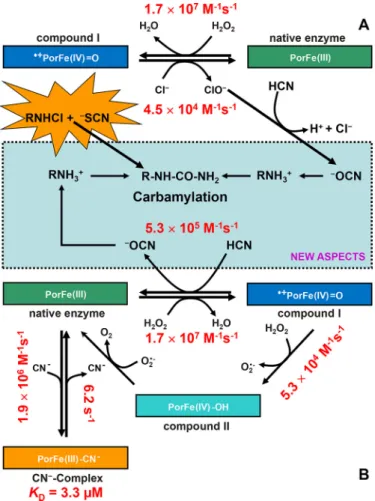
Figure 2. Carbamylation promoted by the system MPO/H2O2/ⴚCN
sys-tem and MPO-typical reaction products. A, MPO-typical oxidation of
chlo-ride to hypochlorous acid (i.e. chlorination cycle). Ferric MPO (PorFe(III)) is oxidized to compound I (䡠⫹PorFe(IV)⫽O; oxoiron(IV) porphyrin radical), which is directly reduced to the resting state by chloride, thereby releasing hypo-chlorous acid (HOCl/⫺OCl). Hypochlorous acid mediates the oxidation of cya-nide to cyanate and the formation of chloramines, which may be also involved in carbamylation by direct reaction with thiocyanate (NEW ASPECTS).
B, two-electron oxidation of cyanide to cyanate by MPO compound I, thereby
restoring the ferric protein and releasing cyanate (5.3⫻ 105
M⫺1s⫺1; NEW
ASPECTS). Ferric MPO binds cyanide, forming the low-spin complex
(Por-Fe(III)-CN⫺, KD⫽ 3.3M). Further MPO-typical reactions include the
one-elec-tron reduction of compound I to compound II by H2O2(14) or by other
one-electron donors followed by compound II reduction to ferric MPO. Kinetic constants were calculated from current experiments (see “Experimental pro-cedures”) or from the literature when already known (13–17).
⫺CN, [13C]⫺CN, or [13C,15N]⫺CN. The reaction products were
analyzed by MS.Fig. 3shows the formation of a reaction prod-uct with an appropriate m/z (i.e. m/z⫽ 43) and a fragment ion at m/z⫽ 27.Fig. 4Aillustrates the formation of cyanate using the three isotopomers. Upon using ⫺CN, [13C]⫺CN, or
[13C,15N]⫺CN, peaks at m/z values of 41.999, 43.002, or 43.999
were found, clearly demonstrating the formation of the isoto-pomers⫺OCN, [13C]⫺OCN or [13C,15N]⫺OCN, respectively. In the absence of cyanide or hydrogen peroxide or MPO, no cyanate was produced except for hydrogen peroxide with cya-nide, where a low cyanate production was detected (around 1%). Furthermore, using a calibration curve of cyanate, we were able to calculate that around 13% of cyanide is transformed to cyanate (n ⫽ 3) after 1 h (see supporting data for detailed results). These findings clearly indicate that the system MPO/ H2O2/cyanide is able to produce cyanate and that the
conver-sion of cyanide to cyanate is incomplete (Fig. 4A).
Carbamylation of taurine, lysine, and LDL
Furthermore, cyanate produced by MPO-mediated cyanide oxidation was probed in its reaction with nucleophilic amino groups in both taurine and lysine. Taurine is a low-molecular weight amine abundant in millimolar concentration in the cytosol of neutrophils (24). In this study, taurine was used as a chemical model of monoamine because it is stable and its chlo-rination is well documented. Taurine (1 mM) was incubated
with MPO (0.3M), hydrogen peroxide (1 mM) and [12C]⫺CN,
[13C]⫺CN, or [13C,15N]⫺CN (1 mM) in ammonium acetate
buffer, pH 7.4, and the reaction mixture was analyzed by
elec-trospray ionization (ESI)-quadrupole TOF (QTOF) MS in neg-ative mode. Peaks at expected m/z values of 167.0109, 168.0284, and 169.0155 corresponding to12C-, 13C-, and 13C,15
N-car-bamylated taurine were found (Fig. 4B). To further confirm the formation of carbamylated taurine, the isolated peaks were fragmented, leading to peaks with m/z values of 124.0074 and 79.9581, respectively, that fully matched with fragments obtained from standard carbamylated taurine (Fig. 4C). Similar experiments performed with lysine demonstrated the forma-tion of Hcit (Fig. S2). In the absence of either hydrogen peroxide or MPO, carbamylation of taurine or lysine was negligible or diminished.
Another tested target for carbamylation was LDL. LDLs were incubated with the MPO/H2O2/cyanide system before being
delipidated and hydrolyzed. Subsequently, the formation of protein-bound Hcit was analyzed by MS and correlated with the abundance of protein-bound lysine by monitoring the ratio Hcit/Lys. A low amount of Hcit was detected in native LDLs. The incubation of LDLs with MPO, H2O2, and increasing
con-centrations of cyanide led to increasing Hcit/Lys ratios (1.5-and 12-fold increases;Fig. 5A). The addition of chloride (150 mM) to the MPO/H2O2system in the presence of 100M
cya-nide even increased the extent of carbamylation. Similar levels of carbamylation were observed when LDLs were incubated with HOCl and cyanide (100M) (Fig. 5A). In the absence of
MPO, the Hcit concentration was almost identical to that of native LDLs (data not shown). Furthermore, a positive control with 100Mof cyanate (⫺OCN) was analyzed. This concentra-Figure 3. Tandem MS/MS determination of cyanate production. MS spectra show a peak around 43 m/z corresponding to13C-cyanate, when MPO (1
M) was added to a mixture of [13C]cyanide (2 m
M) and H2O2(200M) (pink solid line). Results obtained upon the repeated addition of H2O2(three times) are
depicted by the green solid line. MS/MS spectra show a peak around 27 m/z corresponding to the fragmentation of [13C]cyanate into [13C]cyanide (green solid line).
tion was used supposing the full conversion of cyanide to cya-nate. Surprisingly, this experiment showed a lower carbamyla-tion compared with the MPO/H2O2/cyanide/chloride system
but a 3 times higher carbamylation when compared with the MPO/H2O2/cyanide system (Fig. 5A).
The quantitative approach of Hcit formation enabled us to observe that approximately 0.7% of thiocyanate (100M)
medi-ated the formation of Hcit in the presence of MPO and H2O2.
When cyanide was added in the same concentration as thiocy-anate (100 M), the thiocyanate-mediated formation of Hcit
decreased to 0.35%; however, the cyanide-mediated formation of Hcit increased to 0.7%. If the thiocyanate concentration was increased to 400 M, the thiocyanate-mediated formation of
Hcit increased to 0.4%, whereas the cyanide-mediated forma-tion decreased to 0.4%. These results highlight the importance of generated HOCl for the Hcit formation mediated by cyanide. Finally, the positive control using cyanate to carbamylate LDLs at 100Mrevealed that only 0.1% of cyanate was converted into
Hcit under the conditions used.
Finally, we tested the contribution of each anion (thiocya-nate, cyanide, and chloride) in the carbamylation process. Upon the addition of cyanide to the system MPO/H2O2/thiocyanate, the contribution of thiocyanate in the carbamylation process decreased (Fig. 5B, left). However, this was compensated by cyanide-mediated carbamylation, as illustrated by an increase of labeled and total Hcit content (labeled and not labeled;Fig. 5,
B(right) and C). Interestingly, when chloride (150 mM) was added in addition to cyanide to the MPO/H2O2/thiocyanate
system, there was a drop in the formation of Hcit from thiocy-anate (Fig. 5B, left). However, the total carbamylation content increased (total Hcit;Fig. 5B, right) as cyanide-dependent for-mation of Hcit (Fig. 5C) increased.
Role of hypochlorous acid and chloramines in carbamylation pathways
Hypochlorous acid (HOCl) is an important reaction product of MPO deriving from the two-electron oxidation of chloride by compound I. It is well known that HOCl reacts with amines forming chloramines (e.g. taurine chloramines) that in addition contribute to the antimicrobial activity of MPO (25). We could demonstrate that HOCl but not monochloramine is another oxidant of cyanide. Upon mixing hypochlorous acid and cya-nide with taurine, carbamylation of taurine is observed (Fig. S3). Actually, HOCl promotes the formation of cyanate from cyanide (Fig. S3). As demonstrated above, formation of Hcit is observed upon incubation of LDL with HOCl and cyanide (Fig. 5A).
Additionally, carbamylation can derive from the reaction between thiocyanate and monochloramine.Fig. 4Dshows the
formation of carbamylated adducts with m/z values of 167.0115 and 168.0160 by using12C- or13C-labeled thiocyanate and
tau-rine mono-chloramine. Incubating HOCl-oxidized LDL with thiocyanate increases the Hcit/lysine ratio (Fig. 5B).
Cyanide exposure promotes protein-bound carbamylation in atheroma plaque
To confirm that those reactions participate in formation of carbamylated proteins in vivo, we used humanized MPO mice transgenic for the intact human MPO gene (⫺463G allele) (hMPO-TG) with native promoter elements. The rationale for using the hMPO-TG mice is that the human MPO gene is expressed in macrophages in atherosclerotic plaques, whereas the mouse MPO gene is not expressed. This is thought to be due in part to a primate-specific Alu element with binding sites for SP1 and nuclear receptors, including PPAR␥ (26). Prior studies show MPO presence in atherosclerotic plaques in the MPOG-LDLR⫺/⫺ model correlating with increased atherosclerotic plaque and hyperlipidemia (27). This cross was used in an ear-lier study that found increased protein-bound homocitrulline content in atheroplaque laden aorta in huMPOG-LDLR⫺/⫺ mice as compared with LDL receptor– deficient LDLR⫺/⫺ con-trols (7). In this experiment, control mice, human MPO trans-genics (⫺463G allele) (hMPO-TG) mice, and hMPO-TG crossed to the LDLR⫺/⫺mice (all on C57BL/6 background) were treated with [13C,15N]cyanide (0.4 mg/kg everyday,⬃5
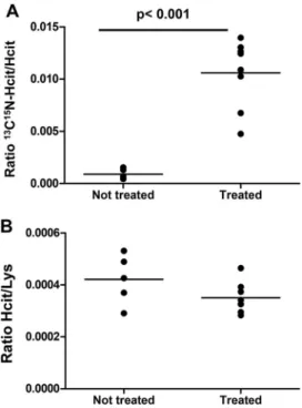
g/day) or PBS as negative control for 8 weeks (see “Experi-mental procedures”). Furthermore, mice were exposed to acute inflammation before sacrifice using intraperitoneal injection of zymosan as in prior studies (Wang et al. (7) with slight modifi-cations). Protein-bound [13C,15N]Hcit was analyzed in plasma, in atheroma plaques, and in the supernatant and cell pellets of peritoneal lavages. Fig. 6A shows that hMPO-TG-LDLR⫺/⫺ mice treated with [13C,15N]cyanide have an increased ratio of
[13C,15N]Hcit/Hcit (0.011 ⫾ 0.003) in the atheroma plaques
compared with animals treated with PBS (ratio of 0.0009 ⫾ 0.0005, p⬍ 0.01). Labeled cyanide was used because carbamy-lation naturally occurs in vivo. Labeling with [13C,15N]Hcit
allowed us to demonstrate that chronic exposure to cyanide causes carbamylation via MPO activity. In this context, the ratio of [13C,15N]Hcit/Hcit was the preferred readout to
express carbamylation. On the other hand, [13C,15N]Hcit was
too low in plasma, cell pellets, and supernatant of peritoneal lavages and was not detected. Nevertheless, it is worth noting that Hcit levels in atheroma plaques were the same in the cya-nide hMPO-TG-LDLR⫺/⫺mice and PBS control hMPO-TG-LDLR⫺/⫺mice (Fig. 6B), showing that it is the chronic exposure to cyanide that led to increased carbamylation by MPO-depen-dent processes.
Figure 4. Mass spectrometric analyses. A, oxidation of cyanide by the MPO/H2O2system. The isotopomers of⫺CN (final concentration⫽ 1 mM) were mixed
with H2O2(1 mM) in 10 mMammonium acetate buffer, pH 7.4, and the reaction was started with 0.3Mhuman recombinant MPO at 37 °C with a reaction time
of 1 h.⫺OCN,⫺O13CN, and⫺O13C15N peaks (at 41.9997, 43.0030, and 43.9999 m/z, respectively) could be detected, which correspond to the oxidation products
of cyanide isotopes. Spectra have been generated by subtracting spectra of a blank (buffer alone). B, taurine carbamylation. Taurine (1 mM) was mixed with 0.3 Mhuman recombinant MPO and isotopic cyanide (1 mM) for 12 h (37 °C) in 10 mMammonium acetate buffer, pH 7.4, and the reaction was started with 1 mM
H2O2. A peak was detected at m/z of 167.0109, which corresponds to [
12C]carbamyltaurine and MS/MS spectra fit with those of the control. In similar
experiments with isotopomers, peaks were detected at m/z⫽ 168.0284 and 169.0155, which correspond to13C- and13C,15N-labeled carbamyltaurine. C, MS
and MS/MS spectra of carbamyltaurine obtained after mixing taurine (1 mM) and cyanate (1 mM) in the acetate buffer and overnight incubation. D, MS and MS/MS spectra for the reaction of taurine chloramines (formed by mixing of 0.5 mMHOCl and 1 mMtaurine) with12C- and13C-labeled thiocyanate after a
reaction time of 12 h. Peaks detected at m/z values 167.0115 and 168.0160 correspond to12C- and13C-labeled carbamyltaurine (absent in the taurine
Discussion
Mounting evidence for the involvement of MPO in athero-genesis has emerged for the past decades. It is now accepted that MPO catalyzes oxidation reactions via the release of reac-tive halogenating and nitrating species that render LDL athero-genic and high-density lipoprotein dysfunctional (28 –33). In mice, Wang et al. (7) showed a marked increase in the content of protein-bound homocitrulline at atherosclerotic plaque-laden aortas of human MPO-transgenic mice as compared with nontransgenic counterparts. In both clinical and exper-imental in vivo situations, MPO seems to be a major catalytic component.
The enhanced consumption of hydrogen peroxide in the presence of low cyanide concentrations and the strict concen-tration dependence of compound I reduction unequivocally demonstrate the role of cyanide as electron donor for MPO. The inability of cyanide to promote the transition of compound I into compound II fits with the high one-electron reduction potential of the 䡠CN/⫺CN couple (1.9 V) (20) that is signifi-cantly higher than that of the redox couple compound I/com-pound II (1.35 V) (34).
The observed spectral transition (Fig. 1C) can be explained by the following reaction sequence that involves cyanide as
two-electron donor of compound I and as LS ligand for ferric MPO.
䡠⫹PorFe(IV)⫽O ⫹⫺C⬅N Ok¡2 PorFe(III)⫹⫺O–C⬅N
PorFe(III)⫹⫺C⬅N O¡
k3
PorFe(III)–C⬅N
Reactions 3 and 4
The calculated biomolecular rate of the reaction between cyanide and MPO(III) was determined to be 1.9⫻ 106M⫺1s⫺1 at pH 7.0 and 25 °C (k3, Reaction 4). Because k3 ⬎ k2, the
measured rate constant of the transition compound I 3 MPO(III)3 LS MPO-cyanide complex reflects k2(Fig. 2). The
two-electron oxidation of cyanide (k2) is fast (5.3⫻ 10 5
M⫺1s⫺1
at pH 7.0 and 25 °C) and reflects the high oxidation capacity of MPO, which is the consequence of heme distortion due to unique heme-to-protein linkages, including an electron-with-drawing vinyl-sulfonium group (19). It is noteworthy that higher concentrations of cyanide favor the MPO-CN⫺complex formation (k3), limiting the MPO activation by reaction with
H2O2(k1). Catalysis of the two-electron oxidation of cyanide by MPO is unique because related human peroxidases like LPO and EPO did not consume hydrogen peroxide in the presence of cyanide.
Mass spectrometric analyses have confirmed the production of cyanate derived from the two-electron oxidation of cyanide by compound I (seeFig. 2). This was supported by consecutive carbamylation reactions of produced cyanate with ⑀-amino groups of taurine and of free or LDL-bound lysine (Figs. 3–5). However, quantitative MS analyses also demonstrated that only 13% of cyanide was converted to cyanate by the MPO/H2O2/
cyanide system (Fig. 4A). Nevertheless, the incubation of cya-nide with HOCl (same concentration as hydrogen peroxide; 1 mM) converted only 20% of the cyanide to cyanate. It is also noteworthy that higher concentrations of cyanide (e.g. 1 mM)
did not increase LDL carbamylation, which corroborates the kinetic studies (Fig. 5A).
In addition, the present work suggests further new routes of carbamylation, namely cyanate formation by HOCl-mediated cyanide oxidation and/or by reaction of stable chloramines with thiocyanate (35, 36). Both pathways follow reactions that only indirectly depend on MPO. Chloramines are relatively stable derivatives and are produced by the reaction of amines with HOCl released from MPO at sites of inflammation (37). In addi-tion to thiocyanate oxidaaddi-tion (7), our findings show that MPO can promote carbamylation in three additional ways: (i) by direct oxidation of cyanide, (ii) by reaction of hypochlorous
Figure 5. Formation of carbamylated LDL. A, box-whisker plots (5th to 95th percentile) showing the ratios between Hcit and Lys residues (ratio Hcit/Lys),
including conditions using only cyanide as substrate. B, box-whisker plots (5th to 95th percentile) showing the ratios between Hcit and Lys residues (ratio Hcit/Lys), including conditions using thiocyanate (SCN⫺) and labeled cyanide ([13C,15N]CN⫺). Left, only monoisotopic Hcit (unlabeled) is considered; right,
total Hcit is considered (Hcit ⫹ [13C,15N]Hcit). C, box-whisker plots showing the ratios between labeled homocitrulline ([13C,15N]Hcit) and Hcit (ratio
[13C,15N]Hcit/Hcit). Native LDLs were incubated overnight at 37 °C in buffer (10 m
Mphosphate, pH 7.4) in the presence of MPO (300 nM), H2O2(1 mM), and/or
chloride anions (150 mM) and/or HOCl (1 mM) and/or thiocyanate (SCN⫺, 100 or 400M) and/or cyanide (CN⫺) and/or [13C,15N]cyanide ([13C,15N]CN⫺). LDLs
were then delipidated, hydrolyzed by microwave under acidic conditions. Amino acid residues were labeled and, finally, analyzed using LC-MS/MS. Each condition was performed in quadruplicate (n⫽ 4). *, statistically significant difference with the native LDL condition (Kruskal–Wallis, p ⬍ 0.01).
Figure 6. In vivo effect of cyanide. Exposure of hMPO-TG-LDLR⫺/⫺mice to [13C,15N]cyanide (0.4 mg/kg/day) led to protein-bound [13C,15N]Hcit
forma-tion in atheroma plaque. Mice were exposed for 8 weeks to isotope-labeled cyanide (n⫽ 8) or to PBS (control, n ⫽ 5). Protein-bound Lys, Hcit, and [13C,15N]Hcit were monitored, and [13C,15N]Hcit/Hcit (A) and Hcit/Lys (B) ratios
were calculated. A statistically significant difference was observed between groups (t test, p⬍ 0.001).
acid with cyanide, or (iii) by reaction of chloramines with thiocyanate.
In vivo, several anions compete with cyanide for reaction with MPO compound I, including chloride (150 mM) and
thio-cyanate (10 –500 M). We observed that in the presence of
chloride, thiocyanate, and cyanide, carbamylation of lysine res-idues was mediated by oxidation of both thiocyanate and cya-nide (Fig. 5B). In the presence of 100Mconcentrations of both
thiocyanate and cyanide, about one-third of carbamylation derived from thiocyanate and two-thirds from cyanide oxida-tion (Fig. 5, B and C). Furthermore, higher concentration of thiocyanate (400M) decreased the carbamylation mediated by
cyanide, but this was compensated by carbamylation mediated by thiocyanate. Under these conditions, the production of HOCl by MPO is lower than the production of HOSCN. How-ever, the quantitative determination of Hcit demonstrated that only⬃0.4% of thiocyanate and cyanide were involved in Hcit formation. Nevertheless, the carbamylation observed in LDLs is about 6 times higher compared with the addition of cyanate only (Fig. 5, A and B). Altogether, these data show a synergic effect of thiocyanate, cyanide, and chloride in the carbamyla-tion process. This synergy might reflect previous reports about the reaction of cyanide with hypothiocyanate to a dicyanosul-fide intermediate (36).
It remains to be discussed whether these reactions are rele-vant in vivo. MPO and⫺CN do exist in soluble forms in blood, and the concentration ratios between these molecules used in the above experiments match those reported in human blood (e.g. [⫺CN]/[MPO] ratio⬃400:800, although the relative con-centrations may vary depending on physiopathological condi-tions (e.g. chronic or acute inflammation, diet, and smoking) (38). Indeed, each cigarette can lead to exposure to 18 –1500g of cyanide (10). However, the concentration measured in plasma even of smokers remains between 0.1 and 1.8M(39).
But this can be explained by the rapid complexation and thio-cyanate transformation (see also below). This led us to investi-gate the impact of chronic exposure to cyanide in a mouse model that expresses human MPO. The results of these exper-iments demonstrate that the postulated pathways are relevant
in vivo. Protein-bound [13C,15N]homocitrulline was indeed
increased in hMPO-TG-LDLR⫺/⫺mice that were exposed to labeled cyanide. Of importance, this increase was observed in atheroma plaque, where MPO and chronic inflammation are present (27), but not in plasma or in the peritoneal lavages (cell pellets and protein supernatant) during the acute inflammation process. These results show that cyanide is involved in chronic inflammatory processes by promoting the accumulation of pro-tein-bound Hcit in atheroma plaque but is definitely not the preferred pathway in acute processes. Moreover, the absence of protein-bound [13C,15N]homocitrulline in plasma could reflect
a dilution effect, making this marker undetectable. Moreover, cyanide oxidation by MPO in plasma may be affected by natural MPO inhibitors, such as the plasma metalloenzyme ceruloplas-min, anti-MPO Igs, and components of the complement system (40). Finally, it is important to highlight that the experiments have been carried out based on a theoretical cyanide exposure of 5g/day for 8 weeks, which could lead to submicromolar concentration of cyanide in blood. A better monitoring of
cya-nide exposure correlated to blood concentrations should help in understanding the impact of cyanide exposure on cardiovas-cular diseases.
On the other hand, cyanide is known to be rapidly metabo-lized by three main routes (41): (i) production of 2-amino-4-thiazoline carboxylic acid from cysteine and⫺CN, (ii) synthesis of cyanocobalamin (CN-B12) via binding to vitamin B12 ana-logues (e.g. hydroxycobalamin or methylcobalamin), and (iii) transformation into thiocyanate by a thiosulfate sulfurtrans-ferase in the presence of a sulfur donor, such as thiosulfate (42). All of these reactions hamper the quantification of MPO-me-diated cyanide oxidation in vivo as well as of HOCl-meMPO-me-diated cyanide oxidation. However, they have no impact on carbamy-lation mediated by chloroamines and thiocyanate. The plasma concentration of the latter is known to be modulated by diet and smoking (7).
Summing up, this is the first work describing cyanide as a substrate for a peroxidase, thereby changing the perception of heme protein– cyanide interaction. The presented data also suggest new pathways of cyanate formation and protein car-bamylation in vivo that involve MPO either directly or its reac-tion products hypochlorous acid and/or chloramines. The in
vivoexperiments show that chronic exposure to cyanide pro-motes the accumulation of carbamylated protein by multiple chemical pathways in atheroma plaque in a mouse model that mimics cardiovascular risk. Chronic exposure to air pollutants produced by combustion of organic materials (e.g. industrial and motor combustions) is reported to increase physiological levels of cyanide (10, 43– 45), and recent data confirm the link between air pollution and cardiovascular risk (46 –48). Future studies should explore the impact of MPO on oxidation of cya-nide derived from pollution or tobacco smoke in human car-diovascular diseases.
Experimental procedures
Materials and reagents
Experiments were performed using both leukocyte and recombinant MPO for which no differences in kinetics of tran-sitions were observed within experimental errors (49). Highly purified MPO of a purity index (A430/A280) of at least 0.85 was
purchased from Planta Natural Products. Transfection of recombinant plasmids into Chinese hamster ovary cells, selec-tion, culture procedures, and protein purification protocols were described previously (50). The recombinant protein had a purity index (A430/A280) of about 0.7. Generally, concentration
of MPO was calculated using ⑀430 nm ⫽ 91 mM⫺1 cm⫺1 as
described previously (51). Eosinophil peroxidase was purified as described (52). Bovine lactoperoxidase and HRP were from Sigma-Aldrich Handels GmbH (Vienna, Austria). Hydrogen peroxide (H2O2) concentration was determined
spectropho-metrically at 240 nm (⑀ ⫽ 39.4M⫺1cm⫺1) (53).
LDLs were isolated from human plasma by ultracentrifuga-tion according to Havel et al. (54). This study conforms with the Declaration of Helsinki, and its protocol was approved by the Ethics Committee of “CHU de Charleroi.” Finally, all subjects gave their written informed consent. Before modification, the LDL fraction (1.019 ⬍ d ⬍ 1.067 g/ml) was desalted by two
consecutive passages through PD10 gel-filtration columns (Amersham Biosciences) using phosphate buffer (10 mM, pH
7.4). The different steps were carried out in the dark, and the protein concentration was measured by an ELISA kit for apoB-100 measurement.
Amperometric analysis of hydrogen peroxide consumption
Hydrogen peroxide consumption by MPO was measured by amperometry using a combined platinum/reference electrode (covered with a hydrophilic membrane) fitted to an Ampero-metric Biosensor Detector 3001 (Universal Sensors, Inc.). The applied electrode potential at pH 7.4 was 650 mV, and the H2O2
electrode-filling solution was freshly prepared twice daily. The electrode was calibrated against known concentrations of H2O2. Consumption of H2O2(100M) in 100 mMphosphate
buffer, pH 7.4, was followed (25 °C), and reactions were started by the addition of either 500 nMMPO, EPO, or LPO.
Transient-state experiments
Multimixing stopped-flow measurements were performed with an SX-18MV spectrofluorometer (Applied Photophysics). As soon as 100l were shot into a flow cell having a 1-cm light path, the fastest time for mixing the solution and recording the first data point was 1.3 ms. The reactions were followed both at fixed wavelength and by using a diode-array detector. At least three determinations (2000 data points) of pseudo-first-order rate constants (kobs) were performed for each substrate
concen-tration (pH 7.0, 25 °C). The mean value was used in the calcu-lation of the second-order rate constants obtained from the slope of the plot of kobsversussubstrate concentration. Concen-trations of substrates were at least 10 times in excess as com-pared with that of the enzyme.
Due to the inherent instability of compound I of MPO, the sequential stopped-flow (multimixing) technique was used for determination of rates of reaction of compound I with cyanide. Typically, MPO (8M) was premixed with a 10-fold excess of
H2O2in the aging loop for 30 ms (100 mMphosphate buffer, pH
7.0). LPO and HRP (8M) were premixed with an equimolar
concentration of hydrogen peroxide for 100 and 1200 ms, respectively. Finally, compound I was allowed to react with varying concentrations of cyanide. Cyanide oxidation was fol-lowed by monitoring the absorbance change at 456 nm (MPO), 430 nm (LPO), and at 403 nm (HRP), respectively.
All reactions were also investigated using the diode-array detector (Applied Photophysics PD.1) attached to the stopped-flow machine. Normal data sets were analyzed using the Pro-K simulation program from Applied Photophysics, which allowed the synthesis of artificial sets of time-dependent spectra as well as spectral analysis of enzyme intermediates.
Carbamylation assays, product analysis, and Hcit quantitation by MS
Cyanate production was carried out at 37 °C in a final volume of 1.0 ml. The reaction mixture contained the following re-agents at the final concentrations indicated in parentheses: ammonium acetate buffer, pH 7.4 (10 mM), H2O2(1 mM), and
cyanide (1 mM). The reaction was started by the addition of
MPO (300 nM). Alternatively, cyanate was obtained by mixing
isotopomers of cyanide (1 mM) and HOCl (1 mM) in the same
buffer. Cyanate concentration was calculated to a calibration curve from 50 to 1000MKOCN. After incubation for 1 h, the
solution was analyzed by MS using an infusion system within an Agilent (Palo Alto, CA) 6520 ESI source QTOF mass spectrom-eter in negative mode with a source temperature at 325 °C, a VCap at 4500 V, a drying gas at 5 liters/min, a nebulizer gas at 10 p.s.i.g., a fragmentor at 150 V, and a skimmer at 80 V. Spectra were accumulated for 2 min.
Tandem MS/MS analysis of the cyanate production was car-ried out by infusion on a Quattro premier XE (Waters, Milford, MA) using an electrospray in negative mode with a source tem-perature at 120 °C, a desolvation temtem-perature at 400 °C, a gas flow at 503 liters/h, a capillary voltage at 2.5 kV, and a cone voltage at 50 V. The fragmentation was allowed with collision energy at 90 eV. The spectra are the results of 30 accumulated scans.
Reactions of carbamylation were performed in triplicate in 10 mMammonium acetate buffer, pH 7.4, at 37 °C with
recom-binant MPO. Usually a 50:50 methanol/solution mixture was directly infused (0.3 ml/min) into the Agilent QTOF described above, and spectra were accumulated for 2 min.
Carbamylation of taurine or lysine was performed using a mixture of taurine (1 mM) or lysine (1 mM), MPO (300 nM), and
cyanide (1 mM) in ammonium acetate buffer, pH 7.4 (37 °C),
and the reaction was started by the addition of H2O2(1 mM).
Chlorotaurine was produced by mixing taurine (1 mM) with
HOCl (500M) at 37 °C. After 4 h, cyanide (1 mM) or
thiocya-nate (1 mM) was added. After 12 h, carbamyltaurine and
homocitrulline formation was analyzed in negative mode on the ESI-QTOF described above with a source temperature at 325 °C, a VCap at 4000 V, a drying gas at 5 liters/min, a nebu-lizer gas at 10 p.s.i.g., a fragmentor at 165 V, and a skimmer at 80 V. MS/MS spectra were obtained by monitoring the collision energy at 1 V. Analysis of carbamyllysine was performed with the same specifications but in positive mode. Standard solu-tions were obtained by mixing in taurine (1 mM) or lysine (1
mM) with cyanate (1 mM) at 37 °C under identical conditions
(incubation time of 12 h).
LDLs (1 mg/ml) were isolated by standard protocol (54) and were incubated overnight in phosphate buffer (10 mM), pH 7.4
(37 °C), with MPO (300 nM) and cyanide (10, 100, or 1000M)
with or without chloride (150 mM). The reaction was started by
the addition of H2O2(1 mM). A control condition without MPO
but with H2O2 (1 mM) and cyanide (100 M) was also
per-formed. LDLs (1 mg/ml) were also incubated with phosphate buffer (10 mM), pH 7.4 (37 °C), and cyanide (100M) before the addition of HOCl (1 mM). A positive control where LDLs (1 mg/ml) were incubated overnight in the same buffer in the presence of cyanate (100M) at 37 °C was performed. LDLs (1
mg/ml) were also incubated for 4 h (37 °C) with HOCl (1 mM) in
phosphate buffer (10 mM), pH 7.4 (37 °C), before the addition
of thiocyanate (100 M) and overnight incubation. Finally,
LDLs (1 mg/ml) were incubated overnight (37 °C) in phosphate buffer (10 mM), pH 7.4 (37 °C), with MPO (300 nM), chloride
anions (150 mM), thiocyanate (100 or 400M), and [13C,15N]
Native LDL and carbamyl-LDL were precipitated with TCA (10%) and centrifuged for 10 min at 16,000⫻ g. Supernatant was discarded, and the process was repeated one more time. The precipitate was then mixed with a solution of water/meth-anol/diethylic ether (1:3:7) to remove lipids. The mixture was centrifuged 10 min at 16,000 ⫻ g, the supernatant was dis-carded, and the process was repeated. The obtained pellets were dried down using a vacuum centrifuge before acid hydro-lysis, derivatization, and MS/MS analysis. The latter was carried out as described below.
Carbamylation was monitored by LC-MS/MS after acidic hy-drolysis as described previously (55). [13C,15N]Lysine (3.4
M,
final concentration) was added as an internal standard. Acid hydrolysis was carried out at 110 °C using a microwave oven; resulting amino acid residues were labeled, and samples were cleaned up as described previously (55). At the end of the pro-cess, samples were reconstituted in 0.1% formic acid and injected into a 1290 Infinity series UHPLC system (Agilent Technologies, Palo Alto, CA). Amino acid residues were resolved on a Poroshell 120 EC-C18 column (2.1⫻ 100 mm, 2.7 m) (Agilent Technologies) using a gradient of 0.2% formic acid and methanol. Lys, Hcit, and [13C,15N]Hcit residues were
quantified by tandem MS on a 6490 series ESI-triple quadru-pole mass spectrometer using a JetStream source (Agilent Technologies, Palo Alto, CA). Parameters were as follows: pos-itive mode; dynamic MRM; gas temperature of 250 °C; drying gas of 14 liters/min; nebulizer pressure of 20 p.s.i.g.; sheath gas heater at 350 °C; sheath gas flow of 7 liters/min; capillary volt-age of 3000 V; high-pressure RF of 150 V; low pressure RF of 60 V; fragmentor at 380 V. Fixed collision energies (CEs) were optimized for each residue and for each product ion and were as follows: Hcit, precursor ion m/z of 246.2, quantifier product ion⫽ 229.1 using CE ⫽ 9 V, qualifier product ion ⫽ 84.1 using CE⫽ 25 V; [13C,15N]Hcit: precursor ion m/z of 248.2,
quanti-fier product ion⫽ 230.1 using CE ⫽ 9 V, qualifier product ion ⫽ 84.1 using CE⫽ 25 V; Lys: precursor ion m/z of 203.2, quanti-fier product ion⫽ 186.2 using CE ⫽ 9 V, qualifier product ion ⫽ 84.1 using CE⫽ 13 V. The precursor ions correspond to the butanolic esters of the amino acid residues. Precursor isolation mode was in unit mode for Lys and enhanced mode for Hcit and [13C,15N]Hcit. For the quantitation of Hcit in samples, a
stan-dard curve of Hcit was performed from 1 to 10,000 nM, and [13C,15N]lysine was used as an internal standard. Data were
acquired using MassHunter Acquisition威 software and ana-lyzed by MassHunter Quantitative Analysis威 software (version B.07, Agilent Technologies).
Animals
Age- and sex-matched C57BL/6, human MPO transgenics (⫺463G allele) (hMPO-TG), and hMPO-TG crossed to the LDLR⫺/⫺ mice on the C57BL/6 background were used in these studies. All animal experiments adhered to the Guide
for the Care and Use of Laboratory Animals (56) and were performed using approved protocols from the Animal Research Committee at Sanford Burnham Prebys Medical Discovery Institute.
Potassium cyanide exposure of mice
Mice were fed a high-fat atherogenic diet (42% kcal from fat) (Envigo, TD 88137) for 12 weeks. After 4 weeks on a high-fat diet, the mice were randomly allocated to treated or control groups. Mice in the first group were injected intraperitoneally with potassium [13C,15N]cyanide (0.4 mg/kg, ⬃5 g/day) in
PBS every day for 8 weeks. Control mice were injected with PBS. Blood collection at sacrifice was by cardiac puncture under Avertin威 anesthetic. Serum was prepared by centrifuga-tion and stored at⫺80 °C until analyzed. The mice were then perfused with ice-cold PBS through the left ventricle, and prox-imal aortae were carefully dissected to remove adventitial fat. The region of the aorta containing the plaque was removed and frozen at⫺80 °C until analyzed. The brain, liver, and kidney were also collected and stored at⫺80 °C until analyzed.
Mouse model of inflammation
The procedure for inflammation was performed essentially as described by Wang et al. (7) except that in this study, we injected potassium [13C,15N]cyanide. Briefly, animals were
injected intraperitoneally with 1 ml of 4% thioglycollate broth. Twenty hours later, mice were injected with zymosan (250 mg/kg) and potassium [13C,15N]cyanide (1.6 mg/kg). Peritoneal
lavage was performed 4 h later with PBS containing 100M
butylated hydroxytoluene and 100M
diethylenetriaminepen-taacetic acid. The samples were centrifuged at 1000 rpm for 5 min at 4 °C to separate the lavage fluid from the peritoneal cells. The samples were frozen and stored at⫺80 °C until analyzed.
Statistics
Data were analyzed using SigmaPlot威 version 12.0 software. Mean values with S.D. were reported because the data were normally distributed (normality tests, Shapiro-Wilk passed). Parametric t tests were the most appropriate for our study design. Outcomes were considered as statistically significant with a two-tailed p ⬍ 0.05. Based on six preliminary mice (treated versus nontreated, difference on mean and S.D.), we determined the number of mice to obtain a power of performed two-tailed test (with␣ ⫽ 0,050) to 0.8. No mice were excluded from the study.
The operator was blinded, as each sample was allocated to a code number, and the corresponding group was revealed after LC-MS data analyses and before statistical analysis.
Author contributions—P. V. A., C. D., P. G. F., R. A. M., W. F. R., V. N., M. D., B. R., A. R., C. N., D. D., F. R., C. C., and M. S. performed experiments; P. V. A., C. D., K. Z. B., W. F. R., R. A. M., P. G. F., L. V., and C. O. designed experiments and analyzed data; C. D., P. V. A., K. Z. B., P. G. F., R. A. M., W. F. R., M. R., N. M., M. V., J. D., J. N. and C. O. wrote the manuscript; K. Z. B., B. R., W. F. R., R. A. M., and P. V. A. provided reagents, notably recombinant MPO, materials, and analysis tools.
Acknowledgment—We thank Dr. Kjell Mortier (Waters, Zeelik, Bel-gium) for helpful advice on the Quattro Premier XE instrument.
References
1. Zeng, J. (1991) Lysine modification of metallothionein by carbamylation and guanidination. Methods Enzymol. 205, 433– 437CrossRef Medline
2. Golemi, D., Maveyraud, L., Vakulenko, S., Samama, J. P., and Mobashery, S. (2001) Critical involvement of a carbamylated lysine in catalytic func-tion of class D-lactamases. Proc. Natl. Acad. Sci. U.S.A. 98, 14280–14285 CrossRef Medline
3. Kraus, L. M., Jones, M. R., and Kraus, A. P., Jr. (1998) Essential carbamoyl-amino acids formed in vivo in patients with end-stage renal disease man-aged by continuous ambulatory peritoneal dialysis: isolation, identifica-tion, and quantitation. J. Lab. Clin. Med. 131, 425– 431CrossRef Medline 4. Kraus, L. M., and Kraus, A. P., Jr. (2001) Carbamoylation of amino acids and proteins in uremia. Kidney Int. Suppl. 78, S102–S107 CrossRef Medline
5. Ok, E., Basnakian, A. G., Apostolov, E. O., Barri, Y. M., and Shah, S. V. (2005) Carbamylated low-density lipoprotein induces death of endothelial cells: a link to atherosclerosis in patients with kidney disease. Kidney Int.
68,173–178CrossRef Medline
6. Jaisson, S., Lorimier, S., Ricard-Blum, S., Sockalingum, G. D., Delevalle´e-Forte, C., Kegelaer, G., Manfait, M., Garnotel, R., and Gillery, P. (2006) Impact of carbamylation on type I collagen conformational structure and its ability to activate human polymorphonuclear neutrophils. Chem. Biol.
13,149 –159CrossRef Medline
7. Wang, Z., Nicholls, S. J., Rodriguez, E. R., Kummu, O., Ho¨rkko¨, S., Barnard, J., Reynolds, W. F., Topol, E. J., DiDonato, J. A., and Hazen, S. L. (2007) Protein carbamylation links inflammation, smoking, uremia and athero-genesis. Nat. Med. 13, 1176 –1184CrossRef Medline
8. Zamocky, M., Jakopitsch, C., Furtmu¨ller, P. G., Dunand, C., and Obinger, C. (2008) The peroxidase-cyclooxygenase superfamily: reconstructed evolution of critical enzymes of the innate immune system. Proteins 72, 589 – 605CrossRef Medline
9. Nicholls, S. J., and Hazen, S. L. (2005) Myeloperoxidase and cardiovascular disease. Arterioscler. Thromb. Vasc. Biol. 25, 1102–1111 CrossRef Medline
10. Mahernia, S., Amanlou, A., Kiaee, G., and Amanlou, M. (2015) Determi-nation of hydrogen cyanide concentration in mainstream smoke of to-bacco products by polarography. J. Environ. Health Sci. Eng. 13, 57 CrossRef Medline
11. Abraham, K., Buhrke, T., and Lampen, A. (2016) Bioavailability of cyanide after consumption of a single meal of foods containing high levels of cya-nogenic glycosides: a crossover study in humans. Arch. Toxicol. 90, 559 –574CrossRef Medline
12. Cooper, C. E., and Brown, G. C. (2008) The inhibition of mitochondrial cytochrome oxidase by the gases carbon monoxide, nitric oxide, hydrogen cyanide and hydrogen sulfide: chemical mechanism and physiological sig-nificance. J. Bioenerg. Biomembr. 40, 533–539CrossRef Medline 13. Marquez, L. A., and Dunford, H. B. (1989) Cyanide binding to canine
myeloperoxidase. Biochem. Cell Biol. 67, 187–191CrossRef Medline 14. Bolscher, B. G., and Wever, R. (1984) A kinetic study of the reaction
between human myeloperoxidase, hydroperoxides and cyanide: inhibi-tion by chloride and thiocyanate. Biochim. Biophys. Acta 788, 1–10 CrossRef Medline
15. Ikeda-Saito, M. (1987) A study of ligand binding to spleen myeloperoxi-dase. Biochemistry 26, 4344 – 4349CrossRef Medline
16. Dolman, D., Dunford, H. B., Chowdhury, D. M., and Morrison, M. (1968) The kinetics of cyanide binding by lactoperoxidase. Biochemistry 7, 3991–3996CrossRef Medline
17. Palcic, M. M., and Dunford, H. B. (1981) The kinetics of cyanide binding by human erythrocyte catalase. Arch. Biochem. Biophys. 211, 245–252 CrossRef Medline
18. Chance, B. (1949) The reaction of catalase and cyanide. J. Biol. Chem. 179, 1299 –1309Medline
19. Furtmu¨ller, P. G., Zederbauer, M., Jantschko, W., Helm, J., Bogner, M., Jakopitsch, C., and Obinger, C. (2006) Active site structure and catalytic mechanisms of human peroxidases. Arch. Biochem. Biophys. 445, 199 –213CrossRef Medline
20. Arnhold, J., Monzani, E., Furtmu¨ller, P. G., Zederbauer, M., Casella, L., and Obinger, C. (2006) Kinetics and thermodynamics of halide and nitrite oxidation by mammalian heme peroxidases. Eur. J. Inorg. Chem. 2006, 3801–3811CrossRef
21. Jantschko, W., Georg Furtmu¨ller, P., Zederbauer, M., Lanz, M., Jakopitsch, C., and Obinger, C. (2003) Direct conversion of ferrous myeloperoxidase to compound II by hydrogen peroxide: an anaerobic stopped-flow study.
Biochem. Biophys. Res. Commun. 312,292–298CrossRef Medline 22. Furtmu¨ller, P. G., Burner, U., Regelsberger, G., and Obinger, C. (2000)
Spectral and kinetic studies on the formation of eosinophil peroxidase compound I and its reaction with halides and thiocyanate. Biochemistry
39,15578 –15584CrossRef Medline
23. Furtmu¨ller, P. G., Jantschko, W., Regelsberger, G., Jakopitsch, C., Arnhold, J., and Obinger, C. (2002) Reaction of lactoperoxidase compound I with halides and thiocyanate. Biochemistry 41, 11895–11900CrossRef Medline 24. Grisham, M. B., Jefferson, M. M., Melton, D. F., and Thomas, E. L. (1984) Chlorination of endogenous amines by isolated neutrophils. Ammonia-dependent bactericidal, cytotoxic, and cytolytic activities of the chlora-mines. J. Biol. Chem. 259, 10404 –10413Medline
25. Malle, E., Marsche, G., Arnhold, J., and Davies, M. J. (2006) Modification of low-density lipoprotein by myeloperoxidase-derived oxidants and re-agent hypochlorous acid. Biochim. Biophys. Acta 1761, 392– 415CrossRef Medline
26. Piedrafita, F. J., Molander, R. B., Vansant, G., Orlova, E. A., Pfahl, M., and Reynolds, W. F. (1996) An Alu element in the myeloperoxidase promoter contains a composite SP1-thyroid hormone-retinoic acid response ele-ment. J. Biol. Chem. 271, 14412–14420CrossRef Medline
27. Castellani, L. W., Chang, J. J., Wang, X., Lusis, A. J., and Reynolds, W. F. (2006) Transgenic mice express human MPO⫺463G/A alleles at athero-sclerotic lesions, developing hyperlipidemia and obesity in⫺463G males.
J. Lipid Res. 47,1366 –1377CrossRef Medline
28. Abu-Soud, H. M., and Hazen, S. L. (2000) Nitric oxide is a physiological substrate for mammalian peroxidases. J. Biol. Chem. 275, 37524 –37532 CrossRef Medline
29. Eiserich, J. P., Baldus, S., Brennan, M. L., Ma, W., Zhang, C., Tousson, A., Castro, L., Lusis, A. J., Nauseef, W. M., White, C. R., and Freeman, B. A. (2002) Myeloperoxidase, a leukocyte-derived vascular NO oxidase.
Sci-ence 296,2391–2394CrossRef Medline
30. Baldus, S., Heitzer, T., Eiserich, J. P., Lau, D., Mollnau, H., Ortak, M., Petri, S., Goldmann, B., Duchstein, H. J., Berger, J., Helmchen, U., Freeman, B. A., Meinertz, T., and Mu¨nzel, T. (2004) Myeloperoxidase enhances nitric oxide catabolism during myocardial ischemia and reperfusion. Free
Radic. Biol. Med. 37,902–911CrossRef Medline
31. Vita, J. A., Brennan, M. L., Gokce, N., Mann, S. A., Goormastic, M., Shishe-hbor, M. H., Penn, M. S., Keaney, J. F., Jr., and Hazen, S. L. (2004) Serum myeloperoxidase levels independently predict endothelial dysfunction in humans. Circulation 110, 1134 –1139CrossRef Medline
32. Zheng, L., Nukuna, B., Brennan, M. L., Sun, M., Goormastic, M., Settle, M., Schmitt, D., Fu, X., Thomson, L., Fox, P. L., Ischiropoulos, H., Smith, J. D., Kinter, M., and Hazen, S. L. (2004) Apolipoprotein A-I is a selective target for myeloperoxidase-catalyzed oxidation and func-tional impairment in subjects with cardiovascular disease. J. Clin.
In-vest. 114,529 –541CrossRef Medline
33. Holzer, M., Gauster, M., Pfeifer, T., Wadsack, C., Fauler, G., Stiegler, P., Koefeler, H., Beubler, E., Schuligoi, R., Heinemann, A., and Marsche, G. (2011) Protein carbamylation renders high-density lipoprotein dysfunc-tional. Antioxid. Redox Signal. 14, 2337–2346CrossRef Medline 34. Berdnikov, V. M., and Bazhin, N. M. (1970) Oxidation-reduction
poten-tials of certain inorganic radicals in aqueous solutions. Russ. J. Phys. Chem.
44,395–398
35. Xulu, B. A., and Ashby, M. T. (2010) Small molecular, macromolecular, and cellular chloramines react with thiocyanate to give the human defense factor hypothiocyanite. Biochemistry 49, 2068 –2074CrossRef Medline 36. Lemma, K., and Ashby, M. T. (2009) Reactive sulfur species: kinetics and
mechanism of the reaction of hypothiocyanous acid with cyanide to give dicyanosulfide in aqueous solution. Chem. Res. Toxicol. 22, 1622–1628 CrossRef Medline
37. Pattison, D. I., Hawkins, C. L., and Davies, M. J. (2009) What are the plasma targets of the oxidant hypochlorous acid? A kinetic modeling ap-proach. Chem. Res. Toxicol. 22, 807– 817CrossRef Medline
38. Lundquist, P., Rosling, H., So¨rbo, B., and Tibbling, L. (1987) Cyanide con-centrations in blood after cigarette smoking, as determined by a sensitive fluorimetric method. Clin. Chem. 33, 1228 –1230Medline
39. Cailleux, A., Subra, J. F., Riberi, P., Tuchais, E., Premel-Cabic, A., and Allain, P. (1988) Cyanide and thiocyanate blood levels in patients with renal failure or respiratory disease. J. Med. 19, 345–351Medline 40. Savenkova, M. L., Mueller, D. M., and Heinecke, J. W. (1994) Tyrosyl
radical generated by myeloperoxidase is a physiological catalyst for the initiation of lipid peroxidation in low density lipoprotein. J. Biol. Chem.
269,20394 –20400Medline
41. Boxer, G. E., and Rickards, J. C. (1952) Studies on the metabolism of the carbon of cyanide and thiocyanate. Arch. Biochem. Biophys. 39, 7–26 CrossRef Medline
42. Ansell, M., and Lewis, F. A. (1970) A review of cyanide concentrations found in human organs: a survey of literature concerning cyanide metab-olism, “normal”, non-fatal, and fatal body cyanide levels. J. Forensic Med.
17,148 –155Medline
43. Matti Maricq, M. (2007) Chemical characterization of particulate emis-sions from diesel engines: a review. J. Aerosol Sci. 38, 1079 –1118CrossRef 44. Baum, M. M., Moss, J. A., Pastel, S. H., and Poskrebyshev, G. A. (2007) Hydrogen cyanide exhaust emissions from in-use motor vehicles. Environ.
Sci. Technol. 41,857– 862CrossRef Medline
45. Moussa, S., Leithead, A., Li, S.-M., Chan, T., Wentzella, J., Stroud, C., Zhang, J., Lee, P., Lu, G., Brook, J., Hayden, K., Narayan, J., and Liggio, J. (2016) Emissions of hydrogen cyanide from on-road gasoline and diesel vehicles. Atmos. Environ. 131, 185–195CrossRef
46. Miller, M. R., Shaw, C. A., and Langrish, J. P. (2012) From particles to patients: oxidative stress and the cardiovascular effects of air pollution.
Future Cardiol. 8,577– 602CrossRef Medline
47. Franchini, M., Guida, A., Tufano, A., and Coppola, A. (2012) Air pollution, vascular disease and thrombosis: linking clinical data and pathogenic mechanisms. J. Thromb. Haemost. 10, 2438 –2451CrossRef Medline 48. Lucking, A. J., Lundba¨ck, M., Barath, S. L., Mills, N. L., Sidhu, M. K.,
Langrish, J. P., Boon, N. A., Pourazar, J., Badimon, J. J., Gerlofs-Nijland, M. E., Cassee, F. R., Boman, C., Donaldson, K., Sandstrom, T., Newby,
D. E., and Blomberg, A. (2011) Particle traps prevent adverse vascular and prothrombotic effects of diesel engine exhaust inhalation in men.
Circu-lation 123,1721–1728CrossRef Medline
49. Furtmu¨ller, P. G., Jantschko, W., Regelsberger, G., Jakopitsch, C., Mogui-levsky, N., and Obinger, C. (2001) A transient kinetic study on the reac-tivity of recombinant unprocessed monomeric myeloperoxidase. FEBS
Lett. 503,147–150CrossRef Medline
50. Moguilevsky, N., Garcia-Quintana, L., Jacquet, A., Tournay, C., Fabry, L., Pie´rard, L., and Bollen, A. (1991) Structural and biological properties of human recombinant myeloperoxidase produced by Chinese hamster ovary cell lines. Eur. J. Biochem. 197, 605– 614CrossRef Medline 51. Furtmu¨ller, P. G., Burner, U., and Obinger, C. (1998) Reaction of
my-eloperoxidase compound I with chloride, bromide, iodide, and thiocya-nate. Biochemistry 37, 17923–17930CrossRef Medline
52. Furtmu¨ller, P. G., Jantschko, W., Regelsberger, G., and Obinger, C. (2001) Spectral and kinetic studies on eosinophil peroxidase compounds I and II and their reaction with ascorbate and tyrosine. Biochim. Biophys. Acta
1548,121–128CrossRef Medline
53. Nelson, D. P., and Kiesow, L. A. (1972) Enthalpy of decomposition of hydrogen peroxide by catalase at 25 degrees C (with molar extinction coefficients of H2O2solutions in the UV). Anal. Biochem. 49, 474 – 478
CrossRef Medline
54. Havel, R. J., Eder, H. A., and Bragdon, J. H. (1955) The distribution and chemical composition of ultracentrifugally separated lipoproteins in hu-man serum. J. Clin. Invest. 34, 1345–1353CrossRef Medline
55. Delporte, C., Franck, T., Noyon, C., Dufour, D., Rousseau, A., Madhoun, P., Desmet, J. M., Serteyn, D., Raes, M., Nortier, J., Vanhaeverbeek, M., Moguilevsky, N., Ne`ve, J., Vanhamme, L., Van Antwerpen, P., and Zouaoui Boudjeltia, K. (2012) Simultaneous measurement of protein-bound 3-chlorotyrosine and homocitrulline by LC-MS/MS after hydroly-sis ashydroly-sisted by microwave: application to the study of myeloperoxidase activity during hemodialysis. Talanta 99, 603– 609CrossRef Medline 56. National Research Council of the National Academies (2011) Guide for
the Care and Use of Laboratory Animals, 8th Ed., National Academies Press, Washington, D. C.