HAL Id: tel-02883407
https://tel.archives-ouvertes.fr/tel-02883407
Submitted on 29 Jun 2020
HAL is a multi-disciplinary open access
archive for the deposit and dissemination of sci-entific research documents, whether they are pub-lished or not. The documents may come from teaching and research institutions in France or abroad, or from public or private research centers.
L’archive ouverte pluridisciplinaire HAL, est destinée au dépôt et à la diffusion de documents scientifiques de niveau recherche, publiés ou non, émanant des établissements d’enseignement et de recherche français ou étrangers, des laboratoires publics ou privés.
ICD Algorithms in the management of arrhythmias :
Pitfalls and advancements
Antonio Frontera
To cite this version:
Antonio Frontera. ICD Algorithms in the management of arrhythmias : Pitfalls and advancements. Human health and pathology. Université de Bordeaux, 2019. English. �NNT : 2019BORD0324�. �tel-02883407�
THÈSE PRÉSENTÉE POUR OBTENIR LE GRADE DE DOCTEUR DE L’UNIVERSITÉ DE BORDEAUX
SCIENCES DE LA VIE ET DE LA SANTE
BIOLOGIE CELLULAIRE ET PHYSIOPATHOLOGIE
Par Antonio Frontera
Né le 30 Juillet 1981 à Catanzaro, ItaliePacemaker and defibrillator algorithms in the management
of arrhythmias. Pitfalls and advancements.
Soutenue le 5 Décembre 2019
Membres du jury
Monsieur le Professeur Jean-Benoit Thambo, CHU Bordeaux, France
Président
Monsieur le Professeur Pasquale Santangeli, University of Pennsylvania, USA
Rapporteur
Monsieur le Professeur Frank Bogun, University of Michigan, USA
Rapporteur
Monsieur le Professeur Pierre Jaïs CHU Bordeaux, France
Membre Invitè
Monsieur le Professeur Pierre Bordacher, CHU Bordeaux, France
Table of Contents
List of abbreviations ………..…….……….... 5
List of scientific publications related to the thesis ………. 6
List of oral presentation, posters related to the thesis ………. 8
Acknowledgements ……… 9
Résumé ……… 10
Introduction
12
Objective of the research
15
Pacemaker undersensing leading to malignant arrhythmia ….………….. 16
Performance of a Specific Algorithm to Minimize Right Ventricular Pacing: A
Multicenter Study ….………. 19
Pacemaker-mediated tachycardia in a real word setup: A clinical study based to
study the accuracy of specific algorithm…... 44
Complications and consequences of inappropriate therapies shocks in a pediatric
population suffering from idiopathic ventricular fibrillation. ... 54
Performance of algorithm in a real-world setup to prevent inappropriate ICD
therapies ( due to oversensing ) ……….………. 67
4
Performance of morphology discrimination algorithm in current defibrillators.
A clinical study based on 3 different companies ………….………. 88
Synthèse/Future perspectives ……….…….... 121
List of abbreviations
AUC Area under the curve
ARVC Arrhythmogenic Right Ventricular Cardiomyopathy CMR Cardiac magnetic resonance
CRT Cardiac Resyncronization Therapy FF Far field
ICD Implantable Cardioverter device ICM Ischaemic cardiomyopathy EGM Electrogram
HCM Hypertrophic Cardiomyopathy IVF Idiopathic Ventricular Fibrillation PMT Pacemaker Mediated Tachycardia NF Near field
NICM Non-Ischaemic Cardiomyopathy RV Right Ventricle
LV Left Ventricle
ROC Receiver Operator Characteristic Curve SCD Sudden Cardiac Death
SVT Supraventricular Tachycardia VA Ventricular – Atrial
VT Ventricular Tachycardia VF Ventricular Fibrillation
6
Manuscripts published
Transient under-sensing of the ventricular lead during abdominal ultrasound as cause of ventricular fibrillation.
Frontera A, Klotz N, Martin R, Haïssaguerre M, Ritter P, Bordachar P.
Pacing Clin Electrophysiology 2018 May 7. doi: 10.1111/pace.13362
Performance of a specific algorithm to minimize right ventricular pacing: A multicenter study.
Strik M, Defaye P, Eschalier R, Mondoly P, Frontera A, Ritter P, Haïssaguerre M, Ploux S, Ellenbogen KA, Bordachar P.
Heart Rhythm. 2016 Jun;13(6):1266-73.
Accuracy of the pacemaker-mediated tachycardia algorithm in Boston Scientific devices.
Strik M, Frontera A, Eschalier R, Defaye P, Mondoly P, Ritter P, Haïssaguerre M, Ploux S, Bordachar P.
J Electrocardiol. 2016 Jul-Aug;49(4):522-9.
Long-Term Follow-Up of Idiopathic Ventricular Fibrillation in a Pediatric Population: Clinical Characteristics, Management, and Complications.
Frontera A, Vlachos K, Kitamura T, Mahida S, Pillois X, Fahy G, Marquie C, Cappato R, Stuart G, Defaye P, Kaski JP, Ector J, Maltret A, Scanu P, Pasquie JL, Deisenhofer I, Blankoff I, Scherr D, Manninger M, Aizawa Y, Koutbi L, Denis A, Pambrun T, Ritter P, Sacher F, Hocini M, Maury P, Jaïs P, Bordachar P, Haïssaguerre M, Derval N.
Multicenter investigation of an implantable cardioverter-defibrillator algorithm to detect oversensing.
Welte N, Strik M, Eschalier R, Mondoly P, Defaye P, Frontera A, Ritter P, Haïssaguerre M, Ploux S, Koneru J, Ellenbogen KA, Bordachar P.
Heart Rhythm. 2017 Jul;14(7):1008-1015.
Real-life assessment of morphology-based algorithms for arrhythmias discrimination by ICDs: a multicenter study.
Frontera A, Strik M, Eschalier R, Biffi M, Pereira B, Welte N, Chauvel R, Mondoly P, Laborderie J, Bernis J, Clementy N, Reuter S; Garrigue S, Deplagne A, Vernooy K, Pillois X, Haïssaguerre M, Dubois R, Ritter P , Bordachar P, Ploux S.
Under review accepted with revision on Heart Rhythm J – First batch of
answers to reviewers sent to on Oct 12th 2019
List of oral presentations,
posters related to the thesis
Poster presentations - Heart Rhythm Society San Francisco May 2016:
Accuracy of the pacemaker-mediated tachycardia algorithm in Boston Scientific devices.
Oral presentation at Heart Rhythm Society Boston May 2018:
Real-life assessment of morphology-based algorithms for arrhythmias discrimination by ICDs: a multicenter study.
8
Acknowledgements
First of all I’d like express my respect and gratitude to Professor Pierre Bordachar for the trust and the opportunity of being a Ph.D student in his department. Excellent teacher, superb cardiologist. Thanks for your lessons, I will think of you everytime I will program a pacemaker/ICD.
I would like to say a huge thank you to Dr. Sylvain Ploux for his supervision, allowing me to lead together very interesting research projects under his auspices. He dedicated me lot of time and patience teaching me that research should be performed in one way: being very rigorous in data collection, analysis and drafting the manuscripts.
Special thanks also to Dr. Philippe Ritter, I am especially grateful to you for the continuous teaching you provided during the cardiac implantation. Lot of CRT implant together, lot of tips, and tricks! I will remember your “Se vuoi andar veloce, vai piano! “.
My sincere gratitude is expressed to Professor Pierre Jaïs, Professor Michel Haïssaguerre and the entire equipe for their outstanding teaching in Cardiac Electrophysiology. Learning from you, pioneers in this field was a blessing and I am grateful for that. If I do have this passion and enthusiasm for my job is thanks to you. These three years together were unforgettable.
Thanks to Nicolas Derval, who taught me how to make research, that preparing a manuscript is like “preparing a surprise”: you must dedicate time, passion and your enthusiasm. Thank you for teaching me that EP is made by tiny EGMs (the “little guy!”). I am really enjoyed our time together. I loved our AT cases!
Thanks to Professor Olivier Bernus and Professor Jean-Benoit Thambo for accepting to be part of the jury members. I recognize your expertise in the field of electrophysiology and I admire your continuous and excellent research output.
Thanks to Professor Pasquale Santangeli and Professor Frank Bogun to act as “rapporteurs” for this defence. It’s an honor for me to have both of you involved in my thesis.
Thanks to all the fellows, especially Karim Mahfouz (cardiac pacing), Ghassen Cheniti, Kostantinos Vlachos, Masateru Takigawa and Takeshi Kitamura (cardiac electrophysiology). We really spent good moments together and I will never forget these. A special thanks to Giovanni (Sgubin): you made my days / nights in Bordeaux, feeling less lonely – a very good friend !
Thanks to Joseph. Thanks for hosting me. You became a very important presence in the first months and then years. You are very patient, helpful, and always available. You are a very special friend for me.
Thanks to my parents, Giuseppe and Rossella, my roots. There is a long distance from Belvedere di Spinello and Trebisacce to Bordeaux but I made it. Thanks for your never ending support. You always with me, wherever I’ll be.
Last but not least I have to say thanks to Claudia, my wife. The adventure comes to the end: how many airplanes, bus, trains to reach one the other? We are now finally back together but +1 ! Elisa you’re finally with us! You’re exactly what we have dreamt during these 5 years, every night, in our beds, far away each from the other thousand of miles. We are Family!
10
Algorithmes de Pacemaker et Défibrillateurs pour traitement
d'arhytmies. Désavantages et optimisation de
programmation.
L'objectif de ma recherche était d'étudier les méthodes de fonctionnement des dispositifs cliniques, tels que les DAI et la PM, pour détecter les arythmies les plus communs rencontrées dans la pratique clinique. Récemment, des algorithmes spécifiques de discrimination sont implémentés dans les dispositifs actuels. Les pièges de la prise en charge des patients souffrant d'arythmie ne sont pas rares. Fréquemment, il s'agit d'érreurs de détection et de discrimination susceptibles de favoriser ou empirer l'arythmie ou de déterminer des thérapies inappropriées tels que des chocs. En fait, la discrimination incorrecte des arythmies malignes pourrait avoir un impact significatif sur la morbidité et la mortalité. La meilleure gestion des arythmies devrait envisager des améliorations des algorithmes actuels des DAI propriétaires implantés dans la pratique clinique.
Keywords: Algorithme; Defibrillateur; Pacemaker, Trouble du rythme, complications; tachycardie ventriculaire; fibrillation ventriculaire.
ICD Algorithms in the management of arrhythmias.
Pitfalls and advancements
The objective of my research was to investigate the manner in which clinical devices, such as ICDs and PMs, detect the most common arrhythmias encountered in clinical practice. Nowadays, specific algorithms of discrimination are implemented in current devices. The pitfalls in the management of patients with arrhythmias are not uncommon; most often these include errors in detection and discrimination which may promote and/or perpetuate the arrhythmia or determine inappropriate therapies such as shocks. In fact, the incorrect discrimination of malignant arrhythmias could have a significant impact on morbidity and mortality. The best management of arrhythmias should consider improvements of current algorithms of proprietary based ICDs implanted in the clinical practice.
Keywords: Algorithms; defibrillator; pacemaker, complications; ventricular tachycardia; ventricular fibrillation.
Inserm - U1045 - Centre de Recherche Cardio-Thoracique de Bordeaux Université Victor Segalen
12
INTRODUCTION
In the last three decades the electrophysiology world changed with the advent of technologies as pacemakers, implantable defibrillators (ICDs) and catheter ablation. This has lead, over the time, to a significant change into the management and treatment of arrhythmias.
Pacemakers have been conceived to “pace” the heart when needed, for example when an heart block is diagnosed. Pacemakers are considered life-saver and are widely used for the treatment of brady arrhythmias. However, even if rarely, these could be pro-arrhythmic(1)(2)(3). One of the potential pitfalls is represented by transient or permanent under-sensing event, with a paced stimuli that could fall into a vulnerable period of repolarization and so may promote ventricular arrhythmias.
Pacemakers can also be responsible for initiating tachycardia. These are called pacemaker-mediated tachycardia (PMT) which are defined as re-entrant rhythm, with one limb of the re-re-entrant loop being the patient’s retrograde conduction, and the other the pacemaker(4). This phenomenon has been observed early after the introduction of dual-chamber pacing in patients having an intrinsic ventriculo-atrial (VA) conduction(5). When PMT occurs this could lead to symptoms ranging from palpitations, light-headness to syncope and chest discomfort. Prolonged PMT may be poorly tolerated or even lead to cardiac decompensation in patients suffering from underlying heart disease(4). The incidence of PMT varies as a function of patient characteristics, programming of the device and the specificities of the algorithms provided by the manufacturers. Few data is available on the
incidence of PMT across pacemaker and defibrillator recipients. Depending on the manufacturer, one must understand the specific means of prevention, diagnosis and termination of PMT in order to optimize the management of device recipients. Each device manufacturer has a proprietary algorithm to detect and terminate PMT, with a large proportion of similarities, including the phases of suspicion, confirmation and termination.
ICDs are indeed the cornerstone of current treatment of malignant arrhythmias and in the prevention of sudden death. They became commercially available in the early ‘90s and their benefit in survival curves was confirmed by prospective trials like the AVID(6), the CASH(7), which compared ICD therapy with antiarrhythmic drug therapy, predominantly with amiodarone, in those patients survivors of life threatening ventricular arrhythmias. Lee et colleagues (8) in their meta-analysis (of these studies) showed a 25% reduction in all cause mortality, entirely attributable to a 50% reduction of sudden cardiac death. Another meta-analysis(9) showed that the effect was primarily limited to those patients with left ventricular ejection fraction (LVEF) <35%. ICDs must comply with correct detection of malignant rhythms such as ventricular tachycardia (VT). In order to fulfill this task, modern ICDs are now equipped with processing speeds capable of running specific algorithms for a faster and more reliable discrimination of arrhythmias. In fact, one of the crucial points of the clinical treatment and subsequent management of arrhythmias is the correct detection of supraventricular and ventricular rhythms.
Rate and rate derived measurements (based on cycle-by-cycle interval measurements) include average/median cycle length, rapid deviation in
14
cycle length (onset), minimal deviation of cycle length (stability). A third algorithm, morphology discrimination, has been implemented in the detection process in order to withhold VT therapy delivery on sinus tachycardia and supraventricular rhythms. Current guidelines suggest the use of morphology-based algorithms, recommended as first-line and stand-alone discriminator for single chamber ICDs(10).
Current research of manufacturers producing implantable defibrillators is to increase the accuracy of discrimination of arrhythmias: differentiating benign from malignant tachycardias is still crucial and difficult to perform. Inappropriate electrical therapy has been reported during documented periods of sinus rhythm, sinus tachycardia, and supraventricular tachycardias including atrial flutter and atrial fibrillation(11). Inappropriate therapy (shocks) due to misclassification of (SVT) as (VT) is the most reported complication. These inappropriate shocks reduce quality of life, endanger patients and, moreover, influence mortality(12). Therefore ameliorating existing VT discrimination algorithms can lead to improved ICD function and targeted appropriate therapy.
In terms of pitfalls, the automatic detection in the ICDs could fall in the double counting and delivery of inappropriate shocks due to T wave oversensing(13). Even if this occurs in less than 4% of transvenous ICD patients, this is usually related to any of these situations: 1) small R waves (ventricular arrhythmogenic cardiomyopathy, new-onset right bundle branch block, fall of the RV signal amplitude due to changes in the lead-tissue inter- face, use of anti-arrhythmic drugs) 2) tall or delayed T waves (hypertrophic cardiomyopathy, long QT, post-paced T waves in CRT-D, electrolytes imbalance). It is usually managed by delaying the increase of ventricular
sensitivity towards its maximum value (Abbott and Biotronik devices), or by detecting the T wave with a dedicated algorithm (Medtronic).
In the setting of inappropriate shocks, a population which request a special consideration is the pediatric because represented by a constantly growing patients. Although these patients represent a small minority of ICD recipients, they may experience inappropriate shocks which are not only due to supraventricular arrhythmias but also due to lead fracture or dislodgement(14)(15)(16).
Inappropriate shocks represent a current issue in the treatment and management of arrhythmias. When delivered inappropriately to patients in the awake state, only a few shocks are enough to induce lasting psychological distress(17). Furthermore shocks can be arrhythmogenic(18).
Objective of the research
My research sought to evaluate the performance of proprietary algorithms of pacemakers and defibrillators in a real world setup by remote monitoring follow-up. The aim was to assess the diagnostic and therapeutic efficacy of the algorithms, to highlight pitfalls, and to investigate whether these algorithms could be optimized for improved performance.
16
Pacemaker under-sensing leading to malignant
arrhythmia
Pacemakers are implanted worldwide in patients with complete heart block or extreme bradycardia. Despite life saving benefits, devices can, rarely, cause potentially lethal arrhythmias. In correctly functioning pacing systems, in the absence of reversible phenomena as electrolyte disorders or acute ischemia, the incidence is very low. During these years, we have encountered a case of an 81 year old man who was referred to our department after an episode of sudden cardiac arrest due to ventricular fibrillation (VF). This case encouraged me to start research on device function, and algorithms implemented in both brady and tachycardia rhythms.
This article is protected by copyright. All rights reserved.
Description. We present the case of an 81-year-old man who was referred to our department after
an episode of sudden cardiac arrest due to ventricular fibrillation (VF). The ECG on admission demonstrated atrial fibrillation with occasional appropriate VVI pacing. He had presented earlier that day to a nearby hospital with epigastric pain. During abdominal ultrasound on the epigastric area, with deep inspiration, he suffered a sudden cardiac arrest. After immediate cardiopulmonary resuscitation, he was cardioverted from VF with a single DC shock after 7 minutes. He was transferred to our center. The serum troponin was elevated at 33ng/ml. Emergency coronary angiography and transthoracic echocardiography were unremarkable. He had a Boston Scientific Accolade MRI pacemaker with a 58 cm active-fix lead (Medtronic 5054), implanted 11 years previously for atrial fibrillation with a slow ventricular response. On interrogation of the pacemaker, lead parameters were within normal ranges. On examination of the memorized EGMs, we identified three episodes immediately before the onset of VF where a normal QRS had been undersensed and followed by stimulation. In the first episode, the coupling interval of the paced beat was 320ms, without sequelae. In the second episode, the coupling interval was 300ms, followed by non-sustained ventricular tachycardia (VT). In the third episode, the coupling interval was 200ms, initiating VF (Figure 1). A chest X-ray showed the course of the RV lead to follow the inferior floor of the right ventricle. At the time of ventricular lead replacement, in our laboratory, we could have demonstrated deep inspiration as cause of repetitive under-sensing of ventricular events. Furthermore, once extracted nothing was noted on the extravascular portion of the ventricular lead to explain intermittent loss of sensing. We speculated that deep inspiration during abdominal ultrasound may have caused a temporary micro-dislodgment of the ventricular catheter causing the transitory under-sensing. Pacemaker and algorithm-induced ventricular arrhythmias has been
described1,2, but our report describe and point out that a transitory under-sensing of the ventricular
18
Figure 1. Endocavitary EGMs recording of the transient episode of undersensing which caused initiation of
ventricular fibrillation. Under-sensed ventricular EGMs are marked with an asterisk.
References
1. Vogelgesang D1, Vogelgesang S. Pacemaker-induced ventricular Europace. 2008 Jan;10(1):46-7. Epub 2007 Dec 14.
2. Galand V, Behar N, Martins RP. Ventricular fibrillation triggered by mediated tachycardia protection algorithm. Europace 2017 Apr 10.1093/europace/eux028.
Performance of a Specific Algorithm to Minimize
Right Ventricular Pacing: A Multicenter Study
1. Study outline
Right ventricular pacing is associated with deterioration of cardiac function and adverse cardiac remodeling (19). Furthermore, clinical trials have shown that long-term right ventricular pacing increases the risk of atrial fibrillation and heart failure(20). In this setting pacemaker manufacturers have developed specific algorithms designed to minimize right ventricular pacing by favoring intrinsic ventricular conduction in non-pacemaker dependent patients. The algorithms differ amongst companies; the one provided by Boston Scientific, RYTHMIQ ™ operates in AAI mode with VVI back-up pacing and switches to DDD mode if a loss of atrioventricular conduction is suspected. We evaluated the performance of this algorithm determining 1) the appropriateness of the switch from the AAI(R) with backup VVI pacing to the DDD(R) mode in case of suspected loss of AV conduction and 2) the rate of recorded pacemaker mediated tachycardia (PMT) when AV hysteresis searches for restored AV conduction. In this multicenter study, we included 157 patients with a dual chamber Boston Scientific device (40 pacemakers and 117 ICDs) without permanent AV-conduction disorder and with the RYTHMIQ algorithm activated.
20
2. Implications
The RYTHMIQ algorithm has some limitations. In the study we documented lot of inappropriate switch and high rate of PMT induction. This may have clinical implications in terms of selection of the patients and suggest improvements in the algorithm architecture.
21
Performance of a Specific Algorithm to
Minimize Right Ventricular Pacing: a
Multicenter Study.
Short title: Performance of algorithm to minimize pacingMarc Strik MD PhD1,2, Pascal Defaye MD3, Romain Eschalier MD PhD4, Pierre Mondoly MD5,
Antonio Frontera MD1, Philippe Ritter MD1, Michel Haïssaguerre MD1, Sylvain Ploux MD
PhD1, Kenneth A Ellenbogen MD FHRS6, Pierre Bordachar MD PhD1
1. Haut-Lévêque Hospital, Centre Hospitalier Universitaire de Bordeaux; LIRYC institute, Pessac, France
2. Maastricht University Medical Center, Cardiovascular Research Institute Maastricht, Maastricht, the Netherlands
3. Centre Hospitalier Universitaire de Grenoble, La Tronche, France
4. Clermont Université, Université d’Auvergne, Cardio Vascular Interventional Therapy and Imaging (CaVITI), Image Science for Interventional Techniques (ISIT), UMR6284, and CHU Clermont-Ferrand, Cardiology Department, F-63003 Clermont-Clermont-Ferrand, France
5. Centre Hospitalier Universitaire de Toulouse, Toulouse, France 6. VCU Health, Richmond Virginia, USA
Corresponding author : Marc Strik
Service de cardiologie-électrophysiologie et stimulation cardiaque.
Hôpital Haut-Lévêque (CHU) de Bordeaux, 5 Av. Magellan, 33600 Pessac, France
Telephone number : +31612906538
Conflict of interests: This study was supported by the French Government, Agence Nationale de la Recherche au titre du programme Investissements d'Avenir (ANR-10-IAHU-04). MS has received research grants from the Dutch Heart Foundation and the Netherlands Heart Institute.
22
2
Abstract
Background
In Boston Scientific dual-chamber devices, the RYTHMIQTMalgorithm aims to minimize right
ventricular pacing.
Objective
We evaluated the performance of this algorithm determining 1) the appropriateness of the switch from the AAI(R) with backup VVI pacing to the DDD(R) mode in case of suspected loss of AV conduction and 2) the rate of recorded pacemaker mediated tachycardia (PMT) when AV hysteresis searches for restored AV conduction.
Methods
In this multicenter study, we included 157 patients with a dual chamber Boston Scientific device (40 pacemakers and 117 ICDs) without permanent AV-conduction disorder and with
the RYTHMIQTM algorithm activated. We reviewed the last 10 remote
monitoring-transmitted RYTHMIQTM and PMT episodes.
Results
We analyzed 1266 episodes of switch in 142 patients (90%): 16% were appropriate and corresponded to loss of AV-conduction, 84% were inappropriate of which 66% were related to compensatory pause (PAC 7%, PVC 56% or both 3%) or to a PVC falling in the post-atrial pacing ventricular refractory period interval (21%) and 10% were related to a pacemaker dysfunction. 154 PMT episodes were diagnosed in 27 patients (17%). In 69% of correctly diagnosed episodes, the onset of PMT was directly related to the algorithm related prolongation of the AV-delay promoting AV-dissociation and retrograde conduction.
Conclusion
This study highlights some of the limitations of the RYTHMIQTM algorithm: high rate of
inappropriate switch and high rate of induction of PMT. This may have clinical implications in terms of selection of the patients and may suggest required changes in the algorithm architecture.
KEY WORDS
pacemaker; RYTHMIQ; dual-chamber; right ventricle; ventricular pacing; dyssynchronopathy; AV block; AV conduction; pacemaker algorithm; Boston Scientific
Introduction
Right ventricular pacing is associated with deterioration of cardiac function and adverse
cardiac remodeling.1, 2 Large clinical trials have shown that long-term right ventricular pacing
increases the risk of atrial fibrillation and heart failure.3-5 Subsequently, pacemaker
manufacturers have developed specific algorithms designed to minimize right ventricular
pacing by favoring intrinsic ventricular conduction in non-pacemaker dependent patients.6, 7
The decreased pacing burden provided by these algorithms has been shown to reduce the
incidence of atrial fibrillation and to increase the anticipated median device longevity.8, 9 On
the other hand, the ANSWER study reports no effects on deaths, syncope or the composite of hospitalization for heart failure, atrial fibrillation, or cardioversion, nor in the individual
components.10 The algorithms provided by each manufacturer vary in design and may be
24
4
The RYTHMIQTM algorithm, implemented in Boston Scientific dual-chamber pacemakers and
implantable cardioverter-defibrillators, operates in AAI mode with VVI back-up pacing and switches to DDD mode if a loss of atrioventricular conduction is suspected. While in DDD mode, the AV Search+™ algorithm is used to search for return of AV conduction with periodic automatic extension of the AV interval. Increasing the AV interval has been
associated with the development of pacemaker-mediated tachycardia.11 However, there is
currently little published data on the performance of the RYTHMIQTM algorithm.12
In this multicenter study, we evaluated the performance of the RYTHMIQTM algorithm
assessing: 1) the appropriateness of the RYTHMIQTM events (switch from AAI with backup
VVI pacing mode to DDD mode in case of suspected loss of AV conduction) and 2) the rate of recorded pacemaker mediated tachycardia (PMT) when AV Search+™ periodically checks for return of intrinsic conduction.
Methods
Selection of patients
In the present study, only patients with a dual-chamber Boston Scientific pacemaker
(ACCOLADETM, VITALIOTM or INGENIOTM) or implantable-cardioverter defibrillator
(AUTOGENTM or INCEPTATM) with RYTHMIQTM algorithm switched ON and followed by
remote monitoring were included in this observational French multicenter study. RYTHMIQ algorithm was activated in patients with sinus node dysfunction, brady-tachycardia syndrome, chronotropic incompetence and paroxysmal AV-block. RYTHMIQ was systematically switched OFF in patients with complete AV block and in patients with dual-chamber ICDs with no pacing needs (who were programmed to VVI mode at 40 beats per minute). Patients with permanent atrial fibrillation or complete permanent AV block were
excluded. All patients gave written, informed consent for analysis of the data providing from remote monitoring and patient information was de-identified prior to analysis of the episodes.
Description of the RYTHMIQTM algorithm
The RYTHMIQTM algorithm is available for DDD or DDD(R) modes only and requires to be switched on.
Primary Pacing Mode: AAI(R) with VVI Back-up
This algorithm provides AAI(R) pacing with asynchronous VVI back-up pacing. The two modes operate nearly independently from one another but to avoid cross-talk, atrial pacing generates ventricular refractory periods (blanking and noise windows). Ventricular pacing is only delivered when the heart rate falls below 15 (fixed value) beats per minute (bpm) slower than the programmed lower rate limit (LRL) with a minimal value of 30 (for LRL < 45 bpm, the VVI back-up LRL will be 30 bpm) and a maximal value of 60 bpm (for LRL > 75 bpm, the VVI back-up LRL will be 60 bpm).
Switch from AAI(R) to DDD(R) mode
The device switches from AAI(R) to DDD(R) mode when 3 slow ventricular beats are detected in a window of 11 beats. A slow ventricular beat is defined as a ventricular paced (VP) beat (by VVI back-up pacing), a VP due to noise response, a VS–VS interval of at least 150 ms longer than the LRL (interval is slower than the atrial rate but not slow enough to trigger a VP) or a VS–VS interval of at least 150 ms longer than the AAIR sensor indicated rate (interval is slower than the atrial rate but not slow enough to trigger a VP).
AV Search+™
In DDD(R) mode, the algorithm uses the AV Search+™ to periodically check for return of intrinsic conduction. After a certain number of cardiac cycles in DDD mode (default value at
26
6
32 cycles), the AV Search Interval is activated and the AV delay is extended to the programmed AV Search+™ value (default value 300 ms). If a V-sense is detected within the 8 cycles period of AV Search+™ (primary phase), hysteresis continues in the secondary phase. The AV Conduction Detector Counter is initialized at “0” and is incremented by each ventricular sensed event (marked as VS-Hy). When the AV Conduction Counter has reached 25, sustained conduction is detected and the device switches back to the primary AAI(R) mode with VVI back-up. The device maintains DDD(R) pacing and does not switch back to the AAI(R) mode when no intrinsic conduction is detected within the first 8 cycles search period (primary phase) or when two out of the last 10 ventricular events are paced (sliding window during secondary phase).
Recording of a RYTHMIQTM switch episode
The mode switch to DDD(R) is recorded in the Arrhythmia Logbook as a RYTHMIQTM episode;
a 20 second EGM is stored (ten seconds before and ten seconds after the switch to DDD). The number of EGMs available for analysis is limited owing to the low memory allocation
priority. The counter for the total number of RYTHMIQTM switch episodes cannot be reset
and indicates the total number of events since device implantation.
Description of the pacemaker-mediated tachycardia termination algorithm
The PMT termination algorithm is applied when 16 consecutive AS-VP cycles occur at the maximum tracking rate and the associated V-A intervals do not vary by more than 32 ms. The post ventricular atrial refractory period (PVARP) is extended to 500 ms for one cycle following the 16th ventricular paced beat aiming to terminate retrograde V-A conduction. Data analysis
We reviewed up to ten of the most recent remote monitoring-transmitted EGMs of
reviewed up to ten of the last remote monitoring-transmitted EGMs of PMT. Statistical analysis
Categorical variables are expressed as absolute numbers and percentages and continuous variables were expressed as mean ± SD. P values < 0.05 were considered statistically significant.
Results
Patient characteristics
Across four French centers, we screened 923 patients with Boston Scientific devices who
were connected to the Boston LatitudeTM system; 644 patients had devices who were
equipped with the RYTHMIQTM algorithm. We included 157 patients who had RYTHMIQTM
activated. The demographics and device specifications of these patients are shown in Table 1.
Switch to DDD mode
142 patients (90%) demonstrated at least 1 episode of RYTHMIQTM episode. We analyzed
1266 RYTHMIQTM episodes (Table 2) of which 16% were diagnosed as appropriate as they
occurred as a result of actual loss of AV conduction (Figure 1). Patients with predominantly
appropriate RYTHMIQTM mode switches experienced 28% (0 to 92%) of cumulative RV
pacing. The remainder of episodes (84%) were considered as inappropriate. Patients who had predominantly inappropriate episodes experienced 10% (0 to 92%) of cumulative RV pacing (p = 0.002 as compared with patients with predominantly appropriate mode switches) with 10 patients who had more than 50% RV stimulation.
28
8
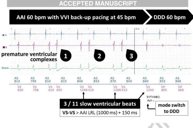
1. Two-thirds of inappropriate episodes (66%) were related to compensatory pauses generated by premature atrial contractions (7%), premature ventricular contractions (56%, Figure 2) or both (3%),
2. 21% were related to premature ventricular contractions falling in the post-atrial pacing ventricular refractory period interval (Figure 3) leading to asynchronous ventricular pacing.
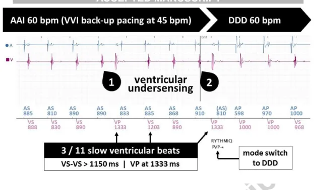
3. 10% were related to a paroxysmal lead dysfunction: atrial undersensing (3%), ventricular undersensing (2%), atrial capture failure (2%) and atrial noise oversensing (3%). An example of a mode switch in the context of ventricular undersensing is shown in Figure 4.
4. 4% were related to ventricular events occurring during the post atrial ventricular blanking period after a very long PR interval.
The average time spent in the secondary DDD mode after an inappropriate RYTHMIQTM event was 4 hours and 48 minutes per episode (median: 1 minute and 3 seconds, minimum 46 seconds and maximum 289 hours).
Pacemaker mediated tachycardia episodes
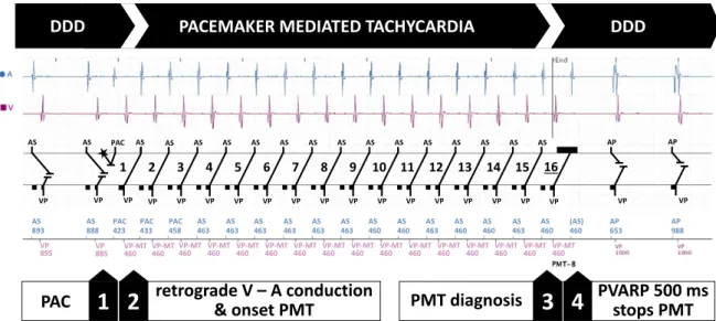
Twenty seven patients (17%) exhibited at least 1 recorded episode labeled as PMT. We analyzed 154 episodes labeled PMT of which 30 (20%) were misdiagnosed during sinus tachycardia (normal tracking of intrinsic atrial events at the maximal tracking rate). In 85 episodes (69% of true PMT episodes) the onset of PMT (Figure 5) was directly related to the prolongation of the AV delay (AV search+™). Indeed, the last ventricular pacing cycle before the onset of the tachycardia was performed with a prolonged AV delay prompting the occurrence of a retrograde atrial activation. The usual triggers for PMT were observed in the remaining episodes; PVC (20%) and PAC (11%). The analysis of the labelled PMT episodes
9
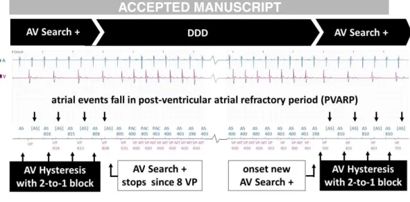
incidentally revealed a deleterious effect of the systematic AV search hysteresis (every 32 cycles) during ventricular tracked sinus tachycardia in ventricular pacing dependent patients. In 20 episodes (across 3 patients) we discovered that the AV Search+™ algorithm induced a 2:1 block during 8 cycles (16 atrial cycles) since every other atrial event occurred during the post-ventricular atrial refractory period (Figure 6).
Discussion
In the present study, we evaluated the RYTHMIQTM algorithm, the specificity of the switches to DDD and its relation with pacemaker-mediated tachycardia by analyzing over one thousand episodes in a large group of patients. The analysis was only possible because mode switch episodes are recorded in Boston Scientific devices. Sorin is the sole other manufacturer which records mode switches (SafeR™) while Medtronic (Managed Ventricular Pacing™), Biotronik (Vp suppression™) and St Jude Medical (Ventricular Intrinsic Preference™) have algorithms designed to reduce ventricular pacing, but do not record the mode switch episodes. Similarly, suspected pacemaker mediated tachycardias are only recorded in Boston Scientific and St Jude Medical devices. The limited recording features of the other device manufacturers renders comparison impossible, therefore the aim of this article is not to suggest the superiority or inferiority of this algorithm compared to the other ones. However, we did observe limitations and repetitive inappropriate switches that may help improve patient selection for this algorithm.
Limitations of the RYTHMIQTM algorithm
Large number of inappropriate AAI to DDD switches
In the present study, the percentage of appropriate switches and thus the specificity of the RYTHMIQTM algorithm was low (16%). However, the percentage of appropriate versus inappropriate switches is not only reliant on the performance of the algorithm but also on
30
10
the indication for implantation and the degree of ventricular pacing dependency. Indeed, selection of patients with a higher degree of AV conduction disorder, would have probably led to a higher percentage of appropriate switches. This is also evidenced by the higher percentage of cumulative RV pacing in the patients with predominantly appropriate switches as compared with patients with predominantly inappropriate switches (28% versus 10%). The large number of analyzed episodes does permit us to draw important conclusions about the triggers for inappropriate switches and about the characteristics of patients less likely to benefit from this algorithm.
Compensatory pauses following atrial or ventricular extrasystoles
In patients with frequent atrial or ventricular extrasystoles, the VVI back-up mode prevents occurrence of prolonged pauses since the minimal ventricular pacing rate is programmed between 30 bpm (pause 2 seconds) to 60 bpm (pause 1 second). On the other hand, the VVI back-up mode renders this algorithm particularly sensitive to switching pacing mode following compensatory pauses. The number of these inappropriate switches may be sustained by additional extrasystoles that can cause a significant increase in percentage of ventricular pacing. This malfunction is specific for this algorithm and is much less frequently observed with other AAI/DDD algorithms. Patients with frequent atrial or ventricular extrasystoles may therefore not be the most suitable candidates for the RYTHMIQTM algorithm.
Ventricular extrasystoles within the refractory period
Ventricular extrasystoles which occur relatively late in the cardiac cycle are also an important contributor to inappropriate commutations. When the late extrasystole occurs in the post atrial pacing ventricular refractory period (blanking or noise window), it does not inhibit ventricular stimulation. The algorithm is designed to function in AAI to reduce
ventricular pacing with a parallel independent VVI back-up mode to prevent pauses. In fact, the AAI and VVI modes do not operate completely independently since during atrial pacing, a ventricular post atrial stimulation-blanking period is mandatory to inhibit cross-talk and the associated risk of asystole. This type of inappropriate mode switch is also observed in other AAI/DDD algorithms such as in Sorin, Medtronic and Biotronik devices (even though these episodes are not recorded in the 2 latter manufacturers pulse generators).
High rate of pacemaker mediated tachycardia
In DDD mode, the AV Search+TM algorithm periodically and systematically prolongs the AV
interval in order to search for spontaneous AV conduction. This method, also chosen by St Jude Medical and Biotronik devices, has as a main advantage: to avoid non-conducted atrial depolarizations and ventricular pauses. In contrast, a blocked-P wave is possible during the search phase with Medtronic and Sorin devices. On the other hand, AV interval prolongation is accompanied with a risk of pacemaker mediated tachycardia induction. Since EGMs of suspected PMTs were recorded, we were able to investigate the underlying mechanism. Besides Boston Scientific, this is only possible for St. Jude Medical devices. In our study the
majority of PMTs (69%) was associated with the AV Search+TM algorithm. An important note
is that PMTs at a rate under the programmed maximal tracking rate (for instance because of prolonged PR interval or programming of a high maximal tracking rate) are not diagnosed as a PMT by the device and will not be saved into the memory. For this reason it is conceivable that the RYTHMIQ algorithm induces more PMTs than apparent from the device memory. Risk of 2-to-1 AV block during exercise
In patients who switched to DDD mode for atrioventricular block, the algorithm searches for spontaneous conduction by prolongation of the AV interval for 8 consecutive cycles. When this search occurs during sinus tachycardia, the increased AV interval favors the risk of
32
12
developing 2-to-1 block with a sudden drop in ventricular pacing rate. In this case, every other atrial event occurs during the post-ventricular atrial refractory period (PVARP) and is thus not followed by a ventricular paced beat. This study is not able to determine the exact incidence of this anomaly since it was accidentally recorded during episodes misdiagnosed as PMT, even though the risk seems important in patients with sinus tachycardia who have been switched to DDD mode for atrioventricular block. The symptomatic importance of these episodes is also yet to be determined.
Diagnosis of pacemaker dysfunction
An important advantage of recording mode-switch episodes is the possibility of diagnosing paroxysmal pacemaker dysfunction (undersensing, loss of atrial capture or oversensing) that induce inappropriate switches to the DDD mode. Many of these episodes were not sustained and would not have been detected by the device which indicates that diagnosing pacemaker dysfunction is one of the pleiotropic effects of the RYTHMIQ algorithm. In our study population, 23 patients (15%) exhibited inappropriate mode switches related to pacemaker
dysfunction. Undersensing, atrial capture failure or oversensing indicate either inadequate
programming of the device (incorrect sensitivity, pacing threshold too low) or lead dysfunction which should alarm the programmer to investigate the issue further.
Suggestions for improving the algorithm
Two-thirds of inappropriate mode switches were related to premature atrial or ventricular contractions. Different options may be proposed to decrease the rate of inappropriate switches. 1) The specific sequence, premature contraction- compensatory pause (short-long cycle), could be recognized by the device, and the compensatory pause would not be considered as a slow ventricular beat and not be integrated in the 3/11 count. When the compensatory pause would not be considered as a slow ventricular beat, we would expect
13
an important decrease in inappropriate mode switches and thus a significant decrease in cumulative RV pacing. 2) The amount of ‘slow ventricular beats’ required for a mode switch is not programmable (fixed at ≥3 out of 11). Having the possibility to program a higher number of required slow beats may be beneficial in patients with frequent extrasystoles. However, increasing the amount of required slow ventricular beats would result in fewer commutations in patients with second degree AV block. In other words, increasing the specificity could result in a decrease in sensitivity.
The other major contributors of inappropriate mode switches, namely refractory period and pacemaker dysfunction, are more complex to improve. A post-atrial pacing refractory period is imperative to protect the ventricular channel from oversensing. The detection of paroxysmal pacemaker dysfunction we actually consider as a desired side-effect of this algorithm.
This study confirmed that prolongation of the AV interval is associated with PMTs since the majority of PMTs were related to the AV Search+TM algorithm. During DDD mode the AV Search+TM algorithm searches for spontaneous conduction every 32 beats. Progressively increasing this amount would also considerably reduce the amount of PMTs. Finally, we suggest to suspend AV Search+TM during tachycardia, as this can initiate 2-to-1 block as we have discovered by chance in the PMT labelled episodes (which were in fact sinus tachycardia at maximal tracking).
Suggestions on patient selection for RYTHMIQ
The RYTHMIQ algorithm is most suited for patients with sinus node dysfunction, brady-tachycardia syndrome and chronotropic insufficiency. In these patients, RYTHMIQ works well to limit the burden of ventricular pacing, however most of the switches were inappropriate
34
14
since the percentage of true AV block is very limited and we demonstrated frequent inappropriate mode switches for other events such as premature contractions. We believe that in these patients, modification of the algorithm may improve the specificity. Patients with intermittent AV block are not the ideal candidates but if the goal is to decrease the burden of ventricular pacing while avoiding long pauses by AV-block, this population would paradoxically show higher accuracy than patients with preserved AV conduction, since more episodes of AV block will increase specificity. Patients with higher degree or permanent AV block should not be programmed to RYTHMIQ. Programming RYTHMIQ in these patients is probably not harmful but it is not logical either.
Limitations
This study has several limitations, namely the retrospective nature of the analysis and the
limited proportion of RYTHMIQTM episodes with stored EGMs (in relation to the total
number of RYTHMIQTM episodes). Nevertheless, based on the large amount of episodes we
can conclude that the specificity of the algorithm is low. Calculating the sensitivity was not possible since we were only able to investigate the events which triggered the algorithm to
perform a mode switch. Finally, we cannot conclude the prevalence of AV Search+TM induced
2-to-1 block since this phenomenon was seen by chance in misdiagnosed PMT episodes. Conclusion
This study highlights some of the limitations of the RYTHMIQTM algorithm: 1) high rate of
inappropriate switch related to premature ventricular or atrial contractions and 2) high rate of induction of PMT. This may have clinical implications in terms of selection of the patients and may suggest required changes in the algorithm architecture.
Acknowledgements
This study was supported by the French Government, Agence Nationale de la Recherche au titre du programme Investissements d'Avenir (ANR-10-IAHU-04). MS has received research grants from the Dutch Heart Foundation and the Netherlands Heart Institute.
References
1. Prinzen FW, Augustijn CH, Arts T, Allessie MA, Reneman RS. Redistribution of myocardial fiber strain and blood flow by asynchronous activation. Am J Physiol Aug 1990;259:H300-308. 2. Prinzen FW, Cheriex EC, Delhaas T, van Oosterhout MF, Arts T, Wellens HJ, Reneman RS. Asymmetric thickness of the left ventricular wall resulting from asynchronous electric activation: a study in dogs with ventricular pacing and in patients with left bundle branch block. Am Heart J Nov 1995;130:1045-1053.
3. Sweeney MO, Prinzen FW. Ventricular pump function and pacing: physiological and clinical integration. Circulation Arrhythmia and electrophysiology Jun 1 2008;1:127-139.
4. Barsheshet A, Moss AJ, McNitt S, Jons C, Glikson M, Klein HU, Huang DT, Steinberg JS, Brown MW, Zareba W, Goldenberg I. Long-term implications of cumulative right ventricular pacing among patients with an implantable cardioverter-defibrillator. Heart Rhythm;8:212-218. 5. Sweeney MO, Prinzen FW. A new paradigm for physiologic ventricular pacing. J Am Coll
Cardiol Jan 17 2006;47:282-288.
6. Sweeney MO, Ellenbogen KA, Casavant D, Betzold R, Sheldon T, Tang F, Mueller M, Lingle J, The Marquis MVPDI. Multicenter, Prospective, Randomized Safety and Efficacy Study of a New Atrial-Based Managed Ventricular Pacing Mode (MVP) in Dual Chamber ICDs. Journal of Cardiovascular Electrophysiology 2005;16:811-817.
7. Olshansky B, Day JD, Moore S, Gering L, Rosenbaum M, McGuire M, Brown S, Lerew DR. Is dual-chamber programming inferior to single-chamber programming in an implantable cardioverter-defibrillator? Results of the INTRINSIC RV (Inhibition of Unnecessary RV Pacing With AVSH in ICDs) study. Circulation Jan 2 2007;115:9-16.
8. Sweeney MO, Bank AJ, Nsah E, Koullick M, Zeng QC, Hettrick D, Sheldon T, Lamas GA. Minimizing Ventricular Pacing to Reduce Atrial Fibrillation in Sinus-Node Disease. New England Journal of Medicine 2007;357:1000-1008.
9. Stockburger M, Defaye P, Boveda S, Stancak B, Lazarus A, Sipotz J, Nardi S, Rolando M, Moreno J. Safety and efficiency of ventricular pacing prevention with an AAI-DDD changeover mode in patients with sinus node disease or atrioventricular block: impact on battery longevity-a sub-study of the ANSWER trial. Europace Nov 26 2015.
10. Stockburger M, Boveda S, Moreno J, Da Costa A, Hatala R, Brachmann J, Butter C, Garcia Seara J, Rolando M, Defaye P. Long-term clinical effects of ventricular pacing reduction with a changeover mode to minimize ventricular pacing in a general pacemaker population. European heart journal Jan 14 2015;36:151-157.
11. Monteil B, Ploux S, Eschalier R, Ritter P, Haissaguerre M, Koneru JN, Ellenbogen KA, Bordachar P. Pacemaker-Mediated Tachycardia: Manufacturer Specifics and Spectrum of Cases. Pacing Clin Electrophysiol Sep 25 2015.
36 16
12. Akerstrom F, Arias MA, Pachon M, Puchol A, Jimenez-Lopez J, Rodriguez-Padial L. The reverse mode switch algorithm: how well does it work? Heart Rhythm Aug 2013;10:1146-1152.
Figure 1. Examples of appropriate RYTHMIQTM episode showing non-conducted P waves. The loss of spontaneous AV conduction results in dropped ventricular rate. When the algorithm discovers three slow ventricular beats in a window of 11 cycles, it executes a switch to DDD mode. Panel A shows 3 non-conducted P waves which result in VS-VS intervals which are slower than 1350 ms (LRL (1200 ms) + 150 ms) but not slow enough to trigger a VP (1714ms). Panel B shows 3 non-conducted P waves with VS-VP intervals of 1333ms (60.000/(LRL 60 - 15)) also considered as a slow ventricular beat.
Figure 2: Example of an inappropriate RYTHMIQTM episode induced by premature ventricular
contractions (PVCs). The PVCs (1, 2 and 3) result in a compensatory pause and the following ventricular event is considered a slow ventricular beat. Since in a window of 11 beats, three
PVCs resulted in three slow ventricular beats, RYTHMIQTM induced a mode switch from AAI
38
18
Figure 3: Example of an inappropriate RYTHMIQTM episode related to post-atrial ventricular refractory periods (blanking and noise window). During atrial stimulation, a post-atrial ventricular blanking period on the ventricular channel is essential to prevent cross-talk. Especially at higher atrial paced rates (sensor), premature ventricular contractions (PVC) can fall within this noise window (1, marked [VS]) and blanking (2, no mark) which triggers backup ventricular pacing. In addition, the stimulated beat acts as a PVC which triggers a compensatory pause which is also classified as slow.
Figure 4. Example of inappropriate RYTHMIQTM episode unmasking ventricular undersensing.
In this example a ventricular event was not sensed (1) which induced a back-up VP (15 bpm slower than LRL at 60 bpm = 1333 ms). In addition, the VP functions as a premature ventricular contraction which induces a compensatory pause. Both events are counted as slow ventricular beats so a second ventricular undersensed event sufficed for mode switch to DDD.
40
20
Figure 5: Example of pacemaker mediated tachycardia induced by AV Search+™. During this
AV Search+™ protocol, the first ventricular events occur after spontaneous AV conduction (Vs-Hy). The fourth atrial event is not conducted to the ventricle within the extended AV delay (300ms) and triggers a VP. This VP has a long AV interval which enabled retrograde ventriculo-atrial conduction. Since 2 cycles were stimulated, AV Search +™ was terminated (≥2/10 VP in the secondary phase terminates AV Search+™). However, retrograde V-A conduction is maintained and the ventricle is stimulated at maximal tracking rate (VP-MT). The electronic re-entry is sustained until the pacemaker correctly labels the event as PMT (16 cycles of VP-MT) and increases the post-ventricular atrial refractory period (PVARP) to 500 ms a single time. The following retrograde atrial conduction falls within the PVARP, thus does not trigger a ventricular stimulation and the PMT is terminated.
21
Figure 6. Example of an episode labeled pacemaker-mediated tachycardia unmasking 2-to-1
AV block during AV Search +™. 16 consecutive ventricular stimulations at the maximal tracking rate (16 X VP-MT) triggered the recording which is labeled PMT but is in fact sinus tachycardia. This tracing reveals that AV Search+™ is activated, since the AV interval is significantly delayed during the first and last events, as compared with the center part. The prolonged AV interval and the following post-ventricular atrial refractory period (PVARP) result in every other atrial event to fall in the blanking. A 2-to-1 AV block occurs with a sudden drop in ventricular rate during sinus tachycardia, which restored when AV Search+™ is stopped (stop criterion is 8 X VP in the primary phase). When AV Search+™ is re-initiated 32 ventricular cycles later, the 2-to-1 block re-occurs in identical fashion.
42
22
Tables
Table 1. Demographics and device specifications of patients with dual chamber Boston Scientific devices and RYTHMIQTM algorithm activated.
RYTHMIQTM ON Patient demographics Age [years] 67±13 Male sex [n] 101 (64%) Device type Pacemaker [n] 40 (25%) ICD [n] 117 (75%) Device programming
Lower Rate Limit [bpm] 55±7
Upper Tracking Rate [bpm] 132±7
Device output
Percentage ventricular pacing [%] 16±28 (0-100)
Percentage atrial pacing [%] 33±35 (0-100)
Median Respiratory Rate [per min]
17±3 (11-26)
Pacemaker-mediated tachycardia in a real word setup:
A clinical study based to study the accuracy
of specific algorithm.
1.
Study outline
A pacemaker-mediated tachycardia (PMT) can be defined as any condition in
which a pacemaker paces the ventricles at rates that are inappropriately fast. This
can be due to a rate response setting that is too sensitive, tracking of atrial noise (such as what may occur with electromagnetic interference), inappropriate pacemaker manipulation with rate response turned on, or tracking of an atrial tachyarrhythmia related to upper rate settings. This phenomenon has been observed and understood early after the introduction of dual-chamber pacing in patients presenting with intrinsic ventriculo-atrial (VA) conduction (21). The majority of patients are asymptomatic but patients may report symptoms ranging from palpitations, rapid heart rates, light-headness, syncope, or chest discomfort. Prolonged PMT may be poorly tolerated or even lead to cardiac decompensation in patients suffering from underlying heart disease (22). The incidence of PMT varies as a function of patient characteristics, programming of the device and the specificities of the algorithms provided by the manufacturers(22). There is few data on the incidence of PMT across pacemaker and defibrillator recipients [5–9]. Studies to compare the respective effectiveness of these various algorithms of detection and termination of PMT are non-existent. In Boston Scientific devices, episodes of PMT are recorded in the memory. In this multicenter study, we included 328 patients implanted with a Boston Scientific device: 157
non-44
with permanent AV-conduction disorder (AV-block group) and 95 Cardiac Resynchronization
Therapy patients (CRT group). For each patient, we reviewed the last 10 remote monitoring transmitted EGMs labelled as PMT.
2.
Implications
We have observed that the AV Search + (RYHTMIQ) algorithm in Boston Scientific devices was associated with PMT induction in a selected group of patients. In this study, we assessed the diagnostic and therapeutic efficacy of the PMT algorithm in Boston Scientific devices. This was associated with a high rate of incorrect PMT diagnosis during exercise resulting in inappropriate therapy with non conducted P-waves, loss of CRT and limited risk of pro-arrhythmic events.
Accuracy of the pacemaker-mediated tachycardia algorithm in Boston
Scientific devices
M. Strik, MD, PhD,a, b,⁎ A. Frontera, MD,a R. Eschalier, MD PhD,c P. Defaye, MD, PhD,d P. Mondoly, MD, PhD,e P. Ritter, MD, PhD,a M. Haïssaguerre, MD, PhD,a
S. Ploux, MD PhD,a P. Bordachar, MD, PhDa
aHaut-Lévêque Hospital, Centre, Hospitalier Universitaire de Bordeaux; LIRYC institute, Pessac, France
bMaastricht University Medical Center, Cardiovascular Research Institute Maastricht, Maastricht, The Netherlands
cClermont Université, Université d'Auvergne, Cardio Vascular Interventional Therapy and Imaging (CaVITI), Image Science for Interventional Techniques
(ISIT), UMR6284, CHU Clermont-Ferrand, Cardiology Department, F-63003, Clermont-Ferrand, France
dCentre Hospitalier Universitaire de Grenoble, La Tronche, France
eCentre Hospitalier Universitaire de Toulouse, Toulouse, France
Abstract Introduction: The incidence of pacemaker-mediated tachycardia (PMT) varies as a function of
patient characteristics, device programming and algorithm specificities. We investigated the efficacy of the Boston Scientific algorithm by reviewing PMT episodes in a large device population. Methods: In this multicenter study, we included 328 patients implanted with a Boston Scientific device: 157 non-dependent patients with RYTHMIQ™ activated (RYTHMIQ group), 76 patients with permanent AV-conduction disorder (AV-block group) and 95 Cardiac Resynchronization Therapy patients (CRT group). For each patient, we reviewed the last 10 remote monitoring-transmitted EGMs diagnosed as PMT.
Results: We analyzed 784 PMT episodes across 118 patients. In the RYTHMIQ group, the diagnosis of PMT was correct in most episodes (80%) of which 69% was directly related to the prolongation of the AV-delay associated with the RYTHMIQ algorithm. The usual triggers for PMT were also observed (PVC 16%, PAC 9%). The remainder of the episodes (20%) in RYTHMIQ patients and most episodes of AV-block (66%) and CRT patients (74%) were incorrectly diagnosed as PMT during sinus tachycardia at the maximal tracking rate. The inappropriate intervention of the algorithm during exercise causes non-conducted P-waves, loss of CRT (sustained in six patients) and may have been pro-arrhythmogenic in one patient (induction of ventricular tachycardia).
Conclusion: Algorithms to minimize ventricular pacing can occasionally have unintended consequences such as PMT. The PMT algorithm in Boston Scientific devices is associated with a high rate of incorrect PMT diagnosis during exercise resulting in inappropriate therapy with non-conducted P-waves, loss of CRT and limited risk of pro-arrhythmic events.
© 2016 The Authors. Published by Elsevier Inc. This is an open access article under the CC BY license (http://creativecommons.org/licenses/by/4.0/).
Keywords: Clinical: implantable devices – pacemaker-bradyarrhythmias; Clinical: implantable devices – biventricular
pacing/defibrillation; Clinical: implantable devices – ventricular tachycardia/fibrillation; Clinical: implantable devices – physiologic pacing; Clinical: cardiac mapping – electrogram analysis
Introduction
Pacemaker mediated tachycardia (PMT) describes a repetitive sequence of sensed retrograde atrial events followed by ventricular pacing. This phenomenon has been observed and understood early after the introduction of dual-chamber pacing in patients presenting with intrinsic ventriculo-atrial (VA) conduction [1–3]. The majority of patients are asymptomatic but patients may report symptoms
Available online atwww.sciencedirect.com
ScienceDirect
Journal of Electrocardiology 49 (2016) 522 – 529
www.jecgonline.com
Funding: This work was supported by the French Government, Agence Nationale de la Recherche au titre du programme Investissements d'Avenir [ANR-10-IAHU-04] and by the Dutch Heart Foundation and the Netherlands Heart Institute [to MS].
⁎ Corresponding author at: Service de cardiologie-électrophysiologie et stimulation cardiaque, Hôpital Haut-Lévêque (CHU) de Bordeaux, 5 Av. Magellan, 33600 Pessac, France.