Publisher’s version / Version de l'éditeur:
The Journal of Physical Chemistry A, 109, 36, pp. 8096-8105, 2005-09-15
READ THESE TERMS AND CONDITIONS CAREFULLY BEFORE USING THIS WEBSITE. https://nrc-publications.canada.ca/eng/copyright
Vous avez des questions? Nous pouvons vous aider. Pour communiquer directement avec un auteur, consultez la première page de la revue dans laquelle son article a été publié afin de trouver ses coordonnées. Si vous n’arrivez pas à les repérer, communiquez avec nous à [email protected].
Questions? Contact the NRC Publications Archive team at
[email protected]. If you wish to email the authors directly, please see the first page of the publication for their contact information.
NRC Publications Archive
Archives des publications du CNRC
This publication could be one of several versions: author’s original, accepted manuscript or the publisher’s version. / La version de cette publication peut être l’une des suivantes : la version prépublication de l’auteur, la version acceptée du manuscrit ou la version de l’éditeur.
For the publisher’s version, please access the DOI link below./ Pour consulter la version de l’éditeur, utilisez le lien DOI ci-dessous.
https://doi.org/10.1021/jp052197t
Access and use of this website and the material on it are subject to the Terms and Conditions set forth at
Conformational Pathways of Saturated Six-Membered Rings. A Static
and Dynamical Density Functional Study
Ionescu, Andrei R.; Berces, Attila; Zgierski, Marek Z.; Whitfield, Dennis M.;
Nukada, Tomoo
https://publications-cnrc.canada.ca/fra/droits
L’accès à ce site Web et l’utilisation de son contenu sont assujettis aux conditions présentées dans le site LISEZ CES CONDITIONS ATTENTIVEMENT AVANT D’UTILISER CE SITE WEB.
NRC Publications Record / Notice d'Archives des publications de CNRC:
https://nrc-publications.canada.ca/eng/view/object/?id=357c916e-19e7-4437-b461-25622e976464 https://publications-cnrc.canada.ca/fra/voir/objet/?id=357c916e-19e7-4437-b461-25622e976464Conformational Pathways of Saturated Six-Membered Rings. A Static and Dynamical
Density Functional Study
Andrei R. Ionescu,*,†Attila Be´rces,‡Marek Z. Zgierski,†Dennis M. Whitfield,§ and
Tomoo Nukada|
Steacie Institute for Molecular Sciences, National Research Council of Canada, Theory and Computation Program, 100 Sussex DriVe, Ottawa, Ontario K1A 0R6, Canada, Computational Chemistry Logic Ltd., Madach. u. 20, 8900 Zalaegerszeg, Hungary, Institute for Biological Sciences, National Research Council of Canada, 100 Sussex DriVe, Ottawa, Ontario K1A 0R6, Canada, and The Institute of Physical and Chemical Research (RIKEN), Wako-shi, Saitama 351, Japan
ReceiVed: April 27, 2005; In Final Form: July 12, 2005
The conformation of the six-membered ring of pyranosyl sugars has pronounced effects on the physical and chemical properties of carbohydrates. We present a method to determine key features of the potential energy surfaces, such as transition states associated with the inversion pathways of the model compounds cyclohexane, tetrahydropyran, p-dioxane, m-dioxane, s-trioxane, and 2-oxanol. Finally, we make the first determination of the pathways for inversion of penta-O-methyl-R-D-glucopyranose and penta-O-methyl-β-D-glucopyranose. For both anomers, a transition state with five coplanar atoms with appreciableOE character was found. The
method is based on constrained Car-Parrinello ab initio molecular dynamics, as implemented in the projector augmented-wave (PAW) method. The constraints are derived from the normal modes of six-membered rings and are described in terms of the canonical conformations1C4chair,1,4B boat, andOS2skew-boat. The PAW
derived trajectories are in agreement with previous suggestions in the literature that pseudorotation is an important feature of such conformational interconversions. The dynamic nature as well as the internal coordinate-based constraints provide a method which can reliably accommodate pseudorotation. To determine semiquantitative energies, we recalculate key conformations using standard quantum mechanical calculations while keeping the ring dihedral angles frozen at their values found in the dynamics. In all cases where experimental barriers are known, our results are in excellent agreement.
1. Introduction
Ring conformation is an essential factor in the hydrolysis and the synthesis of the glycosidic bond. The mechanism of the reactions involved in the synthesis of oligosaccharides using chemical glycosylation1or glycosyl transferases2-4as well as the degradation of oligosaccharides by solvolysis reactions5-7 or glycosidases8 implies changes in the ring conformation. Recent atomic force microscopy experiments were interpreted as indications that polysaccharides owe their elasticity to the chair-boat flips of the pyranose ring.9Our previous studies of the neighboring group-assisted glycosylation reactions, by quantum chemistry calculations, revealed that, in some cases, the relative energies of pyranose conformations determine the transition states between crucial steps.10,11Our previous studies also suggest that chemical modification of the rigidity of the pyranose ring controls the stereochemistry and determines the product distribution.10,12,13
Our present goal is the complete characterization of the conformational potential energy surface of pyranoses. We believe that the mapping of the stationary points on the potential energy surface of pyranoses is a necessary prerequisite for the explanation of the biological functions of carbohydrates. Of most
importance are the saddle points because experimental informa-tion concerning transiinforma-tion states is extremely limited. In the present paper, we developed a new method to explore the potential energy surface of pyranoses with particular emphasis on finding transition states along conformational and reaction pathways.
The application of existing algorithms of quantum chemistry to study the reaction mechanism involving the change in pyranose conformation is not straightforward. The first difficulty lies in the multitude of possible conformations. Pyranoses have 38 distinct basic conformations: 2 chairs, 6 boats, 6 skew-boats, 12 half-chairs, and 12 envelopes.14In addition, there are stable conformations that do not always conform with any of the 38 basic conformations.15New intermediate stable conformations are made possible by different substituents, attached fused rings, and double bonds; e.g, there are 24 additional conformations for 2,6-cis- and 2,6-trans-substituted dyhydropyronones that are intermediates between envelope and half-chair conformations.15 Even the simplest and most symmetrical saturated six-membered ring compound, cyclohexane, has stationary points which cannot be described by any of the chair, boat, skew-boat, half-chair, or envelope conformations.16The brute-force approach to the conformational problem of pyranoses is to optimize the geom-etry of all 38 basic conformations. However, because not all stationary points lie close to one of the 38 basic conformations, it is possible to miss some important conformations.
The second difficulty lies in the nature of the stationary points and the algorithms used to optimize these stationary points. The †Steacie Institute for Molecular Sciences.
‡Computational Chemistry Logic Ltd. §Institute for Biological Sciences.
|The Institute of Physical and Chemical Research. Current address: Department of Fermentation Science, Faculty of Applied Bioscience, Tokyo University of Agriculture, Sakuragaoka, 1-1 Setagayaku, Tokyo 156-8502, Japan.
8096 J. Phys. Chem. A 2005, 109,8096-8105
10.1021/jp052197t CCC: $30.25 © 2005 American Chemical Society Published on Web 08/17/2005
chair and skew-boat conformations are usually minimal, the boat and half-chair conformations are first-order transition states, the envelope conformations are second-order transition states, and there are several exceptions from these rules.
It is not possible to tell a priori the nature of each conforma-tion; furthermore, different algorithms need to be used to optimize stable structures or transition states. It is particularly difficult to find transition states on the ring conformational potential surface because of the nature of the eigenvectors involved in the conformational transitions. Transition-state (TS) optimization methods rely on identifying the eigenvector corresponding to the reaction coordinate. Conformational transi-tions of six-membered rings involve the continuous mixing of two or three eigenvectors. For this reason, the straightforward application of TS optimization algorithms is often unsuccessful. To explore the potential energy surface of ring conformational transition, it was necessary to develop new methods that take into account the physical nature of the conformational degrees of freedom and the possible pathways of converting one conformation into another.
2. Conformational Constraints
The conformational analysis of the potential energy surface of six-membered pyranose rings by quantum chemistry methods requires that the geometry can be constrained to any particular conformation during optimization. The conformational degrees of freedom of the six-membered rings arise from the three out-of-plane normal modes of the regular hexagon. These three normal modes correspond to the distortion of the planar ring into chair, boat, and skew-boat conformations. All pyranose ring conformations can be uniquely described in terms of one chair, one boat, and one skew-boat conformation, but it proves convenient to use the1C4chair,1,4B boat, andOS2skew-boat
conformations, which we call canonical conformations.17 If distortions into 1C4 chair, 1,4B boat, and OS2 skew-boat
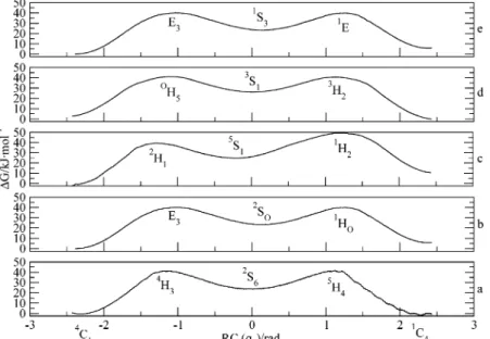
conformations are represented by points on the z, x, and y axes of an orthogonal coordinates system, respectively, all boat and skew-boat conformations derived by permutation of the atom numbering lie on a circle in the xy plane. The half-chair and envelope conformations lie on a smaller radius circle between the xy plane and the chair coordinate on the z axis. Thus, as originally suggested by Hendrickson, all pyranose ring confor-mations are conveniently represented on a sphere and character-ized by spherical polar coordinates (Figure 1).18The radius is the puckering amplitude, and the angular coordinates determine the type of conformation. There have been several proposals
for the mathematical expression of the spherical polar coordi-nates, including our own definition.17The most popular defini-tion was introduced by Cremer and Pople (CP) who expressed the ring conformation as a function of the Cartesian atomic coordinates.19Alternatively, spherical polar coordinates can be derived by Fourier transform from the ring torsion angles.20,21 In the preceding paper,17 we have derived quantitative expressions for the characterization of the pyranose and other six-membered ring conformations which rely heavily on the definitions of the natural internal coordinates introduced by Pulay et al.22,23If τ1, τ2, τ3, τ4, τ5, and τ6 torsion angles are defined by the C1C2C3C4, C2C3C4C5, C3C4C5O, C4C5OC1, C5-OC1C2, and OC1C2C3atom sets, respectively, displacement into the1C4chair conformation is described by the
internal coordinate. Displacement into1,4B boat (q2) andOS2
skew-boat (q3) conformations are described by the internal coordinates
respectively.
The phase angle of pseudorotation can be defined as a function of q2and q3internal coordinates:
The x3/2 term arises from normalization factors. Equation 4 is consistent with the Cremer-Pople definition of φ, provided that the ring atoms are numbered by carbohydrate numbering. In that case, the zero value of the phase angle, φ, corresponds to the1,4B boat conformation.
The internal coordinates defined in eqs 1-3 can be applied as constraints. Although they may not be the most suitable constraints, their availability in quantum chemistry programs makes them an attractive choice. For example, the transforma-tion fromOS2to2S
Oconformations can be studied with q3of
eq 3 as a constraint. The midpoint of the pseudorotational circle (Figure 1) corresponds to the planar conformation, and the radius determines the amplitude of displacement from the planar conformation into theOS2conformation. However, any other
conformation (in this case, any other twistboat) can be described by the internal coordinate q3by permutation of atom numbers in the definition of the internal coordinate. For example, starting the atom numbering with C2instead of C1in the definition of the q3, we can describe distortion into the 1S3conformation. Opposite conformations correspond to the same internal coor-dinate with the opposite sign. By increasing the value of q3from a negative to a positive value, one may induce a conformational change that turns theOS2into the2S
Oconformation. By allowing
the complementary internal coordinates to evolve according to the lowest-energy path, the reaction follows the pseudorotational itinerary instead of going through the energetically forbidden planar conformation. The drawback of using constraints such as q2and q3is that there is no control over whether the transition follows a clockwise or counterclockwise transformation along the pseudorotational itinerary. The direction will be determined by the lower energy of distortion at the starting conformation. However, this direction does not necessarily correspond to the
Figure 1. Spherical mapping of pyranose conformations.
q1)τ1-τ2+τ3-τ4+τ5-τ6 (1) q2)τ2-τ3+τ5-τ6 (2) q3)τ1-12τ2-12τ3+τ4-12τ5-12τ6 (3) φ )arctan
(
x3q2 2q3)
(4)overall lowest-energy reaction pathway because the potential may sharply increase at a subsequent conformation. Another drawback of such constraints is that they do not have high projection onto the intrinsic reaction coordinate (IRC) through-out the reaction. This is a requirement for the calculation of thermodynamic properties along the reaction pathway by constrained reaction dynamics. We used q1to explore the chair inversion process and q2and q3to explore the pseudorotational itinerary.
3. Computational Details
3.1. Dynamical DFT Calculations. Dynamical density
funtional theory (DFT) calculations were carried out with the projector augmented-wave (PAW) method of Blo¨chl,24which is an implementation of the Car-Parrinello ab initio molecular dynamics.25The energy cutoff of the plane wave basis set was set to 30 Ry, and the calculations used the frozen-core approximation. All calculations employed the exchange cor-relation functional of the generalized gradient approximation with the local potential of Perdew and Zunger26augmented by the gradient corrections to the exchange and correlation of Becke and Perdew. Periodic boundary conditions were used with a unit cell defined by lattice vectors ([0.0 10.0 10.0] [10.0 0.0 10.0] [10.0 10.0 0.0]) Å for the test molecules and by lattice vectors ([0.0 14.0 14.0] [14.0 0.0 14.0] [14.0 14.0 0.0]) Å for D-glucopyranose. To prevent
electrostatic interactions between periodic images, a charge-isolation scheme was used.27 A temperature of 300 K was maintained for all simulations by a Nose´ thermostat.28,29The fictitious kinetic energy of the electrons was controlled in a similar fashion by a Nose´ thermostat.30To span large portions of configuration space in a minimum amount of time, the true masses of the nuclei were rescaled to 3.0 (O and C) and 1.5 (H) atomic mass units. Together with an integration step of 7 au (0.17 fs), this choice ensures good energy conservation during dynamics simulations without computational overhead due to heavy atomic nuclei. Because we do not discuss time-dependent properties and because configurational ensemble averages remain unchanged under a rescaling of the masses, this technique is appropriate. To sample space in the vicinity of the transition state, we chose a reaction coordinate (RC) which was kept constrained during the dynamics with the SHAKE algorithm.31 All other degrees of freedom are allowed to evolve naturally in time. By slowly varying the constraint, we can sample phase space in the vicinity of the transition state dynamically, leading to undisturbed dynamics for all motions which are orthogonal to the RC and to the fictitious dynamics along the RC. All simulations consisted of 70 000 (test molecules) and 90 000 time steps which cover 11.9 and 15.3 ps real time, respectively, unless otherwise indicated in the text.
The free-energy difference, ∆F, between two arbitrary points (λ1and λ0) along the reaction coordinate can be evaluated by integrating the average force on λ at a given constant temper-ature, T:
The reaction coordinate can be sampled with discrete values of λ or allowed to vary continuously in what is called a slow-growth simulation. In the latter method, the system is never properly equilibrated unless the change in the RC is infinitesi-mally small. The slow-growth method has been previously demonstrated on several elementary reactions steps in organo-metallic chemistry.32-35In the slow-growth method, the energy
gradient is not averaged at constant values of λ, therefore it does not yield the free energy exactly. However, this method is successful for the qualitative understanding of the reaction mechanism, the location of stationary points on the free-energy surface, and the semiquantitative calculation of free-energy differences.
3.2. Static DFT Calculations.The reported static calculations
were carried out with the Gaussian 98 program package.36All optimized static geometries calculated in this study are based on the generalized gradient approximation (GGA). All calcula-tions were performed with the B3P86 method combining Becke’s 3-parameter exchange functional37with the Perdew 86 correlation functional.38,39The basis set was 6-311+G**. Full geometry optimization followed by complete frequency analysis was performed in all cases.
4. Results and Discussion
Thermodynamical experimentally measured data on saturated six-membered rings consist of values for the energy barriers of the chair inversion and the skew-boat to chair isomerization as well as values for the rate of inversion of the rings. First, we investigated the conformational potential energy surface of cyclohexane, tetrahydropyran, m-dioxane, p-dioxane, and s-trioxane to test various aspects of the constrained dynamics, such as the different constraints, the energy conservation, and the numerical accuracy by two-way simulations. Secondly, we investigated the simplest model for the pyranose ring (2-oxanol) to further test our constrained method and to compare our results with previous high-level theoretical calculations. Finally, we investigated the complete conformational potential energy surface ofD-glucopyranose.
4.1. Benchmark Applications and Tests. Tables 1 and 2
show the experimentally determined free energy and enthalpy of activation of cyclohexane and its derivatives vs our calculated values. The free-energy values were computed by dynamical simulations at 300 K with eq 5 and by static DFT simulations. The enthalpy of activation values were obtained by static DFT thermochemistry calculations at 300 K.
The calculated-energy and free-energy barriers for chair to skew-boat conversion, listed in Table 1, are in excellent ∆F ) F(λ1) - F(λ0) )
∫
λ0λ1
〈
δEδλ
〉
λdλ (5)TABLE 1: Barriers to Chair to Skew-Boat Conversion in kJ·mol-1at 300 Ka
compd (calcd)∆H b (exptl)∆H c (calcd)∆G b (calcd)∆G d (exptl)∆G e
cyclohexane 43.5 38.0-48.0 48.0 41.4 ∼43 tetrahydropyran 42.6 42.0 ( 5.0 41.8 39.7 41.0 ( 1.6
m-dioxane 41.8 40.0 ( 2.1 40.5 40.1 41.0 ( 0.8
p-dioxane 43.0 na 42.6 41.0 ∼41
s-trioxane 41.8 37.0 ( 5.0 35.5 48.5 46.0 ( 0.8
aSee Figure 2 for canonical conformations.bFully optimized at the
b3p86/6-311+g** level.cSee ref 41.dCalculations were carried out
with PAW.eSee ref 42.
TABLE 2: Barriers to Skew-Boat to Chair Conversion in kJ·mol-1at 300 Ka
compd (calcd)∆H b (exptl)∆H (calcd)∆G b (calcd)∆G c (exptl)∆G d
cyclohexane 17.1 na 23.8 18.0 22.5 ( 0.2 tetrahydropyran 18.4 na 19.6 16.7 na
m-dioxane 16.3 na 17.6 23.4 na
p-dioxane 15.5 na 17.6 15.0 na
s-trioxane 20.5 na 16.7 23.8 na
aSee Figure 2 for canonical conformations.bFully optimized at the
b3p86/6-311+g** level.cCalculations were carried out with PAW.dSee
ref 43.
agreement with the experimental values40,41and are within the numerical accuracy of the calculations of the experimental error range.
The calculated-energy and free-energy barriers to skew-boat to chair conversion are listed in Table 2. Experimental values, at room temperature and in gas phase, corresponding to our simulations are not available. The only experimental value for the free-energy barrier of cyclohexane skew-boat-chair isomer-ization was measured at 100 K in an Ar matrix.42
Next, we discuss the conformational interconversion of the different six-membered ring compounds on the basis of dynami-cal and static DFT dynami-calculations.
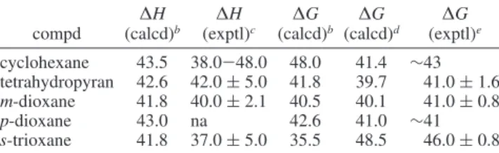
4.1.1. Cyclohexane.Figure 2a shows the free-energy diagram corresponding to the inversion of cyclohexane using the q1 constraint. The dynamical trajectory obtained by constraining the cyclohexane inversion using the q1chair coordinate involves two half-chair transition states (5H4and4H3) and a skew-boat (2S6) secondary minimum. The free-energy diagram is sym-metric with both chair to skew-boat barriers of 41.4 kJ‚mol-1. The first transition state (5H4) normally connects the chair (1C4) conformation to the skew-boat (5S1). The fact that the secondary minimum is the2S6skew-boat suggests that the transition state pseudorotates. This confirms the conclusion of previous ab initio, semiempirical, and molecular mechanics studies that the flat nature of the potential surface around the half-chair conformation gives rise to a fluxional, pseudorotating transition state.
The second half of the trajectory follows the minimum energy pathway by connecting the 2S6 skew-boat with the 4C1 chair conformation through a4H3half-chair transition state. In this case, there is also a strong contribution from pseudorotation. The energy barrier to the skew-boat secondary minimum to the chair conformations of 18.0 kJ‚mol-1was in good agreement with the experimental values. The static calculation free-energy and enthalpy of activation are shown in Tables 1 and 2.
Figure 3 shows the free-energy diagrams corresponding to the inversion of cyclohexane using the q2 and q3constraints. The dynamical trajectories obtained using the q2boat and q3 skew-boat coordinates show that, in both cases, the trajectory is constrained to the pseudorotational itinerary. The skew-boat minimum conformers along the pseudorotational circle are interconverted via boat transition-state conformations. The free-energy barriers are below 4.18 kJ‚mol-1, in very good agreement
with experimental data that found a barrier of interconversion of 3.8 kJ‚mol-1.
4.1.2. Tetrahydropyran. Figure 2b shows the free-energy diagram corresponding to the inversion of tetrahydropyran using the q1chair constraint. The three minima correspond to the two chair conformers (1C4 and 4C1) and to the skew-boat stable secondary minimum (2S
O). The free-energy barriers from1C4
to 2S
O and from 4C1 to 2SO are 33.9 and 39.7 kJ‚mol-1,
respectively. The dynamical trajectory of the tetrahydropyran chair inversion does not follow the lowest skew-boat conforma-tion and passes through an E3envelope transition state, which is essentially isoenergetic with the1H
Oconformation. The
free-energy barriers to the skew-boat to two chair conformations are 16.3 and 16.7 kJ‚mol-1. The static calculation results are shown in Tables 1 and 2 and are in good agreement with the experimental results.
4.1.3. m-Dioxane.Figure 2c shows the free-energy diagram corresponding to the inversion of m-dioxane using the q1chair constraint. The low symmetry of m-dioxane is well represented by the asymmetric free-energy diagram. The dynamical calcula-tions found that the4C1conformer is 10.0 kJ‚mol-1more stable than the1C4conformer. The dynamical trajectory starts from the 4C1 conformer and then passes through a 2H1 half-chair transition state (the free-energy barrier is 40.1 kJ‚mol-1; see
Figure 2. Free-energy diagrams in kJ‚mol-1for the inversion of saturated six-membered rings: (a) cyclohexane, (b) tetrahydropyran, (c) m-dioxane, (d) p-dioxane, and (e) s-trioxane.
Figure 3. Free-energy diagrams in kJ‚mol-1 for the inversion of cyclohexane using (a) the q2constraint and (b) the q3constraint.
Table 1) which leads to a5S1secondary minimum. Interestingly, from the5S1, the trajectory follows the pseudorotational itinerary and passes through the1S5skew-boat and then through the1H2 transition state to finally get to the inverted1C4chair conformer. The detour on the pseudorotational circle costs an additional 9.6 kJ‚mol-1, and the overall free-energy barrier is 23.4 kJ‚mol-1.
4.1.4. p-Dioxane.Figure 2d shows the free-energy diagram corresponding to the inversion of p-dioxane using the q1chair constraint. The dynamical trajectory for the inversion of1C4to 4C1passed through the3S1secondary minimum. The minima were connected by3H2andOH5half-chair transition states. The
free-energy barrier to the chair to skew-boat reaction was 41.0 kJ‚mol-1(Table 1), and the barrier to the skew-boat to chair reaction was 15.0 kJ‚mol-1(Table 2).
4.1.5. s-Trioxane. Figure 2e shows the free-energy diagram corresponding to the inversion of s-trioxane using the q1chair constraint. s-Trioxane inverts through two envelope transition states (1E and E3). The secondary minimum on the free-energy plot is a skew-boat conformer (1S3). The free-energy barrier to the chair inversion is 48.5 kJ‚mol-1(Table 1), and the barrier to the skew-boat to chair interconversion is 23.8 kJ‚mol-1(Table 2).
4.2. Pyranose Systems and Models.The simplest model for
the pyranose ring is 2-oxanol. Previous studies of 2-oxanol have shown that many aspects of the hydrolysis of the glycosidic bond can be interpreted using 2-oxanol as a model system.7,43 Our results showed that the dynamical calculations are in good agreement with previous high-level theoretical calculations and, furthermore, provided a unique insight into the chair inversion process. The next example in this section is the study of
D-glucopyranose.
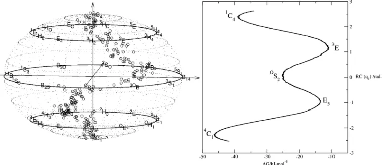
4.2.1. 2-Oxanol. Smith7 has studied the conformational requirements of the hydrolysis of the glycosidic bond using 2-oxanol by very high-level (G2 theory) ab initio calculations. The relevant conformations in his study were selected on the basis of their significance in enzymatic cleavage of the glyco-sidic bond. Smith’s study suggested that the inversion of 2-oxanol from the 1C4 conformation passes through the 3H4 transition state and arrives at the 3S1 conformation on the pseudorotational itinerary. The3H4transition state connects the 1C4 and the 3S1 conformations on the conformational map
(Figure 1). Our constrained dynamical analysis found a different inversion pathway. Starting from1C4conformation (Figure 4), the lowest-energy trajectory passed through the 3E envelope conformation; then, the trajectory involved an evolution in the direction of the3,OB conformer, coupled with pseudorotation, and arrived at theOS2conformation as the secondary minimum.
This was unexpected because theOS2conformation is connected
with the1C4conformation through the3H2half-chair transition state. However, the 3E envelope and the 3H2 half-chair are adjacent structures and are very close in energy. From theOS2
conformation, the dynamical trajectory involved evolution that passed through the E5 envelope conformation and arrived at the inverted4C1chair. We have calculated the total energies of various conformations of 2-oxanol and confirmed that there is a strong preference for the OS2conformation with respect to
the 3S1 conformation. The relevant thermodynamic data are summarized in Table 3. For those conformations characterized by Smith,7the relative energies qualitatively agree between the QCI/631-G(d) and the present DFT calculations.
The dynamical trajectory of 2-oxanol inversion clearly showed that our dynamical approach provided a unique insight into the chair inversion process. The reaction paths suggested by the previous study and the dynamical calculations are different. The dynamical calculation showed a complex evolu-tion: once the reaction path passed the barrier, the chair inversion was combined with significant pseudorotation. The oscillation of the pseudorotational angle around the skew-boat
Figure 4. Free-energy curve and dynamical trajectory on the conformational sphere for the inversion of 2-oxanol.
TABLE 3: Energetics for 2-Oxanol in kJ·mol-1 compd (calcd)G a G (calcd)b (calcd)G c E (calcd)b E (calcd)c 1C 4 0.0 0.0 0.0 0.0 0.0 3H 4 30.9(3E) 30.8 23.7 28.5 35.6 OS 2 11.3 11.1 na 9.1 na 3,OB 18.5 18.8 na 16.1 na 3S1 16.9 15.0 6.9 15.5 13.7 B1,4 na na 14.3 na 20.2 5S1 10.8 14.5 5.9 14.3 14.3 2S O 13.8 17.5 na 20.2 na B3,O 16.4 24.2 na 22.8 na 1S 3 13.7 19.5 na 21.1 na
aCalculations were carried out with PAW.bFully optimized at the
b3p86/6-311+g** level.cSee ref 7.
conformation had a much larger amplitude than that from around the transition states, which suggested a flat potential surface around theOS2secondary minimum.
4.2.2. Glucopyranose.Our main interest was the study of the inversion of R- and β-D-glucopyranose.D-Glucopyranose plays
an essential role in biochemistry, and its structure and confor-mational behavior were intensively studied by theoretical means. There are more than 700 possible conformers based on the different orientations of hydroxyl and hydroxymethyl groups.44 It has been found that both anomers, R- and β-D-glucopyranose,
exist predominantly as 4C1 conformers. Experimentally, the separation energy between the two chair conformers was 46.0 kJ‚mol-1.45Theoretical calculations ranging from semiempiri-cal46-49and molecular mechanics50-52to ab initio,53-57Monte Carlo,58-60 and molecular dynamics61-63 have thoroughly investigated the conformational modifications of glucose rings. However, many of these calculations are based on force fields unsuitable to predict correctly the conformational potential surface of pyranoses. Ab initio calculations found, in vacuo, that the4C1chair is about 33.4 kJ‚mol-1more stable than the inverted1C4. On average, the R anomer is about 1.7 kJ‚mol-1 more stable than the β anomer. Previous ab initio calculations of chair-boat conversions in pyranose monomers have shown transition-energy barriers of approximately 58.5 kJ‚mol-1.64 Many recent studies have concentrated on the study of solvation effects and were based on continuum models. The relative stability changes in aqueous solution, where the population of βanomers is lower in free energy than that of R anomers by 1.2 kJ‚mol-1. Previous ab initio and nanosecond molecular dynamics studies of solvated disaccharides65revealed a com-petition between the intermolecular water-maltose and in-tramolecular hydrogen bonds of comparable strengths in mal-tose, which leads to water-promoting rotation around the glycosidic bond. Atomic force microscope (AFM) manipulations of polyglucose molecules were a different and unique way of inducing conformational transitions by ellastical deformation of the pyranoid ring.9,66
For our studies, we chose per-O-methyl substitution for two reasons: First, the absence of hydroxyls prevents intramolecular hydrogen bonding. Conformations with intramolecular hydrogen bonding are found to be the lowest-energy ones by gas-phase calculations of neutral monosaccharides, and this factor may obscure other factors influencing reactivity.67Secondly, methyl is the smallest possible protecting group and begins to address the important issue of the effect of protecting groups on reactivity. In all cases examined so far, the preferred CH-OCH3 rotamers have a lone pair anti to the sugar methine and to the methyl carbon syn to the methine (Figure 8). This conforma-tional preference has been observed before in calculated structures of permethylated disaccharides.68This syn preference has been found for 2,6-disubstituted-1-methoxycyclohexanes and was ascribed to steric effects.69
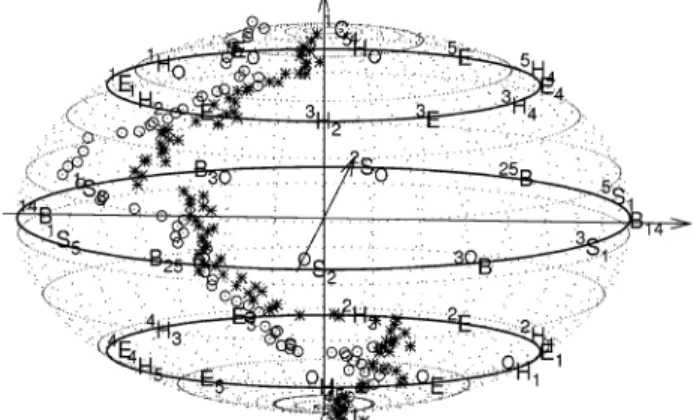
The trajectory that we found to be the minimum energy path between the two chair conformers was determined by con-strained ab initio molecular dynamics and was plotted on the conformational sphere using the Cremer-Pople parameters in Figure 5. The trajectories show that the inversion process is a mix of several modes, with a large contribution from pseudoro-tation.
For the penta-O-methyl R anomer, the MD trajectory followed a complex pathway on the potential energy surface and, in addition to inversion, it also involved pseudorotation (Figure 5). The barrier to pass to get out of the1C4chair minimum was 10.3 kJ‚mol-1and involved a transition state between the 1E
envelope and1H
Ohalf-chair, with the major contribution from
the envelope conformation. From this point, the trajectory continued toward the 1S5 skew-boat minimum and, without reaching it completely, pseudorotated and passed subsequently through the B2,5boat andOS2skew-boat conformations. Once
the conformation settled to the OS2skew-boat minimum, the
pseudorotation ended, and by passing through the OH5
half-chair as a second transition state, the inversion process ended with the4C1inverted chair. The free-energy barrier from the 4C1 chair to theOS2skew-boat was 20.5 kJ‚mol-1, twice the free-energy barrier required to leave the 1C4chair minimum. This is consistent with experimental data that evidenced the greater stability of the4C1chair compared to that of the 1C4 chair.
For the penta-O-methyl β anomer, the MD trajectory had some significant differences from the one found for the R anomer (Figure 5). First, the transition state from the1C4chair was a purely 1E envelope conformation. Consequently, from the envelope-like transition state, the system evolved into the 1,4B minimum. The barrier for this conversion was only 2.1 kJ‚mol-1. Secondly, the pseudorotation was around the 1S5 skew-boat minimum and stopped without reaching theOS2
skew-boat. Thirdly, the transition state to the inverted4C1chair was a mix of envelope and half-chair conformations. The free-energy barrier to leave the4C1chair minimum was 7.2 kJ‚mol-1, nearly twice the free-energy barrier required to leave the1C4 chair minimum.
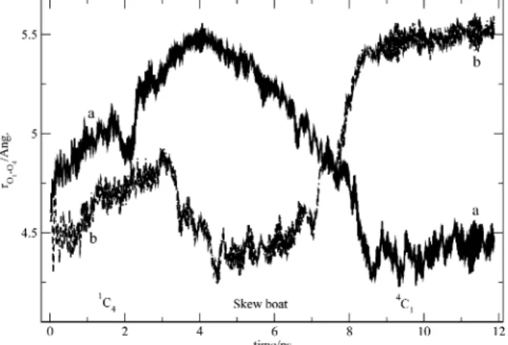
Figure 6 shows the evolution of the O1-O4 distance for the two anomers with time. For the R-D-glucopyranose anomer, the
transition from chair to boat conformations increased the distance between the glycosidic oxygen O1 and the oxygen O4. For the β-D-glucopyranose anomer, the transition from chair to
boat conformations decreased the distance between the two oxygens. These results are in good agreement with AFM studies on amylose, dextran, and cellulose9that found a similar variation of the length of the monomer with the pyranose ring conforma-tion.
Constrained ab initio molecular dynamics can help qualita-tively describe the conformational path, but free energies are not reliable, especially when two different modes dominate a major part of the trajectory. We have conducted a series of static calculations in which we have taken snapshots of the conforma-tions along the MD trajectory at regular spaced intervals. Figure 7 shows the potential energy curves obtained by static calcula-tions. The energy vs constraint plots were calculated by optimizing geometries taken from both R and β anomer MD trajectories, with ring dihedrals kept frozen during the
optimiza-Figure 5. Dynamical trajectory on the conformational sphere for the inversion of penta-O-methyl-R-D-glucopyranose (circles) and penta-O-methyl-β-D-glucopyranose (stars).
tion. The rest of the molecule was allowed to optimize. This permitted us to simulate the dynamical trajectory and to calculate the thermodynamic values associated with the inversion path. Free-energy barriers obtained by both dynamic and static calculations are shown in Table 4. The4C1chair conformation was the most stable for both R and β anomers. An energy plateau was found around the value zero for the constraint and corresponds to the conformations close to the pseudorotational itinerary. The1C4chair was the less-stable conformation. The activation barriers for leaving the4C1chair minimum for R and βanomers had almost equal values. However, once the transition
state was passed, the trajectories followed different energetic pathways, with the β trajectory higher in energy than the R trajectory.
Stationary points were calculated by fully optimizing candi-date structures taken from the static calculation simulated trajectory for each anomer. For the4C1chair, we found that the βconformer was 1.0 kJ‚mol-1more stable than the R conformer. Our static calculations showed that the1C4chair conformations were unstable. Consequently, we calculated only the transitions from the4C1chair to the skew-boat and boat secondary minima. Next, we compared the geometries of the two anomers obtained by optimization of the candidate structures taken from the static potential energy curve. The geometrical parameters that we investigated were the C1-O5 and C1-O1 bonds and the dihedral angle O6-C5-C6-O6. The C1-O5 bond distances for R and β chair minima (Figure 8a,b) were 1.421 and 1.422 Å, respectively. The C1-O1 bonds are longer in R than in β by 0.011 Å. The O6-C5-C6-O6 dihedral angles (-71.2°for both chair anomers) showed that the two conformers are in the g conformation that some experimental NMR studies have shown to be the most populated. The O1-O4 distance is 1.041 Å shorter in the R chair anomer than in its β counterpart.
The investigation of the same geometrical parameters of the transition states (Figure 8c,d) showed that the C1-O bond was 0.021 Å longer in R than in β. Although there was no difference in C1-O1 bonds between anomers, the O1-O4 distance in the Ranomer was 0.967 Å shorter than that in the β anomer. For the two transition states, the C1-C4 and C2-C3-C4-C5 ring dihedrals were 4.8°for R and 17.8°for β and 0.2°
for R and -11.6° for β, respectively. The five contiguous coplanar carbon atoms suggested that, in both cases, the transition followed the route to the skew-boat conformation via an envelope saddle point. Conformation analysis pointed out that both transition structures were ofOE envelope type. This
conformation is adjacent to theOH5half-chair transition state
predicted by the MD trajectory. An attentive look at the MD trajectory revealed oscillations between theOH5half-chair and
theOE envelope conformations. However, the maximum energy
was reached for the half-chair conformation, thus explaining our choice for the MD transition state.
The secondary minima were found to be theOS2skew-boat
for the R-D-glucopyranose and a mix ofOS2skew-boat and1,4B boat for the β-D-glucopyranose (see Table 5 and Figure 8).
Although the C1-O5 and C1-O1 bonds have similar values, the O1-O4 distance is 0.699 Å larger in the R than in the β anomer.
For the R anomer, the transition from chair to skew-boat increased the O1-O4 distance by 0.640 Å and the remaining selected geometrical parameters did not suffer a significant change. The calculated increase is 12.3%, and the experimental measured values are 16.8 ( 1.8% for amylose and 19.5 ( 2.1% for dextran. For the β anomer, the transition from chair to skew-boat significantly modified all the selected geometrical param-eters: the C1-O6 bond is 0.015 Å shorter and the C1-O1 bond is 0.021 Å longer in the skew-boat conformation than in the chair conformation. The transition to the skew-boat decreased the O1-O4 distance by 1.110 Å or 20.6%. The only experi-mental value available of 0% was obtained for cellulose by AFM studies and is consistent with our results: the stretching force compensated the reduction in distance.
5. Discussion and Conclusions
The goal of this paper was to demonstrate how the chosen mathematical constraints enabled us to study the conformational
Figure 6. Time evolution of the distance O1-O4 (in Å) for (a) penta-O-methyl-R-D-glucopyranose and (b) penta-O-methyl-β-D -glucopyra-nose.
Figure 7. Static DFT pathway for the transition from the4C1chair to
the1C
4chair of penta-O-methyl-R-D-glucopyranose (circles) and
penta-O-methyl-β-D-glucopyranose (stars).
TABLE 4: Energy Barriers for Penta-O-methyl-r- and Penta-O-methyl-β-D-glucopyranose Chair to Skew-Boat Conversions in kJ·mol-1at 300 K compd (calcd)∆G a ∆G (calcd)b ∆H (calcd)b ∆E
(calcd)b,c (calcd)∆E d 4C 1to Skew-Boat Re 20.5 51.9 42.6 45.9 65.6 βf 7.2 37.3 30.7 33.5 70.9 1C 4to Skew-Boat Re 10.3 na na na 10.5 βf 2.1 na na na 13.1
aCalculations were carried out with PAW.bFully optimized at the
b3p86/6-311+g** level.cZPE included.dRing dihedrals are frozen;
see Figure 7.ePenta-O-methyl-R-D-glucopyranose.f
Penta-O-methyl-β-D-glucopyranose.
transitions among selected pyranose conformations in both static and dynamical calculations. We have combined the constrained method with ab initio molecular dynamics calculations to sample the conformational space of glucopyranose and some model compounds. Our constrained method allowed us to define the chair, boat, skew-boat, half-chair, and envelope conformers and to map the low-energy paths connecting them.
The inversion process was conducted mainly via the q1 constraint. The inversion trajectory passed through transition states situated on smaller radius circles halfway between the pseudorotational circle (the equator of the conformational sphere) and the two chairs (the poles of the conformational
sphere). The inversion trajectory also evidenced a secondary minimum (usually a skew-boat) situated on the pseudorotational itinerary.
The internal coordinates q2and q3are equivalent and can be used to describe conformational change along the pseudorota-tional itinerary. We only simulated the pseudorotation along the equatorial itinerary for cyclohexane. The results showed that the trajectory followed the surface of the sphere and remained constrained to the pseudorotational itinerary; the skew-boat minima were interconnected along the inversion pathway by boat transition states. The energy barriers along the pseudoro-tational itinerary were about 4 kJ‚mol-1, in good agreement with experimental data.
We have also studied 2-oxanol, which is the simplest model for glucopyranose, as well as the R and β conformers of penta-O-methyl-D-glucopyranose. For 2-oxanol, we have highlighted
a different trajectory than previously calculated. This inversion trajectory, passing through the2S
Oconformer, was confirmed
to be the lowest-energy pathway by static ab initio calculations. For R- and β-D-glucopyranose, the calculations provided
additional insights into the behavior of these anomers. We calculated for the first time a complete inversion trajectory for theD-glucopyranose system. The calculations indicated that a
variety of structures were isoenergetic, suggesting a flat potential energy surface around the skew-boat secondary minima.
Ac-Figure 8. Stationary point geometries (ORTEP representation) for chair to skew-boat transitions of penta-O-methyl-R-D-glucopyranose (a)4C1, (c)
OE, and (e)OS
2and penta-O-methyl-β-D-glucopyranose (b)4C1, (d)OE, and (f)OS2.
TABLE 5: Optimized Canonical Conformations for the Stationary Points on the Trajectory4C1Chair toO
S2 Skew-Boat for Penta-O-methyl-r- and
Penta-O-methyl-β-D-glucopyranose
compd canonical conformations Penta-O-methyl-R-D-glucopyranose 4C 1 4C1, 0.935; B2,5, 0.007;3S1, 0.074 OE 0E, 0.757;O,3B, 0.046;1S 5, 0.087 OS 2 1,4B, 0.181;4C1, 0.062;OS2, 1.009 penta-O-methyl-β-D-glucopyranose 4C1 4C1, 0.926;O,3B, 0.113;5S1, 0.005 OE OE, 0.830;O,3B, 0.253;1S5, 0.098 OS 2 1,4B, 0.801;OH5, 0.110;OS2, 0.850
cording to our calculations, the free-energy barriers for the chair to twist-boat transition in R- and β-D-glucopyranose were 52
and 37 kJ‚mol-1(Table 4), respectively. These values can be compared to the ab initio constrained calculations of O’Donoghue et al.,66which gave a barrier height for the R-D-glucopyranose chair to twist-boat transition of about 59 kJ‚mol-1and gave a barrier height for the β-D-glucopyranose chair to boat transition
of about 53 kJ‚mol-1. These values are internal energies and not free-energy differences and were obtained by a limited conformational search with only one of the three puckering coordinates constrained. Previous MM3 molecular mechanics studies also found a barrier of 50 kJ‚mol-1 for the R-D -glucopyranose chair to twist-boat transition.70 AFM studies suggested that the elastic properties of polyssacharides result from a force-induced elongation of the ring structure and a final transition from a chairlike to a boatlike conformation.9 In contrast to these findings are the most recent AFM experiments, suggesting a mechanism involving relatively low-energy transi-tion barriers for the chair extension, with the dominating process being an anti-syn isomerization of the dihedral angles for the R(1f4) linkage.71
In all of the above MD simulations, the system was thermostated at 300 K and the masses were rescaled. For the test molecules, about 70 000 steps were performed for each simulation, or approximately 12 ps of real time, and for the twoD-glucopyranoses, 90 000 steps were performed for each
simulation, or approximately 15 ps of real time. Both forward and reverse scans were performed to test the rate of change of the reaction coordinate. For example, for 2-oxanol, the calculated hysteresis showed for the forward and reverse estimates of the inversion free-energy barrier a difference of 5.3 kJ‚mol-1. This error arises from the slow-growth simulation, which is a necessary compromise to limit the computational effort to a reasonable level. The comparison of experimental and calculated conversion barriers suggests that this error source is limiting the overall accuracy of the calculations. Furthermore, the stationary points for both forward and reverse scans occurred at the same value of the reaction coordinate. The constrained dynamic calculations we presented above provided us with a tool for finding the lowest-energy pathway for the chair inversion of pyranoses and for calculating the inversion free-energy barrier by means of thermodynamic integration. We have complemented the dynamic simulation by static simulations.
Static simulations were used, first, to validate the free-energy results produced by our constrained dynamics method and, second, to access other thermodynamic properties such as the enthalpy of reaction, usually available from experimental data. Static calculations involved full optimization of reactants. Transition states were calculated by taking snapshots from the MD trajectory; no further optimization was done. Frequency calculations were performed on each of the stationary points. The benchmark tests we have carried out on the enthalpy and the free-energy barrier of chair inversion of the six-membered saturated rings represented a remarkable agreement between experiment and theory for both quantities. For example, the lowering of the enthalpic barrier to inversion by oxygen substitution on going from cyclohexane to tetrahydropyran as well as the small difference in free energy were well reproduced by calculations.
Our constrained method was based on Car-Parrinello (CP) simulations. We emphasize that, although the CP method involves quantum mechanical calculations to determine the electronic structure, the actual dynamic is purely classical; therefore, quantum effects such as zero-point energy correction
are not accounted for. However, the mean absolute deviation between MD and static simulations was 4.8 kJ‚mol-1for the model molecules; as forD-glucopyranoses, the MD simulations
in both cases underestimated the free-energy barriers. We explained this, first, by additional steric and electronic effects not present with the model molecules and, second, by the mixed-inversion pseudorotation process that characterized the MD trajectories ofD-glucopyranoses.
These results clearly demonstrate that our constrained method was able, first, to simulate accurate trajectories for the chair inversion of several pyranose conformations and, second, to calculate free-energy barriers that were in excellent agreement with both established static calculations and experimental data. We consider these results a major step toward understanding the role of conformation change in the kinetic stability of sugar residues involved in glycosylation reactions. Our constrained method was successfully used to characterize the conformational potential energy surface by mapping important stationary points.
Acknowledgment. This work was partly supported by the
iHPC multiscale modeling initiative of the NRC. This is NRC paper # 42504.
References and Notes
(1) Kochetkov, N. K. Stud. Nat. Prod. Chem. 1994, 14, 201-266. (2) Sinnott, M. L. Chem. ReV. 1990, 90, 1171-1202.
(3) Horenstein, B. A.; Bruner, M. J. Am. Chem. Soc. 1998, 120, 1357-1362.
(4) Murray, B. W.; Wittmann, V.; Burkart, M. D.; Hung, S.-C.; Wong, C.-H. Biochemistry 1997, 36, 823-831.
(5) Deslongchamps, P.; Dory, Y. L.; Li, S. Can. J. Chem. 1994, 72, 2021-2027.
(6) Zhu, J.; Bennet, A. J. J. Am. Chem. Soc. 1998, 120, 3887-3893. (7) Smith, B. J. J. Am. Chem. Soc. 1997, 119, 2699-2706. (8) Liras, J.; Anslyn, E. In Molecular Design and Bioorganic Catalysis; Wilcox, C. S., Hamilton, A. D., Eds.; Kluwer Academic Publishing: The Netherlands, 1996.
(9) Marszalek, P. E.; Oberhauser, A. F.; Pang, Y. P.; Fernandes, J. M.
Nature 1998, 396, 661-664.
(10) Nukada, T.; Be´rces, A.; Zgierski, M. Z.; Whitfield, D. M. J. Am.
Chem. Soc. 1998, 120, 13291-13295.
(11) Be´rces, A.; Whitfield, D. M.; Nukada, T.; do Santos, Z. I.; Obuchowska, A.; Krepinsky, J. J. Can. J. Chem. 2004, 82, 1157-1171.
(12) Nukada, T.; Be´rces, A.; Whitfield, D. M. Carbohydr. Res. 2002,
337, 765-774.
(13) Nukada, T.; Be´rces, A.; Wang, L.; Zgierski, M. Z.; Whitfield, D. M. Carbohydr. Res. 2005, 340, 841-852.
(14) Stoddart, J. F. Stereochemistry of Carbohydrates; Wiley-Inter-science: New York, 1971.
(15) Lichtenthaler, F. W.; Ro¨nninger, S.; Lindner, H. J.; Immel, S.; Cuny, E. Carbohydr. Res. 1993, 249, 305-326.
(16) Dixon, D. A.; Komornicki, A. J. Phys. Chem. 1990, 94, 5630-5636.
(17) Be´rces, A.; Whitfield, D. M.; Nukada, T. Tetrahedron 2001, 477-491.
(18) Hendrickson, J. B. J. Am. Chem. Soc. 1967, 89, 7047-7061. (19) Cremer, D.; Pople, J. A. J. Am. Chem. Soc. 1975, 97, 1354-1358. (20) Haasnoot, C. A. G. J. Am. Chem. Soc. 1992, 114, 882-887. (21) Zefirov, N.; Palyulin, V. A.; Dashevskaya, E. E. J. Phys. Org. Chem.
1990, 3, 147-158.
(22) Fogarasi, G.; Zhou, X.; Taylor, P. W.; Pulay, P. J. Am. Chem. Soc.
1992, 114, 8191-8201.
(23) Pulay, P.; Fogarasi, G.; Pang, F.; Boggs, J. E. J. Am. Chem. Soc.
1979, 101, 2550-2560.
(24) Blo¨chl, P. E. Phys. ReV. B 1994, 50, 17953-17979. (25) Car, R.; Parrinello, M. Phys. ReV. Lett. 1985, 55, 2471-2474. (26) Perdew, J. P.; Zunger, A. Phys. ReV. B 1981, 23, 5048-5079. (27) Blo¨chl, P. E. J. Chem. Phys. 1995, 103, 7422-7428. (28) Hoover, W. G. Phys. ReV. A 1985, 31, 1695-1697. (29) Nose, S. Mol. Phys. 1984, 52, 255-268.
(30) Blo¨chl, P. E.; Parrinello, M. Phys. ReV. B 1992, 45, 9413-9416. (31) Ryckaert, J. P.; Ciccotti, G.; Berendsen, H. J. C. J. Comput. Phys.
1977, 23, 327-341.
(32) Margl, P. M.; Ziegler, T.; Blo¨chl, P. E. J. Am. Chem. Soc. 1995,
117, 12625-12634.
(33) Margl, P. M.; Lohrenz, J. C. W.; Ziegler, T.; Blo¨chl, P. E. J. Am.
Chem. Soc. 1996, 118, 4434-4441.
(34) Margl, P. M.; Ziegler, T.; Blo¨chl, P. E. J. Am. Chem. Soc. 1996,
118, 5412-5419.
(35) Yang, S.-Y.; Hristov, I.; Fleurat-Lessard, P.; Ziegler, T. J. Phys.
Chem. A 2005, 109, 197-204.
(36) Frisch, M. J.; Trucks, G. W.; Schlegel, H. B.; Scuseria, G. E.; Robb, M. A.; Cheeseman, J. R.; Zakrzewski, V. G.; Montgomery, J. A., Jr.; Stratmann, R. E.; Burant, J. C.; Dapprich, S.; Millam, J. M.; Daniels, A. D.; Kudin, K. N.; Strain, M. C.; Farkas, O.; Tomasi, J.; Barone, V.; Cossi, M.; Cammi, R.; Mennucci, B.; Pomelli, C.; Adamo, C.; Clifford, S.; Ochterski, J.; Petersson, G. A.; Ayala, P. Y.; Cui, Q.; Morokuma, K.; Malick, D. K.; Rabuck, A. D.; Raghavachari, K.; Foresman, J. B.; Cioslowski, J.; Ortiz, J. V.; Stefanov, B. B.; Liu, G.; Liashenko, A.; Piskorz, P.; Komaromi, I.; Gomperts, R.; Martin, R. L.; Fox, D. J.; Keith, T.; Al-Laham, M. A.; Peng, C. Y.; Nanayakkara, A.; Gonzalez, C.; Challacombe, M.; Gill, P. M. W.; Johnson, B. G.; Chen, W.; Wong, M. W.; Andres, J. L.; Head-Gordon, M.; Replogle, E. S.; Pople, J. A. Gaussian 98, revision A.11.3; Gaussian, Inc.: Pittsburgh, PA, 1998.
(37) Becke, A. D. Phys. ReV. A 1988, 38, 3098-3100. (38) Perdew, J. P. Phys. ReV. B 1986, 33, 8822-8824. (39) Perdew, J. P. Phys. ReV. B 1986, 34, 7406.
(40) Ross, B. D.; True, N. S. J. Am. Chem. Soc. 1983, 105, 1382. (41) Pickett, H. M.; Strauss, H. L. J. Am. Chem. Soc. 1970, 92, 7281. (42) Squillacote, M.; Sheridan, R. S.; Chapman, O. L.; Anet, F. A. L.
J. Am. Chem. Soc. 1975, 97, 3244-3246.
(43) del Carmen Ferna´ndez-Alonso, M.; Asensio, J. L.; Can˜ada, F. J.; Jime´nez-Barbero, J.; Cuevas, G. ChemPhysChem 2003, 4, 754-757.
(44) Cramer, C. J.; Truhlar, D. G. J. Am. Chem. Soc. 1993, 115, 5745-5753.
(45) Joshi, N.; Rao, V. Biopolymers 1979, 18, 2993-3004.
(46) Tvaroska, I.; Kozar, T. J. Am. Chem. Soc. 1980, 102, 6929-6936. (47) Back, D.; Polavarapu, P. J. Comput. Chem. 1987, 8, 772-777. (48) Woods, R.; Szarek, W.; Smith, V. J. Chem. Soc., Chem. Commun.
1991, 5, 334-337.
(49) McNamara, J. P.; Muslim, A.-M.; Abdel-Aal, H.; Wang, H.; Mohr, M.; Hillier, I. H.; Bryce, R. A. Chem. Phys. Lett. 2004, 394, 429-436.
(50) Jeffrey, G.; Taylor, R. J. Comput. Chem. 1980, 1, 99-109. (51) Rasmussen, K. Acta Chem. Scand., Ser. A 1982, 36, 323-327.
(52) Dowd, M.; Reilly, P.; French, A. J. Comput. Chem. 1992, 13, 102-114.
(53) Jeffrey, G. A.; Pople, J. A.; Binkley, J. S.; Vishveshwara, S. J.
Am. Chem. Soc. 1978, 100, 373-379.
(54) Melberg, S.; Rasmussen, K.; Scordamaglia, R.; Tosi, C. Carbohydr.
Res. 1979, 76, 23-37.
(55) French, A.; Schfer, L.; Newton, S. Carbohydr. Res. 1993, 239, 51-60.
(56) Appell, M.; Strati, G.; Willett, J. L.; Momany, F. A. Carbohydr.
Res. 2004, 339, 537-551.
(57) Momany, F. A.; Appell, M.; Strati, G.; Willett, J. L. Carbohydr.
Res. 2004, 339, 553-567.
(58) Rees, D. A.; Smith, P. J. C. Perkin Trans. 1975, 2, 830-835. (59) Rees, D. A.; Smith, P. J. C. Perkin Trans. 1975, 2, 836-840. (60) Peters, T.; Meyer, B.; Stuike-Prill, R.; Somorjai, R.; Brisson, J.-R.
Carbohydr. Res. 1993, 238, 49-73.
(61) Brady, J. W. J. Am. Chem. Soc. 1986, 108, 8153-8160. (62) Brady, J. W. J. Am. Chem. Soc. 1989, 111, 5155-5165. (63) Ha, S.; Gao, J.; Tidor, B.; Brady, J. W.; Karplus, M. J. Am. Chem.
Soc. 1991, 113, 1553-1557.
(64) O’Donoghue, P.; Luthey-Schulten, Z. A. J. Phys. Chem. B 2000,
104, 10398-10405.
(65) Chen, J. Y.-J.; Naidoo, K. J. J. Phys. Chem. B 2003, 107, 9558-9566.
(66) Marszalek, P. E.; Pang, Y.-P.; Li, H.; Yazal, J. E.; Oberhauser, A. F.; Fernandez, J. M. Proc. Natl. Acad. Sci. U.S.A. 1999, 96, 7894-7898. (67) (a) Barrows, S.; Dulles, F.; Cramer, C.; French, A.; Truhlar, D.
Carbohydr. Res. 1995, 276, 219-251. (b) Whitfield, D. M. J. Mol. Struct.:
THEOCHEM 1997, 395, 53-59. (c) Polavarapu, P. L.; Ewig, C. S. J. Comput. Chem. 1992, 13, 1255. (d) Jeffrey, G. A. J. Mol. Struct. 1990, 237, 75-79.
(68) Mendonca, A.; Johnson, G.; French, A.; Laine, R. J. Phys. Chem.
A 2002, 106, 4115-4124.
(69) Anderson, J. E.; Ijeh, A. I. J. Chem. Soc., Perkin Trans. 2 1994, 9, 1965-1968.
(70) Dowd, M. K.; French, A. D.; Reilly, P. J. J. Comput. Chem. 1994,
264, 1-19.