Behavioral and Structural Responses to Chronic Cocaine Require
a Feedforward Loop Involving ΔFosB and
Calcium/Calmodulin-Dependent Protein Kinase II in the Nucleus Accumbens Shell
The MIT Faculty has made this article openly available. Please share
how this access benefits you. Your story matters.
Citation
Robison, A. J., V. Vialou, M. Mazei-Robison, J. Feng, S. Kourrich,
M. Collins, S. Wee, et al. “Behavioral and Structural Responses to
Chronic Cocaine Require a Feedforward Loop Involving FosB and
Calcium/Calmodulin-Dependent Protein Kinase II in the Nucleus
Accumbens Shell.” Journal of Neuroscience 33, no. 10 (March 6,
2013): 4295-4307.
As Published
http://dx.doi.org/10.1523/jneurosci.5192-12.2013
Publisher
Society for Neuroscience
Version
Final published version
Citable link
http://hdl.handle.net/1721.1/81336
Terms of Use
Article is made available in accordance with the publisher's
policy and may be subject to US copyright law. Please refer to the
publisher's site for terms of use.
Neurobiology of Disease
Behavioral and Structural Responses to Chronic Cocaine
Require a Feedforward Loop Involving
⌬FosB and Calcium/
Calmodulin-Dependent Protein Kinase II in the Nucleus
Accumbens Shell
Alfred J. Robison,
1Vincent Vialou,
1Michelle Mazei-Robison,
1Jian Feng,
1Saïd Kourrich,
2Miles Collins,
1Sunmee Wee,
3George Koob,
3Gustavo Turecki,
4Rachael Neve,
5Mark Thomas,
2and Eric J. Nestler
11Fishberg Department of Neuroscience and Friedman Brain Institute, Mount Sinai School of Medicine, New York, New York, 10029,2Departments of
Neuroscience and Psychology, Institute of Human Genetics, University of Minnesota, Minneapolis, Minnesota 55455,3Committee on the Neurobiology of
Addictive Disorders, The Scripps Research Institute, La Jolla, California 920374Depressive Disorders Program, Douglas Mental Health University Institute
and McGill University, Montre´al, Que´bec, Canada, H4H 1R3, and5Department of Brain and Cognitive Sciences, Massachusetts Institute of Technology,
Cambridge, Massachusetts 02139
The transcription factor
⌬FosB and the brain-enriched calcium/calmodulin-dependent protein kinase II (CaMKII␣) are induced in the
nucleus accumbens (NAc) by chronic exposure to cocaine or other psychostimulant drugs of abuse, in which the two proteins mediate
sensitized drug responses. Although
⌬FosB and CaMKII␣ both regulate AMPA glutamate receptor expression and function in NAc,
dendritic spine formation on NAc medium spiny neurons (MSNs), and locomotor sensitization to cocaine, no direct link between these
molecules has to date been explored. Here, we demonstrate that
⌬FosB is phosphorylated by CaMKII␣ at the protein-stabilizing Ser27
and that CaMKII is required for the cocaine-mediated accumulation of
⌬FosB in rat NAc. Conversely, we show that ⌬FosB is both
necessary and sufficient for cocaine induction of CaMKII
␣ gene expression in vivo, an effect selective for D
1-type MSNs in the NAc shell
subregion. Furthermore, induction of dendritic spines on NAc MSNs and increased behavioral responsiveness to cocaine after NAc
overexpression of
⌬FosB are both CaMKII dependent. Importantly, we demonstrate for the first time induction of ⌬FosB and CaMKII in
the NAc of human cocaine addicts, suggesting possible targets for future therapeutic intervention. These data establish that
⌬FosB and
CaMKII engage in a cell-type- and brain-region-specific positive feedforward loop as a key mechanism for regulating the reward circuitry
of the brain in response to chronic cocaine.
Introduction
Increasing evidence supports the view that changes in gene
ex-pression contribute to mechanisms of drug addiction (
Robison
and Nestler, 2011
). One important mediator of these changes is
⌬FosB, a Fos family transcription factor (
Nestler, 2008
). Chronic
administration of virtually any drug of abuse induces the
long-lasting accumulation of
⌬FosB in nucleus accumbens (NAc), a
limbic region essential for reward behaviors. Such induction
ap-pears specific to the class of NAc medium spiny neuron (MSNs)
that expresses D
1dopamine receptors. Inducible overexpression
of
⌬FosB in these D
1-type NAc MSNs increases locomotor and
rewarding responses to cocaine and morphine (
Kelz et al., 1999
;
Zachariou et al., 2006
), including increased cocaine
self-administration (
Colby et al., 2003
). Furthermore, genetic or viral
blockade of
⌬FosB transcriptional activity reduces the rewarding
effects of these drugs (
Zachariou et al., 2006
), indicating that this
sustained induction of
⌬FosB is a critical mediator of the lasting
changes induced in NAc by chronic drug administration.
The unusual stability of
⌬FosB (relative to all other Fos family
proteins) is both an intrinsic property of the molecule,
attribut-able to the truncation of degron domains present in full-length
FosB (
Carle et al., 2007
), and a regulated process.
⌬FosB is
phos-phorylated in vitro and in vivo at Ser27, and this reaction further
stabilizes
⌬FosB, ⬃10-fold, in cell culture and NAc in vivo
(
Ulery-Reynolds et al., 2009
). Although Ser27
⌬FosB has been
shown to be a substrate for casein kinase-2 in vitro (
Ulery et al.,
2006
), its mechanism of in vivo phosphorylation remains
unknown.
Calcium/calmodulin-dependent protein kinase II (CaMKII)
is a highly expressed serine/threonine kinase whose
␣ and 
iso-forms form dodecameric homo- and hetero-holoenzymes in vivo
and are essential for multiple forms of neuroplasticity (
Lisman et
Received Nov. 7, 2012; revised Dec. 31, 2012; accepted Jan. 8, 2013.
Author contributions: A.J.R., G.K., M.T., and E.J.N. designed research; A.J.R., V.V., M.M.-R., J.F., S.K., M.C., S.W., and G.T. performed research; R.N. contributed unpublished reagents/analytic tools; A.J.R., S.W., and E.J.N. analyzed data; A.J.R. and E.J.N. wrote the paper.
This work was supported by the National Institute on Drug Abuse (NIDA) (E.J.N.), NIDA–Yale Proteomics Center Grant DA018343 (A.J.R. and E.J.N.), and the Hartwell Foundation (A.J.R.). We thank Gabby Rundenko for the gen-erous gift of purified⌬FosB and Roger Colbran for the generous gift of purified CaMKII␣.
Correspondence should be addressed to Eric J. Nestler, Fishberg Department of Neuroscience, Room 10-23, 1425 Madison Avenue, New York, NY 10029. E-mail: eric.nestler@mssm.edu.
DOI:10.1523/JNEUROSCI.5192-12.2013
al., 2002
;
Colbran and Brown, 2004
). CaMKII␣ is induced
selec-tively in NAc shell by chronic amphetamine (
Loweth et al., 2010
),
and pharmacological blockade of CaMKII activity in NAc shell
reduces behavioral sensitization to amphetamine (
Loweth et al.,
2008
) and cocaine (
Pierce et al., 1998
), whereas viral
overexpres-sion of CaMKII
␣ in this NAc subregion enhances locomotor
sensitization to and self-administration of amphetamine
(
Loweth et al., 2010
). CaMKII
␣ may affect reward behaviors via
modulation of AMPA glutamate receptor subunits (
Pierce et al.,
1998
), because CaMKII
␣ activity has long been associated with
AMPA receptor function and synaptic targeting in several forms
of neuroplasticity (
Malinow and Malenka, 2002
).
This literature demonstrates several parallels between
⌬FosB
and CaMKII: both are necessary and sufficient for multiple
be-havioral effects of drugs of abuse, both upregulate dendritic
spines in various neuronal cell types in vivo (
Jourdain et al., 2003
;
Maze et al., 2010
), and both exert at least some of their behavioral
effects through modulation of AMPA receptors (
Kelz et al., 1999
;
Malinow and Malenka, 2002
;
Vialou et al., 2010
). Despite these
parallels, no functional link between
⌬FosB and CaMKII is
known. Here, we establish reciprocal regulation between
⌬FosB
and CaMKII and demonstrate that the two proteins form a D
1-type MSN-specific feedforward loop in NAc shell that is induced
by cocaine and regulates a range of cocaine responses in vivo.
Materials and Methods
Experiment 1: isobaric tags for relative and absolute quantitation proteomic analysis of NAc shell and core after cocaine treatment (Fig. 1A). Adult (8 weeks) male rats were administered 20 mg/kg cocaine or saline vehicle intraperitoneally once per day for 7 d. At 24 h after the last injection, NAc shell and core were microdissected (Fig. 1A) and flash frozen. Isobaric
tags for relative and absolute quantitation (iTRAQ) analyses were per-formed as described previously (Ross et al., 2004;Da´valos et al., 2010).
Experiment 2: quantifying protein changes in rat NAc core and shell after cocaine treatment (Fig. 1B–D). Adult (8 weeks) male rats were adminis-tered 10 mg/kg cocaine or saline vehicle intraperitoneally once per day for 7 d in locomotor recording chambers. Locomotor responses to a single injection of cocaine (5 mg/kg, i.p.) were recorded in those animals treated previously with cocaine (called chronic) and a portion of those treated with saline (called acute), and locomotor responses to saline alone was recorded in the remaining chronic saline-treated animals (called saline). Locomotor activity assays were performed as described previously (Hiroi et al., 1997). Briefly, adult male rats were placed in 18⫻ 24 inch photobeam activity system open-field recording boxes (San Di-ego Instruments) for 30 min to habituate, were given a single intraperi-toneal injection of saline and monitored for an additional 30 min, and were given a single intraperitoneal injection of 5 mg/kg cocaine and monitored for 30 min.
At 24 h after this final injection, rats were decapitated without anes-thesia to avoid effects of anesthetics on neuronal protein levels and phospho-states. Brains were serially sliced in a 1.2 mm matrix (Braintree Scientific), and target tissue was removed in PBS containing protease (Roche) and phosphatase (Sigma-Aldrich) inhibitors using a 14 gauge punch for NAc core and a 12 gauge punch of the remaining tissue for NAc shell (Fig. 1A) and immediately frozen on dry ice. Samples were
homog-enized by light sonication in modified RIPA buffer: 10 mMTris base, 150
mMsodium chloride, 1 mMEDTA, 0.1% SDS, 1% Triton X-100, 1%
sodium deoxycholate, pH 7.4, and protease and phosphatase inhibitors as above. After addition of Laemmli’s buffer, proteins were separated on 4 –15% polyacrylamaide gradient gels (Criterion System; Bio-Rad), and Western blotting was performed using the Odyssey system (Li-Cor) ac-cording to the protocols of the manufacturer.
Experiment 3: quantifying protein changes in rat NAc core and shell after cocaine withdrawal (Fig. 1E). Adult (8 weeks) male rats were adminis-tered 10 mg/kg cocaine or saline vehicle intraperitoneally once per day for 7 d. At 14 d after the final injection, animals treated with saline were
given another saline injection (called saline), and animals treated with cocaine were given another saline injection (called 14 d withdrawal) or a single injection of cocaine (called 14 d withdrawal challenge). One hour after the final injection, animals were decapitated, and Western blotting performed as in experiment 2.
Experiment 4: quantifying protein changes in rat NAc core and shell after cocaine self-administration (Fig. 2A–C). Rats were trained to self-administer 0.5 mg/kg per infusion of cocaine in 1 h sessions under a fixed-ratio 1 schedule for 9 d. After nine baseline sessions, the rats were divided into two groups balanced by cocaine intake on the last two ses-sions. One group of rats was allowed to self-administer cocaine (0.5 mg/kg per infusion) in 1 h sessions (short access), whereas the other group of rats self-administered cocaine in 6 h sessions (long access) for 10 additional days (escalation sessions).
Brain sections were processed for immunohistochemistry as described previously (Perrotti et al., 2004). Brains were perfused 18 –24 h after the last exposure to drug, resulting in the degradation of any residual full-length FosB protein such that all remaining immunoreactivity reflects ⌬FosB. This degradation was confirmed by Western blotting, which showed no significant staining with an antibody directed against the C terminus of full-length FosB that does not recognize⌬FosB (data not shown). After slicing into 35m sections, the number of ⌬FosB immu-nopositive cells was quantified by a blinded observer in two sections through the NAc of each rat, and mean values per 40⫻ field were then calculated by region for each animal. Each animal was considered an individual observation for statistical analysis. Regions of interest were identified according to Paxinos and Watson (2007).
Quantification of CaMKII␣ immunoreactivity was performed using a Li-Cor system as described previously (Covington et al., 2009). Inte-grated intensities of CaMKII and GAPDH were determined with Odyssey software. Results are calculated as integrated intensity values per square millimeter and are presented as means⫾ SEM (n ⫽ 4–10 per group). Values for GAPDH were used as reference to normalize CaMKII inten-sity for slice thickness and conditions.
Experiment 5: quantifying protein levels in cocaine-dependent humans
(Fig. 2D). Postmortem human brain tissues were obtained from the
Que-bec Suicide Brain Bank (Douglas Mental Health University Institute, Montreal, Quebec, Canada). The preservation of tissue proceeded essen-tially as described previously (Quirion et al., 1987). Briefly, once ex-tracted, the brain is placed on wet ice in a Styrofoam box and rushed to the Quebec Suicide Brain Bank facilities. Hemispheres are immediately separated by a sagittal cut in the middle of the brain, brainstem, and cerebellum. Blood vessels, pineal gland, choroid plexus, half cerebellum, and half brainstem are typically dissected from the left hemisphere, which is then cut coronally into 1-cm-thick slices before freezing. The latter half cerebellum is cut sagittally into 1-cm-thick slices before freez-ing. Tissues are flash frozen in 2-methylbutane at⫺40°C for ⬃60 s. All frozen tissues are kept separately in plastic bags at⫺80°C for long-term storage. Specific brain regions are dissected from frozen coronal slices on a stainless steel plate with dry ice all around to control the temperature of the environment. Western blotting was performed as described in exper-iment 2.
The cohort was composed of 37 male and three female subjects, rang-ing in age between 15 and 66 years. All subjects died suddenly without a prolonged agonal state or protracted medical illness. In each case, the cause of death was ascertained by the Quebec Coroner Office, and a toxicological screen was conducted with tissue samples to obtain infor-mation on medication and illicit substance use at the time of death. The subject group consisted of 20 individuals who met the Structured Clinical Interview for DSM-IV (Diagnostic and Statistical Manual of Mental Disorders-IV) Axis I Disorders: Clinician Version (SCID-I) criteria for cocaine dependence. The control group comprised 20 subjects with no history of cocaine dependence and no major psychiatric diagnoses. All subjects died suddenly from causes that had no direct influence on brain tissue. Groups were matched for mean subject age, refrigeration delay, and pH. For all subjects, psychological autopsies were performed as de-scribed previously (Dumais et al., 2005), allowing us to have access to detailed case information on psychiatric and medical history, as well as other relevant clinical and sociodemographic data. In brief, a trained
interviewer conducted the SCID-I with one or more informants of the deceased. A panel of clinicians reviewed SCID-I assessments, case re-ports, coroner’s notes, and medical records to obtain consensus psychi-atric diagnoses.
Experiment 6: chromatin immunoprecipitation for rat NAc (Fig. 3A–C). Adult (8 weeks) male rats were administered 10 mg/kg cocaine or saline vehicle intraperitoneally once per day for 7 days. At 24 h after the last injection, NAc shell and core were microdissected. Chromatin immunoprecipitation (ChIP) was performed pooling bilateral NAc punches of shell or core from seven rats per group in 14 total groups (98 animals total, seven cocaine pools, seven saline pools). Tissues were crosslinked, washed, and stored at ⫺80°C until chromatin shearing by sonication. Non-immune IgG was used as a control. After reverse crosslinking and DNA purification, qPCR was used to mea-sure levels of CaMKII␣ promoter DNA. Primers were designed to amplify a region containing an AP-1 consensus sequence located ⬃450 bp before the transcription start site (forward, ACTGACT-CAGGAAGAGGGATA; reverse, TGTGCTCCTCAGAATCCACAA).
Experiment 7: measuring CaMKII transcript and protein expression with cell-type-specific⌬FosB overexpression (Fig. 3D). Male bitransgenic mice derived from NSE–tTA (line A)⫻ TetOp–⌬fosB (line 11) and NSE–tTA (line B)⫻ TetOp–FLAG–⌬fosB (line 11) mice (Chen et al., 1998;Kelz et al., 1999;Werme et al., 2002;Zachariou et al., 2006) were conceived and raised on 100g/ml doxycycline to suppress ⌬FosB expression during development. Littermates were divided at weaning: half remained on doxycycline and half were switched to water, and the animals were used 8 –11 weeks later when transcriptional effects of⌬FosB are maximal (Kelz et al., 1999;McClung and Nestler, 2003). For transcriptional anal-yses, mice were rapidly decapitated, and brains were removed and placed on ice. Dissections of NAc were taken with a 14 gauge needle punch and quickly frozen on dry ice until RNA was extracted. RNA isolation, qPCR, and data analysis were performed as described previously (LaPlant et al., 2009). Briefly, RNA was isolated with TriZol reagent (Invitrogen), fur-ther purified with the RNAeasy micro kit from Qiagen, and checked for quality with Agilent Bioanalyzer. Reverse transcription was performed using iScript (Bio-Rad). qPCR was performed with an Applied Biosys-tems 7900HT RT PCR system with the following cycle parameters: 10 min at 95°C; 40 cycles of 95°C for 1 min, 60°C for 30 s, 72°C for 30 s; graded heating to 95°C to generate dissociation curves for confirma-tion of single PCR products. Immunohistochemical analyses of ⌬FosB and CaMKII␣ protein expression were performed as described in experiment 4.
Experiment 8: effects of intra-NAc D1and D2dopamine receptor antag-onists on cocaine-mediated protein changes (Fig. 3H). Adult (8 weeks) male rats were administered 10 mg/kg cocaine or saline vehicle (vehicle group) intraperitoneally once per day for 7 d. At 30 min before each cocaine injection, rats were intraperitoneally administered either the D1 receptor antagonist SCH 23390 [R( ⫹)-7-chloro-8-hydroxy-3-methyl-1-phenyl-2,3,4,5-tetrahydro-1 H-3-benzazepine hydrochloride] (0.5 mg/ kg), the D2receptor antagonist eticlopride (0.5 mg/kg), or a saline control injection (cocaine group). At 24 h after the final injection, ani-mals were decapitated, and proteins were quantified by Western blotting as per experiment 2.
Experiment 9: effects of adeno-associated viral-mediated⌬FosB overex-pression on protein exoverex-pression (Fig. 4A–C). Stereotaxic surgery was per-formed on adult male rats (8 weeks) to inject adeno-associated viral (AAV)– green fluorescent protein (GFP) or AAV–GFP–⌬FosB (Maze et al., 2010). Thirty-three gauge needles (Hamilton) were used for all sur-geries, during which 0.5l of purified high-titer virus was bilaterally infused over a 5 min period of time, followed by an additional 5 min postinfusion rest period. All distances are measured relative to bregma: 10oangle,⫹1.7 mm anteroposterior (AP), 2.5 mm lateral (L), ⫺6.7 mm dorsoventral (DV). At 14 d after surgery, animals were given a single intraperitoneal injection of 10 mg/kg cocaine in locomotor monitoring chambers to assess the behavioral effects of⌬FosB overexpression. At 24 h after this final injection, rats were decapitated as per experiment 2, and tissue microdissection was performed under fluorescence micro-scopic guidance to obtain GFP-positive NAc tissue. Western blotting was then performed as per experiment 2.
Experiment 10: effects of AAV-mediated ⌬JunD overexpression on cocaine-dependent protein expression (Fig. 4D–F). Stereotaxic injection of AAV–GFP or AAV–GFP–⌬JunD was performed as per experiment 8. At 14 d after surgery, animals were administered 10 mg/kg cocaine or saline vehicle intraperitoneally once per day for 7 d in locomotor recording chambers. Locomotor responses to a single injection of cocaine (5 mg/kg, i.p.) or saline was recorded. At 24 h after this final injection, rats were decapitated, tissue was harvested, and Western blots were performed as in experiment 9.
Experiment 11: in vitro protein kinase assays (Fig. 5A–D). Recombinant CaMKII␣ and ⌬FosB were purified from insect cells (Brickey et al., 1990; Jorissen et al., 2007), and protein kinase assays were performed (Colbran, 1993) as described previously. Briefly, CaMKII was preincubated on ice with 2.5M(or indicated concentration)⌬FosB, 1 mMCa2⫹, 40 mM
Mg2⫹, 15Mcalmodulin, and 200 mMHEPES, pH 7.5. Phosphorylation
was initiated by addition of 200MATP with or without [␥-32P]ATP and
allowed to proceed for 10 min at room temperature (Fig. 5A, B) or 2 min
on ice (Fig. 5C,D). Products were resolved by Western blotting (Fig. 5A, B) or by autoradiogram and scintillation counting (Fig. 5B–D).
Experiment 12: identification of Ser27⌬FosB phosphorylation (Fig. 5E).
In vitro kinase assays were performed as per experiment 11, proteins were
separated by SDS-PAGE, and bands corresponding to⌬FosB were cut out and subjected to tandem mass spectrometry (MS/MS). The mass/ charge (m/z) assignments of the corresponding ion fragments in all of the panels are labeled on top of the ion peaks. Not all fragment ions are labeled because of space limitations. Generally, the text for the fragment ion labels are colored in black except when they directly confirm or add evidence to the presence of the phosphorylation sites of interest, in which case they are marked in red. Evidence for backbone fragmentation prod-ucts are presented in the sequence readout of the phosphopeptide with the detected site of phosphorylation residue indicated in red with a single amino acid letter designation. The numeric description of the observed fragment ions are also marked on the peptide sequence as b and y ions. The zoom factors for the sections of the m/z axis to show the lower-intensity fragment ions are marked at the top of each fragment mass spectra. The fragment ions shown in Figure 5H confirms the presence of Ser27 phosphorylated isoform, but within a mixture of other phosphor-ylated isoforms at sites Ser28, Ser31, Ser34, and Thr37. The presence of pa5, pa5-P, pb5, and pb5-P ions uniquely confirm the phosphorylation of the Ser27 residue.
Experiment 13: quantification of Ser27 phosphorylation (Fig. 5F). Stan-dard peptides were designed mimicking the phospho and non-phospho forms of Ser27⌬FosB. After synthesis and purification, each “heavy” idiotypic peptide was dissolved in a 50:50 acetonitrile/water buffer and sent for amino acid analysis to determine absolute concentration on the synthetic peptide stock solution. Each heavy peptide was then directly infused into the 4000 QTRAP MS to determine the best collision energy for MS/MS fragmentation and two to four multiple reaction monitoring (MRM) transitions. Next, the neat heavy peptides were subjected to liq-uid chromatography (LC) MS on the 4000 QTRAP to ensure peptide separation. The instrument was run in the triple quadrupole mode, with Q1 set on the specific precursor m/z value (Q1 is not scanning) and Q3 set to the specific m/z value corresponding to a specific fragment of that peptide. In the MRM mode, a series of single reactions (precursor/frag-ment ion transitions in which the collision energy is tuned to optimize the intensity of the fragment ions of interest) were measured sequentially, and the cycle (typically 1–2 s) was looped throughout the entire time of the HPLC separation. MRM transitions were determined from the MS/MS spectra of the existing peptides. Two transitions per peptide, corresponding to high-intensity fragment ions, were then selected, and the collision energy was optimized to maximize signal strength of MRM transitions using automation software. Peaks resulting from standard peptides and⌬FosB samples exposed to CaMKII or control were then compared to determine the absolute abundance of each peptide form in the reaction. Data analysis on LC-MRM data is performed using AB Multiquant 1.1 software.
Experiment 14: induction of⌬FosB in CaMKII overexpressing miceFig. 5G,H). Transgenic mice overexpressing T286D CaMKII (Mayford et al., 1996;Kourrich et al., 2012) and wild-type littermates were raised in the
absence of doxycycline to allow transgene expression. Adult mice were administered 20 mg/kg cocaine or saline intraperitoneally once daily for 14 d. At 24 h after the final injection, animals were decapitated, and immunohistochemistry and quantification of⌬FosB expression was per-formed as in experiment 4.
Experiment 15: effects of HSV-mediated⌬FosB overexpression and CaMKII inhibition on NAc dendritic spines (Fig. 6A–E). Adult male mice (8 weeks) were stereotaxically injected in NAc with HSV–GFP, HSV–GFP–⌬FosB (Olausson et al., 2006), HSV–GFPAC3I, or HSV–GFPAC3I–⌬FosB.Inthese constructs, AC3I, a peptide-based inhibitor of CaMKII activity, is fused to the C terminus of GFP. GFPAC3I was cloned by PCR using the pMM400 vector containing GFPAC3I as a template with the following primers: GFP-AC3I forward, 5⬘ CC GCTAGC GCCGCCACC ATGGTGAGCAAGGGC-GAGGAGCTGT 3⬘ (clampNheIKozakmet); GFP-AC3I reverse, 5⬘ CC TCCGGA TTACAGGCAGTCCACGGCCT 3⬘ (clampBspEIstop). The re-sulting PCR product was inserted into the p1005⫹ and p1005⫹–⌬FosB vectors using NheI and BspEI sites. The construct was validated by sequenc-ing. Stereotaxic coordinates were 10oangle,⫹1.6 mm AP, ⫹1.5 mm L, ⫺4.4 mm DV (Barrot et al., 2002). Perfusion and brain sectioning was performed as per experiment 4.
Spine analysis was performed as described previously (Christoffel et al., 2011). Briefly, dendritic segments 50 –150m away from the soma were randomly chosen from HSV-infected cells that express GFP. Images were acquired on a confocal LSM 710 (Carl Zeiss) for morphological analysis using NeuronStudio with the rayburst algorithm. NeuronStudio classifies spines as thin, mushroom, or stubby based on the following values: (1) aspect ratio, (2) head to neck ratio, and (3) head diameter. Spines with a neck can be classified as either thin or mushroom, and those without a significant neck are classified as stubby. Spines with a neck are labeled as thin or mushroom based on head diameter.
Experiment 16: effects of HSV-mediated ⌬FosB overexpression and CaMKII inhibition on cocaine responses (Fig. 6F). Adult male mice were injected with viruses as per experiment 15, and locomotor responses to a single 5 mg/kg injection of cocaine was measured as per experiment 9. Locomotor data are expressed as total beam breaks over 30 min after cocaine injection.
Animal housing. Male Sprague Dawley rats (250 –275 g; Charles
River Laboratories) were housed in pairs. Eight-week-old C57BL/6J male mice (The Jackson Laboratory) were group housed with a max-imum of five animals per cage. All animals were habituated to the animal facility forⱖ1 week before experimental manipulations and housed in climate-controlled rooms (23–25°C) on a 12 h light/dark cycle (lights on at 7:00 A.M.) with access to food and water ad libitum. Experiments were conducted in accordance with guidelines of the Society for Neuroscience and the institutional animal care and use committee at Mount Sinai.
Drugs. Drugs were administered intraperitoneally and dissolved in
sterile saline, including cocaine (5–20 mg/kg per 10l for mice, per 1 ml for rats; National Institute on Drug Abuse) and SCH 23390 or eticlopride hydrochloride (0.5 mg/kg per 1 ml; Tocris Bioscience). For stereotaxic surgery, mice were anesthetized with a mixture of ketamine (100 mg/kg) and xylazine (10 mg/kg) (Henry Schein) in sterile saline.
Antibodies. The following antibodies were used: CaMKII␣ (total),
Mil-lipore catalog #05-532, at 1:5000; CaMKII phospho-Thr286, Promega catalog #V111A, at 1:1000;⌬FosB (total), Cell Signaling Technology cat-alog #5G4, at 1:250;⌬FosB phospho-Ser27, Phosphosolutions, at 1:500; GluA1 (total), Abcam catalog #Ab31232, at 1:1000; GluA1 phospho-Ser831, Millipore catalog #N453, at 1:1000; GluA1 phospho-Ser845, Mil-lipore Bioscience Research Reagents catalog #Ab5849, at 1:2000; GluA2, Millipore catalog #07-598, at 1:2000; NR2A, Sigma catalog #HPA004692, at 1:2500; and NR2B, Millipore catalog #Ab1557P, at 1:1000.
Statistical analyses. All statistical analyses were performed using the
Prism 6 software package (GraphPad Software). Student’s t tests were used for all pairwise comparisons (indicated in Results when t value is given), and one-way ANOVAs were used for all multiple comparisons (indicated in results section when F value is given).
Results
Chronic cocaine induces CaMKII in the NAc shell
Many studies have indicated that MSNs in the NAc shell and core
have different biochemical and physiological responses to
chronic exposure to drugs of abuse (
Kourrich and Thomas, 2009
;
Loweth et al., 2010
) and that the two subregions differentially
regulate drug-seeking behaviors (
Ito et al., 2004
). To determine
the differential effects of cocaine on the protein constituents of
NAc shell versus core, we used iTRAQ and MS/MS. Adult male
rats were injected intraperitoneally with cocaine (20 mg/kg) or
saline daily for 7 d; at 24 h after the last injection, NAc shell and
core were microdissected (
Fig. 1
A) and flash frozen. Proteins in these
samples were then quantified using iTRAQ. All four CaMKII
iso-forms displayed large increases in expression after cocaine treatment
that were specific to NAc shell compared with core. Several protein
phosphatases, including PP1 catalytic and regulatory subunits and
PP2A, which have been associated previously with various CaMKII
substrates in other systems (
Colbran, 2004
), followed a similar
pat-tern. These findings provided novel, unbiased evidence that the
CaMKII signaling pathway is prominently regulated by cocaine in
NAc in a shell-specific manner.
To validate this finding more quantitatively, we treated rats as
above with cocaine (at varying doses) or saline and measured
locomotor responses to a cocaine (5 mg/kg) or saline challenge
dose. Repeated exposure to 10 mg/kg cocaine resulted in the
typical pattern of locomotor sensitization (
Fig. 1
B). Additional
studies with this dosing regimen revealed, by use of Western
blotting, that repeated cocaine induces CaMKII
␣ selectively in
NAc shell 24 h after the final injection of cocaine (
Fig. 1
C,D; p
⫽
0.0019; F
⫽ 7.943; df ⫽ 29). In addition, phosphorylation of the
canonical CaMKII substrate Ser831 of the GluA1 subunit of the
AMPA receptor was significantly increased in NAc shell and not
core ( p
⫽ 0.0261; F ⫽ 4.208; df ⫽ 28), whereas CaMKII␣ Thr286
autophosphorylation had a strong but not significant trend toward
induction in shell only (
Fig. 1
D). Several other glutamate receptors
were unaffected. In contrast to these measures of CaMKII, the
same tissue samples displayed induction of
⌬FosB in both
shell ( p
⫽ 0.0260; F ⫽ 4.189; df ⫽ 29) and core (p ⫽ 0.0350;
F
⫽ 3.807; df ⫽ 29) of the NAc (
Fig. 1
C,D), consistent with
previous findings (
Perrotti et al., 2008
).
Because several previous studies of cocaine regulation of
AMPA receptors analyzed animals after
⬃14 d of withdrawal
from chronic cocaine (Discussion), we repeated these
biochem-ical analyses at this time point. We found that, 14 d after the final
injection of cocaine,
⌬FosB remains elevated in NAc (p ⫽ 0.0288;
F
⫽ 4.258; df ⫽ 22), whereas neither CaMKII nor
phosphoryla-tion of GluA1 Ser831 remains increased (
Fig. 1
E). However, 1 h
after a single 10 mg/kg challenge dose of cocaine, levels of total
CaMKII ( p
⫽ 0.0330; F ⫽ 3.947; df ⫽ 26) and GluA1 Ser831 (p ⫽
0.0213; F
⫽ 4.509; df ⫽ 27) phosphorylation are both elevated to
a degree similar to that found after initial chronic cocaine
expo-sure (
Fig. 1
E). These data indicate that NAc shell neurons are
primed for CaMKII induction during extended periods of
absti-nence, perhaps via direct priming of the CaMKII gene promoter
(Discussion). Moreover, the fact that
⌬FosB induction is more
persistent than CaMKII induction suggests the existence of
addi-tional mechanisms, whether chromatin-based or otherwise, that
exert a “brake” on CaMKII regulation, as covered in Discussion.
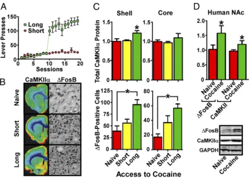
To further strengthen these observations, we explored models
of cocaine self-administration, which involve volitional drug
in-take. Adult male rats were given either short or long access to
cocaine; as expected (
Ahmed and Koob, 1998
), only long access
conditions led to escalating self-administration of the drug (
Fig.
2
A).
⌬FosB was induced to a greater extent by long versus short
access to cocaine in both NAc shell ( p
⫽ 0.0011; F ⫽ 11.12; df ⫽
17) and core ( p
⫽ 0.0004; F ⫽ 13.86; df ⫽ 17). In contrast,
CaMKII
␣ was induced in NAc shell only by long access to cocaine
(
Fig. 2
B, C; p
⫽ 0.0236; F ⫽ 4.957; df ⫽ 16). It is interesting to
compare the average daily cocaine intake across short-access
an-imals (⬃12 mg/kg, i.v.), long-access anan-imals (⬃70 mg/kg, i.v.),
and experimenter-administered animals (10 mg/kg) and ask why
the latter elicits robust induction of
⌬FosB and CaMKII whereas
short-access does not. This discrepancy is likely attributable to
differences in peak cocaine levels (experimenter-administered
cocaine is given as a single bolus intraperitoneally, whereas
self-administered cocaine is delivered via multiple intraperitoneal
doses), or differences in length of drug exposure (7 d for
experi-menter administration, 19 d for self-administration).
Despite the large literature on
⌬FosB and CaMKII in cocaine
action, there are no studies of these proteins in human cocaine
users. Here, we present the first evidence that levels of both
⌬FosB (p ⫽ 0.0316; t ⫽ 1.921; df ⫽ 34) and CaMKII (p ⫽ 0.0444;
t
⫽ 1.755; df ⫽ 32) are increased in NAc of cocaine-dependent
humans (
Fig. 2
D,
Table 1
). These data indicate that our
exami-nation of
⌬FosB and CaMKII induction by cocaine in rodent
NAc is clinically relevant to human cocaine addiction.
⌬FosB regulates CaMKII transcription selectively in D
1-type
MSNs of NAc shell
The finding that both CaMKII and
⌬FosB are upregulated by
cocaine in the rodent NAc led us to determine whether
⌬FosB
might regulate transcription of the CaMKII gene. We previously
reported CaMKII
␣ as a possible target for ⌬FosB in an unbiased
microarray analysis of NAc (
McClung and Nestler, 2003
), but
this finding was not further validated in that study. We first used
quantitative ChIP (qChIP; ChIP followed by quantitative PCR)
to determine whether
⌬FosB binds to the CaMKII␣ gene
pro-moter in NAc of adult male rats and found strikingly that this
binding is significantly increased, by chronic cocaine
administra-tion, in the shell ( p
⫽ 0.0133; t ⫽ 2.901; df ⫽ 12) but not the core
subregion (
Fig. 3
A). To further understand the mechanisms
re-lated to this subregion-specific difference in
⌬FosB binding to the
CaMKII
␣ promoter, we used qChIP to characterize the state of
histone modifications at this genomic region. Previous studies
demonstrated cocaine induction of H3 acetylation at the
CaMKII␣ promoter in total mouse NAc (
Wang et al., 2010
). In
Figure 1. Shell-specific induction of CaMKII in NAc by cocaine. A, Depiction of the “skewed donut” method of dissecting NAc core (blue circle) and shell (remaining half-moon between red and blue circles) from a rat coronal brain slice. B, Locomotor activity analysis reveals that chronic (green) but not acute (yellow) preexposure to cocaine sensitizes animals to a cocaine challenge when compared with a saline control group (red) (n⫽ 10; *p ⬍ 0.05, one-way ANOVA). C, Western blots of NAc shell and core from rats in B. D, Quantitation of Western blots in C shows significant increases in⌬FosB in both NAc shell and core, whereas significant increases in total CaMKII␣ and phospho-Ser831 GluA1 are shell specific (n ⫽ 10; *p ⬍ 0.05, one-way ANOVA). E, Quantitation of Western blot analysis of rat NAc shell 14 d after the last injection of saline or cocaine either before (14d WD) or 1 h after a challenge dose of cocaine (14d WD Chal) (n⫽9–10;*p⬍0.05,two-tailed
contrast, we found that cocaine decreases
H3 acetylation at the CaMKII
␣ promoter
selectively in NAc core (
Fig. 3
B; p
⫽
0.0213; t
⫽ 2.726; df ⫽ 10), with no
change apparent in shell, consistent with
subregion-specific chromatin alterations
beyond
⌬FosB binding. qChIP for the
re-pressive mark, dimethylated H3 lysine 9,
revealed trends for decreases in both the
shell and core subregions (
Fig. 3
C).
To determine whether
⌬FosB
regu-lates CaMKII
␣ transcription in vivo, we
used two bitransgenic mouse lines that
in-ducibly overexpress
⌬FosB specifically in
D
1- versus D
2-type MSNs in a manner
controlled by doxycycline administration
in drinking water (
Chen et al., 1998
;
Kelz
et al., 1999
;
Werme et al., 2002
). Adult
male mice overexpressing
⌬FosB solely in
D
1-type MSNs had significantly increased
levels of CaMKII␣ mRNA in NAc (p ⫽
0.0337; t
⫽ 1.996; df ⫽ 13), an effect not
seen in mice overexpressing
⌬FosB
pre-dominantly in D
2-type MSNs (
Fig. 3
D).
The increase in CaMKII␣ mRNA,
in-duced by
⌬FosB expression in D
1-type
MSNs, was accompanied by a
concomi-tant increase in CaMKII
␣ protein in both
NAc shell ( p
⫽ 0.0030; t ⫽ 3.578; df ⫽ 14) and core (p ⫽ 0.0392;
t
⫽ 2.275; df ⫽ 14;
Fig. 3
E, F ). These data demonstrate that
⌬FosB is capable of driving CaMKII␣ gene expression in D
1-type
MSNs in both subregions, although
Figure 3
B suggests that
cocaine-mediated chromatin changes at the CaMKII␣ promoter
(e.g., reduced acetylation) prevent
⌬FosB from upregulating
CaMKII in the core subregion after cocaine.
Because our transgenic mouse data indicated that
⌬FosB
induc-tion of CaMKII gene expression is specific to D
1-type MSNs in NAc,
we next sought to determine whether cocaine-dependent
upregula-tion of CaMKII requires activaupregula-tion of the D
1dopamine receptor.
Adult male rats were administered chronic cocaine or saline as
be-fore, but 30 min before each injection, rats in the cocaine group were
given an intraperitoneal injection of saline, the D
1antagonist SCH
23390 (0.5 mg/kg), or the D
2receptor antagonist eticlopride (0.5
mg/kg). Animals were analyzed 24 h after the last injection of
co-caine. Western blotting revealed that the D
1, but not the D
2,
antag-onist completely blocked the cocaine-mediated increase in
⌬FosB
(p
⬍ 0.0001; F ⫽ 18.96; df ⫽ 18), as reported previously (
Nye et al.,
1995
), as well as in CaMKII (p
⫽ 0.0005; F ⫽ 10.99; df ⫽ 18;
Fig.
3
G,H). These data support the hypothesis that cocaine engages a
⌬FosB-mediated increase in CaMKII gene expression specifically in
D
1-type MSNs of NAc shell. It would be important in future studies
to demonstrate directly this cell-type-specific effect of cocaine on
CaMKII expression within this brain region.
⌬FosB is both necessary and sufficient for cocaine induction
of CaMKII in NAc shell
To complement the use of bitransgenic mice, we next studied the
role of
⌬FosB in mediating cocaine induction of CaMKII␣ by use of
viral-mediated gene transfer in rats. We bilaterally injected AAV
particles into NAc shell of adult male rats (in which shell can be
selectively targeted) to overexpress
⌬FosB plus GFP or GFP alone.
The animals were then given a single intraperitoneal injection of 10
mg/kg cocaine. The animals overexpressing
⌬FosB/GFP exhibited
an increased locomotor response compared with animals
overex-pressing GFP alone (
Fig. 4
A). At 24 h after the single cocaine
injec-tion, GFP-positive NAc tissue was excised from these animals by
dissection under a fluorescent light source. Western blotting of this
tissue (
Fig. 4
B,C) revealed strong
⌬FosB overexpression as well as a
significant increase in total CaMKII␣ protein compared with GFP
animals (p
⫽ 0.0070; t ⫽ 2.894; df ⫽ 30), similar to the induction
seen with chronic cocaine administration. In addition, CaMKII␣
autophosphorylation at Thr286 (indicative of enzyme activation)
was increased by
⌬FosB overexpression (p ⫽ 0.0330; t ⫽ 2.243; df ⫽
28), as was phosphorylation of the CaMKII substrate, Ser831 of
GluA1 (p
⫽ 0.0540; t ⫽ 2.012; df ⫽ 28), again mimicking the actions
of chronic cocaine (
Fig. 1
C,D). Together, these data provide
addi-tional evidence that
⌬FosB expression in NAc shell is sufficient for
locomotor sensitization to cocaine and for CaMKII induction and
activation in this subregion.
We used a similar approach to determine whether
⌬FosB is
also necessary for cocaine-mediated induction of CaMKII
␣ in the
NAc shell. AAV was used to overexpress a truncated JunD
pro-tein, termed
⌬JunD, which is a negative regulator of ⌬FosB
tran-scriptional activation (
Winstanley et al., 2007
) plus GFP or GFP
alone. Two weeks later, when transgene expression is maximal,
animals were given cocaine (10 mg/kg) or saline daily for 7 d, and
tested for locomotor responses to a cocaine challenge (5 mg/kg)
24 h after the last chronic injection (
Fig. 4
D).
⌬JunD
overexpres-sion prevented locomotor sensitization to cocaine and also
pre-vented CaMKII
␣ induction and activation in NAc shell (
Fig.
4
E, F; p
⫽ 0.0437; F ⫽ 2.997; total df ⫽ 38), indicating that ⌬FosB
transcriptional activity is necessary for cocaine-mediated
induc-tion of CaMKII␣ in this subregion. Interestingly, we found that
⌬JunD reduced levels of ⌬FosB under both saline- and
cocaine-treated conditions ( p
⫽ 0.0004; F ⫽ 8.110; df ⫽ 35), raising the
novel possibility that
⌬FosB depends on AP-1 activity for its own
expression levels.
Figure 2. Induction of CaMKII in NAc shell of self-administering rats and human cocaine addicts. A, Lever presses by rats allowed long or short access to cocaine self-administration. B, Immunohistochemical analysis reveals increased⌬FosBinNAccoreandshell of long-access rats, whereas increases in CaMKII␣ are shell specific; quantified in C (n ⫽ 6; *p ⬍ 0.05, one-way ANOVA). D, Western blots (below) reveal that cocaine-dependent humans display increased⌬FosB and CaMKII␣ levels in shell-enriched NAc samples (n⫽ 18–20; *p ⬍ 0.05, two-tailed t test).
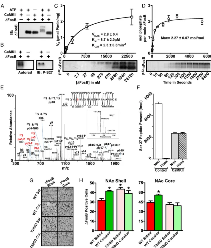
CaMKII phosphorylates
⌬FosB at Ser27
Using in vitro protein kinase assays, we determined that purified
⌬FosB is a robust substrate for CaMKII␣. Incubation of His
6–
⌬FosB with CaMKII␣ and ATP caused an upward shift in
elec-trophoretic mobility of
⌬FosB (
Fig. 5
A); the several resulting
bands suggested multiple sites of phosphorylation. Similar in
vitro kinase assays using [
␥-
32P]ATP showed incorporation of
radiolabeled phosphate into the shifted
⌬FosB bands (
Fig. 5
B),
Table 1. Characterization of samples from human cocaine addicts and matched control group
Group Percentage suicide Age (years) Gender (% male) Brain weight (g) pH Refrigeration delay (h) Control 32% 33.05⫾ 3.193 95% 1508⫾ 37.08 6.569⫾ 0.062 5.934⫾ 1.038 Cocaine dependent 80% 39.80⫾ 2.153 95% 1434⫾ 26.32 6.546⫾ 0.072 8.850⫾ 1.812
p value 0.11 0.18 0.48 0.016
The control group contains 19 individuals, and the cocaine-dependent group contains 20 individuals. All values are mean⫾ SE. p values calculated using two-tailed Student’s t test.
Figure 3. Cell type- and region-specific⌬FosBinductionofCaMKII␣invivo.A,qChIPassaysrevealincreased⌬FosBbindingtotheCaMKII␣genepromoterinratNAcshellbutnotcoreafterchroniccocaine exposure (n⫽ 6–7; *p ⬍ 0.05, two-tailed t test). B, qChIP also reveals decreased H3 acetylation after cocaine exposure in NAc core compared to shell (n ⫽ 6–7; p ⬍ 0.05, two-tailed t test). C, qChIP data suggesting reduced H3K9 dimethylation in both NAc shell and core after chronic cocaine. D, Quantitative PCR shows that mice overexpressing⌬FosBinD1-type, but not in D2-type, MSNs exhibit increased levels of CaMKII␣mRNAinNAc(n⫽8–10;*p⬍0.05two-tailedttest).E,ImmunohistochemicalanalysisshowsthattheD1- and D2-specific mouse lines overexpress⌬FosBtosimilarlevelsinNAcshellandcorebut thatonlyD1-specificoverexpressionof⌬FosBincreasestotalCaMKII␣protein;quantifiedinF(n⫽6–8;*p⬍0.05two-tailedttest).G,WesternblottingrevealsthatD1-specific,butnotD2-specific,antagonist (Antag) coadministration prevents cocaine-mediated⌬FosBandCaMKII␣inductioninratNAcshell;quantifiedinH(n⫽4–5;*p⬍0.05one-wayANOVA,differentfromvehicle).Con,Control.
demonstrating direct phosphorylation
of the protein. We generated a
phospho-specific antibody to the previously
charac-terized Ser27 of
⌬FosB (
Ulery et al., 2006
).
Although this antibody does not produce
a signal against brain extracts that contain
Ser27-phosphorylated
⌬FosB (data not
shown), we were able to detect Ser27
phosphorylation in the in vitro kinase
as-say using CaMKII (
Fig. 5
B). Kinetic
anal-yses of the CaMKII phosphorylation of
⌬FosB indicate that it is a potent substrate
for the kinase (
Fig. 5
C), with an apparent
K
Mof 5.7
⫾ 2.0
Mand K
CATof 2.3
⫾ 0.3
min
⫺1. These results are comparable with
many well-characterized in vivo substrates
of CaMKII (
Colbran and Brown, 2004
).
In addition, we determined that CaMKII
phosphorylates
⌬FosB with a
stoichiome-try of 2.27
⫾ 0.07 mol/mol (
Fig. 5
D),
in-dicating that there are at least three sites
of CaMKII phosphorylation within the
His
6–⌬FosB protein, in agreement with
Figure 5
A.
To investigate individual sites of
phos-phorylation, we used MS analyses of
sam-ples from our in vitro kinase assays.
Figure
5
E demonstrates
⌬FosB phosphorylation
at the previously characterized Ser27 and
at several additional sites (data not
shown). Given the previous functional
characterization of Ser27, we focused on
this site by generating labeled synthetic
peptides mimicking the phospho- and
non-phospho-states of Ser27 and then
used known quantities of these peptides as
standards in MRM analyses of
⌬FosB
be-fore and after in vitro phosphorylation by
CaMKII. Subsequent quantitation (
Fig.
5
F ) confirms that Ser27 is a potent
sub-strate for CaMKII. These results indicate
that, among multiple phosphorylated
res-idues within
⌬FosB, Ser27 is a particularly effective substrate for
CaMKII.
CaMKII mediates cocaine accumulation of
⌬FosB in the
NAc shell
Because CaMKII can phosphorylate
⌬FosB in vitro at a site that
dramatically enhances its stability in vitro and in vivo (
Ulery et
al., 2006
;
Ulery-Reynolds et al., 2009
), we determined whether
CaMKII activity controls
⌬FosB levels in NAc in vivo. To address
this question, we first used a mouse line overexpressing a
calcium-independent mutant of CaMKII␣ (T286D) in multiple
brain regions including NAc (
Mayford et al., 1996
;
Kourrich et
al., 2012
). We injected age-matched adult male mutant and
wild-type littermates with 20 mg/kg cocaine or saline once daily for
14 d and then analyzed the animals 1 d after the final injection.
We found that basal levels of
⌬FosB were increased in the mutant
animals in NAc shell ( p
⫽ 0.0001; F ⫽ 9.207; df ⫽ 37) but not
core (
Fig. 5
G,H ). Surprisingly, cocaine-dependent induction of
⌬FosB was blocked in the mutant animals in both shell and core,
suggesting that, although CaMKII may directly regulate
⌬FosB
stability in NAc shell, it may also lie upstream of
⌬FosB in
cocaine-activated pathways in both NAc subregions.
CaMKII activity is required for
⌬FosB-mediated structural
and behavioral plasticity
Cocaine induction of dendritic spines on NAc MSNs is one of the
best established drug-induced adaptations in this brain region,
and such spine induction has been correlated with sensitized
be-havioral responses to the drug (
Robinson and Kolb, 2004
;
Russo
et al., 2010
) and reported to be selective for D
1-type MSNs (
Lee et
al., 2006
). We demonstrated recently that cocaine induction of
dendritic spines in NAc is dependent on
⌬FosB and its
down-stream transcriptional program (
Maze et al., 2010
). Although
there is an extensive literature concerning the involvement of
CaMKII in dendritic spine morphology and induction in other
brain regions and experimental systems (
Jourdain et al., 2003
;
Penzes et al., 2008
;
Okamoto et al., 2009
), its role in NAc MSN
spine formation has not been studied. Therefore, we determined
whether CaMKII activity is required for
⌬FosB-mediated
induc-tion of MSN dendritic spines by using HSV-mediated
overex-pression of the CaMKII inhibitor peptide AC3I fused to GFP, a
Figure 4. ⌬FosBisbothnecessaryandsufficientforcocaine-mediatedD1receptor-dependent CaMKII␣inductioninNAcshell.
A, AAV-mediated overexpression of⌬FosB in NAc shell promotes locomotor responses to an acute cocaine injection in adult male
rats. B, Western blot analysis of NAc shell shows that⌬FosB is sufficient to increase levels of total CaMKII␣ and both autophos-phorylation of CaMKII␣ and Ser831 phosphorylation of GluA1; quantified in C (n ⫽ 14–18; *p ⬍ 0.05, two-tailed t test). D, AAV-mediated⌬JunD overexpression prevents locomotor sensitization induced by chronic exposure to cocaine. E, ⌬JunD over-expression in NAc shell is sufficient to block cocaine-mediated increases in total and Thr286 phospho-CaMKII and to reduce levels of⌬FosB in both saline- and cocaine-treated animals; quantified in F (n ⫽ 8–10; *p ⬍ 0.05, one-way ANOVA).
Figure 5. ⌬FosB is a potent substrate for CaMKII␣. A, Western blotting shows an ATP-dependent multi-band shift in electrophoretic mobility of ⌬FosB after exposure to CaMKII␣. IB,
Immunoblot. B, Autoradiogram reveals a CaMKII-dependent incorporation of radiolabeled phosphate into⌬FosB(left),anda⌬FosBSer27phospho-specificantibodyshowsphosphorylationofthis site by CaMKII (right). Analyses reveal robust kinase kinetics (C) and incorporation of multiple phosphates into⌬FosB by CaMKII (D). E, The precursor (inset) and fragment spectra of a TiO2enriched phosphopeptide detected from⌬FosBafterinvitrophosphorylationbyCaMKII.AfterusingbothtrypsinandGluCdigestionandenrichmentofthephosphopeptidesamplesbyTiO2, analysis reveals phosphorylation of Ser27 as well as of several other sites not characterized further here. F, MRM analysis of⌬FosBphosphorylatedinvitrobyCaMKII␣revealsthatSer27isapotentCaMKIIsubstrate. Non, Nonphosphorylated peptide; Phos, phosphorylated peptide. G, Immunohistochemical analysis reveals increased⌬FosB in both the NAc shell and core of adult male wild-type (WT) mice exposed to chronic cocaine. Littermates overexpressing a constitutively active form of CaMKII␣ show basal elevation in ⌬FosB in the NAc shell only and show no effect of cocaine on ⌬FosB levels in either region; quantified in H (n⫽ 9–10; *p ⬍ 0.05, one-way ANOVA).
construct shown previously to inhibit
CaMKII activity in vivo (
Zhang et al.,
2005
;
Klug et al., 2012
). Viral
overexpres-sion of
⌬FosB in NAc shell of adult mice
induced a significant increase in MSN
dendritic spine density ( p
⬍ 0.0001; F ⫽
8.558; df
⫽ 59;
Fig. 6
A, B) as reported
pre-viously (
Maze et al., 2010
), and this
increase was driven primarily by thin ( p
⫽
0.0027; F
⫽ 5.319; df ⫽ 59) and stubby
( p
⫽ 0.0378; F ⫽ 2.988; df ⫽ 59) spine
types (both thought to be immature
spines) (
Fig. 6
C–E). No effect was seen on
more mature, mushroom-shaped spines.
However, when GFP–AC3I was
coex-pressed,
⌬FosB induction of spines was
completely abrogated (
Fig. 6
A–E),
indi-cating that CaMKII activity is required for
⌬FosB induction of dendritic spines in
NAc shell.
We next used the same viral tools to
determine whether CaMKII activity is
re-quired for the effects of
⌬FosB on
behav-ioral sensitivity to cocaine. At 72 h after
viral injection into NAc shell, animals
were given a single injection of 5 mg/kg
cocaine and their locomotor activity was
recorded. As shown previously with more
extended AAV overexpression of
⌬FosB
(
Fig. 4
A), HSV-mediated overexpression
of
⌬FosB increased locomotor sensitivity
to cocaine ( p
⫽ 0.0002; F ⫽ 8.823; df ⫽
37;
Fig. 6
F ). As with induction of dendritic
spines, inhibition of CaMKII activity by
co-expression of GFP–AC3I completely
blocked the
⌬FosB-mediated increase in
cocaine sensitivity, indicating that
CaM-KII activity is required for
⌬FosB-induced
alterations in the behavioral effects of
cocaine.
Discussion
The present study delineates a novel
feed-forward mechanism in which cocaine
in-duces
⌬FosB in NAc, which upregulates
transcription of the CaMKII
␣ gene
selec-tively in NAc shell. CaMKII␣ subsequently
phosphorylates and stabilizes
⌬FosB,
lead-ing to greater
⌬FosB accumulation and to
further CaMKII
␣ induction (
Fig. 6
G). The
co-escalating levels of the two proteins
dur-ing chronic exposure to cocaine then
con-tribute in essential ways to sensitized
behavioral responses to the drug. This is a
particularly appealing hypothesis because
both
⌬FosB and CaMKII have each been
demonstrated previously to be required for
increased behavioral responses to cocaine
(
Pierce et al., 1998
;
Peakman et al., 2003
),
and we replicate this finding for
⌬FosBinNAcshellspecificallyusing
a viral approach (
Figs. 4
,
6
).
Although transgenic
⌬FosB overexpression in D
1-type MSNs
can drive CaMKII induction in both NAc shell and core of
cocaine-naive animals, in the context of cocaine, accumulation of
endogenous
⌬FosB, which occurs in both subregions, drives
in-duction of CaMKII specifically in NAc shell. This difference
could relate to the higher levels of
⌬FosB induced in our
bitrans-genic model, but it might also reflect the ability of cocaine to
Figure 6. Blockade of CaMKII activity prevents the morphological and behavioral effects of⌬FosB in NAc. A, Increases in the spine density of MSNs in NAc shell induced by HSV-mediated overexpression of⌬FosBarepreventedbycoexpressionoftheCaMKII inhibitor peptide AC3I (n⫽ 14–16); quantified in B. C–E, ⌬FosB effects on thin and stubby spines are blocked by coexpression of AC3I. F, The⌬FosB-mediated increase in locomotor sensitivity to cocaine is also prevented by AC3I coexpression. G, Model depicting the D1-receptor-dependent induction of a CaMKII/⌬FosB feedforward loop by cocaine, including upstream signaling cascades and physiological processes that may be affected. DA, Dopamine; D1DR, D1dopamine receptor; LTCC, L-type calcium channel (n⫽ 9–10; *p ⬍ 0.05, one-tailed t test).