Publisher’s version / Version de l'éditeur:
Chemistry and Physics of Lipids, 163, 2, pp. 117-126, 2010-02-01
READ THESE TERMS AND CONDITIONS CAREFULLY BEFORE USING THIS WEBSITE. https://nrc-publications.canada.ca/eng/copyright
Vous avez des questions? Nous pouvons vous aider. Pour communiquer directement avec un auteur, consultez la première page de la revue dans laquelle son article a été publié afin de trouver ses coordonnées. Si vous n’arrivez pas à les repérer, communiquez avec nous à PublicationsArchive-ArchivesPublications@nrc-cnrc.gc.ca.
Questions? Contact the NRC Publications Archive team at
PublicationsArchive-ArchivesPublications@nrc-cnrc.gc.ca. If you wish to email the authors directly, please see the first page of the publication for their contact information.
NRC Publications Archive
Archives des publications du CNRC
This publication could be one of several versions: author’s original, accepted manuscript or the publisher’s version. / La version de cette publication peut être l’une des suivantes : la version prépublication de l’auteur, la version acceptée du manuscrit ou la version de l’éditeur.
For the publisher’s version, please access the DOI link below./ Pour consulter la version de l’éditeur, utilisez le lien DOI ci-dessous.
https://doi.org/10.1016/j.chemphyslip.2009.09.003
Access and use of this website and the material on it are subject to the Terms and Conditions set forth at
Preparation of Reconstituted Acetylcholine Receptor Membranes
Suitable for AFM Imaging of Lipid-protein Interactions
Vuong, Ngoc; Baenziger, John E.; Johnston, Linda J.
https://publications-cnrc.canada.ca/fra/droits
L’accès à ce site Web et l’utilisation de son contenu sont assujettis aux conditions présentées dans le site LISEZ CES CONDITIONS ATTENTIVEMENT AVANT D’UTILISER CE SITE WEB.
NRC Publications Record / Notice d'Archives des publications de CNRC:
https://nrc-publications.canada.ca/eng/view/object/?id=996337b9-4571-486e-9d9c-b67cd3f22d81
https://publications-cnrc.canada.ca/fra/voir/objet/?id=996337b9-4571-486e-9d9c-b67cd3f22d81
Chemistry and Physics of Lipids 163 (2010) 117–126
Contents lists available atScienceDirect
Chemistry and Physics of Lipids
j o u r n a l h o m e p a g e :w w w . e l s e v i e r . c o m / l o c a t e / c h e m p h y s l i p
Preparation of reconstituted acetylcholine receptor membranes suitable for
AFM imaging of lipid–protein interactions
Ngoc Vuong
a,1, John E. Baenziger
a,∗, Linda J. Johnston
b,∗∗aDepartment of Biochemistry, Microbiology, and Immunology, University of Ottawa, Ottawa, Ontario K1H 8M5, Canada bSteacie Institute for Molecular Sciences, National Research Council of Canada, 100 Sussex Drive, Ottawa, Ontario K1A 0R6, Canada
a r t i c l e
i n f o
Article history: Received 22 June 2009
Received in revised form 4 September 2009 Accepted 24 September 2009
Available online 2 October 2009 Keywords:
Nicotinic acetylcholine receptor Atomic force microscopy Reconstitution Supported lipid bilayers Proteoliposomes Fluorescence
a b s t r a c t
The nicotinic acetylcholine receptor (nAChR) has been reconstituted in POPC vesicles at high lipid–protein (L/P) ratios for the preparation of supported lipid bilayers with a low protein density for studies of protein–lipid interactions using atomic force microscopy (AFM). Initial reconstitutions using a standard dialysis method with bulk L/P ratios ranging from 20:1 to 100:1 (w/w) gave heterogeneous samples that contained both empty vesicles and proteoliposomes with a range of L/P ratios. This is problematic because empty vesicles adsorb and rupture to form bilayer patches more rapidly than do protein-rich vesicles, resulting in the loss of protein during sample washing. Although it was not possible to find reconstitution conditions that gave homogeneous populations of vesicles with high L/P ratios, an addi-tional freeze–thaw cycle immediately after dialysis did reproducibly yield a fraction of proteoliposomes with L/P ratios above 100:1. These proteoliposomes were separated by sucrose gradient centrifugation and used to prepare supported bilayers with well-separated individual receptors and minimal adsorbed proteoliposomes. AFM images of such samples showed many small features protruding from the bilayer surface. These features range in height from 1 to 5 nm, consistent with the smaller intracellular domain of the protein exposed, and have lateral dimensions consistent with an individual receptor. Some bilayers with reconstituted protein also had a small fraction of higher features that are assigned to nAChR with the larger extracellular domain exposed and showed evidence for aggregation to give dimers or small oligomers. This work demonstrates the importance of using highly purified reconstituted membranes with uniform lipid–protein ratios for AFM studies of integral membrane protein–lipid interactions.
© 2009 Elsevier Ireland Ltd. All rights reserved.
1. Introduction
Atomic force microscopy (AFM) has been widely used to char-acterize the structures of peptides and proteins in supported phospholipid bilayers formed from both reconstituted and native membranes (Frederix et al., 2009). Ordered two-dimensional arrays of membrane proteins including rhodopsin, porins, bacterial hexag-onally packed intermediate layers, photosynthetic core-complexes and connexons are particularly amenable to high resolution AFM imaging (Arechaga and Fotiadis, 2007; Buzhynskyy et al., 2007; Fotiadis et al., 2003; Muller et al., 1997; Scheuring, 2006; Stamouli et al., 2003; Thimm et al., 2005). These studies have provided unprecedented structural data for supramolecular assemblies of membrane proteins in aqueous buffer under close to
physiolog-∗ Corresponding author. Tel.: +1 613 562 5800x8222; fax: +1 613 562 5251. ∗∗ Corresponding author. Tel.: +1 613 990 0973; fax: +1 613 952 0068.
E-mail addresses:John.Baenziger@uottawa.ca(J.E. Baenziger),
Linda.Johnston@nrc-cnrc.gc.ca(L.J. Johnston).
1Tel.: +1 613 562 5800x8222; fax: +1 613 562 5251.
ical conditions, where conformational changes and the effects of factors such as pH and ligand binding can be readily studied. The ability to obtain high lateral resolution, on the order of ∼1 nm, is critically dependent on obtaining samples with a high density of protein over a relatively large and flat membrane area. Reconsti-tution at such high protein density is still largely empirical and generally depends on removing detergent from a fully solubilized lipid/protein/detergent mixture. Interestingly, a recent report has shown that integral membrane proteins such as the bacterial light harvesting complex can be incorporated into existing lipid bilayers that are destabilized by detergent, providing a potentially useful approach for preparing arrays of protein for structural analysis from minimal amounts of protein (Milhiet et al., 2006).
On the other hand, AFM studies aimed at probing protein–lipid interactions require the use of reconstituted samples with much lower levels of protein. Supported bilayers with high lipid–protein ratios are required for characterizing the distribution of individ-ual membrane proteins within the membrane plane and thus for probing the preference of proteins for various lipid environments. Supported films with high lipid–protein ratios are necessary for interrogating individual molecules, as for example in correlated
flu-0009-3084/$ – see front matter © 2009 Elsevier Ireland Ltd. All rights reserved. doi:10.1016/j.chemphyslip.2009.09.003
orescence and AFM-based force spectroscopy measurements. High lipid–protein ratio bilayers are also advantageous for membrane biosensing, where protein availability may be an issue. Unfortu-nately, there are relatively few examples of the use of reconstituted integral membrane receptors in supported bilayers at low pro-tein density for AFM imaging (Periasamy et al., 2009; Slade et al., 2002). Individual insulin receptors reconstituted into a phos-phatidylcholine (PC) membrane have been imaged, with height measurements confirming that the protein is oriented in two direc-tions, with either the larger extracellular or shorter intracellular domains protruding out of the bilayer (Slade et al., 2002). This study illustrates the difficulty of controlling the orientation of the protein in supported membranes, although in some cases this challenge has been addressed by using specific labels (e.g., using histidine or streptavidin tethers) to immobilize the protein in one orientation (Elie-Caille et al., 2005; Giess et al., 2004; Trepout et al., 2007). A second practical limitation is that the conditions necessary for the formation of supported membranes from proteoliposomes may be significantly different than those for lipid vesicles, since the process of vesicle adsorption, rupture, and spreading is known to depend on the nature of the support as well as vesicle composition, lipid con-centration, and temperature (Goksu et al., 2009; Johnston, 2007; Richter et al., 2006).
The nicotinic acetylcholine receptor (nAChR) is one member of a super-family of neurotransmitter-gated ion channels that plays a central role in inter-neuronal communication within the brain (Sine and Engel, 2006). Due to its natural abundance, the nAChR from Torpedo has been used extensively as a model for probing neurotransmitter-gated ion channel–lipid interactions. The func-tional capabilities of the nAChR are highly dependent upon the lipid composition of its surrounding membrane environment (Criado et al., 1984; Fong and McNamee, 1986; Hamouda et al., 2006; Rankin et al., 1997; Ryan et al., 1996). nAChR–lipid interactions may even play a role in nAChR function in humans (Shen et al., 2006). Current models suggest that lipids influence nAChR function by modulating the equilibria between activatable resting and non-activatable desensitized and/or uncoupled conformations (Baenziger et al., 2000; daCosta and Baenziger, 2009). The pres-ence of both anionic lipids and cholesterol in a reconstituted PC membrane is required for optimal stabilization of the activatable resting state. With a structural model approaching atomic resolu-tion now available (Unwin, 2005), potential mechanisms by which lipids influence nAChR conformational equilibria are beginning to emerge (Brannigan et al., 2008; daCosta and Baenziger, 2009).
One unique feature of lipid–protein interactions for the nAChR is that lipids not only influence nAChR structure (and thus func-tion), the nAChR also influences lipid structure, and does so in a lipid-selective manner (daCosta et al., 2002, 2004). Reconstitution of the nAChR into bilayers composed of PC has little effect on the structure of the lipid bilayer. In contrast, incorporation into mem-branes composed of PC and the anionic lipid, phosphatidic acid (PA) leads to a substantial increase in the lateral packing density. The effects of the nAChR on the lateral packing density of lipid bilayers appear to result, at least in part, from an nAChR-induced concentra-tion of caconcentra-tions at the membrane surface (Sturgeon and Baenziger, submitted for publication). The ability of the nAChR to influence the surrounding membrane environment could play a role in lipid raft formation. It has been suggested that the nAChR is associated with lipid rafts during transport to synaptic membranes (Marchand et al., 2002).
Although previous studies have imaged dry native and het-erogeneous reconstituted nAChR membranes (Lal and Yu, 1993; Puu et al., 2000), these high nAChR density membranes (i.e. high protein–lipid ratio) are not suitable for probing nAChR–lipid inter-actions. As a first step towards studying the intriguing interactions that occur between the nAChR and its lipid environment using AFM,
we have developed a method for reconstituting the receptor at high lipid–protein ratios and have optimized conditions for preparing supported lipid bilayers. Our results demonstrate that reconsti-tution protocols often give rise to a heterogeneous population of proteoliposomes with a wide range of lipid–protein ratios. Recon-stitutions performed at high lipid–protein ratios generally lead to the formation of empty vesicles, which are problematic in that they rupture and spread to form supported bilayers more rapidly than densely packed proteoliposomes. By separating vesicle fractions using sucrose density centrifugation, however, we obtained pro-teoliposomes with uniform lipid–protein ratios that reproducibly form supported bilayers with low loadings of receptor. AFM anal-ysis of the resulting membranes reveals many individual receptors with heights that are consistent with predominantly the smaller cytoplasmic domain of the nAChR protruding out of the bilayer. Our results show the importance of using highly purified reconstituted membranes with uniform lipid–protein ratios for AFM studies of integral membrane protein–lipid interactions.
2. Materials and methods 2.1. Materials
Frozen Torpedo californica electroplax tissue was obtained from Aquatic Research Consultants (San Pedro, CA). 1-Palmitoyl-2-oleoyl phosphatidylcholine (POPC) was from Avanti Polar Lipids, Inc. (Alabaster, AL). Cholesterol, carbamylcholine, and sodium cholate were from Sigma–Aldrich. Alpha-bungarotoxin conju-gated to Alexa488 (␣BTx-A488) and Texas Red dihexadecanoyl-phosphatidylethanolamine (TR-DHPE) were from Invitrogen. The Lipofast mini extruder and 400 nm polycarbonate filters were obtained from Avestin (Ottawa, ON).
2.2. Preparation of lipid vesicles
POPC alone in chloroform or in the presence of 0.2 mol% Texas Red DHPE solubilized in chloroform/methanol 1:1 (vol:vol) was vortexed briefly, dried down under a stream of nitrogen gas and then dried further under vacuum for at least 10 h to remove remain-ing solvent. The lipid film was hydrated with Tris dialysis buffer (TDB = 10 mM Tris, 100 mM NaCl, 0.01 mM EDTA, 0.01% NaN3, pH
7.8) to a final lipid concentration of 1 mg/ml and vortexed for 1 min to generate large vesicles. The vesicle solution was then soni-cated in a water bath at room temperature for ∼10 min to generate small unilamellar vesicles. Once the solution was transparent, the vesicles were diluted to 0.1 mg/ml and stored at 4◦C. All small
unilamellar vesicle solutions were used for AFM imaging within 1 week.
2.3. Reconstitution of the nAChR
The nAChR was reconstituted into POPC membranes as described in detail bydaCosta et al. (2002). Briefly, Torpedo synaptic membranes were solubilized in TDB containing 1% cholate and then applied to a bromoacetylcholine-derivatized Affi-Gel 102 column. The column bound nAChR was washed with nine column volumes of TDB containing various concentrations of POPC solubilized in 1% sodium cholate. The nAChR was eluted from the column in three column volumes of TDB containing 10 mM of carbamylcholine and 0.13 mM POPC solubilized by 0.5% sodium cholate. The eluted frac-tions containing protein (A280> 0.05 a.u.) were pooled and dialyzed
against 2 l of TDB (Spectrapo/Por type 2 tubing, 12,000–14,000 Da cut off, Spectrum Laboratories Inc.) with 5 buffer changes, once every ∼12 h. The dialyzed samples were centrifuged at 120,000 × g for 2 h at 4◦C and the pellets resuspended in a small volume of TDB
N. Vuong et al. / Chemistry and Physics of Lipids 163 (2010) 117–126 119
to verify sample purity. TLC analysis showed that the reconstituted proteoliposomes contained only POPC, with no detectable native lipid.
Note that the lipid–protein ratios of reconstituted POPC–nAChR membranes prepared as described above are typically in the 0.5–1.0/1.0 (w/w) range (daCosta et al., 2002). To obtain higher lipid–protein ratios, the solubilized nAChR eluted from the column was mixed with an appropriate volume of 5 mg/ml of POPC solubi-lized in 1% cholate in TDB to make up the desired lipid–protein ratio (see Section3). Final protein concentrations were determined using the BCA assay (Pierce Biotechnology Inc.). Lipid concentration was assayed using the Phospholipid C/choline assay (Wako Chemicals USA Inc.). 2% Triton X-100 was included in both assays.
2.4. Sucrose gradient centrifugation
Reconstituted nAChR vesicles were purified further using discontinuous sucrose gradient centrifugation. Preparative scale (10 ml) step gradients were prepared by layering solutions with decreasing concentrations of sucrose in TDB on top of one another. The nAChR in 1 ml of TDB was then layered on top of the sucrose, and the gradients centrifuged at 41,000 rpm for 20 h at 4◦C in a
SW41 swinging bucket rotor (Beckmann). Fractions of 250 or 400 l were drawn from the gradient (starting from the top) and the lipid and protein content of each analyzed via the Phospholipid C and BCA protein assays. For preparative purposes, those fractions containing the vesicles of interest were isolated from the sucrose gradient directly by inserting a sharp syringe needle through the wall of the sucrose gradient tube at the fraction of interest. Isolated vesicles were then dialyzed against 2 l of TDB with 6 buffer changes, once every ∼12 h. Following dialysis, each sample was diluted to a lipid concentration of 0.1 mg/ml with TDB and stored at −80◦C.
For fluorescence microscopy, the nAChR was labeled with ␣BTx-A488 for 1 h after preparative sucrose gradient centrifugation. The unbound toxin and sucrose in the sample were then removed by dialysis.
2.5. Atomic force microscopy (AFM)
The appropriate volume (between 70 and 125 l) of either pure lipid vesicles or nAChR proteoliposomes in TDB (both at 0.1 mg/ml lipid) was mixed with distilled water containing the desired con-centration of CaCl2 (typically 10 mM) to give a final volume of
500 l. The lipid mixtures were deposited on freshly cleaved mica (high grade mica sheets, Ted Pella) and incubated for varying lengths of time (1–20 h) in an AFM fluid cell. Unbound material was washed away by flowing 100 ml of either water or imaging buffer (2.5 mM HEPES, 50 mM NaCl) into the AFM cell. The sample was allowed to equilibrate for 5 min after washing. Prior to AFM imag-ing, the AFM tip and holder were washed sequentially with 25 ml volumes of 70% ethanol, water, and imaging buffer. Bilayers were imaged using a PicoSPM atomic force microscope (Molecular Imag-ing/Agilent) in MAC (magnetic alternating current)-mode using magnetic coated silicon tips (Type II, spring constants of ∼0.5 N/m) in aqueous solutions. Either a 30 m × 30 m or 5 m × 5 m scan-ner was used with a scan rate between 0.7 and 1.4 Hz. All images shown are flattened raw data.
2.6. Total internal reflection fluorescence (TIRF) microscopy Fluorescence images of bilayers were taken on an Olympus IX81 total internal reflection fluorescence (TIRF) microscope equipped with a high resolution CCD camera (CoolSNAP, Photometrics, US) and a 60×/1.45 NA Plan Apochromat objective. Supported bilayers of either POPC labeled with Texas Red DHPE or proteoliposomes labeled with ␣BTx-A488 were prepared as described above for
the AFM measurements, except that the volumes of sample were increased proportionally to account for the increased surface area of the mica used in the larger TIRF cell. TR-DHPE and ␣BTx-A488 were excited at 543 and 488 nm, respectively.
3. Results and discussion 3.1. Preparation of POPC bilayers
Supported POPC bilayers on mica were prepared by the incuba-tion of POPC vesicles in water in the presence of 10 mM CaCl2for
1 h, followed by extensive washing to remove unadsorbed vesicles.
Fig. 1shows AFM images of bilayer patches and a continuous bilayer membrane, prepared using two different initial lipid concentra-tions. The bilayer patches have a height of 4.5 nm (see cross-section inFig. 1A) consistent with the expected thickness of a fluid POPC bilayer. Increasing either the incubation time (20 h) or the lipid concentration gives a continuous bilayer that is featureless with a low surface roughness (Fig. 1B). By contrast, in the absence of Ca2+, only bilayer patches were formed, even at long incubation
times of 20 h. Neither the use of HEPES buffer nor EDTA washing to remove excess Ca2+(both of which were employed for
bilay-ers with reconstituted protein) had any effect on the uniform and featureless POPC bilayers such as that shown inFig. 1B. Extrac-tion of a POPC bilayer and quantificaExtrac-tion using enzymatic detecExtrac-tion of choline generated by phospholipid hydrolysis indicated that an individual POPC bilayer prepared in our standard AFM fluid cell contains 1–2 g of lipid.
3.2. Reconstitution of nAChR
The receptor was initially reconstituted using a slight modifi-cation of the procedure typically employed for the preparation of nAChR proteoliposomes with low L/P ratios (typically ∼1:2 (w:w)) for FT-IR studies (daCosta et al., 2002; Ryan et al., 2002). Our goal, however, was to obtain samples with L/P ratios of 100:1 (w:w) or higher in order to facilitate imaging of individual receptors. Increasing the lipid concentration (from 0.13 to 1.6 mM) in the lipid-cholate detergent solution used to elute the purified recep-tor from the affinity column only increased the L/P ratio to ∼3:1 (w:w). The solubilized receptor eluted from the column was there-fore further diluted with a lipid-cholate solution prior to dialysis, ultracentrifugation and storage at −80◦C. Several attempts to form
supported POPC bilayers with samples of reconstituted nAChR with a L/P ratio of 100:1 (w:w) using the same conditions as for POPC bilayers gave variable AFM results. In some cases, bilayer patches that showed little or no evidence of raised features expected for reconstituted protein were obtained (Fig. 2A). In other cases, sam-ples appeared to have an intact lipid membrane but with many small roughly spherical particles (typically 10–20 nm in height and >100 nm in diameter) adhering to the surface. This material rapidly contaminated the AFM probe and was easily moved by the tip dur-ing scanndur-ing, makdur-ing such samples very difficult to image.
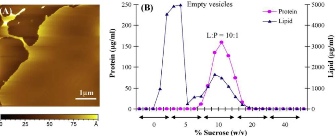
One possible explanation for the variable results for supported bilayers prepared as described above is that the vesicle population has a heterogeneous distribution of L/P ratios. We reasoned that proteoliposomes might rupture to form a bilayer membrane more slowly than empty vesicles, which could account for the observa-tion of patches that did not incorporate protein and large particles adhering to the bilayer. In order to characterize the L/P ratio in the initial proteoliposomes, the sample was separated on a discontinu-ous sucrose gradient and the protein and lipid content of individual fractions was analyzed. As illustrated inFig. 2B, there are two vesi-cle populations in the sample; the band near the top at 0–5% sucrose contains no protein and occurs at a similar density to POPC vesicles
Fig. 1. AFM images of POPC bilayer patches (A) and a continuous POPC bilayer (B). Bilayers were prepared by incubating POPC vesicles (7 and 10 g total lipid for A and B, respectively) in 10 mM CaCl2for 1 h. Cross-sections for the dashed lines are shown below each image.
(data not shown), while a second broad peak at 10% sucrose has a lipid/protein ratio of approximately 10:1. Samples reconstituted using the same method with bulk L/P ratios of 20:1 and 50:1 also gave populations of vesicles with both free and protein-rich vesicles, the latter with L/P ratios much lower than those of the bulk sample. Attempts to make bilayers from these samples had the same problems as described above for the 100:1 (w/w) sample. The observation of a large population of empty vesicles is consistent with an earlier study in which attempts to reconstitute the receptor at L/P ratios >16:1 led to mixtures of proteoliposomes and empty vesicles (Anholt et al., 1981).
The reconstituted sample (100:1 L/P, Fig. 2B) was separated on a preparative scale sucrose gradient and the protein-rich vesi-cle fraction was collected and pooled. This sample was labeled with ␣-bungaratoxin-Alexa 488 in order to visualize the protein by fluorescence microscopy, as well as by AFM, and then used for the preparation of supported bilayers. AFM images of a
sup-ported bilayer prepared using this sample showed bilayer patches with occasional raised features (Fig. 3). Some of these protrude from the membrane by 5–8 nm whereas others are much higher (>14 nm) and larger in diameter as shown by the representative cross-sections inFig. 3B and C. The smaller protrusions are consis-tent with protein incorporated in the membrane, based on a recent electron microscopy study that indicates that the larger extracellu-lar domain of nAChR should extend ∼8.5 nm above the membrane surface (Miyazawa, 2003). The larger particles are assigned to vesicles or protein–lipid aggregates adhering to the bilayer sur-face, consistent with the observation that they can be moved by the tip. Note that AFM images of protein-free POPC bilayers occasionally show a few remaining vesicles on the surface but they can easily be removed with additional washing. By contrast, the material adhering to the bilayer prepared with proteolipo-somes was not readily removed by washing with either water or buffer.
Fig. 2. (A) AFM image of bilayer patches prepared from nAChR proteoliposomes (L/P ratio of 100:1) by incubating 10 g total lipid in 10 mM CaCl2for 1 h. The film was
imaged in HEPES buffer. (B) Sucrose gradient separation of the same proteoliposome sample, showing populations of empty vesicles and proteoliposomes with a L/P ratio of ∼10:1.
N. Vuong et al. / Chemistry and Physics of Lipids 163 (2010) 117–126 121
Fig. 3. (A) AFM image of a supported bilayer prepared from nAChR proteoliposomes with L/P ratio of 10:1 (conditions as forFig. 2A). (B and C) Cross-sections for the dashed lines indicated in image A. (D and E) TIRF images of a similar bilayer. Image D is on the same x–y scale as the AFM image (A); the large bright features correspond to clusters of features observed by AFM (see for example the area at the bottom left in A). Image E corresponds to the boxed region in image D with the intensity scale adjusted to allow visualization of weakly fluorescent features (two of which are marked with arrows) that are not visible in the larger images.
Total internal reflection fluorescence (TIRF) microscopy was used to visualize the distribution of receptors within bilayer films prepared from the same sample of proteoliposomes. TIRF images (Fig. 3D) showed a number of bright features that are too large to be individual receptors, but are consistent with small clusters of proteoliposomes that are too close together to be resolved as individual features with the diffraction-limited resolution of optical microscopy. A zoomed image on a different intensity scale for the boxed region ofFig. 3D shows a number of additional weakly fluo-rescent features that are visible above the surrounding background (Fig. 3E, see arrows). These features have a diffraction-limited spot size (∼400 nm in diameter), as expected for either an indi-vidual labeled receptor or a small oligomer. Thus, both TIRF and AFM images provide evidence for the presence of proteoliposomes adsorbed on the membrane, in addition to a small amount of recon-stituted protein. Based on a L/P ratio of 10:1, we would expect to see a very protein-rich membrane, suggesting that most of the pro-tein is not deposited in the membrane and is lost during the bilayer washing step. Control experiments in which ␣-bungarotoxin-Alexa 488 was added to bilayers prepared from either unlabeled pro-teoliposomes or POPC vesicles indicated a significant amount of non-specific binding of the toxin to mica at bilayer defects. For this reason we avoided labeling nAChR after bilayer formation.
An additional experiment using a higher concentration of Ca2+
during bilayer formation confirmed the hypothesis that a signifi-cant amount of protein was lost during the washing step. In this case, AFM images showed a surface with many adherent parti-cles (>15 nm in height and >150 nm in diameter) and the TIRF image showed relatively large fluorescent areas (data not shown). From this we conclude that higher Ca2+concentration results in
the deposition of large numbers of protein-rich vesicles on the sur-face. Although it is not clear whether the vesicles are adsorbed on a bilayer or on mica, these results indicate that variation of either
the L/P ratio of the proteoliposomes or the bilayer preparation conditions is required to form supported POPC bilayers with recon-stituted protein. Thus, we opted to prepare more homogeneous proteoliposomes with higher L/P ratios and to vary the incubation time over a relatively modest range of Ca2+concentrations in order
to optimize conditions for the formation of bilayer films containing reconstituted protein for AFM imaging. Note that, in principle, one can also vary the incubation temperature or lipid concentration. However, given that the initial experiments were already carried out with close to the minimum lipid concentration required for membrane formation (based on the results for POPC bilayers) and that higher temperatures may be problematic for protein stability, we chose to focus on the factors noted above.
Several alternative reconstitution approaches were investi-gated. First, proteoliposomes with a 3:1 L/P ratio in lipid/cholate were mixed with POPC vesicles and part of the mixture was sub-jected to five freeze–thaw cycles. Analysis of the sample with and without freeze–thaw treatment on a sucrose gradient indi-cated that the freeze–thaw cycles did not promote mixing and thus the formation of a uniform proteoliposome population with a higher L/P ratio. Second, a method based on the addition of detergent-solubilized protein to detergent-destabilized liposomes was examined (Rigaud and Levy, 2003), but with limited suc-cess. In the course of the latter experiments, however, it was observed that at higher lipid-to-protein ratios, freeze–thaw cycles do influence the relative populations of free versus protein-incorporated vesicles (Fong and McNamee, 1986). The effect of freeze–thaw cycles was thus investigated further.
In order to probe the effect of sample treatment on the L/P ratios, we made a direct comparison of samples that were subjected to freeze–thaw treatment and/or ultracentrifugation after dialysis to remove detergent (Fig. 4). Analysis of the initial sample (after dial-ysis) using sucrose gradients showed a mixture of empty vesicles
Fig. 4. Sucrose gradients showing the effect of ultracentrifugation and freeze–thaw treatments on an initial proteoliposome sample after dialysis with an overall L/P ratio of 40:1.
and proteoliposomes with an overall L/P ratio of ∼40:1 (Fig. 4A). A proportion of the empty vesicles disappeared after centrifuga-tion, largely due to the loss of vesicles in the supernatant (Fig. 4C). The pelleted material contained predominantly proteoliposomes and had a lower overall L/P ratio than the original vesicle sample. Freeze–thaw cycles also led to the disappearance of the protein-free vesicles, but in this case yielded a sample containing a population of vesicles with different L/P ratios (Fig. 4B). Pelleting the sample subjected to freeze–thaw cycles had minimal effect on the distri-bution of L/P ratios (Fig. 4D). These results indicate that at high lipid-to-protein ratios, a freeze–thaw cycle does promote vesicle mixing, although the vesicle population is still far from uniform. Fortunately, one of the resulting vesicle populations exhibited a L/P ratio of ∼160:1 (w:w) and this fraction was separated and used further for AFM studies (see below). A second independent recon-stitution in which the sample was subjected to a single freeze–thaw cycle after dialysis yielded a fraction of proteoliposomes with a L/P ratio of 120:1 (w/w), which was also used to prepare bilayers for AFM.
Several recent studies have examined the effect of reconstitu-tion method on the distribureconstitu-tion of sizes and protein densities of proteoliposomes and on protein activity. In one example, SNARE protein mediated lipid mixing was shown to depend on the physi-cal state of the vesicles (Chen et al., 2006). The standard method of co-micellization of proteins and lipids with detergent gave pro-teoliposomes with a large dispersion in size and L/P ratios that promoted variable amounts of lipid mixing at moderate protein densities. However, the direct incorporation of SNARE proteins into preformed liposomes gave a more homogeneous population of proteoliposomes, but no lipid mixing at comparable protein
densities. Similarly, a recent study of rhodopsin reconstituted in asolectin has shown that the homogeneity of the proteolipo-somes and the protein activity depend on the method used for detergent removal after co-solubilization of protein and lipids in detergent micelles (Niu et al., 2009). Detergent dialysis produced a heterogeneous population of proteoliposomes with a signifi-cant population of empty lipid vesicles. By contrast, rapid dilution yielded only proteoliposomes with a higher L/P ratio and a higher level of rhodopsin activity, leading to the conclusion that the rapid dilution method is advantageous for both preserving rhodopsin activity and controlling the L/P ratio of the proteoliposomes. The lipid composition, purification protocol and thermal history of the sample were found to be important in minimizing aggre-gation of the mechanosensitive channel, MscS (Vasquez et al., 2007).
3.3. AFM imaging of nAChR in POPC bilayers
Proteoliposome samples with L/P in excess of 100:1, as pre-pared above, were used to prepare supported lipid bilayers for AFM characterization. Initially we tested a range of lipid and Ca2+
concentrations, and incubation times in order to optimize con-ditions for preparing bilayers suitable for imaging reconstituted protein. These trials indicated that 10 g lipid was sufficient to give complete bilayers with few defects when using short incu-bation times (<1 h) and 10 mM or higher Ca2+. However, bilayers
prepared with short incubation times invariably had a combina-tion of large particles that adhered to the bilayer as well as smaller features with heights <10 nm that are consistent with reconstituted protein. Increasing the lipid concentration increased the problems
N. Vuong et al. / Chemistry and Physics of Lipids 163 (2010) 117–126 123
Fig. 5. AFM images for bilayers prepared from a proteoliposome sample with L/P ratio of 160:1, with cross-sections for the lines marked shown on the right. Images A and B show the same region of a bilayer on two length scales with cross-sections indicating a range of heights (2.3, 3.2 and 3.8 nm for a, b, c) for the raised features. Images C and D show smaller scale images for an area of the same bilayer; the cross-section for line d indicates a height and diameter of 3.9 and 13 nm, respectively.
associated with vesicles adhering to the bilayer. Washing the sam-ples after incubation with an EDTA solution was useful in removing some of the larger particles and reducing the problems with tip contamination. However, we found that using slightly less lipid (5–7.5 g) also gave bilayer patches with equally short incuba-tion times, but led to uniform bilayers that had few, if any, vesicle adhesion problems after incubation of the proteoliposomes for approximately 20 h in the presence of either 10 or 25 mM Ca2+.
In the absence of Ca2+only very small bilayer patches were
gen-erated for the nAChR proteoliposomes (L/P 120:1), even with 20 h incubation times.
Fig. 5shows typical AFM images for a bilayer obtained by incu-bating a minimum concentration of proteoliposomes (L/P ratio of 160:1) for 20 h in the presence of 25 mM Ca2+, followed by
exten-sive washing.Fig. 5A and B shows a uniform bilayer with a number of randomly distributed small features of variable height that pro-trude out of the bilayer. Cross-sections for the lines marked in
Fig. 5B indicate that most are in the range of 2–4 nm in height. Note that the features are considerably smaller in both height and diameter than the large particles, assigned to liposomes, which we observed on POPC bilayers prepared using high concentrations of lipid and on proteoliposome films prepared from low L/P ratios pro-teoliposomes. Furthermore, POPC bilayers prepared from vesicles that were subjected to a similar dialysis and sucrose gradient treat-ment showed uniform featureless bilayers, similar to that shown inFig. 1B.
Fig. 5C shows a smaller scale image of the same bilayer. A zoomed image of one feature with a height of 3.9 nm and a diam-eter of 13 nm (see cross-section for line d) is shown in Fig. 6D.
The roughly circular shape and the lateral dimensions are consis-tent with an individual receptor. Note that the measured lateral size of small features that have similar dimensions to the tip will be significantly overestimated due to tip-sample convolution. The overestimation of the feature size depends on both the feature height and the tip radius and can be assessed using a literature model (Maeda, 1997). For AFM tips with radii between 5 and 10 nm (based on the manufacturer’s specifications), the overesti-mate will be in the range of 6–14 nm for a feature with a height of 4 nm. Therefore, the measured diameter of 13 nm is consistent with an individual receptor, based on recent structural data which indicates a diameter of 8 nm for the membrane-embedded nAChR (Miyazawa, 2003; Unwin, 2005).
A histogram of heights for >400 features in AFM images of two bilayers prepared from a proteoliposome sample with L/P ratio of 160/1 is provided inFig. 6A. The measured heights show a rela-tively broad distribution, ranging from 1 to 6 nm. Based on electron microscopy data, the extracellular domain should extend ∼8.5 nm from the bilayer surface, while the smaller cytoplasmic domain protrudes ∼3 nm from the membrane (Fig. 6B;Miyazawa, 2003; Unwin, 2005). The heights measured by AFM are thus consistent with predominantly the smaller cytoplasmic domain extending above the bilayer surface, with few, if any, proteins with the larger extracellular domain exposed. The heterogeneity in heights is not surprising since all receptors may not be oriented perpendicular to the bilayer and since tip–sample interactions may lead to some dis-tortion or mobility of the protein domains above the bilayer surface. A similar heterogeneous distribution of heights has been observed by AFM for another integral membrane protein, the insulin
recep-Fig. 6. (A) Histogram showing the distribution of heights for raised features measured from multiple images for the bilayer shown inFig. 6. (B) Atomic model of the nAChR (Protein Data Bank code 2BG9). The model defines the location of ∼80% of the residues, with undefined structures being located mainly in the cytoplasmic domain (lower portion of the structure in this orientation). The nAChR is ∼16 nm long with a cross-sectional diameter of roughly 8 nm. The bilayer position is indicated by the grey lines.
tor, reconstituted in phosphatidylcholine bilayers (Slade et al., 2002).
An AFM image of a bilayer containing reconstituted nAChR pre-pared from a second proteoliposome sample (L/P 120:1) is shown inFig. 7A. The sample is qualitatively similar in that a number of small features of variable height are observed. However, in this case, the features show a wider range of heights, as indicated by the cross-sections (Fig. 7B and C) showing receptors that are 10 and 2.8 nm in height. The raised features observed from this sample were frequently larger in the lateral dimension, providing evidence for receptor aggregation. A small scale image (Fig. 7D) of a region in the centre of Fig. 7A illustrates this clearly with two small adjacent protrusions in the lower right of the image. Note that the nAChR is known to form disulfide-linked dimers (daCosta et al., 2005). The lower L/P ratios for this sample may be responsible for the increased evidence for protein aggrega-tion, although additional work under exactly controlled conditions would be required to establish if this is in fact the case. A histogram of heights obtained for multiple images and bilayers (Fig. 7E) indi-cates that this proteoliposome sample has a small but significant fraction of features with heights in excess of 5 nm, as expected for nAChR oriented with the larger extracellular domain on the bilayer surface.
The generally accepted mechanism for the formation of a sup-ported bilayer on hydrophilic substrates such as glass or mica involves adsorption and flattening of vesicles followed by rup-ture and unfolding to give a planar membrane (Goksu et al., 2009; Richter et al., 2006). Previous studies have shown that the nAChR
is reconstituted into vesicles with the large extracellular domain preferentially oriented outwards (Anholt et al., 1981; Fong and McNamee, 1986). Thus one would predict that the smaller cyto-plasmic domain will be exposed on the upper bilayer surface after bilayer rupture and spreading. This is consistent with our AFM observations since both reconstituted samples showed predomi-nantly the smaller cytoplasmic domain on the bilayer surface. The presence of a larger fraction of features consistent with exposed extracellular domains for one proteoliposome sample may indi-cate a larger fraction of receptor with the opposite orientation in the initial vesicles. This could result from differences in either the sample preparation or the length of time the sample was stored prior to the preparation of supported bilayers. An alternate pos-sibility is that vesicle rupture and spreading to give a supported bilayer leads to some scrambling of protein orientation. The vesicle fusion process may also lead to redistribution of proteins within the plane of the membrane.
Proteoliposomes with L/P ratios >100:1 give reproducible supported bilayers with low densities of reconstituted protein. However, the amount of protein is lower than would be expected based on the L/P ratios of the individual samples. For example, using estimated values for the area of POPC and nAChR molecules at the bilayer surface, a bilayer with L/P of 100:1 would have individual proteins separated by only ∼100 nm if we assume that proteins are distributed homogeneously throughout the mem-brane. Although such a protein distribution is unlikely, it is clear that there is a substantially larger average separation between individual reconstituted proteins, based on the AFM images. The
Fig. 7. (A) AFM image for a bilayer prepared from proteoliposomes with L/P of 120:1. (B and C) Cross-sections for the lines marked b and c in image A indicate heights of 10 and 2.8 nm. (D) A zoomed image shows two adjacent features (arrow). (E) The histogram indicates that there is a small but significant fraction of features with heights consistent with the larger extracellular domain of the protein protruding from the bilayer surface.
N. Vuong et al. / Chemistry and Physics of Lipids 163 (2010) 117–126 125
observation of a much lower than expected protein density sug-gests that bilayer formation favors adsorption and rupture of the least protein-rich liposomes within the proteoliposome popula-tion.
The AFM images obtained for the nAChR reconstituted at low density in a fluid POPC bilayer do not allow resolution of the pen-tameric structure of the nAChR. This is not surprising as detailed structural information is generally only obtained from relatively ordered and rigid films formed from membranes at high protein density (Frederix et al., 2009). High resolution structural informa-tion has been obtained from AFM studies of the nAChR in high density dry native membranes (Lal and Yu, 1993). These studies revealed the pentameric structure of the nAChR, consistent with the data obtained from electron microscopy (Jones et al., 1988; Zingsheim et al., 1982). While the data obtained by AFM in this study is clearly of lower resolution, these membrane films are uniquely amenable to studying other aspects of nAChR–lipid inter-actions, such as protein aggregation and the distribution of the nAChR in lipid raft versus non-raft bilayers.
4. Conclusions
Samples of POPC–nAChR proteoliposomes prepared by the stan-dard dialysis method with bulk L/P ratios between 20:1 and 100:1 are heterogeneous, containing both empty vesicles and prote-oliposomes with a range of L/P ratios. The presence of empty vesicles is particularly problematic for preparing supported bilay-ers since empty vesicles adsorb and rupture to form supported bilayer patches more rapidly than do protein-rich vesicles, result-ing in the loss of the majority of protein durresult-ing sample washresult-ing. Attempts to vary conditions to favor adsorption and rupture of protein-rich vesicles were unsuccessful. Although it was not pos-sible to find reconstitution conditions that gave homogeneous populations of vesicles with high L/P ratios, additional freeze–thaw cycles immediately after dialysis did reproducibly yield a fraction of proteoliposomes with L/P ratios in excess of 100:1. Fractions with the desired L/P ratio were separated on a sucrose gradient and used for the preparation of supported bilayers for AFM imag-ing. Optimization of conditions indicated that the use of minimal lipid concentrations with lengthy incubation times in the pres-ence of Ca2+was optimal for the preparation of supported bilayers
containing well-separated individual receptors with a minimum number of adsorbed proteoliposomes. AFM images of such samples showed many small features protruding from the bilayer surface. These features ranged in height from 1 to 5 nm, consistent with the smaller intracellular domain of the protein exposed. The lateral dimensions were in the range expected for an individual recep-tor after taking into account the convolution of probe and feature size. Some bilayers with reconstituted protein also had a small but significant fraction of higher features that are assigned to nAChR with the opposite orientation with the larger extracellu-lar domain exposed and showed evidence for aggregation to give dimers or small oligomers. The predominance of a single protein orientation for reconstituted nAChR is consistent with the known orientation of the protein in reconstituted vesicles. In addition to establishing suitable conditions for preparing reconstituted sam-ples for studies of lipid–protein interactions, this work indicates the importance of characterizing the lipid and protein content of reconstituted proteoliposomes prior to functional or biophysical studies.
Author contributions
This project was initiated by JEB and LJJ. NV, JEB, and LJJ designed all the experiments. NV performed all the experiments. JEB and LJJ wrote the manuscript.
Acknowledgements
LJJ and NV acknowledge partial support from a Natural Sci-ences and Engineering Research Council grant to LJJ. This work was also supported by a grant from the Canadian Institutes of Health Research to JEB.
References
Anholt, R., Lindstrom, J., Montal, M., 1981. Stabilization of acetylcholine receptor channels by lipid in cholate solution and during reconstitution in vesicles. J. Biol. Chem. 256, 4377–4387.
Arechaga, I., Fotiadis, D., 2007. Reconstitution of mitochondrial ATP synthase into lipid bilayers for structural analysis. J. Struct. Biol. 160, 287–294.
Baenziger, J.E., Morris, M.L., Darsaut, T.E., Ryan, S.E., 2000. Effect of membrane lipid composition on the conformational equilibria of the nicotinic acetylcholine receptor. J. Biol. Chem. 275, 777–784.
Brannigan, G., Hénin, J., Law, R., Eckenhoff, R., Klein, M.L., 2008. Embedded choles-terol in the nicotinic acetylcholine receptor. Proc. Natl. Acad. Sci. U.S.A. 105, 14418–14423.
Buzhynskyy, N., Hite, R.K., Walz, T., Scheuring, S., 2007. The supramolecular archi-tecture of junctional microdomains in native lens membranes. EMBO Rep. 8, 51–55.
Chen, X., Arac, D., Wang, T.-M., Gilpin, C.J., Zimmerberg, J., 2006. SNARE-mediated lipid mixing depends on the physical state of vesicles. Biophys. J., 2062– 2074.
Criado, M., Eibl, H., Barrantes, F.J., 1984. Functional properties of the acetylcholine receptor incorporated in model lipid membranes. Differential effects of chain length and head group of phospholipids on receptor affinity states and receptor-mediated ion translocation. J. Biol. Chem. 259, 9188–9198.
daCosta, C.J., Ogrel, A.A., Mccardy, E.A., Blanton, M.P., Baenziger, J.E., 2002. Lipid–protein interactions at the nicotinic acetylcholine receptor. A functional coupling between nicotinic receptors and phosphatidic acid-containing lipid bilayers. J. Biol. Chem. 277, 201–208.
daCosta, C.J.B., Baenziger, J.E., 2009. A lipid dependent uncoupled conformation of the acetylcholine receptor. J. Biol. Chem. 284, 17819–17825.
daCosta, C.J.B., Kaiser, D.E.E., Baenziger, J.E., 2005. Role of glycosylation and mem-brane environment in nicotinic acetycholine receptor stability. Biophys. J. 88, 1755–1764.
daCosta, C.J.B., Wang, I.D., Mckay, M.E., Baenziger, J.E., 2004. Phosphatidic acid and phosphatidylserine have distinct structural and functional interactions with the nicotinic acetylcholine receptor. J. Biol. Chem. 279, 14967–14974.
Elie-Caille, C., Fliniaux, O., Pantigny, J., Maziere, J.-C., Bourdillon, C., 2005. Self-assembly of solid-supported membranes using a triggered fusion of phospholipid-enriched proteoliposomes prepared from the inner mitochondrial membrane. Langmuir 21, 4661–4668.
Fong, T.M., McNamee, M.G., 1986. Correlation between acetylcholine receptor func-tion and structural properties of membranes. Biochemistry 25, 830–840. Fotiadis, D., Liang, Y., Filipek, S., Saperstein, D.A., Engel, A., Palczewski, K., 2003.
Rhodopsin dimers in native disc membranes. Nature 421, 127–128.
Frederix, P.L.T.M., Bosshart, P.D., Engel, A., 2009. Atomic force microscopy of biolog-ical membranes. Biophys. J. 96, 329–338.
Giess, F., Friedrich, M.G., Heberle, J., Naumann, R.L., Knoll, W., 2004. The protein-tethered lipid bilayer: a novel mimic of the biological membrane. Biophys. J. 87, 3213–3220.
Goksu, E.I., Vanegas, J.M., Blanchette, C.D., Lin, W.-C., Longo, M.L., 2009. AFM for structure and dynamics of biomembranes. Biochim. Biophys. Acta 1788, 254–266.
Hamouda, A.K., Sanghvi, M., Sauls, D., Machu, T.K., Blanton, M.P., 2006. Assessing the lipid requirements of the Torpedo californica nicotinic acetylcholine receptor. Biochemistry 45, 4327–4337.
Johnston, L.J., 2007. Nanoscale imaging of domains in supported lipid membranes. Langmuir 23, 5886–5895.
Jones, O.T., Eubanks, J.H., Earnest, J.P., McNamee, M.G., 1988. A minimum num-ber of lipids are required to support the functional properties of the nicotinic acetylcholine receptor. Biochemistry 27, 3733–3742.
Lal, R., Yu, L., 1993. Atomic force microscopy of cloned nicotinic acetylcholine recep-tor expressed in Xenopus oocytes. Proc. Natl. Acad. Sci. 90, 7280–7284. Maeda, H., 1997. An atomic force microscopy study for the assembly structures of
tobacco mosaic virus and their size evaluation. Langmuir 13, 4150–4161. Marchand, S., Devillers-Thiery, A., Pons, S., Changeux, J.-P., Cartaud, J., 2002.
Rap-syn escorts the nicotinic acetylcholine receptor along the exocytic pathway via association with lipid rafts. J. Neurosci. 22, 8891–8901.
Milhiet, P.-E., Gubellini, F., Berquand, A., Dosset, P., Rigaud, J.-L., Le Grimellec, C., Levy, D., 2006. High-resolution AFM of membrane proteins directly incorporated at high density in planar lipid bilayer. Biophys. J. 91, 3268–3275.
Miyazawa, A., 2003. Structure and gating mechanism of the acetylcholine receptor pore. Nature 423, 949–955.
Muller, D., Schoenenberger, C.-A., Schabert, F., Engel, A., 1997. Structural changes in native membrane proteins monitored at subnanometer resolution with the atomic force microscope: a review. J. Struct. Biol. 119, 149–157.
Niu, S.-L., Doctrow, B., Mitchell, D.C., 2009. Rhodopsin activity varies in proteolipo-somes prepared by different techniques. Biochemistry 48, 156–163.
Periasamy, N., Teichert, J.H., Wiese, K., Vogel, R.F., Winter, R., 2009. Effects of tem-perature and pressure on the lateral organization of model membranes with functionally reconstituted multidrug transporter LmrA. Biochim. Biophys. Acta 1788, 390–401.
Puu, G., Artursson, E., Gustafson, I., Lundstrom, M., Jass, J., 2000. Distribution and stability of membrane proteins in lipid membranes on solid supports. Biosens. Bioelectron. 15, 31–41.
Rankin, S.E., Addona, G.H., Kloczewiak, M.A., Bugge, B., Miller, K.W., 1997. The cholesterol dependence of activation and fast desensitization of the nicotinic acetylcholine receptor. Biophys. J. 73, 2446–2455.
Richter, R.P., Berat, R., Brisson, A.R., 2006. Formation of solid-supported lipid bilay-ers: an integrated view. Langmuir 22, 3497–3505.
Rigaud, J.-L., Levy, D., 2003. Reconstitution of membrane proteins into liposomes. Methods Enzymol. 372, 65–86.
Ryan, S.E., Demers, C.N., Chew, J.P., Baenziger, J.E., 1996. Structural effects of neutral and anionic lipids on the nicotinic acetylcholine receptor. An infrared difference spectroscopy study. J. Biol. Chem. 271, 24590–24597.
Ryan, S.E., Hill, D.G., Baenziger, J.E., 2002. Dissecting the chemistry of nicotinic receptor–ligand interactions with infrared difference spectroscopy. J. Biol. Chem. 277, 10420–10426.
Scheuring, S., 2006. AFM studies of the supramolecular assembly of bacterial pho-tosynthetic core-complexes. Curr. Opin. Chem. Biol. 10, 387–393.
Shen, X.M., Deymeer, F., Sine, S.M., Engel, A.G., 2006. Slow-channel mutation in acetylcholine receptor alphaM4 domain and its efficient knockdown. Ann. Neu-rol. 60, 128–136.
Sine, S.M., Engel, A.G., 2006. Recent advances in Cys-loop receptor structure and function. Nature 440, 448–455.
Slade, A., Luh, J., Ho, S., Yip, C.M., 2002. Single molecule imaging of supported planar lipid bilayer-reconstituted human insulin receptors by in situ scanning probe microscopy. J. Struct. Biol. 137, 283–291.
Stamouli, A., Kafi, S., Klein, D.C.G., Oosterkamp, T.H., Frenken, J.W.M., Cogdell, R.J., Aartsma, T.J., 2003. The ring structure and organization of light harvesting 2 complexes in a reconstituted lipid bilayer, resolved by atomic force microscopy. Biophys. J. 84, 2483–2491.
Sturgeon, R.M., Baenziger, J. E., submitted for publication.
Thimm, J., Mechler, A., Lin, H., Rhee, S., Lal, R., 2005. Calcium-dependent open/closed conformations and interfacial energy maps of reconstituted hemichannels. J. Biol. Chem. 280, 10646–10654.
Trepout, S., Mornet, S., Benabdelhak, H., Ducruix, A., Brisson, A.R., Lambert, O., 2007. Membrane protein selectively oriented on solid support and reconstituted into a lipid membrane. Langmuir 23, 2647–2654.
Unwin, N., 2005. Refined structure of the nicotinic acetylcholine receptor at 4 A resolution. J. Mol. Biol. 346, 967–989.
Vasquez, V., Cortes, D.M., Furukawa, H., Perozo, E., 2007. An optimized purification and reconstitution method for the MscS channel: strategies for spectroscopical analysis. Biochemistry 46, 6766–6773.
Zingsheim, H.-P., Neugebauer, D.-C., Frank, J., Hanicke, W., Barrantes, F.J., 1982. Dimeric arrangement and structure of the membrane-bound acetylcholine receptor studied by electron microscopy. EMBO J. 1, 541–547.