Publisher’s version / Version de l'éditeur:
Optics letters, 34, 19, pp. 3026-3028, 2009-09-01
READ THESE TERMS AND CONDITIONS CAREFULLY BEFORE USING THIS WEBSITE. https://nrc-publications.canada.ca/eng/copyright
Vous avez des questions? Nous pouvons vous aider. Pour communiquer directement avec un auteur, consultez la première page de la revue dans laquelle son article a été publié afin de trouver ses coordonnées. Si vous n’arrivez pas à les repérer, communiquez avec nous à [email protected].
Questions? Contact the NRC Publications Archive team at
[email protected]. If you wish to email the authors directly, please see the first page of the publication for their contact information.
NRC Publications Archive
Archives des publications du CNRC
This publication could be one of several versions: author’s original, accepted manuscript or the publisher’s version. / La version de cette publication peut être l’une des suivantes : la version prépublication de l’auteur, la version acceptée du manuscrit ou la version de l’éditeur.
For the publisher’s version, please access the DOI link below./ Pour consulter la version de l’éditeur, utilisez le lien DOI ci-dessous.
https://doi.org/10.1364/OL.34.003026
Access and use of this website and the material on it are subject to the Terms and Conditions set forth at
Frequency resolved high-harmonic wavefront characterization
Frumker, E.; Paulus, G. G.; Niikura, H.; Villeneuve, D. M.; Corkum, P. B.
https://publications-cnrc.canada.ca/fra/droits
L’accès à ce site Web et l’utilisation de son contenu sont assujettis aux conditions présentées dans le site LISEZ CES CONDITIONS ATTENTIVEMENT AVANT D’UTILISER CE SITE WEB.
NRC Publications Record / Notice d'Archives des publications de CNRC:
https://nrc-publications.canada.ca/eng/view/object/?id=b4274a09-6525-444f-9cc3-1e2e3efac04d https://publications-cnrc.canada.ca/fra/voir/objet/?id=b4274a09-6525-444f-9cc3-1e2e3efac04dFrequency-resolved high-harmonic
wavefront characterization
E. Frumker,1,2,*G. G. Paulus,2,3H. Niikura,1,4D. M. Villeneuve,1and P. B. Corkum1,5
1
Joint Laboratory for Attosecond Science, National Research Council of Canada and University of Ottawa, 100 Sussex Drive, Ottawa, ON K1A 0R6, Canada
2
Department of Physics, Texas A&M University, College Station, Texas 77843, USA
3
Institute of Optics and Quantum Electronics, Max-Wien-Platz 1, Jena 07743, Germany
4
PRESTO, Japan Science and Technology Agency, 5 Sanbancho, Chiyodaku, Tokyo 102-0075, Japan
5
*Corresponding author: [email protected]
Received August 11, 2009; accepted August 23, 2009;
posted September 9, 2009 (Doc. ID 115586); published September 30, 2009
We introduce and demonstrate a novel concept of frequency-resolved wavefront characterization. Our ap-proach is particularly suitable for high-harmonic, extreme-UV (XUV) and soft X-ray radiation. The concept is based on an analysis of radiation diffracted from a slit scanned in front of a flat-field XUV spectrometer. With the spectrally resolved signal spread across one axis and the spatially resolved diffraction pattern in the other dimension, we reconstruct the wavefront. While demonstrated for high harmonics, the method is not restricted in wavelength. © 2009 Optical Society of America
OCIS codes: 120.5050, 020.2649, 260.1960.
It is as important to characterize the spatial proper-ties of a high-harmonic (or attosecond) pulse, or of an x-ray free-electron laser beam, as it is to characterize its temporal properties. The high harmonics carry the combined signature of underlying quantum physical processes at the atomic level [1] and of the cooperative phase matching [2]. Accurate reconstruc-tion of the high-harmonic spatial wavefront, along with its temporal profile, gives us a complete range of tools to apply to the fundamental quantum proper-ties and dynamics associated with high-harmonic generation [3]. For many applications it will be nec-essary to accurately measure the beam properties, just as it is important to know the beam characteris-tics for many laser experiments. For example, high harmonics and attosecond pulses are being proposed as a front end for the next generation x-ray free-electron lasers. This oscillator-amplifier-like arrange-ment will require well-characterized high-harmonic sources [4–7].
From a laser perspective, characterizing extreme-UV (XUV) light is particular challenging. There are few high-quality optical components. Sev-eral methods for high-harmonic wavefront character-ization have been reported. Point diffraction interfer-ometry (PDI) [8], uses a semitransparent membrane plate with a small pinhole placed in front of the high-harmonic beam. The beam diffracting from the pin-hole serves as a reference for a main beam transmit-ted through an aluminum film. This requires an extremely high optical-quality (on the scale of XUV radiation) membrane plate. An array of multiaper-tures in a Hartman-like sensor have also been used in combination with an aluminium filter [9]. Both ap-proaches measure averaged wavefront across the full spectral bandwidth of the source. However, high-harmonic (and especially attosecond) pulses cover a very broad bandwidth, often spanning more than
sev-eral octaves. This broad spectrum will smear the in-terference pattern. Smearing limits the existing tech-nique to narrow-bandwidth radiation.
We present a concept of frequency-resolved wave-front characterization that is particularly suitable for characterizing XUV radiation. In keeping with tradi-tion in the area [10] we give it an acronym—SWORD (Spectral Wavefront Optical Reconstruction by Dif-fraction). Our approach is based on an analysis of the diffraction pattern of a slit situated in front of a flat-field spectrometer. As the slit is scanned, the spec-trally resolved diffraction pattern is recorded.
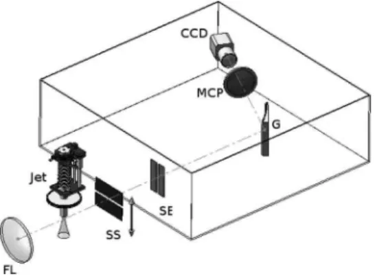
A schematic representation of our experiment is shown in Fig.1. XUV radiation produced in a super-sonic jet is diffracted through a horizontal scanning slit (SS) and then impinges onto the entrance slit (SE) of an XUV spectrometer outlined by the framed box. The vertically diffracted sample of the incoming wavefront passes through the spectrometer slit (SE). The spectrometer is constructed to resolve the
spec-Fig. 1. Schematic of the experimental setup. The scanning slit (SS) is 20 µm wide, and the spectrometer slit (SE) width is 100 µm. The variable groove spacing flat field Hi-tachi imaging diffraction grating (model 001-0266) was used with nominal groove density of 1200 mm−1[11].
3026 OPTICS LETTERS / Vol. 34, No. 19 / October 1, 2009
trum in one direction (horizontal) and to allow essen-tially free-space field propagation in the other. Two-dimensional images are taken for each position of the scanning slit, producing what we call a diffracto-gram. The relative vertical position of the diffraction pattern’s centroid is proportional to the wavefront slope of the sampled wavefront slice. Analyzing the measured diffractogram, we can reconstruct the wavefront using Eq.(1):
␦i=␦z
yi− zi
d . 共1兲
In Eq.(1), yi is the relative position of the centroid of the diffracted pattern, ziis the relative slit position, d is the distance between the scanning slit and the im-aging plane,␦z is the scanning slit iteration step, and
␦i is the optical path difference across the slit. Then the sampled wavefront phase profilenfor the wave-length across the scanning direction is determined by
n=2
兺
i=0n
␦i. 共2兲
The amplitude at each sampling point is determined by the integral intensity of the corresponding diffrac-tion pattern in the diffractogram. The choice of the zero-phase point is arbitrary: it was chosen at the centroid of the intensity pattern. A different choice of the zero-phase point would correspond to a linear phase added to the reconstructed wavefront. This lin-ear phase results in a spatial shift of the entire field pattern in the detection [multichannel plate (MCP)] plane.
The only assumption required for the validity of our approach is that the reconstructed wavefront is spatially coherent within the spectral resolution of the spectrometer. This is a less restrictive require-ment as compared with the prerequisite for the con-ventional (Hartmann–Shack and interferometry-based) wavefront characterization techniques, which require spatial coherence over the full bandwidth of radiation. This makes SWORD applicable to a wide class of light sources.
In our experiment we have used high harmonics produced by focusing a 35 fs, ⬃400 µJ laser beam onto a nitrogen gas jet with a f / # = 80 lens. The laser beam was spatially filtered by propagating it through hollow fiber, and the radial symmetry of the mode was validated. The jet has 250 µm aperture and was operated with a backing pressure of 2.7 atm. Under these conditions we should see primarily short trajec-tory harmonics.
The imaging properties of our diffraction grating were studied in detail by Nakano et al. [12]. Based on their work, we estimated and confirmed experimen-tally resolution of our spectrometer to be 0.25 nm. A 40 mm diameter Burle imaging MCP was used in the imaging plane of the spectrometer, and the backside phosphor screen was imaged onto the CCD camera.
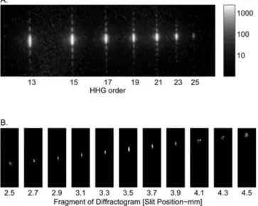
The typical high-harmonic spectrum, diffracted through the scanning slit, is shown on a log-scaled
image in Fig. 2(a). As expected, we can clearly ob-serve the sync-like diffraction structure from rectan-gular aperture in the vertical dimension, while the high harmonics are spectrally resolved in the hori-zontal dimension (harmonics 13 to 25 are shown). To be specific, we will choose harmonic 21 for further analysis. The small fragment of the sparse diffracto-gram (at 100 µm steps) for the 21th harmonic is shown in Fig. 2(b). In the experiment, a diffraction image was taken at each 20 µm step of the scanning slit. For each diffraction picture in the diffractogram, the centroid position and the integral intensity were found, after which the wavefront was reconstructed using Eqs.(1) and(2).
In the case of sources with rotational symmetry, the complete 2D wavefront profile can be recon-structed from a single vertical scan. There are sev-eral important high-harmonic sources that inher-ently have this symmetry, such as semi-infinite gas cells, hollow fibers, small gas cells, etc. In the most general case a complete 2D scan would be required (across the XUV beam ⫹90 deg rotation to measure both projections).
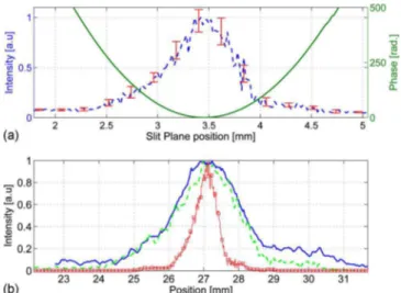
Our jet source does not have rotational symmetry. However, it has reflection symmetry in the vertical plane. This implies that the normal to the wavefront lies in this plane. Therefore, to reconstruct the verti-cal wavefront profile, only one scan across this direc-tion is needed. The phase and amplitude of the recon-structed wavefront profile are shown in Fig.3(a).
In this example, we found the phase to be almost perfectly parabolic, corresponding to the divergent beam with a radius of curvature of 271 mm. For com-parison, the distance between the gas jet and the scanning plane was 245⫾1 mm. In other words, the harmonics appear to originate before the jet,
quanti-Fig. 2. (a) Logarithmic image of a high-harmonic
spec-trum diffracted through scanning slit. (b) The diffracto-gram. Each slice of the diffractogram shows the diffraction pattern for the particular scanning slit position printed be-low each slice. Only a small, sparse fragment of the com-plete diffractogram is shown. At each slice the intensity is normalized to maximum for the given slit position. In the experiment the slit was scanned and the diffraction pattern measured at 20 µm increments.
fying the phase structure placed on the beam by the radial intensity distribution of the fundamental. We repeated the scan measurements with different inte-gration times. The phase reconstruction is repeatable to within 0.3%, even though the intensity noise (due to fluctuations in the laser power and the gas jet) was ⬃15%. Because the phase is measured from the cen-troid of the diffracted pattern it is not directly sensi-tive to amplitude fluctuations. Taking into account all relevant tolerances, we estimate the systematic error of our measurement to be better than 1%.
We have performed a similar reconstruction for all harmonics. The radius of curvature varies from 240 mm to 275 mm from harmonic 13 to harmonic 25. Re-ferring to harmonic 21, the phase and amplitude pro-files of the wavefront are shown in Fig. 3(a). We at-tribute the slight asymmetry in the amplitude of the wavefront profile to the density gradient of the jet in the vertical direction. This gradient result in varying density of molecules across the generating beam that influences both the harmonic generation process and the propagation of the fundamental beam.
To validate our results, we propagated the recon-structed wavefront profile from the scanning slit po-sition to the imaging plane and compared the calcu-lated profile with the high-harmonic profile measured without the scanning slit. We use the Fresnel transform [13] because the Fresnel number
D2/ Z ⬃ 25ⱖ 1, where D ⬃ 700m is the high-harmonic extension size at the scanning slit position, ⯝38 nm, and Z⫽513 mm is the distance between the scanning slit to the MCP. To account for the MCP and optical system resolution of 90 µm, we have con-volved the numerically propagated field at the MCP position with the corresponding point spread function kernel. Both the measured and calculated profile are presented in [Fig. 3(b)]. For comparison, if we use
only the amplitude information and assume a flat phase front, we find the central curve in the figure. It differs substantially from the measured results.
Because we know the size of our slit and relevant distances, we can compare the diffraction pattern with the theoretically expected sync function with shift. Doing so, we achieve at least two goals: first, a more accurate estimate of the linear term for the wavefront. Second, we can analyze the width of the sync function to estimate the quadratic term in the wavefront (phase variations across the slit). Thus we further increase the accuracy and/or reduce the num-ber of the required sampling points.
In summary, we have demonstrated an approach of frequency-resolved wavefront characterization that is particularly suitable for characterization of high-harmonic and XUV radiation. The technique can be easily extended beyond the XUV spectral region. When combined with temporal characterization tech-niques [14,15], all information about the beam can be measured. This paves the way to temporal–spatial coupling studies of high-harmonic and attosecond pulses.
We are grateful for discussions and help of Andrei Naumov, Doug Moffatt, and Adrian Pegoraro. We ac-knowledge financial support of MURI grant W911NF-07-1-0475, Marie Curie IOF, and Japan Sci-ence and Technology Agency.
References
1. P. B. Corkum, Phys. Rev. Lett. 71, 1994 (1993). 2. E. Constant, D. Garzella, P. Breger, E. Mevel, C.
Dorrer, C. Le Blanc, F. Salin, and P. Agostini, Phys. Rev. Lett. 82, 1668 (1999).
3. J. Itatani, J. Levesque, D. Zeidler, H. Niikura, H. Pepin, J. C. Kieffer, P. B. Corkum, and D. M. Villeneuve, Nature 432, 867 (2004).
4. R. A. Bartels, A. Paul, H. Green, H. C. Kapteyn, M. M. Murnane, S. Backus, I. P Christov, Y. W. Liu, D. Attwood, and C. Jacobsen, Science 297, 376 (2002). 5. R. Neutze, R. Wouts, D. van der Spoel, E. Weckert, and
J. Hajdu, Nature 406, 752 (2000).
6. W. L. Chao, B. D. Harteneck, J. A. Liddle, E. H. Anderson, and D. T. Attwood, Nature 435, 1210 (2005). 7. H. M. Quiney, A. G. Peele, Z. Cai, D. Paterson, and K.
A. Nugent, Nat. Phys. 2, 101 (2006).
8. D. G. Lee, J. J. Park, J. H. Sung, and C. H. Nam, Opt. Lett. 28, 480 (2003).
9. C. Valentin, J. Gautier, L. P. Goddet, C. Hauri, T. Marchenko, E. Papalazarou, G. Rey, S. Sebban, O. Scrick, P. Zeitoun, G. Dovillaire, X. Levecq, S. Bucourt, and M. Fajardo, J. Opt. Soc. Am. B 25, B161 (2008). 10. D. J. Kane and R. Trebino, J. Quantum Electron. 29,
571 (1993).
11. T. Kita, T. Harada, N. Nakano, and H. Kuroda, Appl. Opt. 22, 512 (1983).
12. N. Nakano, H. Kuroda, T. Kita, and T. Harada, Appl. Opt. 23, 2386 (1984).
13. J. W. Goodman, Introduction to Fourier Optics (McGraw-Hill, 1996).
14. P. M. Paul, E. S. Toma, P. Breger, G. Mullot, F. Auge, Ph. Balcou, H. G. Muller, and P. Agostini, Science 292, 1689 (2001).
15. J. Itatani, F. Quéré, G. L. Yudin, M. Yu. Ivanov, F. Krausz, and P. B. Corkum, Phys. Rev. Lett. 88, 173903 (2002).
Fig. 3. (Color online) (a) Experimental results: phase and amplitude reconstruction of the 21st harmonic at the scan-ning slit position. Error bars for the reconstructed phase (⬃0.3%) are unobservable on the figure scale. (b) Intensity profile on the MCP plane. The solid curve shows the mea-sured profile. The dashed curve represents reconstructed intensity. The reconstructed intensity profile for a flat phase is shown by the solid curve with markers in the center.