HAL Id: hal-01793926
https://hal.archives-ouvertes.fr/hal-01793926
Submitted on 10 Apr 2019
HAL is a multi-disciplinary open access
archive for the deposit and dissemination of
sci-entific research documents, whether they are
pub-lished or not. The documents may come from
teaching and research institutions in France or
abroad, or from public or private research centers.
L’archive ouverte pluridisciplinaire HAL, est
destinée au dépôt et à la diffusion de documents
scientifiques de niveau recherche, publiés ou non,
émanant des établissements d’enseignement et de
recherche français ou étrangers, des laboratoires
publics ou privés.
Very low concentration of cerium dioxide nanoparticles
induce DNA damage, but no loss of vitality, in human
spermatozoa
L. Préaubert, V. Tassistro, M. Auffan, I. Sari-Minodier, Jérôme Rose, B.
Courbiere, J. Perrin
To cite this version:
L. Préaubert, V. Tassistro, M. Auffan, I. Sari-Minodier, Jérôme Rose, et al.. Very low concentration
of cerium dioxide nanoparticles induce DNA damage, but no loss of vitality, in human spermatozoa.
Toxicology in Vitro, Elsevier, 2018, 50, pp.236-241. �10.1016/j.tiv.2018.03.013�. �hal-01793926�
Contents lists available atScienceDirect
Toxicology in Vitro
journal homepage:www.elsevier.com/locate/toxinvit
Very low concentration of cerium dioxide nanoparticles induce DNA
damage, but no loss of vitality, in human spermatozoa
L. Préaubert
a, V. Tassistro
a, M. Au
ffan
b, I. Sari-Minodier
a, J. Rose
b, B. Courbiere
a,c, J. Perrin
a,c,⁎aAix Marseille Univ, CNRS, IRD, Avignon Univ, IMBE UMR 7263, 13397 Marseille, France bCNRS, Aix Marseille Univ, IRD, CEREGE UM34, UMR 7330, 13545 Aix en Provence, France
cCentre Clinico-Biologique d'Assistance Médicale à la Procréation - CECOS, Pôle Femmes Parents Enfants, AP-HM La Conception, Marseille, Cedex 05, France
A R T I C L E I N F O
Keywords:
Cerium dioxide nanoparticles Human spermatozoa DNA damage Comet assay Environment
A B S T R A C T
Cerium dioxide nanoparticles (CeO2NP) are widely used for industrial purposes, as in diesel, paint, wood stain
and as potential therapeutic applications. The Organization for Economic Cooperation and Development in-cluded CeO2NP in the priority list of nanomaterials requiring urgent evaluation. As metal nanoparticles can cross
the blood-testis barrier, CeO2NP could interact with spermatozoa. The genotoxicity of CeO2NP was
demon-strated in vitro on human cell lines and mouse gametes. However, the effects of CeO2NP on human spermatozoa
DNA remain unknown. We showed significant DNA damage induced in vitro by CeO2NP on human spermatozoa
using Comet assay. The genotoxicity was inversely proportional to the concentration (0.01 to 10 mg·L−1). TEM showed no internalization of CeO2NP into the spermatozoa. This study shows for thefirst time that in vitro
exposure to very low concentrations of cerium dioxide nanoparticles can induce significant DNA damage in human spermatozoa. These results add new and important insights regarding the reproductive toxicity of priority nanomaterials, which require urgent evaluation.
1. Introduction
Due to the extensive industrial production of nanoparticles, human exposure has been increasing exponentially. The reproductive toxicity of nanoparticles is a particularly important issue (Ema et al., 2010;
Greco et al., 2015a), as toxic effects can be transgenerational. Indeed,
Yoisungnern et al. showed that silver nanoparticles could be inter-nalized into mouse sperm in vitro and subsequently alter fertilization and compromise embryo development (Yoisungnern et al., 2015). Moretti et al. incubated human sperm with high-concentration sus-pensions of silver and gold nanoparticles in vitro, observed a significant dose-dependent decrease of motility and viability, and described the internalization of Au-NP in sperm cells (Moretti et al., 2013). Taylor et al. recently emphasized the need for a better understanding of the reproductive toxicity of nanoparticles (Taylor et al., 2015).
Cerium dioxide nanoparticles (CeO2NP) are commonly used for
industrial purposes, including as a diesel additive (Cassee et al., 2011;
Park et al., 2008) or wood stain, and have potential medical applica-tions such as protection against radiation-induced damage (Giri et al., 2013; Sack et al., 2014;Tarnuzzer et al., 2005). CeO2NP are on the
priority list of nanomaterials requiring urgent evaluation as declared by the Organization for Economic Cooperation and Development (OECD's
guidelines, 2011). Indeed, the toxicity/safety assessment of CeO2NP is
still incomplete, with only a few studies available. It has been shown in rats that CeO2NP can cross the blood-testis barrier and accumulate into
the testis after in vivo exposure (Geraets et al., 2012); CeO2NP could
then interact with spermatozoa. The impact of oral administration of citrate-coated 2–5-nm CeO2NP on semen parameters was recently
in-vestigated in aged rats in vivo (Kobyliak et al., 2015), and an im-provement in sperm concentration, motility and morphology was ob-served in treated rats compared to control rats. Similarly, Falchi et al. reported no intracellular uptake and no impairment of the functional and morphological characteristics of ram sperm after in vitro exposure to high concentrations of CeO2NP (Falchi et al., 2016). Conversely, the
genotoxicity of CeO2NP has been shown in human cell lines, mouse
oocytes and spermatozoa (Benameur et al., 2015; Courbiere et al., 2013;Mittal and Pandey, 2014; Preaubert et al., 2015) after in vitro exposure. However, the effects of CeO2NP on human sperm DNA
re-main unknown. Our objective was to study the in vitro genotoxicity of well-characterized CeO2NP on human spermatozoa at low doses.
https://doi.org/10.1016/j.tiv.2018.03.013
Received 22 February 2017; Received in revised form 18 March 2018; Accepted 28 March 2018
⁎Corresponding author at: Laboratoire Biogénotoxicologie, sous sol aile rouge, Faculté de Médecine, Aix-Marseille Université, 27 bd Jean Moulin, 13005 Marseille, France.
E-mail address:jeanne.perrin@imbe.fr(J. Perrin).
Available online 04 April 2018
0887-2333/ © 2018 Published by Elsevier Ltd.
2. Materials and methods
2.1. Physico-chemical characterization of CeO2nanoparticles
As the characterization of nanomaterial is of utmost importance for in vitro testing of toxicity (Love et al., 2012), we performed a thorough physico-chemical characterization of the cerium dioxide nanoparticles. CeO2NP (Rhodia chemicals) were synthesized by aqueous precipitation
of Ce4+(NO
3−)4salt at an acidic pH (Spalla and Cabane, 1993). They
are ellipsoidal crystallites with a mean diameter of ~7 nm and a specific surface area evaluated at 400 m2/g (Spalla and Cabane, 1993; Thill et al., 2006). Their hydrodynamic diameters were measured either in a stock suspension using dynamic light scattering (DLS) (NanoZS, Mal-vern Instruments® Inc., UK) with an optimal measurement range of 1 to 1000 nm or in FertiCult® medium using laser diffraction (Malvern3000, Malvern Instruments® Inc., UK).
The local atomic environment and oxidation states before and after incubation in FertiCult® were assessed by X-ray absorption spectro-scopy at the Ce L3-edge (5723 eV). A concentration of 100 mg·L−1
CeO2NP were incubated for 1 h in abiotic FertiCult® medium and then
ultracentrifuged at 200,000g for 1 h. The solid phase was freeze-dried and analyzed by XAS (CRG-FAME beamline at the ESRF, France). Samples were diluted in BN, pressed into thin pellets, and analyzed at liquid helium temperatures in fluorescence mode with a 30-element solid-state Ge detector. The spectra were compiled from the merge of three scans. XANES (X-ray absorption near edge structure) data were obtained after performing standard procedures for pre-edge subtraction and normalization.
The dissolution of CeO2NP after a 1-h incubation in FertiCult®
medium was assessed by inductively coupled plasma mass spectrometry (ICP-MS) (PerkinElmer, Nexion 300×). The contaminated media (e.g., 0.01 mg·L−1to 10 mg·L−1of CeO2) were ultracentrifuged at 200,000g
for 1 h and the cerium levels were detected in the supernatant.
2.2. Experimental design
According to the Organization for Economic Co-operation and Development guideline (OECD, 2014;Lovell and Omori, 2008;Wiklund and Agurell, 2003), we performed 3 independent in vitro experiments and analyzed 3 replicate slides from each experiment.
2.3. Culture media and reagents
We used FertiCult IVF® culture medium (FertiPro, Beernem, Belgium), which is specifically designed for in vitro human sperm and embryo culture. All other reagents were provided by Sigma-Aldrich® (St-Quentin-Fallavier, France) unless otherwise mentioned.
2.4. Collection and in vitro exposure of human spermatozoa
We used frozen human spermatozoa from 3 healthy fertile donors. The semen samples had been diluted in cryoprotectant medium ac-cording to the manufacturer's instructions (Spermfreeze; JCD, La Mulatiere, France), transferred to high security straws with a capacity of 300μL (Cryo Bio System, L'Aigle, France), and then stored in liquid nitrogen until further use. These spermatozoa were purchased from GERMETHEQUE biobank, which obtained informed consent from each donor for inclusion of samples in the biobank and for their use in re-search experiments regarding human fertility in accordance with the 1975 Helsinki Declaration on human experimentation. The GERMET-HEQUE biobank (BB-0033-00081 Marseille, France) Scientific Committee approved the present study design (number 20130102).
Straws were placed in a 37 °C water bath for 5 min; then, the cryoprotectant was removed by progressively diluting the thawed sample with 1 mL of FertiCult® culture medium at 37 °C. The prepara-tion was aliquoted and centrifuged for 10 min at 1500 rpm (420 g), and
the supernatants were discarded. Then, 150μL of the subsequent sus-pensions were carefully disposed on each pellet. As the genotoxicity of CeO2NP on human spermatozoa had not been studied before, we
as-sessed a wide range of CeO2NP concentrations. The suspensions used
for in vitro exposure of spermatozoa were a) FertiCult® culture medium (negative control); b) FertiCult® containing 0.01, 0.1, 1 or 10 mg·L−1
CeO2NP; c) the supernatant obtained after ultracentrifugation
(60,000 rpm (16,000g) for 1 h) of the suspensions from b); d) a 110μM H2O2 solution in FertiCult® (positive control); e) the same CeO2NP
suspensions as described in b) with 5 mM L-ergothioneine. L
-Ergothioneine (L-erg) is an anti-oxidant commonly used in toxicological studies as a powerful scavenger of free radicals to explore the in-volvement of oxidative stress in the induction of DNA damage (Franzoni et al., 2006). We incubated the preparations for 1 h at 37 °C in 5% CO2, after which we analyzed the supernatants containing
motile-selected spermatozoa. We therefore studied spermatozoa motile-selected by swim-up and included all motile sperm. The in vitro culture conditions we chose corresponded to the IVF-based culture conditions, which are close to those of the female genital tract environment.
For each CeO2NP concentration, 3 different conditions were
stu-died: 1) CeO2NP suspensions in culture medium; 2) the supernatants of
the same suspensions (containing dissolved Ce3+); and 3) CeO 2NP
suspensions withL-erg. Each condition of each concentration was
re-peated 3 times.
2.5. Sperm vitality
We assessed the viability of spermatozoa before performing each triplicate of the Comet assay to exclude sperm DNA damage associated with cytotoxicity. According to the World Health Organization manual, (World Health Organization, 2010), we combined 10μL of a 0.5% eosin
solution (Gilbert®, Hérouville-Saint-Clair, France) containing 0.9% NaCl with 10μL of the exposed sperm sample and observed the pre-paration at ×400 under a contrast microscope. The percentage of live (unstained) and dead (red-stained) spermatozoa was assessed blindly from at least 100 evaluated cells in each condition.
2.6. Human sperm comet assay
The Comet assay is a common genotoxicity test that detects and quantifies DNA primary lesions of eukaryotic cells (Olive and Banáth, 2006). The Comet assay has been validated for the testing of chemicals to determine mutagenicity (Eastmond et al., 2009;Parry, 2000). It is highly sensitive, adaptable and does not require a large number of cells (Baumgartner et al., 2009). We performed a procedure adapted from
Baumgartner et al. (2012), each condition and concentration in tripli-cates.
Each spermatozoa suspension obtained after in vitro exposure was mixed with an equal volume of 2% low melting point (LMP) agarose. The obtained suspension was spread on 3 glass slides that were pre-coated with 1% normal melting point (NMP) agarose. A third layer of 0.5% LMP agarose was added. Slides were immersed in a lysis buffer (100 mM EDTA, 10 mM Tris base, 2.5 M NaCl, 1% Triton X-100, 10% DMSO, 5 mM dithiothreitol, 0.01 mg·mL−1proteinase K; pH 10) for 1 h and then transferred to an electrophoresis tank. Slides were covered by freshly prepared electrophoresis buffer (1 mM EDTA, 300 mM NaOH; pH 13.5) at 4 °C, rested for 20 min for equilibration, and then electro-phoresed at 25 V at 4 °C for 20 min in a 30 cm-long electrophoresis tank. Slides were rinsed with a Tris buffer (0.4 M Tris base; pH 7.4) and de-hydrated with methanol.
2.6.1. Scoring of comet slides
Slides were read blindly after staining with 0.1 mg·mL−1propidium iodide. For each condition, at least 50 spermatozoa were evaluated per slide (i.e., at least 150 spermatozoa over 3 slides per condition). All experiments were repeated three times (i.e., at least 450 spermatozoa in
L. Préaubert et al. Toxicology in Vitro 50 (2018) 236–241
3 independent experiments per condition). Quantitative image analysis was performed using a CCD camera (Andor Technology, Belfast, UK) attached to the microscope (Olympus, Rungis, France) and linked to the comet analysis software (version 6.0; Andor Bioimaging, Nottingham, UK). Sperm DNA damage was expressed as the percentage of tail DNA (% tail DNA), which is total DNA that migrates from the nucleus into the comet tail during electrophoresis (Baumgartner et al., 2012).
2.7. Transmission electron microscopy (TEM)
We performed TEM analysis on spermatozoa exposed to 0.01 mg·L−1CeO2NP for 1 h in order to explore the interaction between
CeO2NP and spermatozoa at the lowest studied concentration. Samples
were washed two times in phosphate buffer, fixed with 2.5% glutar-aldehyde for 30 min, dehydrated in a graded series of ethanol and fi-nally embedded with an Embed-812 kit using a standard procedure. Ultrathin sections of 60 nm were examined with a JEOL/JEM 1400 apparatus, and images were obtained with a MegaView III CCD camera (SIS-Olympus, Munster, Germany).
2.8. Statistics
Each experiment contained at least 150 raw values of % tail DNA by condition and was replicated 3 times: the data for each condition are presented as the 3 means of % Tail DNA median values form the 3 independent experiments. For each condition, we summarized the va-lues obtained for every experiment and performed a one-way ANOVA test using StatView 5.1 software (Abacus Concept, Berkeley, CA, USA)., to compare DNA damage between the various concentration levels and control, between the various concentrations and, for each concentra-tion, between the 3 conditions (Krzywinski et al., 2014). Differences
were considered statistically significant at p < 0.05. 3. Results
3.1. Colloidal and chemical behavior of CeO2NP in FertiCult® medium
Fig. 1a shows the distribution of the hydrodynamic diameters (Dh)
of 100 mg·L−1CeO2NP after a 1-h incubation in FertiCult® medium.
Although CeO2NP are colloidally stable in their stock suspension (with
a Dhdistribution centered at ~7 nm), significant aggregation occurred
in the FertiCult® with a Dhcentered at 3.6μm (volume distribution).
Once expressed as a number, the distribution of most of these ag-gregates had a Dhcentered at 190 nm. Even if this number distribution
is based on assumptions regarding the shape, the density of the ag-gregates, etc., it highlights that most of the CeO2NP interact with the
spermatozoa as small aggregates.
The chemical stability of the nanoparticles was studied in FertiCult® medium in terms of dissolution and local-scale environment. The ICP-MS measurements show that < 2% and 0.02% of the CeO2NP were
dissolved after 1 h from initial CeO2concentrations of 0.01 mg·L−1and
10 mg·L−1, respectively (Fig. 3). This is in agreement with the low so-lubility expected for Ce oxy-hydroxide (as KspCe(OH)3= 6.3 × 10⁻²⁴at
25 °C) (Söhnel and Garside, 1992). This chemical stability was also observed by XANES at the Ce L3-edge (Fig. 1b). The experimental
spectra of CeO2NP before and after the 1-h incubation in FertiCult® are
superimposed and indicate that the atomic structure of CeO2NP is not
affected. XANES is not sensitive to minor Ce species (i.e., < 10%). Consequently, the detection of < 2% Ce dissolution is not contradictory with the structural stability observed by XANES. Such local-scale sta-bility and slow dissolution suggests that the CeO2NP surface interaction
with molecules in the FertiCult® medium is not associated with major surface complexation or reduction into Ce(III).
3.2. Human sperm comet assay
The spermatozoa viability rates were all > 58%, which is the nor-mality threshold for human spermatozoa as stated by the WHO criteria (World Health Organization, 2010).
DNA damage in the spermatozoa was quantified by % tail DNA. The variability of biological data in the 3 independent experiments and in each 3 replicate slide of each independant experiment is presented in
Table 1.
The comet assay showed that compared to the negative control, there was a significant increase of DNA damage in human spermatozoa after in vitro exposure to CeO2NP at all concentrations (Fig. 2), the
supernatants of the CeO2NP suspensions (Fig. 3) and theL-erg
condi-tions (Fig. 2), (p < 0.0001).
We detected a significant increase in DNA damage in spermatozoa exposed to the lowest nanoparticle concentrations, i.e., 0.01 mg·L−1 CeO2NP (mean median = 69.8) versus higher concentrations i.e.
0.1 mg·L−1, 1 and 10 mg·L−1 CeO2NP (55.0, 53.2 and 46.5,
respec-tively) (p < 0.001).
We observed no significant difference among the 3 conditions (CeO2NP, supernatants andL-erg).
(a)
0 5 10 15 20 25 30 35 1 10 100 1000 10000 % Hydrodynamic diameter (nm)Stock suspension (volume) Stock suspension (number) in Ferticult (volume) in Ferticult (number) 0 1 2 3 5700 5720 5740 5760 5780 No rm al iz ed ab sor b anc e (a.u.) Energy (eV) Initial CeO2 NPs CeO2 NPs in Ferticult
Fig. 1. Colloidal behavior of CeO2NP in abiotic culture medium. (a)
Distribution of the hydrodynamic diameters of CeO2NP in their stock
suspen-sion and after 1 h in FertiCult® at 100 mg·L−1of CeO
2. (b) Structural stability of
CeO2NP after 1 h of incubation in FertiCult® medium as assessed by XANES at
3.3. TEM analysis of spermatozoa
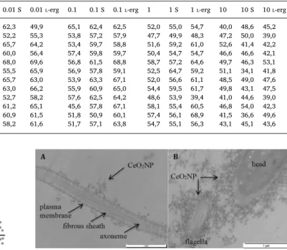
After a 1 h incubation in 0.01 mg·L−1 CeO2NP suspension in
FertiCult® culture medium, TEM showed the accumulation of CeO2NP
on the plasma membranes of exposed human spermatozoa (Fig. 4), particularly along theflagellum. We did not observe any internalization of CeO2NP in spermatozoa.
4. Discussion
We showed that in vitro exposure of human spermatozoa to CeO2NP
significantly increased DNA damage at all concentrations as assessed by
Table 1
Biological variability of the data. Median values of % Tail DNA of each slide (and means of median values of each independent experiment). Concentrations of CeO2NP suspensions (0.01, 0.1, 1, 10) are expressed in mg·L−1. S: exposure to supernatant (obtained after ultracentrifugation of the CeO2NP suspensions);L-erg:
adjunction ofL-ergothioneine (anti-oxidant) in the CeO2NP suspension.
Negative control Positive control 0.01 0.01 S 0.01L-erg 0.1 0.1 S 0.1L-erg 1 1 S 1L-erg 10 10 S 10L-erg Experiment 1 Slide 1 36,9 74,5 73,9 62,3 49,9 65,1 62,4 62,5 52,0 55,0 54,7 40,0 48,6 45,2 Slide 2 37,3 73,3 72,5 52,2 55,3 53,8 57,2 57,9 47,7 49,9 48,3 47,2 50,0 39,0 Slide 3 32,5 75,6 69,4 65,7 64,2 53,4 59,7 58,8 51,6 59,2 61,0 52,6 41,4 42,2 Mean of medians 35,6 74,5 71,9 60,0 56,4 57,4 59,8 59,7 50,4 54,7 54,7 46,6 46,6 42,1 Experiment 2 Slide 1 34,7 74,8 73,1 68,0 69,6 56,8 61,5 68,8 58,7 57,2 64,6 49,7 46,3 53,1 Slide 2 35,7 75,3 78,3 55,5 65,9 56,9 57,8 59,1 52,5 64,7 59,2 51,1 34,1 41,8 Slide 3 32,0 68,3 69,8 65,7 63,0 53,9 63,3 67,1 52,0 56,6 61,1 48,5 49,0 47,6 Mean of medians 34,1 72,8 73,7 63,0 66,2 55,9 60,9 65,0 54,4 59,5 61,7 49,8 43,1 47,5 Experiment 3 Slide 1 32,6 69,2 62,7 52,7 58,2 57,6 62,5 64,2 48,6 53,9 39,4 41,0 44,6 39,0 Slide 2 34,0 65,9 61,5 61,2 65,1 45,6 57,8 67,1 58,1 55,4 60,5 46,8 54,0 42,3 Slide 3 39,8 63,7 66,7 60,9 61,5 51,8 50,9 60,1 57,4 56,1 68,9 41,5 36,6 49,6 Mean of medians 35,4 66,3 63,6 58,2 61,6 51,7 57,1 63,8 54,7 55,1 56,3 43,1 45,1 43,6
Fig. 2. The comet assay shows, compared to the negative control, a significant increase of DNA damage in human sperm after a 1-hour in vitro exposure to CeO2NP at all concentrations as well as in the presence of an anti-oxidant,L-erg (*) (p < 0.0001). The results are presented as the 9 median values of % Tail DNA obtained from 9 different slides from 3 independent experiments for each condition.L-erg:L-ergothioneine (anti-oxidant). a: vs. 0.1 mg·L−1CeO2NP. b: vs.
1 mg·L−1CeO2NP. c: vs. 10 mg·L−1CeO2NP (p < 0.0001). Positive control:
110μM H2O2.
Fig. 3. Chemical behavior of CeO2NP in the culture medium and genotoxicity of the corresponding supernatant. Release of Ce from the nanoparticles after a 1-h
incubation in FertiCult® medium. [CeO2]initial= 0.01, 0.1, 1, or 10 mg·L−1; ambient temperature. The comet assay showed a significant increase in DNA damage in
human sperm after a 1-h in vitro exposure to the supernatants (S, obtained after ultracentrifugation) of the CeO2NP suspensions compared to negative control (***)
(p < 0.0001). The results are presented as the 9 median values of % Tail DNA obtained from 9 different slides from 3 independent experiments for each condition. Fig. 4. The TEM aspect of human spermatozoa exposed to 0.01 mg·L−1CeO2NP
in vitro. One-hour exposure at 0.01 mg·L−1: accumulation of nanoparticles (→) on the plasma membrane. A) Longitudinal section of theflagella. B) Transversal section of theflagella (on the left) and head.
L. Préaubert et al. Toxicology in Vitro 50 (2018) 236–241
the comet assay. We did not observe any internalization of CeO2NP in
spermatozoa. Interestingly, the lowest concentrations of nanoparticles were associated with the highest amount of DNA damage.
A strong point of our study design is the wide range of CeO2NP
concentrations explored, including very low concentrations (0.01 to 10 mg·L−1). Indeed, the majority of recent studies about the genotoxi-city of CeO2NP on human cells analyzed the impact of nanoparticle
concentrations ranging from 0.1 to 100 mg·L−1(Franchi et al., 2015;
Mittal and Pandey, 2014;Strobel et al., 2015;Verstraelen et al., 2014;
Yoisungnern et al., 2015). We addressed lower exposure doses that are probably closer to the expected in vivo concentrations and remain poorly investigated. Nevertheless, to the best of our knowledge, no information is available about the expected human in vivo concentra-tions in the testes, seminal ducts, uterus or Fallopian tubes. Another strong point is that we performed an extensive chemical and physical characterization of the nanoparticles; this type of data are essential for toxicity studies in vitro and in vivo (Love et al., 2012).
The inverse dose-effect relationship observed has been described in previous nanotoxicity studies. Sergent et al. studied the toxicity of 100 nm SiO2 nanoparticles (concentrations ranging from 10 to
150 mg·L−1) on the HT-29 human intestine cell line and showed inverse dose-dependent relationships between the nanoparticle concentration and cell viability and genotoxicity (Sergent et al., 2012). The authors hypothesized that the phenomenon of phagocytosis of damaged cells by undamaged cells could occur to maintain the integrity of the islet profile. Since sperm have no phagocytic abilities, it is unlikely that such a mechanism was involved in our study. Our results could be explained by dose-dependent changes in the physico-chemical behavior. At low doses, the probability of contact between two particles (homo-aggregation) decreases, and it is likely that CeO2NP are more dispersed
at lower concentrations. Indeed, agglomeration increases the contact surface between NP. Consequently, CeO2NP dispersion could lead to small aggregates with a large surface area available to interact with cells, which could enhance the biotransformation, biological and tox-icological effects (Nel et al., 2009;Yoshida et al., 2012). Future work should include genotoxicity assessments at lower concentrations.
This inverse dose response could also be related to different geno-toxicity mechanisms. At the lowest concentration (0.01 mg·L−1), the adjunction of an anti-oxidant (L-erg) in the exposure medium decreased
DNA damage in the sperm. This indirectly highlights that oxidative stress is one mechanism involved in the observed DNA damage. With their high redox potential, CeO2NP can easily become reduced once in
close contact with spermatozoa, thereby oxidizing the nearby organic molecules (Auffan et al., 2009a;Xu and Qu, 2014). Oxidative stress induced by in vitro exposure to CeO2NP has been described in human
lung cells (leading to cytotoxicity (Mittal and Pandey, 2014)) and dermalfibroblasts (leading to DNA and chromosome damages (Auffan
et al., 2009b)). Oxidative stress is known to induce DNA damage in sperm by oxidizing DNA bases (Aitken and Iuliis, 2010). A higher oxi-dative stress at very low concentration could be related to a higher contact surface between sperm and CeO2NP, which could enhance the toxicological effect (Nel et al., 2009;Yoshida et al., 2012). These results are in agreement with previous nanotoxicity studies performed on mouse gametes (Greco et al., 2015b;Preaubert et al., 2015) and cor-roborate a mechanism of DNA damage occurring at low concentrations of nanoparticles (0.01 mg·L−1), thus requiring a close interaction be-tween the cells and the CeO2NP.
At higher concentrations (0.1, 1 and 10 mg·L−1), no difference was observed between the applied nanoparticles and DNA damage induced among the 3 experimental conditions (exposure to CeO2NP suspension,
supernatant or CeO2NP suspension + anti-oxidant), suggesting that the
genotoxicity mechanisms are different and that oxidative stress is not involved. At all studied concentrations, the dissolved cerium con-centration in the supernatant did not increase proportionally to the nanoparticles concentration: for example, whereas the concentration of nanoparticles increased 1000-fold, the dissolved cerium increased only
8-fold (Fig. 3). We assume that at all concentrations, dissolved cerium could be involved in the observed genotoxicity. Indeed, the Ce3+ions present in the nanoparticle stock suspension related to cerium dis-solution in the culture medium could diffuse through the plasma membrane and indirectly stress the spermatozoa. Our results confirm the importance of a careful assessment of the physico-chemical beha-vior of nanoparticles to better understand the mechanisms of geno-toxicity towards germ cells.
Our study is limited by its in vitro nature. However, these results of in vitro exposure are interesting, as spermatozoa can encounter nano-particles in the female genital tract or in IVF plots. Indeed, metal na-noparticles can transfer to various organs in animals after in vivo ex-posure (Blum et al., 2012;Gao et al., 2012;Tassinari et al., 2014), and nanomaterials have been increasingly studied as future medical appli-cations (Barkalina et al., 2014).
In mice, we previously found that in vitro exposure to CeO2NP was
associated with decreased fertilization rates (Preaubert et al., 2015). DNA damage in spermatozoa could be partly responsible for the de-creased fertilization rates. The present results show that very low concentrations of CeO2NP also induce DNA damage in human sperm.
For obvious ethical reasons, the impact of CeO2NP on human in vitro
fertilization could not be analyzed.
5. Conclusion
This study shows for thefirst time that in vitro exposure to very low concentrations of cerium dioxide nanoparticles induces significant DNA damage in human spermatozoa. These results add new and important insights regarding the reproductive toxicity of priority nanomaterials requiring urgent evaluation and warrant further in vivo animal studies examining exposure to low concentrations of CeO2NP.
Acknowledgements
This work was supported by the LABEX SERENADE (No. ANR-11-LABX-0064) and has been completed with the support of the A*MIDEX project « CREER » (No. ANR-11-IDEX-0001-02) funded by the « Investissements d'Avenir », French Government Program of the French National Research Agency (ANR) through the A*MIDEX project (No. ANR-11-IDEX-0001-02).
We are particularly grateful to Bernard Angeletti for his technical help to achieve this project, Joël Courageot for his expert preparation and analysis of TEM samples, and GERMETHEQUE biobank (BB-0033-00081 Marseille, France).
Conflict of interest
The authors report no conflicts of interest. References
Aitken, R.J., Iuliis, G.N.D., 2010. On the possible origins of DNA damage in human spermatozoa. Mol. Hum. Reprod. 16, 3–13.
Auffan, M., Rose, J., Wiesner, M.R., Bottero, J.Y., 2009a. Chemical stability of metallic nanoparticles: a parameter controlling their potential cellular toxicity in vitro. Environ. Pollut. Barking Essex 1987 (157), 1127–1133.
Auffan, M., Rose, J., Orsiere, T., Meo, M.D., Thill, A., Zeyons, O., Proux, O., Masion, A., Chaurand, P., Spalla, O., Botta, A., Wiesner, M.R., Bottero, J.Y., 2009b. CeO2 nano-particles induce DNA damage towards human dermalfibroblasts in vitro. Nanotoxicology 3, 161–171.
Barkalina, N., Charalambous, C., Jones, C., Coward, K., 2014. Nanotechnology in re-productive medicine: emerging applications of nanomaterials. Nanomed. Nanotechnol. Biol. Med. 10, 921–938.
Baumgartner, A., Cemeli, E., Anderson, D., 2009. The comet assay in male reproductive toxicology. Cell Biol. Toxicol. 25, 81–98.
Baumgartner, A., Kurzawa-Zegota, M., Laubenthal, J., Cemeli, E., Anderson, D., 2012. Comet-assay parameters as rapid biomarkers of exposure to dietary/environmental compounds—an in vitro feasibility study on spermatozoa and lymphocytes. Mutat. Res. Toxicol. Environ. Mutagen. 743, 25–35.
Tassistro, V., Bottero, J.-Y., Rose, J., Botta, A., Pietri, S., 2015. DNA damage and oxidative stress induced by CeO2nanoparticles in human dermalfibroblasts: evidence of a clastogenic effect as a mechanism of genotoxicity. Nanotoxicology 9, 696–705.
Blum, J.L., Xiong, J.Q., Hoffman, C., Zelikoff, J.T., 2012. Cadmium associated with in-haled cadmium oxide nanoparticles impacts fetal and neonatal development and growth. Toxicol. Sci. Off. J. Soc. Toxicol. 126, 478–486.
Cassee, F.R., van Balen, E.C., Singh, C., Green, D., Muijser, H., Weinstein, J., Dreher, K., 2011. Exposure, health and ecological effects review of engineered nanoscale cerium and cerium oxide associated with its use as a fuel additive. Crit. Rev. Toxicol. 41, 213–229.
Courbiere, B., Auffan, M., Rollais, R., Tassistro, V., Bonnefoy, A., Botta, A., Rose, J., Orsière, T., Perrin, J., 2013. Ultrastructural interactions and genotoxicity assay of cerium dioxide nanoparticles on mouse oocytes. Int. J. Mol. Sci. 14, 21613–21628.
Eastmond, D.A., Hartwig, A., Anderson, D., Anwar, W.A., Cimino, M.C., Dobrev, I., Douglas, G.R., Nohmi, T., Phillips, D.H., Vickers, C., 2009. Mutagenicity testing for chemical risk assessment: update of the WHO/IPCS harmonized scheme. Mutagenesis 24, 341–349.
Ema, M., Kobayashi, N., Naya, M., Hanai, S., Nakanishi, J., 2010. Reproductive and de-velopmental toxicity studies of manufactured nanomaterials. Reprod. Toxicol. 30, 343–352.
Falchi, L., Bogliolo, L., Galleri, G., Ariu, F., Zedda, M.T., Pinna, A., Malfatti, L., Innocenzi, P., Ledda, S., 2016. Cerium dioxide nanoparticles did not alter the functional and morphologic characteristics of ram sperm during short-term exposure. Theriogenology 85, 1274–1281.e3.
Franchi, L.P., Manshian, B.B., de Souza, T.A.J., Soenen, S.J., Matsubara, E.Y., Rosolen, J.M., Takahashi, C.S., 2015. Cyto- and genotoxic effects of metallic nanoparticles in untransformed humanfibroblast. Toxicol. in Vitro 29, 1319–1331.
Franzoni, F., Colognato, R., Galetta, F., Laurenza, I., Barsotti, M., Di Stefano, R., Bocchetti, R., Regoli, F., Carpi, A., Balbarini, A., Migliore, L., Santoro, G., 2006. An in vitro study on the free radical scavenging capacity of ergothioneine: comparison with re-duced glutathione, uric acid and trolox. Biomed. Pharmacother. 60, 453–457 International Congress, Pisa, October 10–13, 2006.
Gao, G., Ze, Y., Li, B., Zhao, X., Zhang, T., Sheng, L., Hu, R., Gui, S., Sang, X., Sun, Q., Cheng, J., Cheng, Z., Wang, L., Tang, M., Hong, F., 2012. Ovarian dysfunction and gene-expressed characteristics of female mice caused by long-term exposure to tita-nium dioxide nanoparticles. J. Hazard. Mater. 243, 19–27.
Geraets, L., Oomen, A.G., Schroeter, J.D., Coleman, V.A., Cassee, F.R., 2012. Tissue dis-tribution of inhaled micro- and nano-sized cerium oxide particles in rats: results from a 28-day exposure study. Toxicol. Sci. 127, 463–473.
Giri, S., Karakoti, A., Graham, R.P., Maguire, J.L., Reilly, C.M., Seal, S., Rattan, R., Shridhar, V., 2013. Nanoceria: a rare-earth nanoparticle as a novel anti-angiogenic therapeutic agent in ovarian cancer. PLoS One 8, e54578.
Greco, F., Courbière, B., Rose, J., Orsière, T., Sari-Minodier, I., Bottero, J.-Y., Auffan, M., Perrin, J., 2015a. Toxicity of nanoparticles on reproduction. Gynécologie Obstétrique Fertil. 43, 49–55.
Greco, F., Perrin, J., Auffan, M., Tassistro, V., Orsière, T., Courbiere, B., 2015b. A new approach for the oocyte genotoxicity assay: adaptation of comet assay on mouse cumulus-oocyte complexes. Lab. Anim. 49, 251–254.
Kobyliak, N.M., Falalyeyeva, T.M., Kuryk, O.G., Beregova, T.V., Bodnar, P.M., Zholobak, N.M., Shcherbakov, O.B., Bubnov, R.V., Spivak, M.Y., 2015. Antioxidative effects of cerium dioxide nanoparticles ameliorate age-related male infertility: optimistic re-sults in rats and the review of clinical clues for integrative concept of men health and fertility. EPMA J. 6, 12.
Krzywinski, M., Altman, N., Blainey, P., 2014. Points of significance: nested designs. For studies with hierarchical noise sources, use a nested analysis of variance approach. Nat. Methods 11 (10), 977–978.
Love, S.A., Maurer-Jones, M.A., Thompson, J.W., Lin, Y.S., Haynes, C.L., 2012. Assessing nanoparticle toxicity. Annu. Rev. Anal. Chem. Palo Alto Calif. 5, 181–205.
Lovell, D.P., Omori, T., 2008. Statistical issues in the use of the comet assay. Mutagenesis 23 (3), 171–182.
Mittal, S., Pandey, A.K., 2014. Cerium oxide nanoparticles induced toxicity in human lung cells: role of ROS mediated DNA damage and apoptosis. Biomed. Res. Int. 2014, 891934.
Moretti, E., Terzuoli, G., Renieri, T., Iacoponi, F., Castellini, C., Giordano, C., Collodel, G., 2013. In vitro effect of gold and silver nanoparticles on human spermatozoa.
Andrologia 45, 392–396.
Nel, A.E., Mädler, L., Velegol, D., Xia, T., Hoek, E.M.V., Somasundaran, P., Klaessig, F., Castranova, V., Thompson, M., 2009. Understanding biophysicochemical interactions at the nano-bio interface. Nat. Mater. (7), 543–557.
OECD's Guidelines, 2011. OECD's Meeting on Safety Testing of Manufactured Nanomaterials and Test Guidelines. March 2011, available on.http://www.oecd.org. OECD's Guidelines, 2014. Guideline for the Testing of Chemicals by In Vivo Mammalian
Comet Assay. November 2017, available on.http://www.oecd.org.
Olive, P.L., Banáth, J.P., 2006. The comet assay: a method to measure DNA damage in individual cells. Nat. Protoc. 1, 23–29.
Park, B., Donaldson, K., Duffin, R., Tran, L., Kelly, F., Mudway, I., Morin, J.-P., Guest, R., Jenkinson, P., Samaras, Z., Giannouli, M., Kouridis, H., Martin, P., 2008. Hazard and risk assessment of a nanoparticulate cerium oxide-based diesel fuel additive - a case study. Inhal. Toxicol. 20, 547–566.
Parry, J.M., Parry, J., 2000. Guidance on a strategy for testing of chemicals for muta-genicity. In: Committee on Mutagenicity of chemicals in Food, Consumer Products and the Environment, available on internet.http://www.cometassayindia.org/COM %20GUIDELINES.pdf.
Preaubert, L., Courbiere, B., Achard, V., Tassistro, V., Greco, F., Orsiere, T., Bottero, J.Y., Rose, J., Auffan, M., Perrin, J., 2015. Cerium dioxide nanoparticles affect in vitro fertilization in mice. Nanotoxicology 10, 111–117.
Sack, M., Alili, L., Karaman, E., Das, S., Gupta, A., Seal, S., Brenneisen, P., 2014. Combination of conventional chemotherapeutics with redox-active cerium oxide nanoparticles - a novel aspect in cancer therapy. Mol. Cancer Ther. 13, 1740–1749.
Sergent, J.A., Paget, V., Chevillard, S., 2012. Toxicity and genotoxicity of Nano-SiO2 on human epithelial intestinal HT-29 cell line. Ann. Occup. Hyg. 56, 622–630.
Söhnel, O., Garside, J., 1992. Precipitation. In: Butterworth-Heinemann (Ed.), Basic Principles and Industrial Applications. Boston, pp. 149.
Spalla, O., Cabane, B., 1993. Growth of colloidal aggregates through polymer bridging. Colloid Polym. Sci. 271, 357–371.
Strobel, C., Oehring, H., Herrmann, R., Förster, M., Reller, A., Hilger, I., 2015. Fate of cerium dioxide nanoparticles in endothelial cells: exocytosis. J. Nanopart. Res. 17, 1–14.
Tarnuzzer, R.W., Colon, J., Patil, S., Seal, S., 2005. Vacancy engineered ceria nanos-tructures for protection from radiation-induced cellular damage. Nano Lett. 5, 2573–2577.
Tassinari, R., Cubadda, F., Moracci, G., Aureli, F., D'Amato, M., Valeri, M., De Berardis, B., Raggi, A., Mantovani, A., Passeri, D., Rossi, M., Maranghi, F., 2014. Oral, short-term exposure to titanium dioxide nanoparticles in Sprague-Dawley rat: focus on re-productive and endocrine systems and spleen. Nanotoxicology 8, 654–662.
Taylor, U., Tiedemann, D., Rehbock, C., Kues, W.A., Barcikowski, S., Rath, D., 2015. Influence of gold, silver and gold–silver alloy nanoparticles on germ cell function and embryo development. Beilstein J. Nanotechnol. 6, 651–664.
Thill, A., Zeyons, O., Spalla, O., Chauvat, F., Rose, J., Auffan, M., et al., 2006. Cytotoxicity of CeO2nanoparticles for Escherichia coli. Physico-chemical insight of the cytotoxicity mechanism. Environ. Sci. Technol. 40, 6151–6156.
Verstraelen, S., Remy, S., Casals, E., De Boever, P., Witters, H., Gatti, A., Puntes, V., Nelissen, I., 2014. Gene expression profiles reveal distinct immunological responses of cobalt and cerium dioxide nanoparticles in two in vitro lung epithelial cell models. Toxicol. Lett. 228, 157–169.
Wiklund, S.J., Agurell, E., 2003. Aspects of design and statistical analysis in the comet assay. Mutagenesis 18, 167–175.
World Health Organization, 2010. WHO Laboratory Manual for the Examination and Processing of Human Semen, Fifth Edition, Available on the Internet.
Xu, C., Qu, X., 2014. Cerium oxide nanoparticle: a remarkably versatile rare earth na-nomaterial for biological applications. NPG Asia Mater. 6, e90.
Yoisungnern, T., Choi, Y.J., Woong Han, J., Kang, M.-H., Das, J., Gurunathan, S., Kwon, D.-N., Cho, S.-G., Park, C., Kyung Chang, W., Chang, B.-S., Parnpai, R., Kim, J.-H., 2015. Internalization of silver nanoparticles into mouse spermatozoa results in poor fertilization and compromised embryo development. Sci. Rep. 5, 11170.
Yoshida, T., Yoshioka, Y., Matsuyama, K., Nakazato, Y., Tochigi, S., Hirai, T., Kondoh, S., Nagano, K., Abe, Y., Kamada, H., Tsunoda, S.I., Nabeshi, H., Yoshikawa, T., Tsutsumi, Y., 2012. Surface modification of amorphous nanosilica particles suppresses nanosi-lica-induced cytotoxicity, ROS generation, and DNA damage in various mammalian cells. Biochem. Biophys. Res. Commun. 427, 748–752.
L. Préaubert et al. Toxicology in Vitro 50 (2018) 236–241