Scientific paper
Crystal Structure, Hirshfeld Surface Analysis
and Computational Studies of Thiazolidin-4-one derivative:
(Z)-5-(4-Chlorobenzylidene)-3-(2-ethoxyphenyl)
-2-thioxothiazolidin-4-one
Nawel Khelloul,
1Khaled Toubal,
2Nadia Benhalima,
1Rachida Rahmani,
1Abdelkader Chouaih,
1,* Ayada Djafri
2and Fodil Hamzaoui
11Laboratory of Technology and Solid’s Properties, Faculty of Sciences and Technology,
University Abdelhamid Ibn Badis of Mostaganem, 27000 Mostaganem, Algérie
2Laboratoire de Synthèse Organique Appliquée (LSOA), Département de Chimie, Faculté des Sciences,
Université d’Oran 1 - Ahmed Ben Bella, 31000 Oran, Algérie * Corresponding author: E-mail: achouaih@gmail.com
Received: 18-02-2016
Abstract
The title compound (Z)-5-(4-chlorobenzylidene)-3-(2-ethoxyphenyl)-2-thioxothiazolidin-4-one (CBBTZ) was charac-terized by X-ray single crystal diffraction, 1H NMR and 13C NMR spectra. Theoretical investigations were carried out using HF and DFT levels of theory at 6-31G(d,p) basis set. The X-ray structure is compared with that computed. The calculated geometrical parameters are in good agreement with those determined by X-ray diffraction. The dihedral an-gle between the two benzene rings is 16.89(5)° indicating that the structure is non planar. The molecule exhibits intra-and intermolecular contacts of type C–H···O, C–H···S intra-and C–H···Cl. The intercontacts in the crystal structure are explo-red using Hirshfeld surfaces analysis method.
Keywords: Structure, thiazolidin-4-one, theoretical calculations, intermolecular interactions, Hirshfeld surface
1. Introduction
Heterocyclic compounds containing five membered rings with nitrogen, sulfur, and oxygen atoms have been investigated since a long time for their important proper-ties. Several theoretical and experimental investigations were performed to determine the absolute molecular con?guration of organic compounds containing five-membered heterocyclic derivatives.1–3Among these types
of compounds, 4-thiazolidinones have been shown to ha-ve various significant activities. Furthermore, thiazolidi-none derivatives have been developed into useful mate-rials for a variety of applications, such as of biological in-terest, nonlinear optical field and photovoltaic cells.4–10
Organic photovoltaic compounds (OPVs) have attracted significant attention as low-cost alternatives to conventio-nal semiconductor photovoltaic devices.11–13
Conse-quently, (Z)-5-(4-chlorobenzylidene)
-3-(2-ethoxyp-henyl) -2-thioxothiazolidin-4-one (CBBTZ) is a intere-sting member of the above-mentioned molecules contai-ning delocalized π electrons with donor and acceptor groups. Appropriate electron donor and acceptor groups and π-conjugated system allow the CBBTZ to exhibit the asymmetric electronic distribution which leads to an in-creased charge transfer.
Recently, some studies have been carried out on CBBTZ. The optical, electrochemical and X-ray pho-toelectron spectroscopy (XPS) characterization of CBBTZ has been explored and CBBTZ thin films with electronic properties were also studied as an exciton blocking layer in CuPc/C60 heterojunction solar cells.14,15
In this context, and in continuation of our works on thiazolidinones molecules, this study was aimed to report the structural properties and intermolecular interactions of CBBTZ.1,16
2. Experiment and Computational
Methods
2. 1. Experimental
Synthesis, Spectral Data and Spectral Analysis of (Z)-5-(4-Chlorobenzylidene)-3-(2-ethoxyphenyl)-2-thio-xothiazolidin-4-one (CBBTZ)
Synthesis and spectral data (IR, 1H NMR, 13C
NMR) of CBBTZ are reported in previous work.16 The structure of CBBTZ is presented in Scheme 1.
set.26,27The spatial coordinate positions of the title
com-pound, as obtained from X-ray structural investigation, were used as initial coordinates for the theoretical calcu-lations.
3. Results and Discussion
3. 1. Description of the Crystal Structure
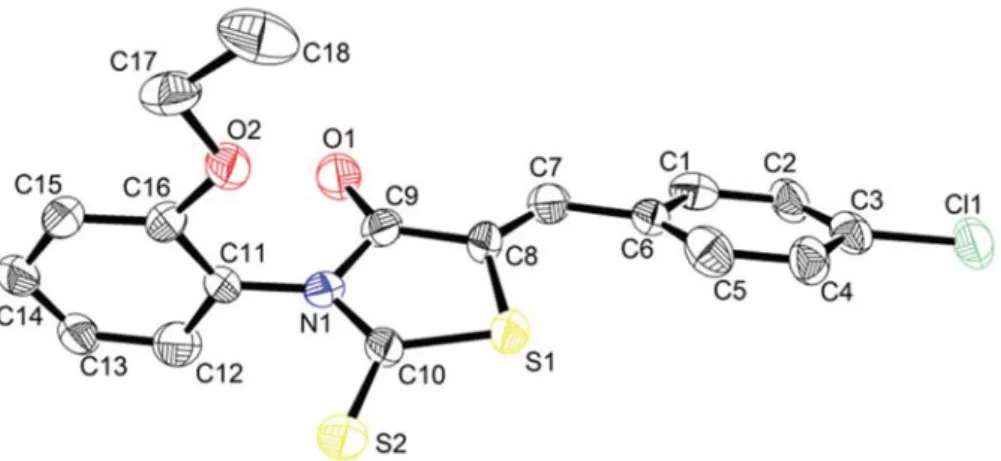
Selected experimental geometrical parameters are summarized in Tables 2, 3 and 4. The molecular structu-re with atomic labelling (thermal ellipsoids astructu-re drawn at 50% probability) is depicted in Fig. 1. The thioxothiazol ring is essentially planar. The full molecule has a Z con-figuration about the C7=C8 double bond (Figure 1). This Z configuration of CBBTZ crystal is stabilized by intra-molecular hydrogen bonds C–H···O and C–H···S. The CC bond lengths in the phenyl rings have average value of 1.38 Å obtained by X-ray diffraction and calculated mean values of 1.40 Å and 1.39 Å obtained with B3LYP and HF, respectively. The double bond of C7=C8 is cha-racterized by the experimental distance of 1.332(8) Å. The thiazole ring contains two C–S bonds, namely S1–C8 [1.729(7) Å] and S1–C10 [1.717(7) Å]. C9–O2 distance shows a typical double bond character with bond length of 1.198(7) Å. The bond lengths are consi-stent with previous phenyl ring-containing studies.1 In
the thiazole moiety formed by C8, C9, C10, N1 and S1
Table 1. Crystallographic details and refinement data
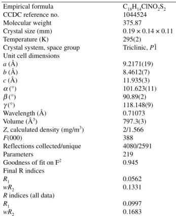
Empirical formula C18H14ClNO2S2 CCDC reference no. 1044524 Molecular weight 375.87
Crystal size (mm) 0.19 × 0.14 × 0.11 Temperature (K) 295(2)
Crystal system, space group Triclinic, P1 -Unit cell dimensions
a (Å) 9.2171(19) b (Å) 8.4612(7) c (Å) 11.935(3) α (°) 101.623(11) β (°) 90.89(2) γ (°) 118.148(9) Wavelength (Å) 0.71073 Volume (Å3) 797.3(3) Z, calculated density (mg/m3) 2/1.566 F(000) 388 Reflections collected/unique 4080/2591 Parameters 219 Goodness of fit on F2 0.945 Final R indices R1 0.0562 wR2 0.1331
R indices (all data)
R1 0.0997
wR2 0.1683
Scheme 1. (Z)-5-(4-Chlorobenzylidene)-3-(2-ethoxyphenyl)-2-thioxothiazolidin-4-one (CBBTZ)
2. 2. X-Ray Structure Determination
X-Ray diffraction study was done on single crystal diffractometer Kappa CCD Nonius. X-Ray data have been measured using graphite monochromated MoKα ra-diation (λ = 0.71073 Å) at ambient temperature. The pro-gram SHELXS-97 was used to solve the structure by di-rect methods.17 Then, full-matrix least-squares
refine-ment using SHELXL-97 revealed the final structure.18 Hydrogen atoms were located in their calculated posi-tions. Figure 1 shows the structure of CBBTZ along with the atomic labeling using ORTEP visualization pro-gram.19For highlighting intra- and intermolecular
inte-ractions, Hirshfeld Surface analyses were performed and fingerprint plots were drawn using Crystal Explorer.20
Crystallographic details and refinement data are summa-rized in Table 1.
2. 3. Theoretical Approach
Throughout this study, Gaussian 03 software21and
Gauss-View program22have been used to perform mole-cular modelling. B3LYP23,24 and HF25 methods were
used to optimize the molecular structure of the title com-pound in the ground state using the 6-31G(d,p) basis
atoms, the average value of bond angles is 108(5)°. In addition, delocalization of the π electrons in CBBTZ is confirmed by C–C–C, C–N–C and C–C–N bond angles which are around 120°. The torsion angle between the two benzene (C1–C6) and (C11–C16) rings is 16.89(5)°. The ethoxyphenyl group is twisted slightly, with a C9–N1–C11–C12 torsion angle of 82.9(12)°. The two moieties chlorobenzene and thioxothiazolidinone are nearly planar according to the dihedral angle C5–C6–C7–C8 of 16.4(18)° (X-ray diffraction) and from 172.5° to 179.5° (theoretical calculation). As men-tioned in our previous work,16the chirality of this kind
of compounds is highlighted by the value of the dihedral angle formed by the heterocyclic ring and the aryl bound at the nitrogen atom. In the present study, this angle is 95.8°. As we can easily see from the above results, there is a good correlation between the experimental and theo-retical structural results. The observed differences are due to the fact that experimental results belong to the so-lid phase, while theoretical calculations belong to the
gas phase of isolated molecules. The molecular packing in the crystal structure of CBBTZ is stabilized by inter-molecular interactions forming a three-dimensional net-work (Figure 2).
3. 2. DFT and HF Optimized Geometry
Figure 3 depicts the calculated molecular structure of CBBTZ using the B3LYP/6-31G(d,p) level of theory. Theoretical geometric parameters by HF and DFT levels of theory using 6-31G(d,p) basis set are given in Tables 2, 3 and 4 together with the experimental ones. The theoretical structural results of CBBTZ have a little different values compared with corresponding experimental results. Thus, bond length values of the thiazole ring N1–C9 and N1–C10 are 1.411 and 1.377 Å computed with B3LYP, with respect to the X-ray results 1.379(8) and 1.330(7) Å, respectively. In the same context, calculated distances S1–C8 (1.765 Å) and S1–C10 (1.763 Å) are comparable to the experimental values (1.729(7) and 1.716(7) Å). The C–C distances in the two aromatic cycles vary from 1.380(8) to 1.401(8) Å compared to the theoretical values which vary from 1.389 to 1.415 Å. According to the above results, deviations of 0.01 Å for bond lengths and 3° for bond and torsion angles are found between experimental
Figure 1. Structure of CBBTZ with atomic labeling scheme (ellipsoids are drawn at 50% probability). For clarity, the hydrogen atoms are omitted.
Figure 2. A perspective view of the crystal packing in the unit cell. View along the c axis.
Figure 3. Theoretical crystal structure of CBBTZ with B3LYP/6-31G(d,p) level.
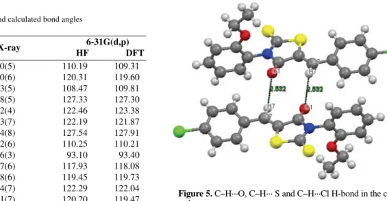
and theoretical geometries. This can be explained by con-sidering that the theoretical calculations were carried out in a gaseous phase, whereas the X-ray diffraction study was performed on the compound in the solid form. Figure 4 compares the calculated structure to that obtained by X-ray diffraction.
Table 2. Experimental and calculated bond lengths
Bond distances X-ray 6-31G(d,p) (Å) HF DFT S1C8 1.729(7) 1.775 1.765 S1C10 1.716(7) 1.745 1.763 S2C10 1.600(7) 1.634 1.642 O1C9 1.198(7) 1.209 1.213 O2C16 1.356(7) 1.340 1.357 O2C17 1.404(12) 1.411 1.431 N1C9 1.379(8) 1.395 1.411 N1C10 1.330(7) 1.354 1.377 N1C11 1.403(7) 1.432 1.436 C7C8 1.332(8) 1.336 1.352 C8C9 1.467(8) 1.486 1.482
Table 3. Experimental and calculated bond angles
Bond angles X-ray 6-31G(d,p) (Å) HF DFT S1C10N1 123.0(5) 110.19 109.31 S1C8C7 124.0(6) 120.31 119.60 S1C8C9 111.3(5) 108.47 109.81 S2C10N1 120.8(5) 127.33 127.30 S2C10S1 116.2(4) 122.46 123.38 O1C9N1 113.3(7) 122.19 121.87 O1C9C8 132.4(8) 127.54 127.91 N1C9C8 114.2(6) 110.25 110.21 C8S1C10 85.6(3) 93.10 93.40 C9N1C10 105.7(6) 117.93 118.08 C9N1C11 123.8(6) 119.45 119.73 C10N1C11 130.4(7) 122.29 122.04 C7C8C9 124.1(7) 120.20 119.47
Table 4. Experimental and calculated dihedral angles
Torsion angles X-ray 6-31G(d,p) (Å) HF DFT S1C8C9O1 179.8(10) 178.9 179.5 S1C8C9N1 –3.1(10) –1.8 –0.9 S1C8C7C6 –9.2(15) –1.9 –0.2 S1C10N1C9 3.5(10) –2.2 –1.6 S1C10N1C11 –172.4(7) –175.6 –177.1 S2C10S1C8 177.1(6) –179.6 –179.5 S2C10N1C9 –178.3(7) 178.2 178.8 S2C10N1C11 5.8(13) 4.8 3.2 O1C9N1C10 177.6(9) –178.1 –178.8 O1C9N1C11 –6.1(13) –4.4 –3.2 O1C9C8C7 –8.8(18) –2.4 –0.7 N1C9C8C7 168.3(9) 176.7 178.7 N1C10S1C8 –4.6(8) 3.1 1.8 C6C7C8C9 –179.5(9) 1.5 0.2 C10S1C8C7 –167.5(9) –178.1 –179.6 C10S1C8C9 3.8(7) 0.6 0.1 C10N1C9C8 –0.1(11) 2.6 1.7 C11N1C9C8 176.2(7) 176.3 177.3 C9N1C11C12 –82.9(12) –84.7 –86.9 C10N1C11C12 92.4(12) 88.6 88.5 C9N1C11C16 96.5(11) 94.4 95.4 C10N1C11C16 –88.2(12) –90.2 –89.6
Figure 4. Atom-by-atom superimposition of the structures calcu-lated (solid line) over the X-ray structure (dashed line) for CBBTZ.
Figure 5. C–H···O, C–H··· S and C–H···Cl H-bond in the
cry-stal.
3. 3. Intermolecular H-Bonds
C–H···O, C–H···S and C–H···Cl intra- and intermo-lecular interactions are present in the crystal structure. These interactions are responsible for the stability of the crystal structure. C atoms, namely C2, C4, C5, C7, C14 and C18 act as donors and O atoms, namely C2, C4, C5, C7, C14 and C18 act as acceptors. H-Bond interactions are presented in Table 5. Figure 5 shows C7H7···O1 H-bond in the crystal.
contacts (24.4% of the HS) results in a symmetrical pair of wings, see Fig. 8(c). The prominent spikes at de= di= 1.8 Å are due to S···H contacts. These contributions are highlighted in the fingerprint plot, Fig. 8(d). For the title compound, H···O contacts, which are attributed to CH···O H-bond interactions, occur as two sharp symmetric spikes in the two dimensional fingerprint map. The presence of
3. 4. Hirshfeld Surface Analysis
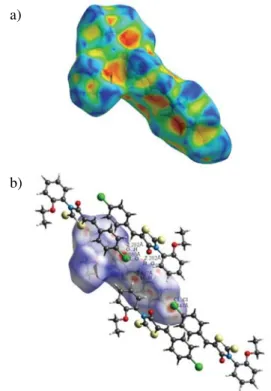
Hirshfeld surface (HS) analysis represents a unique approach towards an understanding of different interac-tions in the crystal structure and is a necessary tool in cry-stal engineering. In addition to the HS analysis, the fin-gerprint plots also provide some useful quantitative infor-mation about the individual contribution of each intermo-lecular interaction in the crystal packing. The intra- and in-termolecular interactions of CBBTZ crystal are quantified using HS analysis. The three-dimensional HS generated for structure of CBBTZ crystal is presented in Fig. 6. The red contacts highlight the intermolecular interactions with distances closer than the sum of the van der Waals radii, while white indicates contacts near the van der Waals sepa-ration, and blue depicts longer contacts.28Figure 7 shows
Hirshfeld surfaces mapped for CBBTZ compound with the shape index property (a) and with dnormselected intermole-cular contacts (b). The full fingerprint plot for the CBBTZ crystal and the contribution of each type of interaction to the total HS are presented in Figure 8 displaying surfaces that were mapped over dnorm(0.242 to 1.414).
As seen in Figure 7, the deep red colour indicates hydrogen-bonding contacts. For example, a deep red spot indicated the presence of a CH···O H-bond (between H7 and O1). The other colour spots are observed due to the presence of other close contacts, such as H···H, C···H, S···H, O···H and Cl···H. The fingerprint plots of CBBTZ are dominated by H···H and C···H contacts. The remaining area of the fingerprint plot is occupied by C···C (3.2%), Cl···C (2.9%), Cl···O (2%), S···S (1.5%), C···S (0.9%), Cl···Cl (0.8%), C···O (0.6%), Cl···N (0.2%), Cl···S (0.1%) and O···O (0.1%) contact regions. The molecular stacking, in spite of having a considerable energetic stabilization, contributes much less [C···C (3.2%)] towards the crystal packing. The H···H contacts, which are prominent in the molecular packing, appear as the scattered points along with double broad peaks in the middle of the region of the fingerprint plot. The positions of the peaks marked with (2) in Fig. 8(a), are at de= di= 1.2 and 1.0 Å, and the per-cent contribution is 28.4%. The contribution from C···H
Table 5. C–H···O, C–H···S and C–H···Cl H-bonds in CBBTZ cry-stal.
DH···A DH H···A D···A DH···A
C5H5···S1 0.93 2.48 3.082 (8) 122.5 C2H2···S1i 0.93 2.96 3.647 (11) 131.9 C2H2···Cl1ii 0.93 2.83 3.313 (11) 113.1 C4H4···O2iii 0.93 2.77 3.575 (13) 145.3 C7H7···O1iv 0.93 2.53 3.110(13) 134.5 C14H14···S1v 0.93 2.98 3.354 (14) 105.4 C18H18B···S1vi 0.96 2.92 3.747 (13) 144.7 Symmetry codes:
(i) –x+1, –y, –z;(ii) –x, –y–1, –z; (iii) x–1,+y,+z; (iv) –x+2, –y, –z; (v) x+1, +y, +z; (vi) –x+2, –y, –z+1.
Figure 6. View of the HS for CBBTZ molecule.
Figure 7. HS mapped for CBBTZ compound with (a) the shape in-dex property (b) dnormselected intermolecular contacts.
a)
these long spikes (indicated by (1) in Fig. 8) is characteri-stic of strong hydrogen bonds. The intermolecular O···H and H···O contacts, Fig. 8(a and e) and Fig. 9, provide contribution of 8.3% to the HS of the CBBTZ crystal. Fi-gure 8(f) shows the contribution of Cl···H intermolecular contacts to the HS.
The quantitative results of the HS analysis for the CBBTZ crystal are presented in Fig. 9 which gives a
de-tailed quantitative analysis of all intra- and intermolecular contacts contributing to the HS.
4. Conclusion
A novel thiazolidinone derivate, (Z)-5-(4-chloro-
benzylidene)-3-(2-ethoxyphenyl)-2-thioxothiazolidin-4-Figure 8. The 2D fingerprint plots showing the percentage contribution of the individual types of interaction to the total HS area.
Figure 9. Quantitative results of different intra- and intermolecular interactions contributing to the HS.
a)
d) e) f)
one (CBBTZ) has been investigated for the first time. Its structural properties have been examined by theoretical cal-culations using HF and DFT methods and X-ray diffraction technique. CBBTZ crystallizes in the triclinic system with the space group P-1. Obtained results indicate that the theo-retical calculations can reproduce the experimental results. In the crystal packing, the molecules are connected by in-tra- and intermolecular H-bonds of the type C–H···O, C–H···S and C–H···Cl. In general, a good agreement was observed between the calculated geometrical parameters (with B3LYP) and that of reported similar derivatives. All the calculated data and experimental results of the studied molecule are useful in the application in fundamental re-search in chemistry and photovoltaic cells in the future. Fi-nally, HS analysis and fingerprint plots are a unique way for understanding the contribution of individual types of inte-ractions within the crystal structure. More theoretical calcu-lations can be performed on this compound to assess other properties especially in the photovoltaic field.
5. Supplementary Material
Crystallographic data for the structure reported in this article have been deposited with Cambridge Crystal-lographic Data Center, CCDC 1044524. Copies of this in-formation may be obtained free of charge from the Direc-tor, CCDC, 12 Union Road, Cambridge, CBZ IEZ, UK. Facsimile (44) 01223 336 033, E-mail: deposit@ccdc. cam.ac.uk or http//www.ccdc.com.ac.uk/deposit.
6. References
1. N. Benhalima, K. Toubal, A. Chouaih, G. Chita, S. Maggi, A. Djafri, F. Hamzaoui, J. Chem. Crystallogr. 2011, 41, 1729–1736. http://dx.doi.org/10.1007/s10870-011-0165-9 2. N. Özdemir, M. Dinçer, A. Çukurovalý, J. Mol. Model. 2010,
16, 291–302. http://dx.doi.org/10.1007/s00894-009-0552-8
3. R. Anbazhagan, K. R. Sankaran, J. Mol. Struct. 2013, 1050, 73–80. http://dx.doi.org/10.1016/j.molstruc.2013.07.019 4. N.K. Fuloria, V. Singh, M.S. Yar, M. Ali, Acta Pol. Pharm.
Drug Res. 2009, 66, 141–147.
5. C. Nandagokula, B. Poojary, S. Vittal, S. Shenoy, P. Shetty, A. Tangavelu, Med Chem Res. 2013, 22(1), 253–266. http://dx.doi.org/10.1007/s00044-012-0028-8
6. R. V. Patel, S. W. Park, Res. Chem. Intermed. 2015, 41, 5599–5609. http://dx.doi.org/10.1007/s11164-014-1684-8 7. P. Chawla, R. Singh, S. K. Saraf, Med Chem Res. 2012, 21,
2064–2071. http://dx.doi.org/10.1007/s00044-011-9730-1 8. V. Smokal, B. Derkowska, R. Czaplicki, O. Krupka, A.
Ko-lendo, B. Sahraoui, Opt. Mater. 2009, 31, 554–557. http://dx.doi.org/10.1016/j.optmat.2007.10.019
9. M. M. Oliva, J. Casado, M. M. M. Raposo, A. M. C. Fonse-ca, H. Hartmann, V. Hernández, et al., J. Org. Chem. 2006,
71, 7509–7520.
http://dx.doi.org/10.1021/jo060318v
10. L. Duan, J. Qiao, Y.D. Sun, Y. Qiu, Adv. Mater. 2011, 23(9), 1137–1144.
http://dx.doi.org/10.1002/adma.201003816
11. P. V. Kamat, M. Haria, S. Hotchandani, J. Phys. Chem. B,
2004, 108, 5166–5170. http://dx.doi.org/10.1021/jp0496699
12. C. Brabec, S. Gowrisanker, J. M. M. Halls, D. Laird, S. J. Jia, S. P. Williams, Adv. Mater. 2010, 22, 3839–3856.
http://dx.doi.org/10.1002/adma.200903697
13. A. Lakhdar Toumi, A. Khelil, J. C. Bernède, Y. Mouchaal, A. Djafri, K. Toubal, N. Hellal, L. Cattin, Surface Review and Letters 2015, 22(2), 1550025–1550033.
http://dx.doi.org/10.1142/S0218625X15500250
14. A.S. Yapi, L. Toumi, Y. Lare, G.M. Soto, L. Cattin, K. Tou-bal, A. Djafri, M. Morsli, A. Khelil, M.A. Del Valle, et al.,
Eur. Phys. J. Appl. Phys. 2010, 50, 30403–30411.
http://dx.doi.org/10.1051/epjap/2010062
15. Y. Mouchaal, A. Lakhdar Toumi, A. S. Yapi , Y. Lare , G. M. Soto, L. Cattin , K. Toubal, A. Reguig, A. Khelil, A. Djafri, M. Morsli, M. A. Del Valle, J. C. Bernède, EPJ Web of
Con-ferences 2012, 29, 00030–00036.
http://dx.doi.org/10.1051/epjconf/20122900030
16. K. Toubal, A. Djafri, A. Chouaih, A. Talbi, Molecules 2012, 17, 3501–3509.
http://dx.doi.org/10.3390/molecules17033501
17. G. M. Sheldrick, SHELXS97 Program for crystal structure determination, University of Göttingen, Göttingen, Ger-many, 1997.
18. G. M. Sheldrick, SHELXL97 Program for crystal structure refinement, University of Göttingen, Göttingen, Germany, 1997.
19. L. J. Farrugia, J. Appl. Crystallogr. 1997, 30, 565–565. http://dx.doi.org/10.1107/S0021889897003117
20. S. K. Wolff, D. J. Grimwood, J. J. Mckinnon, D. Jayatilaka, M. A. Spackmann, “Crystal Explorer 3.0”, University of We-stern Australia, Perth, WeWe-stern Australia, 2007.
21. M. J. Frisch, G. W. Trucks, H. B. Schlegel, G. E. Scuseria, M. A. Robb, J. R. Cheeseman, J. A. Montgomery, Jr., T. Vre-ven, K. N. Kudin, J. C. Burant, J. M. Millam, S. S. Iyengar, J. Tomasi, V. Barone, B. Mennucci, M. Cossi, G. Scalmani, N. Rega, G. A. Petersson, H. Nakatsuji, M. Hada, M. Ehara, K. Toyota, R. Fukuda, J. Hasegawa, M. Ishida, T. Nakajima, Y. Honda, O. Kitao, H. Nakai, M. Klene, X. Li, J. E. Knox, H. P. Hratchian, J. B. Cross, V. Bakken, C. Adamo, J. Jaramillo, R. Gomperts, R. E. Stratmann, O. Yazyev, A. J. Austin, R. Cammi, C. Pomelli, J. W. Ochterski, P. Y. Ayala, K. Moroku-ma, G. A. Voth, P. Salvador, J. J. Dannenberg, V. G. Zakr-zewski, S. Dapprich, A. D. Daniels, M. C. Strain, O. Farkas, D. K. Malick, A. D. Rabuck, K. Raghavachari, J. B. Fore-sman, J. V. Ortiz, Q. Cui, A. G. Baboul, S. Clifford, J. Cio-slowski, B. B. Stefanov, G. Liu, A. Liashenko, P. Piskorz, I. Komaromi, R. L. Martin, D. J. Fox, T. Keith, M. A. Al-La-ham, C. Y. Peng, A. Nanayakkara, M. Challacombe, P. M. W. Gill, B. Johnson, W. Chen, M. W. Wong, C. Gonzalez, and J. A. Pople, Gaussian 03, Revision C.02, Gaussian, Inc., Wal-lingford CT, USA, 2004.
22. A. E. Frisch, A. B. Nielsen, A. J. Holder, Gaussview, Gaus-sian Inc., Pittsburg, USA, 2003.
23. A. D. Becke, J. Chem. Phys., 1997, 107, 8554–8560. http://dx.doi.org/10.1063/1.475007
24. G. Rauhut, P. Pulay, J. Phys. Chem. 1995, 99, 3093–3100. http://dx.doi.org/10.1021/j100010a019
25. H. D. Cohen, C. C. Roothaan, J. Chem. Phys. 1965, 43, S34.
http://dx.doi.org/10.1063/1.1701512
26. R. Fletcher, M. J. D. Powell, Comput. J. 1963, 6, 163–168. 27. R. F. Bader, Atoms in Molecules. A Quantum Theory,
Cla-rendon Press, Oxford, GB, 1990.
28. J. J. Mckinnon, D. Jayatilaka, M. A. Spackman, Chem.
Com-mun. 2007, 37, 3814–3816.
http://dx.doi.org/10.1039/B704980C
Povzetek
Naslovno spojino (Z)-5-(4-klorobenziliden)-3-(2-etoksifenil)-2-tioksotiazolidin-4-on (CBBTZ) smo ka-rakterizirali z rentgensko difrakcijo na monokristalu ter 1H in 13C NMR spektri. Teoreti~ne izra~une smo
izvedli s pomo~jo teorije na nivoju HF in DFT z uporabo 6-31G(d,p) baznega seta. Rentgensko strukturo smo primerjali z izra~unano in ugotovili, da se izra~unani geomterijski parametri dobro ujemajo s tistimi, ki smo jih dobili z rentgensko difrakcijo. Dihedralni kot med dvema benzenovima obro~ema je 16.89(5)°, kar nakazuje, da struktura ni planarna. Molekula izkazuje tudi intra- in intermolekularne kontakte, npr. C–H···O, C–H···S in C–H···Cl. Interakcije v kristalni strukturi smo raziskali s pomo~jo metode Hirschfel-dovih povr{in.