HAL Id: cea-01936753
https://hal-cea.archives-ouvertes.fr/cea-01936753
Submitted on 13 Dec 2018HAL is a multi-disciplinary open access archive for the deposit and dissemination of sci-entific research documents, whether they are pub-lished or not. The documents may come from teaching and research institutions in France or abroad, or from public or private research centers.
L’archive ouverte pluridisciplinaire HAL, est destinée au dépôt et à la diffusion de documents scientifiques de niveau recherche, publiés ou non, émanant des établissements d’enseignement et de recherche français ou étrangers, des laboratoires publics ou privés.
Rhodobacter sphaeroides methionine sulfoxide reductase P
reduces R ‑ and S ‑diastereomers of methionine sulfoxide
from a broad‑spectrum of protein substrates
Lionel Tarrago, Sandrine Grosse, Marina Siponen, David Lemaire, Béatrice
Alonso, Guylaine Miotello, J. Armengaud, Pascal Arnoux, David Pignol,
Monique Sabaty
To cite this version:
Lionel Tarrago, Sandrine Grosse, Marina Siponen, David Lemaire, Béatrice Alonso, et al..
Rhodobacter sphaeroides methionine sulfoxide reductase P reduces R ‑ and S ‑diastereomers of
me-thionine sulfoxide from a broad‑spectrum of protein substrates. Biochemical Journal, Portland Press, 2018, 475 (23), pp.3779-3795. �10.1042/BCJ20180706�. �cea-01936753�
1 Rhodobacter sphaeroides methionine sulfoxide reductase P reduces R- and S-diastereomers of 1
methionine sulfoxide from a broad-spectrum of protein substrates 2
Lionel Tarrago1,*, Sandrine Grosse1, Marina I. Siponen1, David Lemaire2, Béatrice Alonso1, Guylaine
3
Miotello3, Jean Armengaud3, Pascal Arnoux1, David Pignol1, Monique Sabaty1,*
4
1Aix Marseille Univ, CEA, CNRS, BIAM, Laboratoire de bioénergétique cellulaire, Saint
5
Paul-Lez-Durance, France F-13108; 2Aix Marseille Univ, CEA, CNRS, BIAM, Laboratoire des Interactions
6
Protéine-Métal, Saint Paul-Lez-Durance, France F-13108; 3Laboratoire Innovations technologiques pour la
7
Détection et le Diagnostic (Li2D), Service de Pharmacologie et Immunoanalyse (SPI), CEA, INRA, 8
F-30207 Bagnols sur Cèze, France. 9
10
*Correspondence: Lionel Tarrago (lioneltarrago@msn.com) or Monique Sabaty (monique.sabaty@cea.fr) 11
12 13
Keywords: Enzyme kinetics, methionine sulfoxide reductase, microbiology, molybdoenzyme, oxidative 14
stress, periplasm, protein oxidation, proteomics 15
2 Abstract
16
Methionine (Met) is prone to oxidation and can be converted to Met sulfoxide (MetO), which exists as 17
R- and S-diastereomers. MetO can be reduced back to Met by the ubiquitous methionine sulfoxide reductase
18
(Msr) enzymes. Canonical MsrA and MsrB were shown to be absolutely stereospecific for the reduction of 19
S- and R-diastereomer, respectively. Recently, a new enzymatic system, MsrQ/MsrP which is conserved in
20
all gram-negative bacteria, was identified as a key actor in the reduction of oxidized periplasmic proteins. 21
The haem-binding membrane protein MsrQ transmits reducing power from the electron transport chains to 22
the molybdoenzyme MsrP, which acts as a protein-MetO reductase. The MsrQ/MsrP function was well 23
established genetically, but the identity and biochemical properties of MsrP substrates remain unknown. In 24
this work, using the purified MsrP enzyme from the photosynthetic bacteria Rhodobacter sphaeroides as a 25
model, we show that it can reduce a broad spectrum of protein substrates. The most efficiently reduced 26
MetO are found in clusters of amino acid sequences devoid of threonine and proline on the C-terminal side. 27
Moreover, R. sphaeroides MsrP lacks stereospecificity as it can reduce both R- and S- diastereomers of 28
MetO, similarly to its Escherichia coli homolog, and preferentially acts on unfolded oxidized proteins. 29
Overall, these results provide important insights into the function of a bacterial envelop protecting system, 30
which should help understand how bacteria cope in harmful environments. 31
32
Abbreviations: BV, benzyl viologen; DMSO, dimethylsulfoxide; DTT, dithiothreitol; ESI-MS, 33
Electrospray Ionization-Mass spectrometry; H2O2, hydrogen peroxide;
34
HEPES4-(2-hydroxyethyl)-1-piperazineethanesulfonic acid; MES, 2-(N-morpholino)ethanesulfonic acid; 35
MetO, methionine sulfoxide; Met-R-O, R-diastereomer of MetO; Met-S-O, S-diastereomer of MetO; MS, 36
mass spectrometry, Msr, MetO reductase, NADPH, Nicotinamide adenine dinucleotide phosphate; NaOCl, 37
sodium hypochlorite. 38
3 Introduction
39
Aerobic life exposes organisms to reactive oxygen species (ROS) derived from molecular oxygen, such 40
as hydrogen peroxide (H2O2) or singlet oxygen (1O2). Bioenergetic chains are important sources of these
41
intracellular ROS. H2O2, is principally produced during respiration [1] and 1O2 arises from photosynthesis
42
[2]. In most organisms, these oxidative molecules act as signaling messengers playing major roles in 43
numerous physiological and pathological states. Furthermore, their production and elimination are tightly 44
regulated [3]. However, numerous stresses can affect ROS homeostasis and increase their intracellular 45
concentration to excessive values leading to uncontrolled reactions with sensitive macromolecules [4]. For 46
instance, photosynthetic organisms, such as plants or the purple bacteria Rhodobacter sphaeroides can 47
experience photo-oxidative stress in which unbalance between incident photons and photosynthetic electron 48
transfer generate detrimental accumulation of 1O
2 [5]. Moreover, production of ROS could be used
49
advantageously in a defensive strategy against potential pathogenic invaders. For instance, neutrophils 50
produce the strong oxidant hypochlorite (ClO-) from H
2O2 and chlorine ions to eliminate bacteria and fungi
51
[3]. Because of their abundance in cells, proteins are the main targets of oxidation [6]. Methionine (Met) is 52
particularly prone to oxidation and the reaction of Met with an oxidant leads to the formation of Met 53
sulfoxide (MetO), which exists as two diastereomers R (Met-R-O) and S (Met-S-O). Further oxidation can 54
then lead to the formation of Met sulfone (MetO2) [7,8]. As opposed to most oxidative modifications on
55
amino acids, the formation of MetO is reversible, and oxidized proteins can be repaired thanks to methionine 56
sulfoxide reductases (Msr) enzymes that principally exist in two types, MsrA and MsrB. These enzymes, 57
present in almost all organisms, did not evolve from a common ancestral gene and possess an absolute 58
stereospecificity towards their substrates. Indeed, MsrA can reduce only Met-S-O [7,9–12] whereas MsrB 59
acts only on Met-R-O [10–14]. This strict stereospecificity was enzymatically demonstrated using 60
chemically prepared Met-R-O and Met-S-O from racemic mixtures of free MetO or by using HPLC methods 61
allowing discrimination of both diastereomers. A structural explanation was also provided by deciphering 62
the mirror images of their active sites, in which only one MetO diastereomer can be accommodated [10]. 63
4 While MsrA can reduce Met-S-O, whether as a free amino acid or included in proteins, MsrB is specialized 64
in the reduction of protein-bound Met-R-O, and both are more efficient on unfolded oxidized proteins 65
[15,16]. Eukaryotic Msrs are important actors in oxidative stress protection, aging and neurodegenerative 66
diseases in animals [17], during environmental stresses and seed longevity in plants [18,19]. In bacteria, 67
MsrA and MsrB are generally located in the cytoplasm [3], except in Neisseria or Streptococcus species, 68
for which MsrA and MsrB enzymes can be addressed to the envelope [20,21]. They play a role in protecting 69
against oxidative stress and as virulence factors [3]. 70
Beside these stereotypical Msrs found in all kinds of organisms, several other enzymes can catalyze 71
MetO reduction, principally in bacteria. For instance, numerous bacteria as well as unicellular eukaryotes, 72
such as Saccharomyces cerevisiae, possess another type of absolutely stereospecific Msr, called free-R-Msr 73
(fRMsr) or MsrC, which is specialized in the reduction of the free from of Met-R-O [22,23]. As MsrA and 74
MsrB, the fRMsr uses thiol-based chemistry and the reducing power coming from NADPH to reduce its 75
substrate [22,23]. 76
In bacteria, several molybdenum cofactor-containing enzymes were also shown to be able to reduce 77
oxidized Met. Particularly, the biotin sulfoxide reductase BisC, or its homolog TorZ/BisZ, specifically 78
reduce the free form of Met-S-O, in the Escherichia coli cytoplasm and the Haemophilus influenza 79
periplasm, respectively [24,25]. Moreover, E. coli DMSO reductase reduces a broad spectrum of substrates, 80
including MetO [26], while the R. sphaeroides homolog was shown to be absolutely stereospecific towards 81
S-enantiomer of several alkyl aryl sulfoxides [27]. Finally, another molybdoenzyme, MsrP (formerly known 82
as YedY), was recently identified as a key player of MetO reduction in the periplasm [28,29]. MsrP was 83
shown to be induced by exposure to the strong oxidant hypochlorite (ClO-) and to reduce MetO on several
84
abundantly present periplasmic proteins in E. coli [28] or on a Met-rich protein in Azospira suillum [29]. A 85
most striking feature of E. coli MsrP (EcMsrP) is that, contrary to all known methionine sulfoxide 86
reductases, it seems capable of reducing both Met-R-O and Met-S-O [28]. The cistron, msrP, belongs to an 87
operon together with the cistron encoding the transmembrane protein MsrQ, which is responsible for the 88
5 electron transfer to MsrP from the respiratory chain. Of note, the cytosolic flavin reductase Fre was proposed 89
as a potential alternative electron-carrier to MsrQ [30]. The operon is conserved in the genome of most 90
gram-negative bacteria suggesting that the MsrP/Q system is very likely a key player for general protection 91
in the bacterial envelop against deleterious protein oxidation [28,29]. R. sphaeroides MsrP (RsMsrP) shares 92
50% of identical amino acid residues with EcMsrP and transcriptomic analyses evidenced that RsmsrP is 93
strongly induced under high-light conditions, suggesting a putative role in protecting the periplasm against 94
1O 2 [31].
95
In this paper, we describe the biochemical characterization of RsMsrP regarding its substrate specificity. 96
Using kinetic activity experiments and mass spectrometry analysis, we show that RsMsrP is a very efficient 97
protein-bound MetO reductase, which lacks stereospecificity and preferentially acts on unfolded oxidized 98
proteins. Proteomic analysis indicates that it can reduce a broad spectrum of proteins in the R. sphaeroides 99
periplasm, and that Met sensitive to oxidation and efficiently reduced by RsMsrP are found in clusters and 100
in specific amino acids sequences. 101
6 Material and methods
103
Production and purification of recombinant proteins 104
Recombinant MsrP was produced similarly to the previously described protocol [32]. Briefly, R. 105
sphaeroides f sp. denitrificans IL106 dmsA- strain carrying the pSM189 plasmid for production of a
106
periplasmic MsrP with a 6-His N-terminal tag was grown in 6-liter culture under semi-aerobic conditions 107
in Hutner medium until late exponential phase. The periplasmic fraction was extracted and loaded on a 108
HisTrap column (GE Healthcare), MsrP was then eluted by an imidazole step gradient. MsrP solution was 109
concentrated using 15-ml Amicon® Ultra concentrators with 10-kDa cutoff (Millipore), desalted with
110
Sephadex G-25 in PD-10 Desalting Columns (GE Healthcare). The protein concentration was adjusted to 1 111
mg.ml-1 in 30 mM Tris-HCl pH 7.5, 500 mM NaCl, the Tobacco Etch Virus (TEV) protease was added
112
(1:80 TEV:RsMsrP mass ratio) and the solution incubated overnight at room temperature to remove the 113
polyhistidine tag. Untagged RsMsrP was purified on a second HisTrap column, then concentrated and 114
desalted in 50 mM 4-(2-hydroxyethyl)-1-piperazineethanesulfonic acid (HEPES) pH 8.0. Protein solution 115
was then loaded on a SuperdexTM 200 10/30 gel filtration column equilibrated with 30 mM Tris-HCl pH
116
7.5. Main fractions were pooled and applied to a MonoQTM 4.6/100 PE (GE Healthcare). RsMsrP was then
117
eluted using a linear NaCl gradient (0 to 500 mM). Fractions were analyzed on SDS-PAGE using 118
NuPAGETM, 10 % Bis-Tris gels with MES-SDS buffer (ThermoFisher). Recombinant MsrA, MsrB,
119
Thioredoxin Reductase (TR) 1, Thioredoxin 1 (Trx1) from Saccharomyces cerevisiae with polyhistidine 120
tags, as well as the glutathione-S-transferase (GST) from Schistosoma japonicum, were produced and 121
purified as previously described [15]. Protein concentrations were determined spectrophotometrically using 122
specific molar extinction coefficients at 280 nm: 6-His-RsMSRP, 56,380 M-1.cm-1; untagged RsMsrP,
123
54,890 M-1.cm-1; MsrA, 34,630 M-1.cm-1; MsrB, 24,325 M-1.cm-1; TR1, 24,410 M-1.cm-1; Trx1, 9,970
124
M-1.cm-1; GST, 42,860 M-1.cm-1, bovine β-casein (Sigma-Aldrich), 11,460 M-1.cm-1 and chicken lysozyme
125
(Sigma-Aldrich), 32,300 M-1.cm-1. Protein solutions were stored at -20°C until further use.
126 127
7 Peptides
128
Ser-Met(O)-Ser, QWGAGM(O)QAEED and TTPGYM(O)EEWNK peptides were obtained from 129
GenScript® (Hong-Kong). 130
Preparation of oxidized bovine β-casein and its Met-R-O and Met-S-O containing counterparts 131
For oxidation, bovine β-casein was prepared in Phosphate Buffered Saline (PBS) at 1 mg.ml-1 in the
132
presence of 200 mM H2O2 and incubated overnight at room temperature. H2O2 was removed by desalting
133
using a PD-10 column and the protein solution was concentrated with 10-kDa cutoff Amicon® Ultra
134
concentrator. Oxidized GST was similarly prepared using 100 mM H2O2. To prepare Met-R-O containing
135
β-casein, a solution of oxidized β-casein was incubated in 30 mM Tris-HCl pH 8 at a final concentration of 136
6.5 mg.ml-1 (260 µM) in the presence of 25 mM dithiothreitol (DTT) with 10 µM MsrA and incubated
137
overnight at room temperature. The solution was diluted 10-fold in 30 mM Tris-HCl pH 8 and passed over 138
a HisTrap column to remove the his-tagged MsrA. After concentration, the DTT was removed by desalting 139
using a PD-10 column. Met-S-O containing β-casein was prepared similarly replacing the MsrA by the 140
MsrB (14 µM). The protein solutions were concentrated with a 10-kDa cutoff Amicon® Ultra concentrator
141
and the final concentration was determined spectrophotometrically. Protein solutions were stored at -20°C 142
until further use. 143
Enzymatic activity and apparent stoichiometry measurements 144
RsMsrP reductase activity was measured as described in ref. [32] with a few modifications. Benzyl 145
viologen (BV) was used as an electron donor and its consumption was followed at 600 nm using an UVmc1®
146
spectrophotometer (SAFAS Monaco) equipped with optic fibers in a glovebox workstation (MBRAUN 147
Labstar) flushed with nitrogen. We determined the specific molar extinction coefficient of benzyl viologen 148
at 8,700 M-1.cm-1 in 50 mM 2-(N-morpholino)ethanesulfonic acid (MES), pH 6.0 buffer. Each reaction
149
mixture (1 ml or 0.5 ml) contained 0.2 mM BV reduced with sodium dithionite, and variable concentrations 150
of substrates in 50 mM MES, pH 6.0 buffer. 151
8 Reactions were started by the addition of RsMsrP enzyme (10 to 46 nM). Reduction of MetO rates were 152
calculated from the A600 nm slopes respecting a stoichiometry of 2 (2 moles of BV are oxidized for 1 mole
153
of MetO reduced). Thus, the activity values presented as kcat or kobs (s-1) represent the number of moles of
154
MetO reduced per mole of enzyme per second. 155
The apparent stoichiometry was determined similarly, using subsaturating concentrations of substrates: 156
1–10 μM oxidized β-casein, 1–10 μM Met-R-O containing β-casein and 1.5–15 μM Met-S-O containing 157
β-casein. The amount of oxidized BV was determined 1 hour after the addition of the RsMsrP (46 nM) by 158
subtracting the final A600 nm value from the initial one. Controls were done without the RsMsrP enzyme, or
159
without the MetO-containing substrate. Quantities of MetO reduced were plotted as a function of substrate 160
quantities and the apparent stoichiometry was obtained from the linear regression slope. 161
MsrA and MsrB activities were measured following the spectrophotometrical consumption of NADPH 162
at 340 nm using the thioredoxin system similarly to the previously described protocol [15]. A 500-µl reaction 163
cuvette contained 200 µM NADPH, 2 µM TR1, 25 µM Trx1 and 5 µM MsrA or MsrB and 100 µM oxidized 164
β-casein. Production of Met was calculated respecting a stoichiometry of 1 (1 mole of NADPH is oxidized 165
for 1 mole of Met produced). 166
Analysis and kinetics parameters determination were done using GraphPad® Prism 4.0 software (La 167
Jolla, CA, USA). 168
169
Electrospray ionization/Mass spectrometry analysis of purified proteins 170
For oxidation, bovine β-casein (5 mg.ml-1) in 50 mM HEPES, pH 7.0, was incubated overnight at room
171
temperature with H2O2 (50 mM). H2O2 was removed by desalting using a PD-10 column and the protein
172
solution was concentrated with a 10-kDa cutoff Amicon® Ultra concentrator. Oxidized β-casein (100 µM)
173
was reduced by addition of 44 nM MsrP in a reaction mixture containing 50 mM HEPES pH 7.0, 0.8 mM 174
BV and 0.2 mM sodium dithionite. After two hours of reaction in the glove-box, the repaired β-casein was 175
analyzed by electrospray ionization/mass spectrometry in comparison to non-oxidized and oxidized 176
β-casein. Analyses were performed on a MicroTOF-Q Bruker (Wissembourg, France) with an electrospray 177
9 ionization source. Samples were desalted in ammonium acetate buffer (20 mM) and concentrated with a 178
30-kDa cutoff Amicon® Ultra concentrator prior to analyses. Samples were diluted in CH
3CN/H2O
179
(1/1-v/v), 0.2% Formic Acid (Sigma). Samples were continuously infused at a flow rate of 3 µL.min-1. Mass
180
spectra were recorded in the 50-7000 mass-to-charge (m/z) range. MS experiments were carried out with a 181
capillary voltage set at 4.5 kV and an end plate off set voltage at 500 V. The gas nebulizer (N2) pressure 182
was set at 0.4 bars and the dry gas flow (N2) at 4 L.min-1 at a temperature of 190 °C. Data were acquired in
183
the positive mode and calibration was performed using a calibrating solution of ESI Tune Mix (Agilent) in 184
CH3CN/H2O (95/5-v/v). The system was controlled with the software package MicrOTOF Control 2.2 and
185
data were processed with DataAnalysis 3.4. 186
187
Generation of R. sphaeroides 2.4.1 msrP- mutant 188
The msrPQ operon was amplified from R. sphaeroides 2.4.1 genomic DNA with the primers 189
5’-AGATCGACACGCCATTCACC-3’ and 5’-TCGGTGAGGCGCTATCTAGG-3’. The 2.2 kb PCR 190
product was cloned into pGEMT Easy (Promega). An omega cartridge encoding resistance to streptomycin 191
and spectinomycin [33] was then cloned into the BamHI site of msrP. The resulting plasmid was digested 192
with SacI and the fragment containing the disrupted msrP gene was cloned into pJQ200mp18 [34]. The 193
obtained plasmid, unable to replicate in R. sphaeroides, was transferred from E. coli by conjugation. The 194
occurrence of a double-crossing over event was confirmed by PCR and absence of the protein from the 195
SDS-PAGE profile. 196
Preparation of periplasmic samples for proteomics analysis 197
R. sphaeroides 2.4.1 msrp- mutant was grown under semi-aerobic conditions. Periplasmic extract was
198
prepared as previously described [32] by cells incubation in 50 mM HEPES pH 8.0, 0.45 M sucrose, 1.3 199
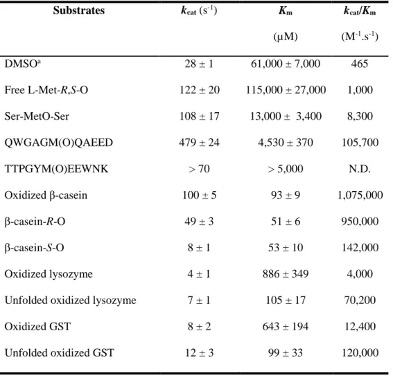
mM Ethylenediaminetetraacetic acid (EDTA) and 1 mg.ml-1 chicken lysozyme. For Met oxidation, the
200
periplasmic extract (0.7 mg.ml-1) was incubated with 20 mM N-Ethylmaleimide (NEM) and 2 mM NaOCl
201
(Sigma-Aldrich) in 50 mM HEPES pH 8.0, 50 mM NaCl for 10 min at room temperature. NaOCl was 202
10 removed by desalting using a PD-10 column and buffer was changed to 50 mM MES pH 6.0. The protein 203
solution was concentrated with a 3-kDa cutoff Amicon® Ultra concentrator. Three reaction mixtures were
204
prepared in the glove box containing 35 µl of periplasmic extract, 1 mM benzyl viologen, 2 mM dithionite 205
in 50 mM MES pH 6.0. The protein concentration in each reaction was 2.5 mg.ml-1. The first reaction
206
contained non-oxidized periplasmic extract, the second and third ones contained oxidized periplasmic 207
extract. For the third reaction (repaired periplasm) 10 µM RsMsrP was added. The reactions were incubated 208
for three hours at room temperature. 209
Trypsin proteolysis and tandem mass spectrometry 210
Protein extracts were immediately subjected to denaturing PAGE electrophoresis for 5 min onto a 4– 211
12% Bis-Tris gradient 10-well NuPAGETM gel (Thermofisher). The proteins were stained with Coomassie
212
Blue Safe solution (Invitrogen). Polyacrylamide bands corresponding to the whole proteomes were sliced 213
and treated with iodoacetamide followed by trypsin as previously recommended [35]. Briefly, each band 214
was destained with ultra-pure water, reduced with DTT, treated with iodoacetamide, and then proteolyzed 215
with Gold Mass Spectrometry Grade Trypsin (Promega) in the presence of 0.01% ProteaseMAX surfactant 216
(Promega). Peptides were immediately subjected to tandem mass spectrometry as previously recommended 217
to avoid methionine oxidation [36]. The resulting peptide mixtures were analyzed in a data-dependent mode 218
with a Q-Exactive HF tandem mass spectrometer (Thermo) coupled on line to an Ultimate 3000 219
chromatography system chromatography (Thermo) essentially as previously described [37]. A volume of 220
10 µL of each peptide sample was injected, first desalted with a reverse-phase Acclaim PepMap 100 C18 221
(5 µm, 100 Å, 5 mm x 300 µm i.d., Thermo) precolumn and then separated at a flow rate of 0.2 µL.min-1
222
with a nanoscale Acclaim PepMap 100 C18 (3 µm, 100 Å, 500 mm x 300 µm i.d., Thermo) column using 223
a 150 min gradient from 2.5 % to 25 % of CH3CN, 0.1% formic acid, followed by a 30 min gradient from
224
25% to 40% of CH3CN, 0.1% formic acid. Mass determination of peptides was done at a resolution of
225
60,000. Peptides were then selected for fragmentation according to a Top20 method with a dynamic 226
11 exclusion of 10 sec. MS/MS mass spectra were acquired with an AGC target set at 1.7 105 on peptides with
227
2 or 3 positive charges, an isolation window set at 1.6 m/z, and a resolution of 15,000. 228
MS/MS spectrum assignment, peptide validation and protein identification 229
Peak lists were automatically generated from raw datasets with Proteome Discoverer 1.4.1 (Thermo) and 230
an in-house script with the following options: minimum mass (400), maximum mass (5,000), grouping 231
tolerance (0), intermediate scans (0) and threshold (1,000). The resulting .mgf files were queried with the 232
Mascot software version 2.5.1 (Matrix Science) against the R. sphaeroides 241 annotated genome database 233
with the following parameters: full-trypsin specificity, up to 2 missed cleavages allowed, static modification 234
of carbamidomethylated cysteine, variable oxidation of methionine, variable deamidation of asparagine and 235
glutamine, mass tolerance of 5 ppm on parent ions and mass tolerance on MS/MS of 0.02 Da. The decoy 236
search option of Mascot was activated for estimating the false discovery rate (FDR) that was below 1%. 237
Peptide matches with a MASCOT peptide score below a P value of 0.05 were considered. Proteins were 238
validated when at least two different peptides were detected. The FDR for proteins was below 1% as 239
estimated with the MASCOT reverse database decoy search option. 240
Ice logo analysis 241
Ice logo analysis were performed using the IceLogo server 242
(http://iomics.ugent.be/icelogoserver/index.html) [38]. 243
244 245
12 Results
246
The R. sphaeroides MsrP is an efficient protein-MetO reductase 247
The results showing that the EcMsrP is a protein-bound MetO reductase, able to reduce both R- and 248
S-diastereomer of MetO [28] prompted us to evaluate whether these properties are conserved for RsMsrP. 249
As the EcMsrP was determined to be 5-fold less efficient in reducing the Met-S-O than Met-R-O, and 250
knowing that all previously identified MetO reductases were absolutely stereospecific towards one 251
enantiomer, we thought that it cannot be excluded that a protein contamination might explain the apparent 252
ability of the EcMsrP to reduce the Met-S-O [28]. Such potential Met-S-O reductase contaminant should be 253
able to use benzyl viologen (BV) as electron provider in activity assays and a good candidate is the 254
periplasmic DMSO reductase [26,27]. Thus, we prepared the recombinant RsMsrP from a R. sphaeroides 255
strain devoid of the dorA gene encoding the catalytic subunit of the DMSO reductase [32]. After purification 256
on Ni-affinity column and removal of the polyhistidine tag, the mature enzyme was purified by gel filtration, 257
followed by strong anion exchange, yielding a highly pure enzyme (Supplementary Figure S1). 258
After optimal pH determination showing that RsMsrP acts efficiently between pH 5.5 and 8.0 259
(Supplementary Figure S2), we determined the kinetic parameters of RsMsrP using BV as an electron 260
provider and several model substrates: the free amino acid MetO, a synthetic tripeptide Ser-MetO-Ser and 261
the oxidized bovine β-casein (Table 1). The β-casein contains 6 Met, it is intrinsically disordered, and was 262
shown as an efficient substrate for the yeast MsrA and MsrB, after oxidation [15] (see also Supplementary 263
Figure S3). Commercial β-casein contains a mixture of genetic variants, appearing as multiple peaks on 264
mass spectrometry (MS) spectra (Figure 1A). After oxidation with H2O2, MS analysis confirmed an increase
265
in mass of 96 Da for each peak, very likely corresponding to the addition of 6 oxygen atoms on the Met 266
residues (Figure 1B). Using the free MetO, we determined a kcat of ~ 122 s-1 and a Km of ~ 115,000 µM,
267
yielding a catalytic efficiency (kcat /Km) of ~ 1,000 M-1.s-1 (Table 1). With the Ser-MetO-Ser peptide, the kcat
268
and the Km values were ~ 108 s-1 and ~ 13,000 µM, and thus the kcat/Km was ~ 8,300 M-1.s-1. Compared to
269
free MetO, the ~ 8-fold increase in catalytic efficiency is due to the lower KM, and thus this indicates that
13 the involvement of the MetO in peptide bonds increases its ability to be reduced by RsMsrP. With the 271
oxidized β-casein, the kcat and the Km were ~ 100 s-1 and ~ 90 µM, respectively. The kcat/Km was thus ~
272
1,000,000 M-1.s-1. This value, 4 orders of magnitude higher than the one determined with free MetO,
273
indicates that the oxidized protein is a far better substrate for RsMsrP. Moreover, even assuming that all 274
MetO in the oxidized β-casein were equal substrates for the RsMsrP and thus multiplying the KM by 6, the
275
catalytic efficiency obtained (~ 175,000 M-1.s-1) remained ~ 175-fold higher for the oxidized protein than
276
for the free amino acid. These results indicate that the RsMsrP acts effectively as a protein-MetO reductase. 277
RsMsrP reduces both Met-R-O and Met-S-O of an oxidized model protein 278
To determine whether the RsMsrP can reduce both MetO diastereomers, we chose the oxidized bovine 279
β-casein as a model substrate because it was efficiently reduced by the yeast MsrA and MsrB indicating the 280
presence of both R and S diastereomers of MetO [15]. After oxidation with H2O2, we treated the protein
281
with MsrA and MsrB, taking advantage of their stereospecificity, to obtain protein samples containing only 282
the Met-R-O (“β-casein-R-O”) or the Met-S-O (“β-casein-S-O”), respectively. The absence of one or the 283
other diastereomer of MetO was validated by the absence of remaining Msr activity (Supplementary Figure 284
3). These three forms, containing either two or only one diastereomer of MetO, were tested as substrate for 285
RsMsrP (Figure 2). We measured a kcat of ~ 45 s-1 with the oxidized β-casein, which decreased to ~ 30 and
286
to 5 s-1 for the β-casein containing the R or the S sulfoxide, respectively. This result shows that RsMsrP can
287
reduce both diastereomers of MetO, but appears 6-fold less efficient to reduce Met-S-O than Met-R-O. 288
From this result, we postulated that RsMsrP should be able to reduce all MetO in the oxidized β-casein, 289
as this protein was intrinsically disordered and thus all MetO were very likely accessible. We evaluated this 290
hypothesis by mass spectrometry analysis. When incubated with RsMsrP, the mass of the oxidized protein 291
decreased by 96 Da, showing that all MetO were reduced (Figure 1C). Altogether, these results clearly 292
showed that RsMsrP was able to reduce both R- and S-diastereomers of MetO contained in the oxidized 293
β-casein, and thus lacked stereospecificity. 294
14 The RsMsrP preferentially reduces Met-R-O but acts effectively on Met-S-O too
296
To gain insight into the substrate preference of RsMsrP toward one of the diastereomers of MetO, we 297
performed kinetics analysis using the oxidized β-casein containing the R or S diastereomers of MetO (Table 298
1; Supplementary Figure S4). With the protein containing only the R-diastereomer of MetO 299
(“β-casein-R-O”), we determined a kcat of ~ 50 s-1, a Km of ~ 50 µM and thus a catalytic efficiency of ~
300
950,000 M-1.s-1. In the case of the protein containing only the Met-S-O (“β-casein-S-O”), the k
cat and Km
301
were ~ 8 s-1 and of ~ 50 µM, respectively. This yielded a catalytic efficiency of 142,000 M-1.s-1. This value,
302
~ 7 fold lower than the one obtained with the β-casein-R-O, was due to the lower kcat as the Km was not
303
changed. These values seem to indicate that the RsMsrP preferentially reduced the R than the S diastereomer 304
of MetO in the oxidized β-casein. However, as we could not exclude that the proportion of Met-R-O was 305
higher than the proportion of Met-S-O in the protein, we developed an assay to estimate the number of MetO 306
reduced by RsMsrP in the three forms of oxidized β-casein. We measured the total moles of BV consumed 307
for the reduction of all MetO using subsaturating concentrations of the oxidized protein. Practically, the 308
absorbance at 600 nm was measured before and 90 min after substrate addition. As two moles of BV are 309
consumed per mole of MetO reduced, we obtained the apparent stoichiometry of RsMsrP toward the 310
oxidized protein by performing a linear regression on the straight part of the line and taking the slope, which 311
defines the amount of MetO reduced as a function of substrate concentration (Figure S5). The values 312
determined were ~ 4.6, ~ 3.2 and ~ 1.8 for the oxidized β-casein, the β-casein-R-O and the β-casein-S-O, 313
respectively. In the case of the oxidized β-casein, we expected a value of 6 based on the data obtained by 314
mass spectrometry (Figure 1). This may have been due to the heterogeneity of the oxidized β-casein (all 315
Met were not initially fully oxidized) and/or to a too short time of incubation (all MetO were not fully 316
reduced, as indicated by the presence of a peak corresponding to a portion of β-casein not fully reduced in 317
Figure 1C). To compare the catalytic parameters, the data was normalized by multiplying the Km by the
318
apparent stoichiometries, yielding values per reduced MetO, thereby allowing the removal of variation due 319
to the different numbers of reduced Met-R-O or Met-S-O. The catalytic efficiencies were thus 230,000, 320
300,000 and 80,000 M-1.s-1 for the oxidized β-casein, the β-casein-R-O and the β-casein-S-O, respectively
15 (Table 1). The highest value was that obtained for the β-casein containing only the R form of MetO, 322
indicating that this diastereomer was the preferred substrate for RsMsrP. However, the value obtained with 323
the β-casein-S-O was only less than 4-fold lower, showing that RsMsrP can also act effectively on the 324
Met-S-O. 325
RsMsrP can reduce a broad spectrum of periplasmic proteins 326
To identify potential periplasmic substrates of RsMsrP and gain insight into its substrate specificity, we 327
applied a high-throughput shotgun proteomic strategy. Periplasmic proteins from the msrP- R. sphaeroides
328
mutant were extracted, oxidized with NaOCl and then reduced in vitro with recombinant RsMsrP. Untreated 329
periplasmic proteins, oxidized periplasmic proteins and RsMsrP-treated oxidized periplasmic proteins were 330
analyzed by semi-quantitative nanoLC–MS/MS. All experiments were done systematically for 3 biological 331
replicates and resulted in the identification of 362,700 peptide-to-spectrum matches. From all 11,320 332
individual peptide sequences, we identified 2,553 unique Met belonging to 720 proteins. The overall 333
percentages of Met oxidation were ~ 35%, ~ 71% and ~ 40% for proteins from the periplasm extract, the 334
oxidized periplasm extract and the RsMsrP-repaired proteins, respectively (Supplementary Table 1). This 335
first result indicates that RsMsrP is very likely able to reduce MetO from numerous proteins and to restore 336
an oxidation rate similar to the one of the periplasmic extract that has not undergone any oxidation. 337
The identification of preferential RsMsrP substrates requires the precise comparison of the oxidation 338
state of Met residues from periplasmic proteins before and after the action of the enzyme. After tryptic 339
digestion, since most of the Met/MetO-containing peptides were found in low abundance (i.e. with very low 340
spectral counts), we focused on the proteins robustly detected in all samples. We selected the Met-containing 341
peptides for which at least 10 spectral counts were detected in two replicates for each condition (i.e. 342
untreated periplasm, oxidized periplasm and repaired oxidized periplasm) and at least 7 spectral counts were 343
found in the third replicate. This restricted the dataset to 202 unique Met belonging to 70 proteins 344
(Supplementary Table 2). Overall percentage of Met oxidation (calculated as the number of spectral counts 345
for a MetO-containing peptide vs. the total number of spectral count for this peptide) varied from 2% to 346
16 87%, from 9% to 100% and from 4% to 91% in the periplasm, oxidized periplasm and repaired oxidized 347
periplasm, respectively. Comparison of Met-O containing peptides between oxidized and RsMsrP treated 348
samples indicates that the percentage of reduction varied from 100 % to no reduction at all. Eleven MetO 349
were not reduced and 22 were reduced at more than 75 % (only 2 at 100 %). The percentage of reduction 350
for the remaining majority of MetO was almost uniformly distributed between inefficient (less than 25 %) 351
to efficient (75% or more) reduction (Figure 3A). 352
No clear evidence of sequence or structure characteristic arose from these 70 identified proteins, neither 353
in term of size or in Met content (Supplementary Table 2). The periplasmic chaperone SurA, the 354
peptidyl-prolyl cis-trans isomerase PpiA, the thiol-disulfide interchange protein DsbA, the 355
spermidine/putrescine-binding periplasmic protein PotD and the ProX protein were previously proposed as 356
potential substrates of EcMsrP [28]. All these proteins contain at least one MetO amongst the most 357
efficiently reduced by RsMsrP (Supplementary Table 2), indicating that they are potential conserved 358
substrates of the MsrP enzymes in E. coli and R. sphaeroides, and very likely in numerous other 359
gram-negative bacteria. 360
The sensitivity to oxidation of the Met belonging to these 70 proteins, and their efficiency of reduction 361
by RsMsrP show a wide range of variation, from Met highly sensitive to oxidation and efficiently reduced 362
to Met barely sensitive to NaOCl treatment and not reduced by RsMsrP (Supplementary Table 2). Moreover, 363
this diversity could be visible within a single protein, in which all Met may not be uniformly oxidized and 364
reduced. For instance, the ABC transporter DdpA, along with another putative ABC transporter (Figure 365
3B,C), contained one of the two only MetO found to be fully reduced in the dataset (Met-230 and Met-353, 366
respectively), although DdpA also contained the Met-243 that was neither efficiently oxidized nor reduced. 367
This is also illustrated by the case of the peptidyl-prolyl cis-trans isomerase, which possessed the Met found 368
to have the higher decrease in oxidation in the entire dataset (Met-172) but also a Met almost not reduced 369
by RsMsrP (Met-190) (Figure 3D). The Met-539 of the PQQ dehydrogenase XoxF illustrates the case in 370
which a Met was highly sensitive to NaOCl-oxidation and very efficiently reduced (Figure 3E). Twenty-one 371
Met were oxidized at 50 % or more and reduced by 50 % or more by RsMsrP (Supplementary Table S2). 372
17 Altogether, these results show that RsMsrP can reduce a broad spectrum of apparently unrelated proteins 373
(only 11 Met among 202 were not reduced). However, since all MetO were not reduced with similar 374
efficiency, some structural or sequence determinants could drive the ability of MetO to be reduced by 375
RsMsrP. 376
The nature of the amino acids surrounding a MetO influences the RsMsrP efficiency 377
Having in hand a relatively large dataset of oxidized and reduced Met prompted us to search for 378
consensus sequences that could favor or impair the oxidation of a Met or the reduction of a MetO by RsMsrP. 379
For all identified Met, we extracted, the surrounding 5 amino acids on the N- and C-terminal sides to obtain 380
an 11-amino acid sequence with the considered Met centered at the 6th position. As shown for bacterial
381
MsrB [39], this length might be sufficient to encompass the amino acids in physical contact with RsMsrP 382
during reduction. We then performed an IceLogo analysis aiming to identify whether some residues were 383
enriched or depleted around the target Met. The principle is to compare a ‘positive’ dataset of peptides, to a 384
‘negative’ one [38]. To find potential consensus sequences of oxidation, we first compared all unique 385
MetO-containing peptides from both the untreated and the NaOCl-oxidized periplasmic extracts (our 386
positive dataset) to the theoretical R. sphaeroides proteome (our negative dataset). The IceLogo presented 387
in Figure 4A shows that MetO-containing sequences were mainly depleted of His and aromatic or 388
hydrophobic residues (Trp, Phe, Tyr, Leu, Ile) and were mainly enriched in polar or charged amino acids 389
(Asn, Gln, Asp, Glu and Lys). This suggests that Met in a polar environment, as commonly found at the 390
surface of proteins, are very likely more susceptible to oxidation than those located in hydrophobic 391
environments such as those in the protein core. We then compared all these unique MetO-containing 392
peptides to all the Met-containing peptides from the same samples (Figure 4B), and we observed that Trp, 393
along with His, Tyr and Cys, were principally depleted around the potentially oxidized Met. Strikingly, the 394
only amino acid significantly more abundant around an oxidized Met was another Met in position -2 and 395
+2. These results indicate that oxidation sensitive Met might be found as clusters. 396
18 To identify potential consensus sequence favorable to MetO reduction by RsMsrP, we performed a 397
precise comparison of the oxidation percentage before and after the action of the enzyme. We thus defined 398
two criteria to characterize the reduction state of each Met: i) the percentage of reduction calculated using 399
the formula described in Supplementary Table S2 and based on the comparison of the oxidation percentages 400
in oxidized versus repaired oxidized periplasm. For instance, a Met found oxidized at 25 % in the oxidized 401
periplasm and at 5 % in the repaired oxidized periplasm was considered enzymatically reduced at 80 %. ii) 402
the decrease in percentage of oxidation by comparison of the 2 samples. For instance, the same Met found 403
oxidized at 25 % in the oxidized periplasm and 5 % in the repaired extract had a decrease in the oxidation 404
percentage of 20 %. This second criterion was used to avoid bias in which very little oxidized Met were 405
considered as efficient substrates (i.e. a Met oxidized at 5 % in the oxidized periplasm extract and at 1 % in 406
the repaired oxidized periplasm was reduced at 80 %, similarly to one passing from 100 % to 20 %, which 407
intuitively appears as a better substrate than the previous one). We selected as efficiently and inefficiently 408
reduced MetO those for which both criterions were higher than 50 % and lower than 10 %, respectively. 409
Comparison of the sequences surrounding the efficiently reduced MetO to the theoretical proteome of R. 410
sphaeroides showed no depletion of amino acid, but mainly enrichment of polar amino acids (Gln, Lys, and
411
Glu) around the oxidized Met (Figure 5A). Similar analysis with the inefficiently reduced MetO indicated 412
the enrichment of Thr and Ser in the far N-terminal positions (-5 and -4) and of a Tyr in position -2 (Figure 413
5B). The C-terminal positions (+ 1 to +5) were mainly enriched in charged amino acids (Gln, Lys, and Glu), 414
similarly to efficiently reduced MetO. This apparent contradiction may indicate that the amino acids in the 415
C-terminal position of the considered MetO did not really influence the efficiency of RsMsrP but were 416
observed simply because of the inherent composition of the overall identified peptides. We then compared 417
the variation of amino acids composition of the MetO-containing peptides between both datasets, using the 418
inefficiently reduced MetO as a negative dataset (Figure 5C). The results were similar to those obtained by 419
comparison with the entire theoretical proteome of the bacterium, i.e. most enriched amino acids were polar 420
(Glu, Gln, Asp and Lys) at most extreme positions (-5, -4 and + 2 to + 5). Of note, the conserved presence 421
of a Gly in position -1, and the presence of several other Met around the central Met. This potential 422
19 enrichment of Met around an oxidation site is consistent with the result found for the sensibility of oxidation 423
(Figure 4B), and indicates that potential clusters of MetO could be preferred substrates for RsMsrP. We 424
found 16 peptides containing 2 or 3 MetO, reduced at more than 25 % by RsMsrP (Supplementary Table 425
S2). This was illustrated, for example, by the cell division coordinator CpoB which possesses two close Met 426
residues (66 and 69) highly reduced by the RsMsrP, or by the uncharacterized protein (YP_353998.1) having 427
4 clusters of MetO reduced by the RsMsrP (Supplementary Table S2). 428
From this analysis, the only depleted amino acids appeared to be Thr and Pro in positions -4 and -3 429
(Figure 4C). To validate these results, we designed two peptides, QWGAGM(O)QAEED and 430
TTPGYM(O)EEWNK, as representative of most efficiently and most inefficiently RsMsrP-reduced 431
peptide-containing MetO, respectively. We used them as substrates to determined reduction kinetics 432
parameters for RsMsrP (Table 1; Supplementary Figure S6). The results showed that the peptide 433
QWGAGM(O)QAEED was efficiently reduced, with the highest kcat value from all the substrates we tested
434
(~ 480 s-1)and a K
m of~ 4,500 µM. This yielded a kcat/Km of ~ 100,000 M-1.s-1, which is 2 orders of magnitude
435
higher than the one determined for the free MetO, and 10-fold lower than for the oxidized β-casein (Table 436
1). On the contrary, the peptide TTPGYM(O)EEWNK was not efficiently reduced by RsMsrP (Table 1; 437
Supplementary Figure S6). Indeed, we could not determine the kinetic parameters as the activity value curve 438
never reached an inflection point using concentrations as high as 5,000 µM. The maximal kcat value was
439
determined at ~ 70 s-1 at 5,000 µM of peptide, which is ~ 3.5-fold less than the one determined with the
440
same concentration of the other peptide (~ 250 s-1) (Supplementary Figure S6). These results are in full
441
agreement with the proteomics analysis and confirm that the nature of the amino acids surrounding a MetO 442
in a peptide or a protein strongly influences its ability to be reduced by RsMsrP. 443
The RsMsrP preferentially reduces unfolded oxidized proteins 444
To test whether structural determinants affect RsMsrP efficiency of MetO reduction, we compared its 445
activity using oxidized model proteins, either properly folded or unfolded. We started with chicken 446
lysozyme as it is a very well folded protein highly stabilized by four disulfide bonds [40]. We oxidized it 447
20 with H2O2 and checked its oxidation state by mass spectrometry (Supplementary Figure S7). Surprisingly,
448
using a protocol similar to the one allowing the complete oxidation of the 6 Met of β-casein, we observed 449
only a weak and incomplete oxidation of the protein. The major peak corresponded to the non-oxidized form 450
and a small fraction had an increase in mass of 16 Da, likely corresponding to the oxidation of one Met. 451
Nevertheless, we prepared from this oxidized sample, an unfolded oxidized lysozyme by reduction with 452
dithiothreitol in 4M urea followed by iodoacetamide alkylation of cysteines, and both samples (oxidized 453
and unfolded oxidized), were used as substrates for RsMsrP (Figure 6). We also used 454
glutathione-S-transferase (GST) which possesses 9 Met and is highly structured. After oxidation with H2O2,
455
GST was incubated with 4 M of the chaotropic agent urea, a concentration sufficient to induce complete 456
unfolding of the protein [15]. For both oxidized proteins, we observed a dramatic increase in activity after 457
unfolding. Indeed, the RsMsrP activity increased 7-fold with the unfolded oxidized lysozyme compared to 458
the folded one, and 6-fold in the case of the unfolded oxidized GST compared to the folded oxidized GST 459
(Figure 6). As the unfolded oxidized protein solutions of lysozyme or GST contained a substantial amount 460
of urea, we performed controls in which the urea was added extemporaneously in the cuvette during the 461
measurements, showing that urea did not influence the RsMsrP activity (Supplementary Figure S8). 462
Mass spectrometry analysis showed that the RsMsrP was able to completely reduce the oxidized 463
lysozyme in these conditions (Supplementary Figure S7), suggesting that observed differences of repair 464
between the folded- and unfolded-oxidized lysozyme were not due to the incapacity of RsMsrP to reduce 465
some MetO, but were due to kinetic parameters. We thus determined the kinetics of reduction of these 466
proteins by RsMsrP (Table 1, Supplementary Figure S8). For the oxidized lysozyme, the kcat and the Km
467
were ~ 4 s-1 and~ 900 µM, respectively. Using the unfolded oxidized lysozyme, the k
cat increased to ~ 7 s-1
468
andthe Km decreased to ~ 100 µM. The catalytic efficiency determined with the unfolded oxidized lysozyme
469
was thus ~ 18-fold higher than the one determined using the oxidized lysozyme before unfolding (70,200 470
vs. 4,000 M-1. s-1). Similar results were obtained with GST. Indeed, with the oxidized GST, we recorded k cat
471
and Km values of ~ 8 s-1 and ~ 640 µM, respectively. Whereas for the unfolded oxidized GST, the kcat was
472
slightly higher (~ 12 s-1), and the K
m was ~ 6-fold lower (~ 100 µM). The catalytic efficiency was 10-fold
21 higher for the unfolded oxidized GST than for its folded counterpart (Table 1; Supplementary Figure S8). 474
Altogether, these results showed that RsMsrP is more efficient at reducing MetO in unfolded than in folded 475
oxidized proteins. Moreover, as evidenced with lysozyme that contained only one MetO in our conditions, 476
the increase in activity using unfolded substrate is not dependent on the number of MetO reduced. 477
22 Discussion
478
All organisms have to face harmful protein oxidation and almost all possess canonical Msrs that protect 479
proteins by reducing MetO. Bacteria also have molybdoenzymes able to reduce MetO, as a free amino acid 480
for the DMSO reductase [26] or the biotin sulfoxide reductase BisC/Z [24,25], but also included in proteins 481
in the case of MsrP [28,29]. Genetic studies and the conservation of MsrP in most gram-negative bacteria 482
indicate that it is very likely a key player in the protection of periplasmic proteins against oxidative stress 483
[28,29] However, an in-depth characterization of its protein substrate specificity is still lacking. In this work, 484
we chose the MsrP from the photosynthetic purple bacteria R. sphaeroides as a model enzyme to uncover 485
such specificity. Using purified oxidized proteins and peptides, we showed that RsMsrP is a very efficient 486
protein-containing MetO reductase, with apparent affinities (Km) for oxidized proteins 10 to 100-fold lower
487
than for the tripeptide Ser-MetO-Ser or the free MetO (Table 1). As reported for canonical MsrA and MsrB 488
[15], we observed important variations in the reduction kcat of different oxidized proteins, arguing for the
489
existence of sequence and structural determinants affecting the enzyme efficiency (Table 1). 490
To find potential physiological substrates of RsMsrP and uncover their properties, we used a proteomic 491
approach aiming at comparing the oxidation state of periplasmic proteins after treatment with the strong 492
oxidant NaOCl, followed by RsMsrP reduction of these proteins. We found 202 unique Met, belonging to 493
70 proteins, for which the sensitivity of oxidation and the ability to serve as an RsMsrP substrate varied 494
greatly (Figure 3, Supplementary Table S2). MetO efficiently reduced by RsMsrP belong to structurally and 495
functionally unrelated proteins, indicating that RsMsrP very likely does not possess specific substrates and 496
acts as a global protector of protein integrity in the periplasm. Interestingly, we observed from our IceLogo 497
analysis that Met sensitive to oxidation are generally presented in a polar amino acid environment and can 498
be found in clusters (Figure 4). These properties might be common to all Met in proteins as similar results 499
were found in human cells [41,42] and plants [43]. Moreover, oxidized Met efficiently reduced by the 500
RsMsrP were also found clustered in polar environments and our analysis shows that the presence of Thr 501
and Pro in N-terminal side of a MetO strongly decrease RsMsrP efficiency (Table 1, Figure 5 and 502
23 Supplementary Figure S6). To our knowledge, the presence of a Thr close to a MetO was not previously 503
shown to influence any Msr activity, but the presence of a Pro was shown to decrease or totally inhibit MetO 504
reduction by the human MsrA and MsrB3, depending on its position [41]. 505
The presence of oxidation-sensitive Met efficiently reduced by the RsMsrP in clusters on polar parts of 506
proteins should facilitate the oxidation/reduction cycle aiming to scavenge ROS as previously proposed for 507
canonical Msrs [44]. This is also illustrated by the methionine-rich protein MrpX proposed as main substrate 508
of the A. suillum MsrP, which is almost only composed of Met, Lys, Glu and Asp [29]. The presence of 509
numerous MetO on a single molecule of protein substrate should increase the RsMsrP efficiency as one 510
molecule of the substrate allows several catalytic cycles, potentially without breaking physical contact 511
between the enzyme and its substrate. 512
Comparison of the RsMsrP activity using folded or unfolded protein substrates (lysozyme and GST) 513
showed that it is far more efficient to reduce unfolded oxidized proteins (Figure 6). Similar results were 514
found for canonical Msrs [15]. In the case of MsrB it was because more MetO were accessible for reduction 515
whereas for MsrA this increase was independent of the number of MetO reduced. Here, the use of lysozyme 516
containing only one MetO (Supplementary Figure S7) undoubtedly showed that the increase in activity is 517
not related to the unmasking of additional MetO upon protein denaturation (Table 1; Figure 6). This could 518
indicate that the RsMsrP has better access to the MetO in the protein or that the MetO is more easily 519
accommodated in the active site of the enzyme because of increased flexibility. This should provide a 520
physiological advantage to the bacteria during oxidative attacks, which could occur during other stresses 521
such as acid or heat, hence promoting simultaneous oxidation and unfolding of proteins. Particularly, 522
hypochlorous acid, which was shown to induce msrP expression in E. coli [28] and A. suillum [29], has 523
strong oxidative and unfolding effect on target proteins [45]. 524
Finally, previous work indicated that the E. coli MsrP lacks stereospecificity and can reduce both R- and 525
S-diastereomers of MetO chemically isolated from a racemic mixture of free L-Met-R,S-O [28]. This 526
discovery is of fundamental importance as it breaks a paradigm in Met oxidation and reduction knowledge, 527
24 and very likely for all enzymology as non-stereospecific enzymes were very rarely described. Indeed, to our 528
knowledge, all previously characterized enzymes able to reduce Met sulfoxide or related substrates were 529
shown to be absolutely stereospecific. This was the case for the canonical MsrA and MsrB, which reduce 530
only the S-diastereomer and the R-diastereomer, respectively [7,9–14], as well as for the free Met-R-O 531
reductase [22,23] and for the DMSO reductase [26,27] and BisC/Z molybdoenzymes [24,25]. To evaluate 532
the potential lack of stereospecificity of the RsMsrP, we chose to use a different strategy than the one used 533
for E. coli MsrP [28] and prepared oxidized β-casein containing only one or the other MetO diastereomer 534
using yeast MsrA and MsrB to eliminate the S- and the R-diastereomers, respectively. Activity assays and 535
kinetic experiments using a highly purified RsMsrP demonstrated that it can efficiently reduce the β-casein 536
containing only the R- or the S-diastereomer (Table 1; Figure 2 and Supplementary Figure S4). Moreover, 537
this lack of stereospecificity was undoubtedly confirmed by the ability of the RsMsrP to reduce all 6 MetO 538
formed on the oxidized β-casein (Figure 1). These results, consistent with Gennaris and coworkers finding, 539
indicate that this lack of stereospecificity is very likely common to all MsrP homologs. Together with the 540
apparent ability of the enzyme to repair numerous unrelated oxidized proteins, the capacity to reduce both 541
diastereomers of MetO, argues for a role of MsrP in the general protection of envelope integrity in gram 542
negative bacteria. However, it raises questions regarding the structure of its active site as the enzyme should 543
be able to accommodate both diastereomers. From this, we wondered whether the RsMsrP could reduce the 544
Met sulfone, which can be imagined as a form of oxidized Met containing both R- and S-diastereomers, but 545
we did not detect any activity (Supplementary Figure S9). Although it could be because of an incompatibility 546
in redox potential, it may indicate that this form of oxidized Met cannot reach the catalytic atom. The 547
three-dimensional structure of the oxidized form of E. coli MsrP indicated that the molybdenum atom, which 548
is supposed to be the catalytic center of the enzyme, is buried 16 Å from the surface of the protein [46]. The 549
next challenge will be to understand the MsrP reaction mechanism and will require the determination of the 550
enzyme structure in its oxidized and reduced forms bound to its MetO-containing substrates. 551
25 Acknowledgment: We are very grateful to Prof. Vadim, N. Gladyshev (Brigham’s and Women Hospital 553
and Harvard Medical School) for the gift of pET28a-MsrA, pET21b-MsrB, pET15b-TR1, pET15b-Trx1 554
and pGEX4T1 expression vectors. We thank Dr. Benjamin Ezraty and the members of his team (Laboratoire 555
de Chimie Bactérienne, Institut de Microbiologie de la Méditerranée) for fruitful discussions. Pascaline 556
Auroy-Tarrago (Laboratoire de Bioénergétique et Biotechnologie des Bactéries et Microalgues, CEA, 557
BIAM) is acknowledged for her help with proteomics analysis. 558
559
Competing Interests: The Authors declare that there are no competing interests associated with the 560
manuscript. 561
562
Funding: This work was supported by the Commissariat à l'Energie Atomique et aux Energies Alternatives 563
(CEA) and by the project METOXIC (ANR 16-CE11-0012). 564
565
Author contribution: LT, PA, DP and MS designed the study. LT, SG, MIS and MS purified RsMsrP. LT 566
and MS prepared all other proteins. LT, SG, MIS, MS performed biochemical characterization of RsMsrP. 567
LT, MS and DL performed β-casein and lysozyme mass spectrometry analysis and analyzed the data. SG 568
and MS prepared R. sphaeroides 2.4.1 msrP- mutant and periplasmic proteins samples. BA, GM and JA
569
performed proteomics analysis of periplasmic proteins and LT, MS, GM and JA analyzed the data. LT wrote 570
the manuscript with contribution of MIS, DL, PA, DP, JA and MS. All authors approved the final 571
manuscript. 572
26 References
573
![[PDF] Introduction au Pc et au compilateur Visual C++ | Cours informatique](data:image/gif;base64,R0lGODlhAQABAIAAAP///wAAACH5BAEAAAAALAAAAAABAAEAAAICRAEAOw==)