Acta Palaeontol. Pol. 60 (3): 711–731, 2015 http://dx.doi.org/10.4202/app.00015.2013
Devonian antiarch placoderms from Belgium revisited
SÉBASTIEN OLIVE
Olive, S. 2015. Devonian antiarch placoderms from Belgium revisited. Acta Palaeontologica Polonica 60 (3): 711–731. Anatomical, systematic, and paleobiogeographical data on the Devonian antiarchs from Belgium are reviewed, updated and completed thanks to new data from the field and re-examination of paleontological collections. The material of
Bothriolepis lohesti is enhanced and the species redescribed in more detail. An undetermined species of Bothriolepis is
recorded from the Famennian of Modave (Liège Province), one species of Asterolepis redescribed from the Givetian of Hingeon and another one described from the Givetian of Mazy (Namur Province). Grossilepis rikiki sp. nov. is recorded from the Famennian tetrapod-bearing locality of Strud (Namur Province) and from the Famennian of Moresnet (Liège Province). It is the first occurrence of Grossilepis after the Frasnian and on the central southern coast of the Euramerican continent. Its occurrence in the Famennian of Belgium may be the result of a late arrival from the Moscow Platform and the Baltic Depression, where the genus is known from Frasnian deposits. Remigolepis durnalensis sp. nov. is described from the Famennian of Spontin near Durnal (Namur Province). Except for the doubtful occurrence of Remigolepis sp. in Scotland, this is the first record of this genus in Western Europe. Its occurrence in Belgium reinforces the strong faunal affinities between Belgium and East Greenland and the hypothesis of a hydrographical link between the two areas during the Late Devonian.
Key words: Placodermi, Asterolepis, Bothriolepis, Grossilepis, Remigolepis, palaeobiogeography, Devonian, Belgium.
Sébastien Olive [[email protected]], Royal Belgian Institute of Natural Sciences, O.D. Earth and History of Life, Laboratory of Palaeontology, Rue Vautier 29, 1000 Brussels, Belgium; and Liege University, Geology Department, Laboratory of Animal and Human Palaeontology, B18, Allée du 6 Août, 4000 Liège, Belgium.
Received 1 August 2013, accepted 20 November 2013, available online 26 November 2013.
Copyright © 2015 S. Olive. This is an open-access article distributed under the terms of the Creative Commons Attribu-tion License, which permits unrestricted use, distribuAttribu-tion, and reproducAttribu-tion in any medium, provided the original author and source are credited.
Introduction
Early studies assigned antiarchs to jawless ostracoderms (Woodward 1891; Patten 1912; Eastman 1917). Stensiö (1931) and Gross (1931) reinterpreted, independently, the antiarch remains as belonging to jawed vertebrates, to the class Placodermi, and this idea has since been widely accept-ed and confirmaccept-ed (Moy-Thomas and Miles 1971; Denison 1975; Goujet and Young 1995). Johanson (2002) argued, based on the vascularisation of the pectoral fins, that an-tiarchs were not placoderms, but Young (2008) rejected that hypothesis.
Antiarchs are characterised by box-like dermal armour covering the head and thorax and by highly modified pec-toral fins enclosed in interlocking dermal plates. Their geo-logical range extends from the early Silurian (Wang 1991) to the Late Devonian (e.g., Johanson 1997a, b; Lukševičs 2001) and they include some genera with a worldwide dis-tribution during the Late Devonian, e.g., Remigolepis and Bothriolepis.
There is no widely accepted classification of antiarchs, but the most consensual one (Janvier and Pan 1982; Young 1984;
Zhu 1996; Lukševičs 2001) groups the asterolepidoids, both-riolepidoids, and sinolepids in the clade Euantiarcha (Janvier and Pan 1982). This clade, characterised by the presence of a brachial process, makes the bothriolepidoids sister group of the asterolepidoids or the sinolepids, according to different authors. The yunnanolepids are the sister group of the Euan-tiarcha. Lukševičs (2001) places the procondylolepids in the Euantiarcha but their position is still not resolved.
To date, Belgian antiarchs were poorly known and only few taxa have been published or mentioned in the literature. Bothriolepis lohesti Leriche, 1931 was the first described one but was partially known. Gross (1965) described a species of the genus Asterolepis from the Givetian of Belgium, and Clément and Prestianni (2009) misinterpreted the presence of Bothriolepis in the Belgian Famennian of two localities (i.e., Strud and Spontin).
During the Devonian, Belgium was located on the south-eastern margin of Laurussia. This was a time of important sea level variations, but the progression of the sea towards the north never reached the north of Belgium. This is why Devonian strata in Belgium are located only in the south of the country, the source of the localities studied in this article (Fig. 1).
Institutional abbreviations.—IRSNB, Institut Royal des Sci-ences Naturelles de Belgique, Brussels, Belgium; PALULG, Paleontological collections of the Université de Liège, Liège, Belgium; UCL, Université Catholique de Louvain-la-Neuve, Louvain-la-Neuve, Belgium.
Anatomical abbreviations.—ADL, anterior dorso-lateral plate; adlc, anterior dorso-lateral corner of PVL lateral lam-ina; AMD, anterior median dorsal plate; AVL, anterior ven-tro-lateral plate; CD1, dorsal central plate 1; CV1–4, ventral central plates; DM1–4, plates of the dorso-medial marginal series; La, lateral plate; ML1–5, plates of the lateral marginal series; MM1–4, plates of the medial marginal series; MV, median ventral plate; MxL, mixilateral plate; Nu, nuchal plate; PDL, posterior dorso-lateral plate; Pmg, postmarginal plate; Pn, paranuchal plate; Pp, postpineal plate; Prm, preme-dian plate; PVL, posterior ventro-lateral plate; Sm, semilu-nar plate; SM, submarginal plate; T, terminal plate; VM1–3, plates of the ventro-medial marginal series.
Material and methods
All specimens have been mechanically prepared. Some have been whitened with ammonium chloride to make the obser-vation of sensory line grooves easier. Some others have been photographed under immersion to enhance certain characters.
The limited number of samples for each taxon makes it unnecessary to use dermal plate measurement indices be-cause they are more representative of individuals than of the taxon (Long and Werdelin 1986; Johanson 1998). Thus, the taxon descriptions are qualitative and not quantitative.
The ornament characterization (i.e., reticulate, nodose, and tuberculate) follows the definitions given by Young (1988).
The dermal skeleton of antiarchs is exemplified in Fig. 2, based on the four genera found in Belgium.
Systematic palaeontology
Placodermi McCoy, 1848
Euantiarcha Janvier and Pan, 1982
Bothriolepidoidei Miles, 1968
Bothriolepididae Cope, 1886
Genus Bothriolepis Eichwald, 1840
Type species: Bothriolepis ornata Eichwald, 1840, subsequently
des-ignated by Woodward (1891); Priksha River, Russia, upper Famennian Lnyanka Beds, Devonian.
Bothriolepis lohesti Leriche, 1931
Fig. 3.
1888 Bothriolepis or Pterychtis sp.; Lohest 1888: 60. 1889 Bothriolepis; Lohest 1889: 58.
1895 Bothriolepis canadensis; Lohest 1895: 39. 1931 Bothriolepis; Leriche 1931: 15, pl. 3. 1932 Bothriolepis lohesti; Gross 1932: 29. 1948 Bothriolepis lohesti; Stensiö 1948: 513. 1978 Bothriolepis lohesti; Denison 1978: 110.
Type material: No holotype was defined by Leriche (1931). Gross
(1932) assigned as lectotype an AMD (Leriche 1931: pl. 3: 4), bearing now the number PALULG-2011.12.14.14A.
Type locality: Chèvremont, Liège Province, Belgium
Type horizon: Montfort/Evieux Formation, Famennian, Upper
Devo-nian.
Material.—Partial headshield: UCL-P.V.L.10.621 (cast); AMD: PALULG-2011.12.14.15, PALULG-2011.12.14.16, PALULG-2011.12.14.17A; ADL: PALULG-2011.12.14.17B; ?ADL: IRSNB vert 5179-001; PMD: PALULG-2011.12.14.19, PALULG-2011.12.14.18; MxL: P.V.L.10.533, UCL-P.V.L.10.668; AVL: PALULG-2011.12.14.20; pectoral fin: PALULG-2011.12.14.14B, PALULG-2011.12.14.21, PA L ULG- 2011.12.14.22; CV1: UCL-P.V.L. 10.656, PA-L ULG- 2011.12.14.23A, IRSNB vert 5179-002; ML2: PALULG-2011.12.15.9, PALULG-2011.12.15.10, PALU LG 2011.12.15.11, PALPALULG2011.12.15.12, PALPALULG -- 2011.12.15.13, PALULG-2011.12.14.23B. From Chèvre-mont and Vaux-sous-ChèvreChèvre-mont, both in Liège Province, Belgium, Montfort/Evieux Formation, Famennian, Upper Devonian. Chèvremont is the hill close to Vaux-sous-Chèvre-mont city. Old quarries and outcrops stretch between both localities. In old collections, there is often confusion between these two localities.
Bruxelles THE NETHERLANDS Li geè Namur Spontin StrudModave Vaux-sous-Ch vremontè Vaux-sous-Ch vremontè Moresnet Moresnet MazyHingeon LUXEMBOURG FRANCE 60 km North Sea GERMANY BELGIUM
Fig. 1. Locality map of Belgian antiarch occurrences. Black fish symbols for Givetian finds; white for Famennian.
Fig. 2. Reconstructed dermal skeleton of the antiarch placoderm genera found in Belgium. A. Bothriolepis canadensis, dorsal view, after Werdelin and Long (1986: fig. 2). B. Asterolepis maxima, dorsal view, after Tra-quair (1914). C. Grossilepis tuberculata, dorsal view, after Gross (1941).
D. Remigolepis walkeri, dorsal (D1), lateral (D2), and ventral (D3) views, after Johanson (1997a). Abbreviations: CD1–4, plates of the dorsal central series; CV1, ventral central plate 1; DM1–4, plates of the dorso-medial marginal series; ML1–4, plates of the lateral marginal series; MM1–4, plates of the medial marginal series; VM1–3, plates of the ventro-medial marginal series.
postmarginal plate paranuchal plate premedian plate lateral
plate postpinealplate nuchal plate anterior median dorsal plate anterior dorso-lateral plate mixilateral
plate posterior mediandorsal plate
premedian plate postmarginal plate paranuchal plate lateral plate semilunar plate terminal plate postpineal plate nuchal plate anterior dorso-lateral plate anterior median dorsal plate posterior median dorsal plate premedian plate lateral plate postmarginal plate nuchal plate postpineal plate paranuchal plate anterior dorso-lateral plate anterior median dorsal plate mixilateral plate mixilateral plate
posterior median dorsal plate
premedian plate semilunar plate semilunar plate paranuchal plate lateral plate postpineal plate nuchal plate anterior dorso-lateral plate anterior median dorsal plate anterior median dorsal plate anterior dorso-lateral plate anterior ventro-lateral plate posterior ventro-lateral plate posterior lateral plate posterior ventro-lateral plate posterior median dorsal plate posterior median dorsal plate posterior dorso-lateral plate posterior dorso-lateral plate median ventral plate
A
B
C
CD1 ML2 MM2 CD2 CD1 ML2 MM2 CD2 MM3 ML3 CD3 MM4 ML4 CD1 ML2 CD4 ML4 CD2 MM2 ML3 CD3 MM4 terminal plate ML1 ML2 ML3 ML4 CD1 DM1 CD2 CD3 DM3 CD4 DM4 DM2 anterior ventro-lateral plate CV1 ML1 VM1 ML2 VM2 ML3 VM3 ML4 terminal plate 2D
D
3D
1 10 cm 5 cm 10 cm 5 cmDescription
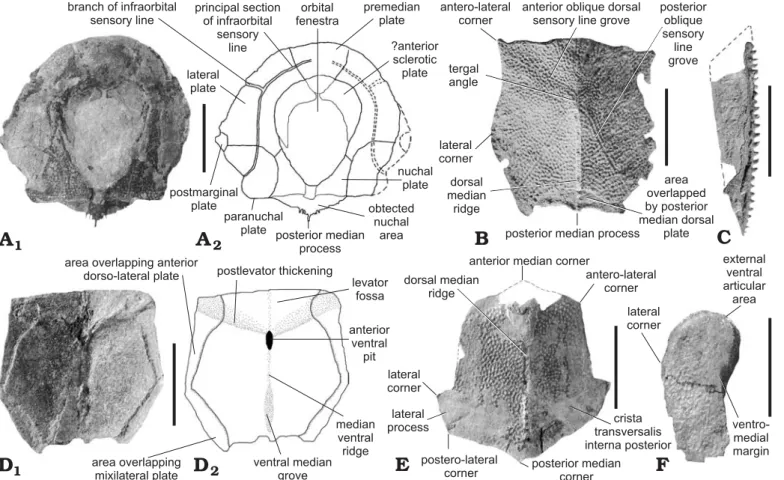
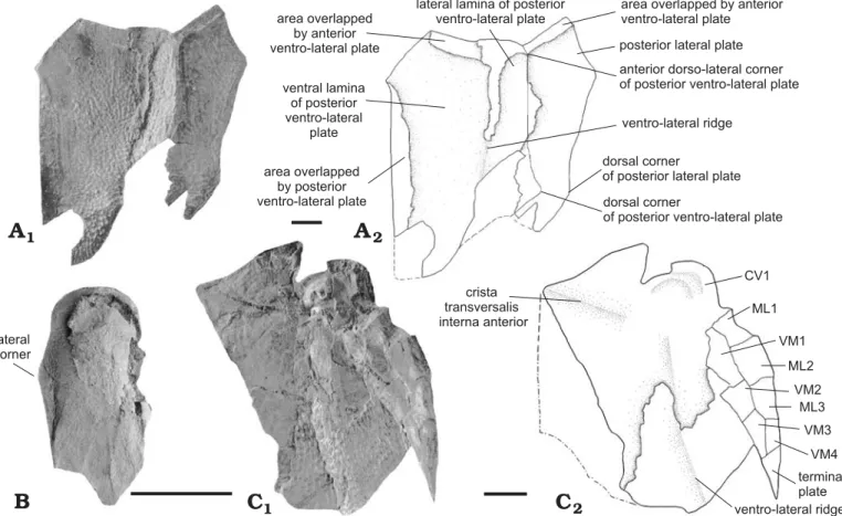
Headshield.—Partial headshield (Fig. 3A): UCL-P.V.L.10.621 displays a partial headshield with the nuchal, paranuchal, pre-median, and lateral plates in connection. It is badly preserved and the plate margins are difficult to observe. However it seems that the nuchal plate is excluded from the orbital fenes-tra. The postpineal notch forms a strong angle posteriorly, i.e., almost 90°. The central sensory line grooves and the principal section of infraorbital sensory line are well observed.
Trunkshield.—Anterior median dorsal plate, AMD (Fig. 3B, C): This plate is as long as wide. Externally, it is almost flat, with only a slight bulge on the dorsal median ridge. The latter is only defined by a longitudinal row of fused tubercles on PALULG-2011.12.14.16. The tergal angle is well marked and situated between the anterior and middle thirds of the plate. It is roughly of 45°. The posterior oblique dorsal sensory line grooves are usually well defined. On PALULG-2011.12.14.14, the right posterior oblique dorsal sensory line groove is shortened or interrupted. The posterior oblique dorsal sensory line grooves run from the tergal angle and cut the posterior division of the lateral margin at the junction between its anterior and middle thirds. The anterior margin is fairly straight, except on PALULG-2011.12.14.14 where it is slightly concave and not convex as Stensiö (1948: 514) noticed. It is twice the length of the posteri-or margin. The antero-lateral and lateral cposteri-orners are well defined. The posterior division of the lateral margin is as long as the anterior division except on the biggest AMD (PALULG-2011.12.14.14), where the posterior division is one third smaller than the anterior division. Only the right area overlapped by the mixilateral plate is observable (PAL-ULG-2011.12.14.14). It is quite small. The posterior lateral margin displays a sigmoid shape, as observed in a specimen of B. canadensis (Stensiö 1948: fig. 182) and suggesting that the AMD both overlaps and is overlapped by the MxL. Internally, the AMD shows a triangular-shaped levator fossa stretching on all the anterior third length. It is limited lateral-ly by fairlateral-ly conspicuous postlevator thickenings, observable directly on PALULG-2011.12.14.16 and indirectly on im-pressions of the visceral surface (PALULG-2011.12.14.15 and PALULG-2011.12.14.17A). The postlevator cristae are quite sharp and high. Their confluence is located at the poste-rior edge of the anteposte-rior ventral pit . The median ventral ridge and ventral median groove are well marked.
Posterior median dorsal plate, PMD (Fig. 3D): The anteri-or margin is convex, with an obtuse median canteri-orner. The lateral process is well developed and extended. In fact, the margin between the lateral corner and postero-lateral corner is quite long compared to what is observed in Bothriolepis canadensis (Stensiö 1948: text-fig. 138A–R). The lateral margin situated in front of the lateral corner and running to the antero-lateral corner is straight. The posterior margin is convex, with an obtuse median angle. The plate is slightly arched with a low dorsal median ridge. The dorsal median ridge extends almost straight from the anterior angle to the posterior angle area.
Anterior dorso-lateral plate, ADL (Fig. 3E): Only the an-terior part of a left ADL, in internal view, is preserved in the material. Both lateral and dorsal anterior laminae are present. The anteriormost part of the plate is hidden by a displaced AMD. The articulation area with the headshield is thus not observable. The ventral margin is convex on the preserved anterior part. The notch, for the external postlevator process of the AMD, is smooth and the postnuchal corner strongly developed. The dorso-medial margin is concave along all its preserved length. The anterior part of the lateral lamina is twice less wide than the median part. The dorso-lateral ridge is well pronounced, and the lateral lamina forms an angle of almost 150° with the dorsal lamina, but that value may be incorrect as a result of compression. The main lateral line groove is not directly observed, but suggested by a tiny edge running along the length of the lateral lamina that tends to join the dorso-lateral ridge anteriorly and to remain parallel to the dorso-lateral ridge posteriorly.
Mixilateral plate, MxL (Fig. 3F): As Stensiö (1948) no-ticed for Bothriolepis canadensis, the lateral lamina of MxL is often missing, crushed, compressed or damaged. It is the case in the available material of Bothriolepis lohesti, where the lateral lamina is crushed and damaged. Because of its bad preservation, it is difficult to evaluate the angle between the lateral and dorsal laminae. However, the specimen UCL-P.V.L.10.668 shows quite a well preserved area of transition between these two laminae and suggests that the angle is approximately 150°, as noticed for the ADL, with a sharp prominent dorso-lateral ridge. The crushed lateral lamina, in that specimen, presents a broad area overlapped by the PVL. The area overlapped by the posterior median dorsal plate is long and narrow. The posterior oblique dorsal sensory line groove crosses the anterior part of the dorso-medial margin and ends in the posterior third of the dorsal lamina.
Anterior ventro-lateral plate, AVL (Fig. 3G): This plate is badly preserved, with the anterior part and the lateral lamina damaged. The length of this plate is at least twice the width. The angle between the ventral and lateral laminae is roughly of 160°, and the ventro-lateral ridge is smooth. The subce-phalic division is not well preserved. The posterior margin of the ventral lamina is straight and the posterior ventro-lateral corner well defined. The different portions of the medial margin seem straight but this is not obvious, because they are badly preserved. The brachial process is almost entirely damaged but the axillary foramen is observable and quite small. Internally, the crista transversalis interna anterior is well developed, as well as the transverse thickening on the visceral surface and the depression between the two cristae. Pectoral fin (Fig. 3H).—PALULG-2011.12.14.22 and PA-LULG-2011.12.14.21 present only a part of the pectoral fin proximal segment, in ventral view. It is a long segment with the estimated width being approximately one quarter the length. It is quite incomplete because only the ventral central plate 1 and lateral marginal plate 2 are clearly preserved and connected. The margins of these plates are not well defined and the presence or absence of the ventral central plate 2
cannot be checked. The mesial marginal plates 1 and 2 are not observable. The material also comprises isolated plates referred to CV1 and ML2. The ventral central plate 1 is more than twice longer than broad. The external ventral articular area is well developed. The ventro-medial margin is prom-inent on the largest specimen (UCL-P.V.L. 10.656). Lateral spines are numerous and closely set. These spines are not very elongated but strong and quite sharp. Basally all the spines are fused to a lamina, forming a tiny lateral crest. Ornament.—On the dorsal elements, the ornament is coarse, with well-marked tubercles. “Short pieces of ridges with
very few anastomoses” can also be observed on the material. “The pieces of ridges are clearly nodose and somewhat ver-miculating” (Stensiö 1948: 515). The ornament is the same on all the plates. However, the ornament of the pectoral fin proximal segment, for specimens of the same size, can be either reticulate or nodose (PALULG-2011.12.14.21 and PALULG-2011.12.14.22), a retention of immature charac-ter already noticed by Stensiö (1948) for B.canadensis. The uniformity of the ornamentation and the equivalent size of all the plates suggest that all the material belongs to the same species.
A
1A
2B
C
D
E
F
G
H
lateral plate principal section of infraorbital sensory line paranuchal plate nuchal plate postpineal plate central sensory line grove premedian plate lateral plate orbital fenestra tergal angle posterior oblique dorsal sensory grove antero-lateral corner dorsal median ridge lateral corner area overlaped by mixilateral plate levator fossa postlevator thickening postlevator crista anterior ventral pit median ventral ridge ventral median grove postero-lateralcorner posterior median
corner dorsal median ridge antero-lateral corner anterior median corner lateral corner lateral process postnuchal corner notch in the dorsal margin dorso-lateral ridge main lateral line grove area overlaped by posterior ventro-lateral plate dorso-lateral ridge posterior oblique dorsal sensory line grove area overlaped by posterior median dorsal plate axillary foramen ventro-lateral ridge crista transversalis interna anterior medial marginal plate 2? lateral marginal plate 2 ventral central plate 1 transverse thickening ventro-lateral corner
Fig. 3. Dermal plates of antiarch placoderm Bothriolepis lohesti Leriche, 1931, from Vaux-sous-Chèvremont and Chèvremont (Liège Province), Famen-nian, Devonian. A. UCL-P.V.L.10.621, partial headshield, external view. Photograph, whitened with ammonium chloride (A1), explanatory drawing (A2).
B. PALULG-2011.12.14.14A, AMD, external view. C. PALULG-2011.12.14.17A, AMD, internal view. D. PALULG-2011.12.14.18, PMD, external view. E. PALULG-2011.12.14.17B, ADL, internal view. F. UCL-P.V.L.10.668, MxL, external view. G. PALULG-2011.12.14.20, AVL, external and internal
Remarks.—On the AMD, the strongly developed ventral me-dian ridge, the slightly marked dorsal meme-dian ridge and the lack of anterior oblique dorsal sensory line, indicate that these plates belong to an adult specimens (Stensiö 1948). The relative breadth of the proximal segment of the pectoral fin and numerous few developed lateral spines argue for the same conclusion (Stensiö 1948).
The remains of this taxon were first noticed by Lohest (1888: 60), who referred them to the genera “Bothriolepis or Pterychtis”, and then to the genus Bothriolepis (Lohest 1889: 58). Later, the same author (Lohest 1895: 39) assigned these remains to Bothriolepis canadensis. They consisted of “une tête complète, des organes natatoires et de nombreuses plaques dorsales et ventrales” (Lohest 1895: 39), i.e., a com-plete head, swimming appendages and numerous dorsal and ventral plates. Finally, Leriche (1931) studied this material and found only several anterior median dorsal plates, dor-so-lateral plates and pectoral fins. In the present study, I have considered the historical material examined by Lohest (1888, 1889, 1895), Leriche (1931), and new material in the Univer-sité de Liège and in the UniverUniver-sité Catholique de Louvain-la-Neuve collections. There is no trace of the complete head, never figured, and of the ventral plates studied by Lohest (1895). Leriche (1931) did not assign the historical material to B. canadensis because of (i) the more anastomosed orna-mentation of B. canadensis, (ii) the different internal side of the anterior median dorsal plate, and (iii) the less developed lateral spines of the lateral marginal plate 2 of B. canadensis. He therefore assigned this material to a new species, Both-riolepis lohesti, and concluded that B. lohesti is a relative of B. canadensis and Bothriolepis hydrophila. According to Stensiö (1948), B. lohesti resembles B. canadensis by the de-velopment of the ornament, whereas it recalls B. hydrophila by the general shape of the AMD.
In the adult stage, B. canadensis could reach a total armour length of 18–19 cm (Stensiö 1948: 224), which corresponds approximately to a trunk armour length of 12–13 cm. This is different from B. lohesti, which reached a maximum trunk armour length of 8 cm. The shape of the anterior median dor-sal plate of B. lohesti is quite similar to that of B. canadensis except that the dorsal median ridge is more pronounced in B. canadensis. Internally, and as noticed by Leriche (1931: 17), the postlevator cristae of B. lohesti are rather sharper than those of B. canadensis. Concerning the posterior median dorsal plate, both plates of both species are quite similar, except that the lateral process of B. lohesti is longer and more extended. On the anterior dorso-lateral plate of B. canaden-sis, the notch for the external postlevator process of AMD is pronounced. This is not the case for B. lohesti, where this notch is smooth. Leriche (1931: 18) argues that the spines of the pectoral fin lateral margin are more developed in B. lohesti than in B. canadensis. According to the variability shown by B. canadensis in this respect, it seems that several specimens of this species present spines that are similar to those observed in B. lohesti (i.e., Stensiö 1948: 355, fig. B). This criterion can consequently not discriminate the two
spe-cies. Concerning the ornament, Stensiö (1948: 515) argued that the development of the ornament of B. lohesti “reminds mostly of large individuals of B. canadensis”. In fact, in the adult stage, both can present a coarse ornament with tubercles and short pieces of ridges with very few anastomoses on the dorsal elements. Even if the adult ornament of B. canadensis can be more complicated, with frequent stellate tubercles at their bases, in general the ornament in both species is quite similar. Concerning the ornament on the proximal segment of the pectoral fin, B. canadensis often retains immature, reticular characters (Stensiö 1948). It is also observed for B. lohesti, with proximal segments showing a reticulate or nodose ornament (PALULG-5011 and PALULG-5012).
Bothriolepis hydrophila was certainly described on the basis of juvenile material (Stensiö 1948; Miles 1968). The comparison with other Bothriolepis species is therefore dif-ficult. The trunk armour length of that species is maximally about 9 cm (Stensiö 1948). This corresponds to the trunk armour length of B. lohesti, which is of 8 cm. Contrary to the quite flat dorsal wall of B. lohesti, the dorsal wall of B. hydrophila is somewhat elevated, with a strongly developed dorsal median ridge. In both species, the anterior median dorsal plate is approximately as long as broad and the tergal angle is situated between the anterior and middle thirds of this plate. In both species, the postnuchal notch and external postlevator process are slightly developed or absent. The anterior median dorsal plate of B. lohesti overlaps the mixi-lateral plate but is also overlapped by that plate. This pattern is also observed in certain specimens of B. hydrophila (Sten-siö 1948: 508, fig. 262B; Miles 1968: fig. 30). The posterior median dorsal plate of B. lohesti possesses stronger and more extended lateral processes than in B. hydrophila. The lateral spines of the pectoral fin proximal segment of both species are quite similar, i.e., broad and rather sharp. They are more numerous in B. lohesti than in B. hydrophila. Concerning the ornament, it is of a reticulate type and of uniform distribution in B. hydrophila. In one specimen (Miles 1968: pl. 19: 5) the ornament is both reticulate and tubercular. The ornament of B. lohesti is tuberculate but corresponds to the ornament of adult specimens, contrary to that of B. hydrophila.
Lukševičs (2001) noticed that B. lohesti and Bothriolepis jani Lukševičs, 1986 differ in that the AMD is less arched and the ornament consists of tubercles and nodose ridges in B. lohesti. He also noticed that the two species are similar in the shape and proportions of the AMD and in the sutural connections between AMD and MxL and concluded that they may be phylogenetically very close. The new material of B. lohesti, studied in this article, brings new insights in the com-parison of the two species. B. lohesti differs also from B. jani by (i) the postpineal notch of the nuchal plate, which forms a strong angle posteriorly (rounded in B. jani), (ii) the well developed lateral processes of the PMD, (iii) the less arched dorsal median ridge (on the AMD and on the PMD), and (iv) the well developed postnuchal corner of the ADL. Therefore, the new material studied here slightly modifies Lukševičs’
(2001) conclusion: both species seem phylogenetically less close than previously thought.
The material characterising B. lohesti has been complet-ed with new specimens in collections. Even if this materi-al is poor, and B. lohesti is only known by some elements of the trunk armour and pectoral fin, this species appears quite different from the other species of Bothriolepis, e.g., B. canadensis, B. jani, and B. hydrophila, and its status is not reconsidered. B. hydrophila is defined on juvenile material and, as noticed by Stensiö (1948) and Werdelin and Long (1986) for B. canadensis, ontogenetic changes are numer-ous from juvenile to adult. Therefore it is injudicinumer-ous to use such material for morphological comparisons. However, B. hydrophila continues to be widely used for comparisons with other Bothriolepis species defined on adult characters and is included in phylogenetic analyses (Young 1988; Lukševičs 2001), whereas the data included in the matrix might reflect the growth stage. More data on adult material is necessary for comparisons with B. lohesti.
Stratigraphic and geographic range.—Famennian, Upper Devonian of Belgium.
Bothriolepis sp.
Fig. 4.
Material.—AMD: PALULG-2011.12.15.13, PALULG- 2011.12.15.14, PALULG-2011.12.15.15, UCL-P.V.L.10.646, UCL.P.V.L.10.697; MxL: UCL-P.V.L.10.624; AVL: PAL-ULG-2011.12.15.16; ?CV1: PALULG-2011.12.15.17. From Modave quarry, also known as Pont-de-Bonne, Bonne Val-ley, Liège Province, Belgium, upper part of the Evieux For-mation, Famennian, Upper Devonian.
Description
Trunkshield.—Anterior median dorsal plate, AMD (Fig. 4A, B): This plate is as long as wide. Externally, it is smooth-ly arched. The tergal angle is situated slightsmooth-ly in front of the transition between the anterior and middle thirds of the plate. It is of roughly 45°. The posterior oblique dorsal sen-sory line grooves are clearly observed. The dorsal median ridge is weakly defined and runs from the tergal angle to the posterior margin. The anterior margin is concave and slight-ly longer than the posterior one. The external postlevator process is well defined and prominent. The lateral and the postero-lateral corners are clearly observable. The posterior median process is well defined. The maximal width of the plate is at the lateral corners. The width at the postlevator processes is slightly inferior. The anterior part of the lateral margin is twice the length of the posterior part. Internally, the postlevator thickenings are low but well denoted, the triangular-shaped levator fossa gently deep and the anterior ventral pit oval and tiny. The anterior part of the area over-lapping ADL is well extended. These internal characters are only observable on the largest AMD (Fig. 3A2), which is the only one being preserved in internal view.
Mixilateral plate, MxL (Fig. 4C): The lateral lamina is not preserved and the dorsal lamina presents a damaged surface.
The anterior part of the dorso-medial margin is straight and measures half the length of the posterior part. The dorso-me-dial corner, that separates them, is well defined. The length of the straight posterior margin equals the length of the slightly convex anterior margin. The posterior oblique dorsal sensory line groove is slightly marked and runs from the dorso-me-dial corner area to the limit between the anterior and middle thirds of the plate. Such a course is unusual for the posterior oblique dorsal sensory line. Thus, the identification of this plate could be misinterpreted.
Anterior ventro-lateral plate, AVL (Fig. 4D): This plate is only known in ventral view and is moderately well pre-served. The ventral lamina is quite elongated and equals three times its width. The subcephalic division is roughly one third of the total plate length. The crista transversalis interna ante-rior is well developed, as is also the transverse thickening on the visceral surface and the depression between both cristae. The lateral lamina is so compressed that it is impossible to evaluate the angle made posteriorly by the ventro-lateral ridge. The pectoral joint area is observable but quite difficult to interpret because of the bad preservation. Nevertheless, quite a large axillary foramen is observable.
Pectoral fin.—Only one element of the pectoral fin is pre-served. It is a fragment of the proximal segment, a ventral or a dorsal central plate 1 (Fig. 4E). The only element allowing this attribution is the rounded articular surface. An accurate attribution is impossible due to the very poor preservation. Ornament.—The ornament is the same for all the plates de-scribed above, that is, a reticulate ornament composed of a smooth and regular network of interconnecting ridges. Remarks.—The material described above is not sufficient to erect a new species, because there is not enough material, and because much of it is considered as belonging to half-mature individuals. This is the case of 5 out of 6 AMDs. Actually, they display several juvenile characters such as the presence of oblique transverse depressions and oblique transverse ridges, a developed posterior median process and a reticulate ornament. The length of these plates does not exceed 1.5 cm. However, they also display adult characters, such as a weak dorsal median ridge and the absence, at least not observable, of anterior oblique dorsal sensory line grooves. This mixture of juvenile and adult characters argues for half-mature indi-viduals. AMD PALULG-2011.12.15.14 is bigger, roughly twice the width of the other AMDs, but its poor preservation does not provide much information. It could represent ei-ther adult material or belong to a different species. They are grouped here for convenience only.
The other trunk and pectoral plates are not in the same size range as the half mature AMDs. However they present exactly the same ornamentation, and this is why they are considered conspecific with the AMDs.
As Stensiö (1948: 211) noticed in the “Bothriolepinae”, “certain formations were entirely reduced during the growth, whereas others appeared”. The ornament is also modified during the growth. “In consequence of these changes young
and mature individuals always differ considerably from each other in several of their characters” (Stensiö 1948). Long and Werdelin (1986), Werdelin and Long (1986) and Johanson (1998) quantified these variations. Thus, it seems inappropri-ate to erect a new species of bothriolepid on the basis of juve-nile or half-mature material. Therefore the Bothriolepis ma-terial from Modave, which is clearly half-mature (see below) and too fragmentary, is put in open nomenclature in this paper.
Genus Grossilepis Stensiö, 1948
Type species: Grossilepis tuberculata (Gross, 1941); Bank of Pērse
River near Koknese, Latvia, Lower Frasnian Pļaviņas Formation, De-vonian.
Grossilepis rikiki sp. nov.
Fig. 5.
2009 Bothriolepis; Clément and Prestianni 2009: 107: pl. 3: 2.
Etymology: From French colloquial language word rikiki, small;
refer-ring to the small size of the new species. According to article 31.2.3 of the International Code of Zoological Nomenclature, the name “rikiki” is to be treated as indeclinable because it is not a Latin or Latinized word.
Type material: Holotype: AMD: IRSNB P.9255a, b. Paratypes: Skull
roof: IRSNB P.9253a, b; AMD: IRSNB P.9254; PMD: IRSNB P.9256a, b; CV1: IRSNB P.9258a, b; ML2: IRSNB P.9257.
Type locality: Strud, Namur Province, Belgium (IRSNB P.9255, IRSNB
P.9254, IRSNB P.9258, IRSNB P.9257) and Moresnet, Liège Province, Belgium (IRSNB P.9253, IRSNB P.9256).
Type horizon: Evieux Formation, upper Famennian, Upper
Devoni-an (IRSNB P.9255, IRSNB P.9254, IRSNB P.9258, IRSNB P.9257); Montfort/Evieux Formation, upper Famennian, Upper Devonian (IRSNB P.9253, IRSNB P.9256).
Material.—AMD: IRSNB vert 31594-001a, b; PMD: IRSNB vert 6845-001, PALULG-2011.12.15.18; CV1: IRSNB vert 31594-008a, b, IRSNB vert 32164-002a, b; ML2: IRSNB
vert 31594-004a, b, IRSNB vert 31594-005a, b, IRSNB vert 31594-006a, b, IRSNB vert 31594-007a, b, IRSNB vert 31594-009a, b, IRSNB vert 31913-001a, b, IRSNB vert 31913-002a–c, IRSNB vert 32164-001. From Strud, Namur Province, Belgium, Evieux Formation, upper Famennian, Upper Devonian. IRSNB vert 15025-001a, b, from Mores-net, Liège Province, Belgium, Famennian, Upper Devonian. Diagnosis.—Small bothriolepidoid with estimated length of dorsal wall of trunk-armour reaching about 4–5 cm. Head-shield moderately vaulted, with a large orbital fenestra. Lat-eral plates elongated. Nuchal plate with an obtected nuchal area extending along the entire breadth of the plate, with a medial, complex posterior median process displaying several straight fingerings. Lateral division of the paranuchal plate almost as broad as the median division. Dorsal wall of trunk armour quite arched, with a well developed dorsal median ridge. Tergal angle situated between the anterior and mid-dle thirds of the plate. Levator fossa well marked. Anterior margin of the anterior median dorsal plate sinusoidal and 1.5 times the length of the posterior margin. Area of the AMD overlapped by the posterior median dorsal plate quite extend-ed, with a strong posterior median process. Area of the AMD overlapping the anterior dorso-lateral plate extended, and area overlapping the mixilateral plate elongated. Lateral processes of the posterior median dorsal plate quite extended. Lateral marginal plate 2 very elongate, up to six times longer than broad, with the mesial corner located very anterior. Ornament clearly nodose on the skull roof and anterior median dorsal plate and reticulate to nodose on the pectoral fin bones.
Description
Headshield (Fig. 5A).—The anterior portion of the nuchal plate and the postpineal plate are not preserved, but it seems, however, that the orbital fenestra reaches a large size. Two
Fig. 4. Dermal plates of antiarch placoderm Bothriolepis sp., from Modave (Liège Province), Famennian, Devonian. A. UCL-P.V.L.10.646, AMD, exter-nal view. B. PALULG-2011.12.15.14, AMD, interexter-nal view. C. UCL-P.V.L.10.624, MxL, exterexter-nal view. D. PALULG-2011.12.15.16, AVL, interexter-nal view.
E. ?CV1, PALULG-2011.12.15.17, internal view. Scale bars 10 mm.
posterior median process dorsal median ridge postero-lateral corner posterior oblique dorsal sensory line grove tergal angle external postlevator process lateral corner
A
B
C
D
E
area overlapping anterior dorso-lateral plate levator fossa postlevator thickening anterior ventral pit dorso-medial corner posterior oblique dorsal sensory line grove axillary foramen ventro-lateral ridge crista transversalis interna anterior transverse thickeningsymmetric elements are badly preserved in the fenestra. They could belong to the sclerotic ring and could be anterior scle-rotic plates based on their position in the fenestra.
Premedian plate, Prm.—This plate is broader than long. The breadth is maximal at the anterior margin and gently de-creases until it reaches half the value at the posterior margin. The principal section of infraorbital sensory line is not easily observed, because of the damaged bone.
Lateral plate, La.—This plate is rather long. It is narrow in its anterior part and broad in its posterior part. The orbital mar-gin is concave, with the highest flexure at the middle length of the plate. The posterior portion of the plate, adjoining the nuchal and paranuchal plates, is quite narrow. The principal section of infraorbital sensory line (runs in the medial part of the plate. The branch of infraorbital sensory line is quite long. There is no trace of central sensory line grooves.
Nuchal plate, Nu.—The anterior part of the plate is not pre-served. The posterior portion of the lateral margin is long and concave. The objected nuchal area extends along the entire breadth of the plate. It bears medially a posterior median process with several posterior straight fingerings. There is no observable trace of central sensory line grooves.
Paranuchal plate, Pn.—The principal section of the infraor-bital sensory line crosses the plate in its middle. The lateral division of the plate is almost as broad as the median divi-sion. The postmarginal plate completes the lateral corner of the skull roof.
Trunkshield.—Anterior median dorsal plate, AMD (Fig. 5B, D): This plate is slightly longer than broad. Externally, the plate is arched with a salient and well-developed dorsal me-dian ridge forming an obtuse angle, and running from the ter-gal angle backwards to the posterior margin. The terter-gal angle is situated between the anterior and middle thirds of the plate. The top of the dorsal median ridge forms a tiny flat strip on IRSNB P.9254, probably resulting from the post-mortem wear of a sharper dorsal median ridge. This strip is absent on the other AMDs. The anterior margin, 1.5 times the length of the posterior margin, is sinusoidal and the antero-lateral corners are well marked. The breadth at the level of the antero-lateral corners is slightly shorter than at that of the medio-lateral corners. The anterior part of the lateral margin, thus joining both corners, is concave. The posterior part of the same margin is quite straight and slightly shorter than the anterior part. The posterior margin is clearly concave. Un-derneath, the area overlapped by the posterior median
dor-E
F
C
2A
A
1B
2D
D
1 principal section of infraorbital sensory line branch of infraorbital sensory line orbital fenestra lateral plate postmarginal plate paranuchal plate premedian plate ?anterior sclerotic plate nuchal plate obtected nuchal area posterior median processanterior oblique dorsal sensory line grove
lateral corner dorsal median ridge antero-lateral corner tergal angle posterior oblique sensory line grove
posterior median process area overlapped by posterior median dorsal plate postlevator thickening area overlapping anterior
dorso-lateral plate area overlapping mixilateral plate levator fossa anterior ventral pit median ventral ridge ventral median grove
anterior median corner dorsal median ridge lateral corner lateral process postero-lateral
corner posterior mediancorner crista transversalis interna posterior antero-lateral corner lateral corner ventro-medial margin external ventral articular area
Fig. 5. Dermal plates of antiarch placoderm Grossilepis rikiki sp. nov., from Moresnet, Liège Province (A, E), Strud, Namur Province (B–D, F), Famen-nian, Devonian. A. IRSNB P.9253a, skull roof, external view; photograph (A1), under water immersion; explanatory drawing (A2), dotted line obtained by mirror image. B. IRSNB P.9254, AMD, external view, whitened with ammonium chloride. C. IRSNB P.9257, ML2, external and internal views, dotted line based on IRSNB vert 31594-007. D. IRSNB P.9255a, AMD, internal view; photograph (D1), explanatory drawing (D2). E. IRSNB P.9256a, PMD, external and internal views, dotted line based on the counterpart IRSNB P.9256b. F. IRSNB P.9258a, CV1, external view. Scale bars 10 mm.
sal plate is quite extended and the posterior median process salient. On IRSNB P.9254, the slightly marked left anterior oblique dorsal sensory line groove runs in a smooth depres-sion corresponding internally to the area between the levator fossa and postlevator thickening, from the tergal angle to-ward the lateral margin. It seems to stop gently before the lateral margin. The right anterior oblique dorsal sensory line groove is not observable. On IRSNB vert 15025-001, both anterior oblique dorsal sensory line grooves are present but very slightly marked, whereas the posterior oblique dorsal sensory line grooves are strongly marked. There is a slight bilateral asymmetry of the posterior oblique dorsal sensory line grooves of IRSNB P.9254. The right dlg2 forms a sharp-er angle with the median dorsal ridge than does the left one. Internally the levator fossa is well marked and the postlevator thickenings quite hilly. The anterior ventral pit (is oval and elongated, the median ventral ridge sharp and the ventral median groove (grm) elongated. The areas overlapping the anterior dorso-lateral plates (cf. ADL) and those overlapping the mixilateral plates are quite extended.
Posterior median dorsal plate, PMD (Fig. 5E): The PMD is slightly wider than long. The maximum width is across the lateral corners but the plate is quite uniform in breadth throughout its extent. The anterior margin is convex and the anterior median corner obtuse. The antero-lateral corner is located at the first anterior third of the plate. The lateral process is well marked with prominent lateral and poste-ro-lateral corners. The posteposte-ro-lateral corners are obtuse. The posterior margin is convex and the posterior median corner protruding. The plate is quite arched with a well-defined dorsal median ridge. The crista transversalis interna posterior is smooth and quite hilly.
Pectoral fin.—Ventral central plate 1, CV1 (Fig. 5F): The posterior part is not preserved but this plate is at least twice longer than broad. The external ventral articular area is well preserved. The lateral corner is smooth. The ventro-medial margin is well marked.
Lateral marginal plate 2, ML2 (Fig. 5C): This plate is at least six times longer than broad. This is a very elongate bone. The obtuse mesial corner, usually located at the middle length of the plate, occupies here a very anterior position (well observed on IRSNB vert 31594-007). The anterior part of the mesial margin is thus short. The slight incision on the posterior part of the mesial margin corresponds certainly to the anterior end of the notch for the dorsal central plate 2. The lateral margin is convex and bears spines that are numerous, slightly spaced out and mostly turned proximally.
Ornament.—The ornament on the anterior median dorsal plate is nodose; composed of a delicate and regular network of interconnecting ridges with tubercles developed at the junctions between ridges. On the ML2 plates, the ornament is reticulate (IRSNB vert 31913-002) or nodose (IRSNB vert 31594-009).
Remarks.—Bothriolepis canadensis is known for all its growth stages (Stensiö 1948). Referring to Stensiö’s
obser-vations, and considering that the genus Bothriolepis is close-ly related to the genus Grossilepis, the material described here is certainly from half-mature individuals. In fact, the material presents a combination of (i) juvenile characters, i.e., ornament reticulate to nodose, large orbito-nasal fenestra (Werdelin and Long [1986] and Cloutier [2010] made the same observation), dorsal median ridge well developed, an-terior oblique dorsal sensory line grooves present on several AMDs, anterior dorsal depression corresponding ventrally to the position of the postlevator thickening and posterior me-dian process strongly developed; and (ii) mature characters, i.e., absence of central sensory line grooves on the skull roof, slightly-marked anterior oblique dorsal sensory line grooves, absence of the second anterior oblique dorsal sensory line groove on IRSNB P.9254 and presence of some tubercles in the ornament.
The bothriolepidid material from Strud and Moresnet is assigned to the genus Grossilepis based on the AMD over-lap relations with the surrounding plates. Indeed, the AMD overlapping both the ADL and MxL is one of the diagnostic characters of the genus Grossilepis. Since no MxL or ADL of this taxon has been found, it is impossible to check the presence of the other diagnostic characters of this genus.
The genus Grossilepis includes three named species. G. tuberculata, from the Frasnian of Latvia, Lithuania and Rus-sia (Gross 1941; Stensiö 1948; Lukševičs 2001), is the oldest described species of the genus, and the most complete one. It was described on the basis of numerous adult plates and its validity is not discussed here. G. spinosa, from the Middle Frasnian of Latvia (Gross 1942; Lukševičs 2001), was first described as Bothriolepis spinosa Gross, 1942. It was reas-signed, with reservations, to the genus Grossilepis by Stensiö (1948: 615) in regard to “the shape of certain of its dermal bones, the sutural connection of the AMD and MxL plate, and the ornament”. Lukševičs (2001) confidently assigned this species to the genus Grossilepis. G. brandi from the Frasnian of Scotland (Miles 1968) was described on the basis of very few remains. The AMD, ADL, and MxL are absent, so the diagnostic characters of the genus are impossible to check. The attribution of G. brandi to Grossilepis is therefore doubtful and it is proposed here to consider this species as a nomen dubium.
Grossilepis rikiki from Strud and Moresnet differs chief-ly from G. tuberculata by (i) the shape of the lateral plate, which is larger in G. tuberculata, (ii) the shape of the orbital fenestra, larger in G. rikiki, (iii) the complex obtected nuchal area of the nuchal plate, (iv) the shape of the AMD, which is narrower in G. tuberculata, (v) the absence of central sensory line grooves on the nuchal and lateral plates, (vi) the over-lapping areas of the AMD, which are clearly more developed in G. rikiki, (vii) the size of the dorsal wall of the trunk ar-mour, G. tuberculata possesses a longer trunk armour roof, and (viii) by the ornament, tuberculate for G. tuberculata and mainly nodose for the Grossilepis species from Strud and Moresnet. It differs from G. spinosa in characters (iii), (v), (vi), (vii), (viii) and by the size and shape of the lateral
spines of the proximal segment of the pectoral fin, which are strongly developed in G. spinosa.
Matukhin et al. (1980) described a species of Grossile-pis (remaining in open nomenclature) from the Frasnian of Russia in the Marshrutninskaya locality, Krasnoyarsk region (northwestern Siberian Platform). Moloshnikov (2012) states that this species differs from the other species of Grossilepis by its narrow AMD and the short posterior margin of the AMD. It differs from G. rikiki by those characters too, but also by a more elongated levator fossa, less extended areas overlapping MxLs and ADLs, smoother lateral corners and the absence of antero-lateral corners.
Although the material from Moresnet and Strud seems to be half-mature nature, the differences with the few other species of Grossilepis, from a morphological and a strati-graphical point of views, are enough to justify the erection of a new species.
Stratigraphic and geographic range.—Type locality and horizon only.
Asterolepidoidei Miles, 1968
Asterolepidae Traquair, 1888
Genus Asterolepis Eichwald, 1840
Type species: Asterolepis ornata Eichwald, 1840; Baltic States, Gauja
Formation, Upper Givetian, Devonian.
Asterolepis sp. 1
Figs. 6, 7.
1965 Asterolepis; Gross 1965: 3–5, fig. 1.
Material.—Nu: IRSNB P.1456; Pp: IRSNB P.1457; PMD: IRSNB P.1458; AVL: IRSNB P.1459a, b; CV1: IRSNB P.1460, IRSNB P.1461a, b; MM1: IRSNB P.1463; distal part of the pectoral fin: IRSNB P.1462a, b. From Hingeon, Namur Province, Belgium, Mazy Member, Bois de Bordeaux For-mation, Upper Givetian, Middle Devonian.
Description
Headshield.—Nuchal plate, Nu (Fig. 6B, C).—Only the middle part of this plate is preserved. The posterior margin is convex with a small posterior median process on the objected nuchal area. A portion of the anterior margin is preserved. It constitutes a part of the postpineal notch. The middle pit-line groove is well marked and connected to the supratemporal pit-line groove, in turn linked to the external openings for the endolymphatic duct.
Postpineal plate, Pp (Fig. 6A, C): It is preserved almost entire, on the same block as the nuchal plate, but discon-nected. It fits well with the shape of the preserved part of the postpineal notch of the nuchal plate. Thus, both plates certainly belonged to the same individual. The postpineal plate is broader than long. Anteriorly, it shows the limits of a smaller plate corresponding to a younger growth stage. Trunkshield.—Posterior median dorsal plate, PMD (Fig. 6D): The PMD is relatively small compared to the rest of the
material and might correspond to a juvenile individual. The dorsal median ridge and median ventral ridge are very sharp, forming an angle of about 90°. This plate tapers towards the anterior end. It displays an extended lateral process and a smooth posterior angle . The area overlapping the AMD (is extended, contrary to the narrow areas overlapping MxLs.
Anterior ventro-lateral plate, AVL (Fig. 6E, F): The sub-cephalic part is missing and the posterior part is badly pre-served. The articular area with the pectoral girdle is missing too. Only the axillary foramen is present as a mold in the matrix. The ventral lamina is better preserved than the lateral one. The ventro-lateral ridge forms an angle of about 140°. The area overlapped by the right AVL is slightly observable medio-anterally. Concentric lines are present on the visceral surface of the ventral and lateral laminae.
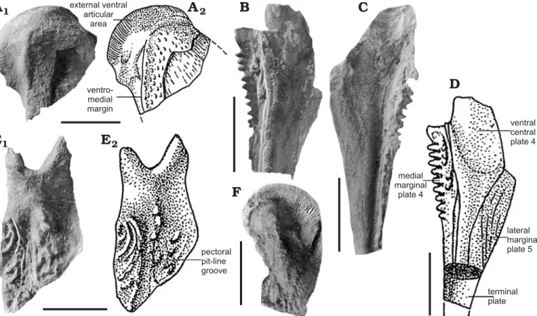
Pectoral fin.—Ventral central plate 1, CV1 (Fig. 7A, F): There are two specimens from Hingeon: a small one and a large one. They are both incomplete in the posterior portion. The ventro-medial margin is prominent and the external ven-tral articular area quite extended.
Medial marginal plate 1, Mm1 (Fig. 7E): This plate is entirely preserved and relatively short. The proximal part is forked and unornamented whereas the distal part is orna-mented. It is crossed proximo-distally by the pectoral pit-line groove. The posterior margin is slightly sinusoidal.
Distal part of the pectoral fin (Fig. 7B–D): A large part of the pectoral fin distal segment is preserved in ventral view. Neither the anteriormost part, nor the posteriormost part is preserved. The medial marginal plate 4 displays laterally short, spaced spines that are absent on the distal part of the plate. Gross (1965) noticed that a medial marginal plate 5 could be attached to the Mm4 but this is not observable. On the contrary, it is linked to the ventral central plate 4 (Cv4) by a strong edge. The posterior part of this plate is narrow, whereas the anterior part is large and flattened. It certainly corresponds to the insertion area of the pectoral fin proximal segment. Immersed in water or alcohol, the limit between the Cv4 and the lateral marginal 5 (ML5) is clearly observed. The ML5 is a long plate with a regular width. Its posterior limit is neither observable nor preserved. The terminal plate is not preserved, but a fragment of its impression might be present on the counterpart of the pectoral fin distal segment. However, its limits are not distinguishable.
Ornament.—The ornament is tuberculate on the nuchal plate. On the postpineal plate, some tubercles fuse to form small ridges. Some of them radiate from the centre of the plate. On the posterior median dorsal plate, Gross (1965: 4) defined the ornament as forming ramparts (“Wallbildung”). It is rather a reticulate ornament with some tubercles present. On the ante-rior ventro-lateral plate, the ornament is clearly tuberculate, with tubercles arranged in parallel rows. As for the ventral central plate 1, it bears few small, spaced tubercles, and the medial marginal plate bears huge, rough tubercles sometimes associated. The distal segment of the pectoral fin is devoid of ornament.
Remarks.—Compared to what Gross (1965) published the present work provides further details and the counterparts of some plates are figured here. The drawings made by Gross are very accurate and have been used here for the figures.
Gross (1965) described an indeterminate species of an-tiarch from the Givetian of Hingeon (Asterolepis sp. 1 in this paper). In spite of the small number of plates preserved, he assigned this form to Asterolepis, because all the bones matched that genus, and not those of other genera of antiarch. In fact, Pterichthyodes and Byssacanthus display shorter and wider postpineal plates. In Gerdalepis, the same plate bulg-es forward. The genus Bothriolepis is excluded because its medial marginal plate 1 is longer and narrower. The genus Remigolepis was not considered by Gross (1965), but the species from Hingeon clearly differs from that genus, notably by the organization of the pectoral fin plates. To date, and without supplementary material, the attribution of this materi-al to Asterolepis by Gross (1965) is not reappraised and seems
correct. He considered that the Asterolepis material from Hin-geon resembles more A. ornata than any other species, but that the material was insufficient to assign it to any particular species of Asterolepis. His cautiousness is here followed.
Asterolepis sp. 2
Fig. 8.
Material.—Unassigned plate of the trunk shield: IRSNB P.9260; Cd1: IRSNB P.9259. From Burtaux quarry, local-ity Alvaux, Mazy, Gembloux, Namur Province, Belgium. Alvaux Member, Bois de Bordeaux Formation, Givetian, Middle Devonian.
Description
Trunkshield.—Unassigned plate (Fig. 8A): The assignment of that plate is not possible due to the very bad preservation. It is a lateral plate of the trunk shield as indicated by the pres-ence of two laminae separated by a strong angle.
A
B
E
F
1F
2 middle pit-line groove obtected nuchal area supratemporal pit-line groove endolymphatic duct opening posterior median process postpineal notch growth line posterior median corner lateral process dorsal median ridge median ventral ridge area overlapping anterior median dorsal plate area overlapping mixilateral plate area overlapping anterior ventro-lateral plate ventro-lateral ridge axillary foramenC
2D
D
1Fig. 6. Dermal plates of antiarch placoderm Asterolepis sp. 1, from Hingeon (Namur Province), Upper Givetian, Devonian. A. IRSNB P.1457, Pp, external view. B. IRSNB P.1456, Nu, external view. C. IRSNB P.1456 and 1457, Pp and Nu, external view. D. IRSNB P.1458, PMD, external and internal views; photograph (D1), explanatory drawing (D2). E. IRSNB P.1459b, AVL, external and internal views, counter-part of IRSNB P.1459a. F. IRSNB P.1459a, AVL, external and internal views; photograph (F1), explanatory drawing (F2). Drawings from Gross 1965. Scale bars 10 mm.
Pectoral fin.—Dorsal central plate 1, Cd1 (Fig. 8B): A well preserved specimen is available. It is rather long. The exter-nal dorsal articular area anterior to the unornamented area, is finely covered with tiny meshes. The posterior margin, articulating with the dorsal central plate 2, is concave. The margin, articulating with the medial marginal plate 2 is elon-gated and strongly concave, as in Asterolepis ornata. On the lateral margin, the lateral corner is quite prominent. The medial margin is slightly damaged but no spine is observable. Ornament.—The ornament of Asterolepis sp. 2 is rather re-ticulate. On the unassigned plate, the ornament also consists
of fine, rounded and flat tubercles arranged in a concentric and radiating pattern.
Remarks.—The shape of the dorsal central plate and the rounded, flat tubercles of the ornamentation support the Asterolepis identification. The dorsal central plate strongly resembles the dorsal central plate of A. ornata (Gross 1931: pl. 5: 1a). Possibly, the specimens from Mazy could belong to the same form as the one from Hingeon. The amount and quality of the material in both localities is unfortunately not sufficient to confirm this conclusion.
Remigolepidae Stensiö, 1931
Genus Remigolepis Stensiö, 1931
Type species: Remigolepis incisa (Woodward, 1900); Ymer Island,
south of the Dusen Fjord, East Greenland, Famennian, Devonian.
Remigolepis durnalensis sp. nov.
Figs. 9–12.
2009 Bothriolepis; Clément and Prestianni 2009: 107.
Etymology: In reference to the locality, where the material has been
found.
Type material: Holotype: AMD: IRSNB P.9266a, b. Paratypes: Prm:
IRSNB P.9262; La: IRSNB P.9263a, b; Pp: IRSNB P.9264a, b; AMD: IRSNB P.9265a, b; PMD: IRSNB P.9267a, b; IRSNB P.9268a, b; ADL: IRSNB P.9269a, b; PDL: IRSNB P.9270a, b; IRSNB P.9271a, b;
external ventral articular area ventro-medial margin pectoral pit-line groove ventral central plate 4 lateral marginal plate 5 terminal plate medial marginal plate 4 2
A
A
1 2E
E
1C
D
F
B
B
2A
A
1 lateral corner external dorsal articular areaFig. 7. Dermal plates of antiarch placoderm Asterolepis sp. 1, from Hingeon (Namur Province), Upper Givetian, Devonian. A. IRSNB P.1460, CV1, external view; photograph (A1), explanatory drawing (A2). B. IRSNB 1462a, distal part of the pectoral fin, external view. C. IRSNB 1462b, distal part of the pectoral fin, external view, counterpart of IRSNB 1462a. D. Drawing based on IRSNB 1462a and b, distal part of the pectoral fin, external view. E. IRSNB P.1463, MM1, external view; photograph (E1), explanatory drawing (E2). F. IRSNB P.1461a, CV1, internal view. Drawings from Gross 1965. Scale bars 10 mm.
Fig. 8. Dermal plates of antiarch placoderm Asterolepis sp. 2., from Mazy (Namur Province), Givetian, Devonian. A. IRSNB P.9260, un assigned plate, external view; photograph (A1); explanatory drawing (A2). B. IRSNB P.9259, CD1, external view. Scale bars 10 mm.
PVL: IRSNB P.9272; PVL and PL: IRSNB P.9273a–d; CV1: IRSNB P.9261a, b; pectoral fin and AVL: IRSNB P.9274a, b.
Type locality: Tienne-des-Marteaux quarry also called Durnal 2, Bocq
valley, Spontin, Namur Province, Belgium.
Type horizon: Montfort/Evieux Formation, Famennian, Upper
Devo-nian.
Material.—PMD: IRSNB vert 32049-001a, b; ADL: IRSNB vert 32049-002 from the type locality.
Diagnosis.—Species of moderate size. Anterior edge of the Prm displaying a strong notch. Short postpineal plate. Dorsal wall of trunk-armour quite flat and reaching an estimated length of 15-16 cm. Tergal angle situated in the middle of the anterior median dorsal plate. Anterior margin of the anterior median dorsal plate narrow and representing one quarter of the posterior margin. Internal surface of the AMD displaying a median ventral ridge. Anterior and posterior oblique dorsal sensory line grooves well defined. Posterior median dorsal plate as long as broad. Anterior median angle of the posterior median dorsal plate prominent. Crista interna transversalis
posterior slightly developed. Processus obstans well devel-oped and articular fossa of the anterior dorso-lateral plate ex-tended. Crista transversalis interna anterior oblique and high. Pectoral fin massive, almost three times as long as broad and stretching slightly less far than the posterior margin of the anterior ventro-lateral plate. Vermiculate ornament forming on several specimens a radiating and concentric network. Tuberculate ornament on the largest plates.
Description
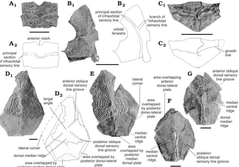
Headshield.—Premedian plate, Prm (Fig. 9A): It is almost as long as broad and crossed medially by the principal sec-tion of infraorbital canal. The anterior edge is marked in its middle by a not extended but well marked notch.
Lateral plate, La (Fig. 9B): According to its size, the only preserved lateral plate (IRSNB P.9263a, b) is most probably that of a juvenile. This plate is rather narrow with the pos-terior part broader than the anpos-terior one. The principal and secondary sections of infraorbital sensory line are deeply incised. The orbit seems small.
area overlapped by posterior median dorsal plate 2
A
A
1 2D
D
1 2B
B
1 2C
C
1 principal section of infraorbitalsensory line infraorbitalbranch of sensory line orbital fenestra principal section of infraorbital sensory line anterior notch growth line posterior oblique dorsal sensory line groove median ventral ridge dorsal median ridge area overlapping anterior dorso-lateral plate median ventral ridge posterior oblique dorsal sensory line groove lateral corner
dorsal median ridge area overlapped by posterior median dorsal plate
area overlapped by posterior dorso-lateral plate anterior oblique dorsal sensory line groove tergal angle lateral corner area overlapped by posterior dorso-lateral plate median ventral ridge anterior oblique dorsal sensory line groove
E
F
G
Fig. 9. Dermal plates of antiarch placoderm Remigolepis durnalensis sp. nov., from Spontin (Namur Province), Famennian, Devonian. A. IRSNB P.9262, Prm, external view, photographed under water immersion. B. IRSNB P.9263a, La, external view, photographed under water immersion. C. IRSNB P.9264a, Pp, external view, dotted line based on the counterpart IRSNB P.9264b, photographed under water immersion. D. IRSNB P.9265a, AMD, exter-nal and interexter-nal views, dotted line based on IRSNB P.9265b. E. IRSNB P.9265b, AMD, exterexter-nal and interexter-nal views. F. IRSNB P.9266b, AMD, exterexter-nal and internal views. G. IRSNB P.9266a, AMD, external and internal views. Photographs (A1–D1, E–G); explanatory drawings (A2–D2). Scale bars 10 mm.
Postpineal plate, Pp (Fig. 9C): The postpineal plate is wid-er than long. The antwid-erior margin is slightly convex whwid-ereas the posterior margin is strongly convex. The antero-lateral margin forms a 160° angle with the postero-lateral margin. The postpineal plate shows anteriorly the limits of a smaller plate corresponding probably to a younger growth stage. Trunkshield.—Anterior median dorsal plate, AMD (Fig. 9D–G): This plate is elongate. Externally, it is quite flat, with a slight arch constituted by the dorsal median ridge. The latter forms an obtuse angle and runs from the tergal angle, situated in the middle of the plate, backwards to the posterior margin. The dorsal median ridge is sharper on the smallest AMD (IRSNB P.9266) than on the largest one (IRSNB P.9265) where it is almost absent. The length of the anterior division of the plate is roughly the same as the pos-terior one. The anpos-terior straight margin is narrow and
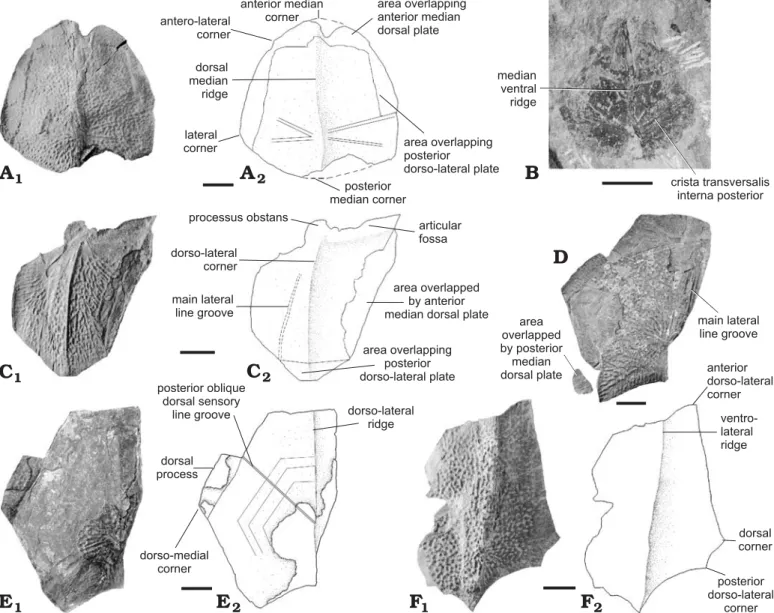
rep-resents one quarter of the straight posterior margin length. The lateral corners are more or less marked depending on the specimen considered. They are quite rounded on IRSNB P.9266 and more angular on IRSNB P.9265. The anterior part of the lateral margin is about twice longer than the pos-terior one. There is a dissymmetry of the lateral corners of IRSNB P.9266. The right lateral corner is situated more an-teriorly than the left one and is therefore at half the length of the lateral margin. The posterior portion of the lateral margin has the typical sigmoid shape of the genus Remigolepis. It overlaps the posterior dorso-lateral plate anteriorly and is overlapped by the posterior dorso-lateral plate posteriorly. The posterior oblique dorsal sensory line grooves are well defined. They form an angle of about 45° with the dorsal me-dian ridge. The posterior oblique dorsal sensory line grooves run from the tergal angle to the middle of the posterior part antero-lateral corner dorsal median ridge lateral corner anterior median corner area overlapping anterior median dorsal plate area overlapping posterior dorso-lateral plate posterior median corner median ventral ridge crista transversalis interna posterior processus obstans dorso-lateral corner area overlapping posterior dorso-lateral plate area overlapped by anterior median dorsal plate main lateral line groove articular fossa main lateral line groove area overlapped by posterior median dorsal plate posterior oblique dorsal sensory line groove dorsal process dorso-medial corner dorso-lateral ridge dorsal corner posterior dorso-lateral corner ventro-lateral ridge anterior dorso-lateral corner 2
A
A
1B
2C
C
1 2E
E
1F
1F
2D
Fig. 10. Dermal plates of antiarch placoderm Remigolepis durnalensis sp. nov., from Spontin (Namur Province), Famennian, Devonian. A. IRSNB P.9267a, PMD, external view, dotted line based on IRSNB P.9268. B. IRSNB P.9268a, PMD, internal view, photographed under water immersion. C. IRSNB P.9269a, ADL, external view. D. IRSNB P.9271a, PDL, external and internal views. E. IRSNB P.9270a, PDL, external and internal views. F. IRSNB P.9272, PVL, external view. Photographs (A1, B, C1, D, E1, F1); explanatory drawings (A2, C2, E2, F2). Scale bars 10 mm.