HAL Id: hal-02549520
https://hal.umontpellier.fr/hal-02549520
Submitted on 21 Apr 2020
HAL is a multi-disciplinary open access archive for the deposit and dissemination of sci-entific research documents, whether they are pub-lished or not. The documents may come from teaching and research institutions in France or abroad, or from public or private research centers.
L’archive ouverte pluridisciplinaire HAL, est destinée au dépôt et à la diffusion de documents scientifiques de niveau recherche, publiés ou non, émanant des établissements d’enseignement et de recherche français ou étrangers, des laboratoires publics ou privés.
Postoperative Patients: A Crossover Randomized Study
Yannaël Coisel, Gerald Chanques, Boris Jung, Jean-Michel Constantin, Xavier
Capdevila, Stefan Matecki, Salvatore Grasso, Samir Jaber
To cite this version:
Yannaël Coisel, Gerald Chanques, Boris Jung, Jean-Michel Constantin, Xavier Capdevila, et al.. Neurally Adjusted Ventilatory Assist in Critically Ill Postoperative Patients: A Crossover Randomized Study. Anesthesiology, Lippincott, Williams & Wilkins, 2010, 113, pp.925 - 960. �10.1097/ALN.0b013e3181ee2ef1�. �hal-02549520�
Neurally Adjusted Ventilatory Assist in Critically Ill
Postoperative Patients: A Crossover Randomized Study
Yannael Coisel, M.D.,* Gerald Chanques, M.D.,† Boris Jung, M.D.,† Jean-Michel Constantin, M.D., Ph.D.,‡ Xavier Capdevila, M.D., Ph.D.,§ Stefan Matecki, M.D., Ph.D.,! Salvatore Grasso, M.D., Ph.D.,#Samir Jaber, M.D., Ph.D.**
ABSTRACT
Background: Neurally adjusted ventilatory assist (NAVA) is
a new mode of mechanical ventilation that delivers ventilatory assist in proportion to the electrical activity of the diaphragm. This study aimed to compare the ventilatory and gas exchange effects between NAVA and pressure support ventilation (PSV) during the weaning phase of critically ill patients who required mechanical ventilation subsequent to surgery.
Methods: Fifteen patients, the majority of whom underwent
abdominal surgery, were enrolled. They were ventilated with PSV and NAVA for 24 h each in a randomized crossover order. The ventilatory parameters and gas exchange effects produced by the two ventilation modes were compared. The variability of the ventilatory parameters was also evaluated by the coefficient of variation (SD to mean ratio).
Results: Two patients failed to shift to NAVA because of
postoperative bilateral diaphragmatic paralysis, and one pa-tient interrupted the study because of worsening of his
sick-ness. In the other 12 cases, the 48 h of the study protocol were completed, using both ventilation modes, with no signs of in-tolerance or complications. The PaO2/FIO2(mean ! SD) ratio in NAVA was significantly higher than with PSV (264 ! 71 vs. 230 ! 75 mmHg, P " 0.05). PaCO2did not differ significantly between the two modes. The tidal volume (median [interquar-tile range]) with NAVA was significantly lower than with PSV (7.0 [6.4–8.6] vs. 6.5 [6.3–7.4] ml/kg predicted body weight, P " 0.05).Variability of insufflation airway pressure, tidal vol-ume, and minute ventilation were significantly higher with NAVA than with PSV. Electrical activity of the diaphragm vari-ability was significantly lower with NAVA than with PSV.
Conclusions: Compared with PSV, respiratory parameter
variability was greater with NAVA, probably leading in part to the significant improvement in patient oxygenation.
P
RESSURE support ventilation (PSV) is the most widely used assisted mode of ventilation during the weaning pro-cess in medical and surgical critically ill patients.1However, PSV provides a fixed end-inspiratory pressure (i.e., level of assistance), regardless of the patient’s ventilatory demand or gas exchange, which limits breathing pattern variability.2–5Given the high variability in disease processes and states, the application of predefined, uniform values for ventilator parameters, such as a fixed end-inspiratory pressure or tidal volume (VT), is un-likely to provide optimal assist at all times. Compared with the monotonous breathing pattern resulting from the limited variability of end-inspiratory pressure, variations of the breathing pattern may be useful to improve gas exchange.6* Research Fellow, † Assistant Professor, ** Professor and Chairman, Intensive Care Unit, Anesthesiology and Critical Care Department B, Saint Eloi Teaching Hospital, Montpellier, France. § Professor and Chairman, Anesthesia and Critical Care Department A, Lapeyronie Teaching Hospital, Montpellier, France.! Professor, Clinical Physiology Center, Arnaud de Villeneuve Teaching Hospital, Equipe soutenue par la Re´gion et l’Institut National de la Sante´ et de la Recherche Me´dicale 25, Universite´ Montpellier 1, Centre Hospitalier Universitaire Montpel-lier, MontpelMontpel-lier, France. ‡ Professor, Intensive Care Unit, Anesthesia and Critical Care Department, Hotel-Dieu Hospital, University Hospital of Clermont-Ferrand, Clermont-Ferrand, France. # Associate Professor of Anesthesiology and Critical Care, Dipartimento dell’Emergenza e Trapianti d’Organo, Sezione di Anestesiologia e Rianimazione, Univer-sita` degli Studi di Bari, Bari, Italy.
Received from the Intensive Care Unit, Anesthesia and Critical Care Department B, Saint Eloi Teaching Hospital, Montpellier, France. Submitted for publication September 13, 2009. Accepted for publication June 4, 2010. Maquet Critical Care France provided the ventilator and the EAdi catheter used in the study (Maquet Critical Care, Sölna, Sweden). Presented in part at the 2009 Annual Meeting of French Society of Anesthesiology and Intensive Care, Paris, France, September 23–26, 2009.
Address correspondence to Dr. Jaber: Anesthesia and Critical Care Department B, University Montpellier, 80 Avenue Augustin Fliche, 34295 Montpellier, Cedex 5, France. [email protected]. Informa-tion on purchasing reprints may be found at www.anesthesiology.org or on the masthead page at the beginning of this issue. ANESTHESIOLOGY’s articles are made freely accessible to all readers, for personal use only, 6 months from the cover date of the issue.
What We Already Know about This Topic
❖Neurally adjusted ventilatory assist (NAVA) is one of several modes of ventilation that permits variations in breathing pat-terns and perhaps more patient-ventilator synchrony.
What This Article Tells Us That Is New
❖In a prospective, randomized crossover study of 12 surgical patients requiring prolonged ventilation, there was improved oxygenation and increased respiratory variability when the pa-tients were on NAVA for 24 consecutive hours compared with these parameters when they received pressure support ventilation.
This observation was mainly obtained with animal studies,6–8 which evaluated new ventilatory modes, including variability in their function, and more recently in a human study with the Neu-rally Adjusted Ventilatory Assist (NAVA) mode.9,10
NAVA is a new ventilatory mode wherein the ventilator delivers positive pressure during inspiration in proportion to the electrical activity of the diaphragm (EAdi) obtained by a
naso-gastric tube covered by electrodes that record and analyze trans-esophageal electromyography.11,12The amount of assistance for a given EAdi depends on a user-gain factor called “NAVA level.” Each change in the patient’s ventilatory demand can theoreti-cally be rewarded by the ventilator. Like proportional assist ven-tilation,2,13,14NAVA ensures a positive relationship between the ventilator assistance and the patient’s effort. Unique to NAVA is the identification of the start of neural exhalation, which is not recognized by assist-control ventilation or PSV. The NAVA characteristics could have clinical implications, such as better patient-ventilator synchrony and a more natural (“noisy”) breathing pattern, leading to improved comfort and oxygenation. NAVA has been studied in animals,15,16healthy subjects17, and critically ill patients9,10,18but only for 20 min10 to 3 h.9To our knowledge, no physiologic study has been per-formed to evaluate the use of NAVA for a prolonged mechanical ventilation (MV) period in selected critically ill patients.
The aim of this prospective, randomized, crossover study was to investigate, in a homogenous group of postoperative patients, during the weaning phase of their illness, the 24-h effects of NAVA on ventilatory parameters and gas exchange and to compare these with those observed with PSV. We hypothesized that in this group of patients, characterized by respiratory modifications related to surgery, NAVA would improve oxygenation because it offers a more variable venti-lation, which is a more physiologic ventilation.19,20
Materials and Methods
The experimental protocol was approved by the Ethics Com-mittee of the Saint-Eloi Teaching Hospital (Comite´ de Protec-tion des Personnes Sud Me´diterrane´e IV, Montpellier, France), and written informed consent was provided by the patient or next of kin. Our study followed the CONSORT recom-mendations concerning the report of randomized trials.21
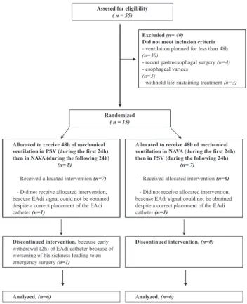
Assesed for eligibility ( n = 55)
Excluded (n= 40) Did not meet inclusion criteria
- ventilation planned for less than 48h
(n=30)
- recent gastroesophagal surgery (n=4) - esophageal varices
(n=3)
- withhold life-sustaining treatment (n=3)
Randomized Randomized ( n = 15) Allocated to receive 48h of mechanical ventilation in PSV (during the first 24h) then in NAVA (during the following 24h)
Allocated to receive 48h of mechanical ventilation in NAVA (during the first 24h) then in PSV (during the following 24h) then in NAVA (during the following 24h)
(n= 8)
- Received allocated intervention (n=7) - Did not receive allocated intervention, beacuse EAdi signal could not be obtained
then in PSV (during the following 24h) (n= 7)
- Received allocated intervention (n=6) - Did not receive allocated intervention, beacuse EAdi signal could not be obtained beacuse EAdi signal could not be obtained
despite a correct placement of the EAdi catheter (n=1)
Discontinued intervention because early
beacuse EAdi signal could not be obtained despite a correct placement of the EAdi catheter (n=1)
Discontinued intervention (n=0) Discontinued intervention, because early
withdrawal (2h) of EAdi catheter because of worsening of his sickness leading to an emergency surgery (n=1)
Discontinued intervention, (n 0)
Analyzed, (n=6) Analyzed, (n=6)
Fig. 1. Trial profile. EAdi # electrical activity of the dia-phragm; NAVA # neurally adjusted ventilatory assist; PSV # pressure support ventilation.
Table 1. Characteristics of the 12 Patients Studied
Patient Sex Age, yr Height, cm Weight, cm SAPSII Procedure
Time Between Total Duration of Ventilation, Days Outcome Surgery and Inclusion, Days Weaning and Inclusion, Days 1 F 43 150 50 68 Laparotomy for hemoperitoneum 6 1 13 D 2 M 76 175 70 36 Pulmonary lobectomy 35 20 43 D 3 M 77 168 58 47 Colectomy 4 3 6 S 4 F 71 155 68 55 Abdominal parietal hematoma 0.5 3 8 D 5 M 70 176 120 80 Peritonitis 6 1 47 S 6 M 73 175 97 33 Rachis surgery 3 1 84 S 7 F 68 160 48 38 Peritonitis 47 42 37 S 8 M 75 160 50 85 Hepatectomy 16 1 12 D 9 M 86 170 85 71 Peritonitis 1 1 4 S 10 F 84 145 90 47 Cardiac surgery 6 1 15 S 11 F 77 170 90 70 Peritonitis 6 1 40 D 12 M 85 180 123 45 Peritonitis 3 1 21 S — 76 [71—79] 169 [159—175] 78 [56—92] 51 [43—70] — 6 [3–9] 1 [1–3] 18 [11–41] — Summary data are presented as median [interquartile range].
Patients
Fifteen patients were prospectively enrolled from March 2009 to June 2009. They had been mechanically ventilated via an endotracheal tube for more than 48 h with PSV levels of 6 to 15 cm H2O above 2 to 10 cm H2O of positive end-expiratory pressure. The following inclusion criteria were used: ventilation planned for more than 48 h and pa-tient alert and calm corresponding to a Richmond Agita-tion–Sedation Scale (RASS) between $2 and 0.22–24The following exclusion criteria were mainly related to the clinical contraindication for the use of NAVA: contraindications for an EAdi catheter placement (e.g., esophageal varices, upper gastrointestinal bleeding, gastroesophageal surgery) and clin-ical instability for any reason. Patients for whom the decision to withhold life-sustaining treatment had been made, preg-nant women, and children were also not considered.
Methods
The two ventilatory modes (PSV and NAVA) were delivered by the same ventilator (Servo-I; Maquet Critical Care, So¨lna, Sweden) and were set to provide similar MV.4In PSV, there was only a flow inspiratory trigger. In NAVA, the ventilator can be cycled on by two different algorithms, based on either EAdi, or Paw or flow, according to a hierarchy that follows the principle that “first-serves-first.” There were flow and neural inspiratory triggers that detected first-caused activa-tion of the pressure assist. The fracactiva-tion of inspired oxygen (FIO2) was set to achieve oxygen saturation greater than 95%. The positive end-expiratory pressure level was set between 2 and 10 cm H2O and kept constant throughout the study.
The PSV level was first applied for 5 min to determine the inspiratory pressure level required to obtain a VT between 6 and 8 ml/kg predicted body weight (PBW)25,26(as calculated with the following formulas for men and women, respec-tively: PBW (kg) # 50 % 2.3 [(height (cm)/2.54) $ 60] and PBW (kg) # 45.5 % 2.3 [(height (cm)/2.54) $ 60]) with a respiratory rate (RR) between 20 and 30 breaths/min; the resulting values of minute ventilation (VE, calculated as the product of VT and RR) were used to set NAVA.4
As described previously,17,27EAdi was obtained through a nasogastric tube with a multiple array of electrodes placed at its distal end (EAdi catheter; Maquet Critical Care). Correct posi-tioning of the EAdi catheter was assured by means of a specific function of the ventilator (“EAdi catheter positioning”). The Table 2. Ventilatory Settings and Main Monitored Ventilatory Parameters Obtained at the Baseline of Each
Ventilatory Period for PSV and NAVA
Parameters PSV (n # 12) NAVA (n # 12) P Value
Ventilatory Settings — — —
Pressure Support Level, cm H2O 11 ! 3 NA —
NAVA Level, cm H2O/!V NA 1.9 ! 1.5 —
Flow Inspiratory Trigger, l/min 2 ! 0 2 ! 0 NS
Neural Inspiratory Trigger, !V NA 0.5 ! 0 —
Flow Expiratory Trigger, % of maximal peak flow value 30 ! 0 NA — Neural Expiratory Trigger, % of maximal peak EAdi value NA 30 ! 0 —
Inspiratory Rise, % 5 ! 0 NA —
Oxygen Inspired Fraction, % 49 ! 13 46 ! 13 NS
PEEP, cm H2O 6 ! 2 6 ! 2 NS
Monitored Ventilatory Parameters — — —
EAdi, !V 7.5 [6.4–12.4] 8.5 [6.3–14.2] NS Maximal Pinsp, cm H2O 18 [15–21] 19 [15–24] NS Mean Pinsp, cm H2O 9 [8–10] 9 [7–11] NS RR, breaths/min 27.4 [18.1–28.4] 25.1 [21.9–28.8] NS VT, ml 460 [376–527] 399 [338–445] NS VT, ml/kg PBW 7.5 [5.6–8.4] 6.0 [5.1–8.0] NS VE, l/min 10.4 [8.5–11.7] 10.1 [9.2–11.5] NS PETCO2, mmHg 30.6 [23.3–33.1] 30.4 [24.5–33.0] NS P0.1, cm H2O 1.6 [1.1–2.2] 0.9 [0.7–1.1] NS
Data are presented as mean ! SD for ventilatory settings and as median [interquartile range] for monitored ventilatory parameters. EAdi # electrical activity of the diaphragm; NA # not applicable; NS # not significant; NAVA # neurally adjusted ventilatory assist; P0.1# occlusion pressure; PBW # predicted body weight; PEEP # positive end-expiratory pressure; PETCO2 # end-tidal partial
pressure of carbon dioxide; Pinsp# inspiratory airway pressure; PSV # pressure support ventilation; RR # respiratory rate; VE # minute
ventilation; VT # tidal volume.
Table 3. Gas Exchange Parameters PSV (n # 11) NAVA (n # 11) pH 7.45 ! 0.06 7.44 ! 0.06 PaCO2, mmHg 41 ! 9 39 ! 7 PaO2, mmHg 108 ! 27 117 ! 32 HCO3$, mM 29 ! 7 27 ! 6 SaO2, % 98 ! 2 98 ! 2 PaO2/FIO2, mmHg 230 ! 75 264 ! 71*
Data are presented as mean ! SD.
*P " 0.05 significantly different from the value with PSV.
FIO2# oxygen inspired fraction; NAVA # neurally adjusted ven-tilatory assist; PaCO2# partial pressure of arterial carbon dioxide;
PaO2 # partial pressure of arterial oxygen; PSV # pressure
EAdi signal was processed according to the American Thoracic Society recommendations28and filtered by algorithms designed to provide the highest possible signal-to-noise ratio. To avoid interference secondary to variations in lung volume and chest wall configuration,28changes in diaphragm position along the array were also considered.17,29EAdi was quantified every 16 ms using the root mean square.12,17Portions of signal with residual disturbances were removed and replaced by the values of the previous segment.15 The amount of pressure instantaneously applied by the ventilator to the airway opening throughout in-spiration was determined by the processed EAdi, expressed in microvolts, multiplied by a user-controlled gain factor (“NAVA level”) expressed as centimeters of H2O per microvolt. The amount of assistance depended on the magnitude of both the EAdi signal and the NAVA level. In NAVA, the ventilator can be cycled on inspiration to expiration by two different algo-rithms, based on EAdi or fluid dynamics (airway pressure or flow), according to a hierarchy that follows the principle of “first-come, first-served.” During NAVA, the ventilator was cy-cled off when the EAdi decreased at 70% of its peak inspiratory value.10In the case of a disturbance or a disappearance of the
EAdi signal during ventilation in NAVA (e.g., EAdi catheter moving, accidental removal of EAdi catheter), the ventilator automatically converted to PSV (independently of the EAdi signal). When the EAdi signal became valid and useable, the ventilator automatically switched from PSV to NAVA. As mentioned previously, the NAVA level was set to obtain the same amount of assistance (corresponding to the same VE and RR) as determined by prior use of PSV during 5 min.
Protocol
We applied a crossover study design very similar to that pre-viously reported by Dojat et al.30and Sydow et al.31 Deter-mination of the type of ventilatory mode used was performed weekly using a cluster randomization, the randomized type of ventilatory mode being used during 7 consecutive days. Each patient was consecutively ventilated for 24 h with the PSV mode and with the NAVA mode in random order. At inclusion, the patients were ventilated using settings previ-ously adjusted by the attending physician. In the PSV mode, the physician in charge modified the PSV level by 2 cm H2O, per the standard of care of the unit. In the NAVA mode,
PSV
NAVA
300 400 m Hg ) 300 400 m Hg ) NS P < 0.01 100 200 PaO 2 /FiO 2 (m m 100 200 Pa O 2 /F iO 2 (m m 0 Baseline H-24 P 0 Baseline H-24A
B
Fig. 2. Individual variations in PaO2/FIO2ratio for 11 of 12 patients after mechanical ventilation with pressure support ventilation
(PSV;A) and with neurally adjusted ventilatory assist (NAVA; B). The horizontal bars represent the mean values. The unbroken
line indicates NAVA then PSV; the dashed line indicates PSV then NAVA. FIO2# inspired oxygen fraction; PaO2# partial
pressure of arterial oxygen.
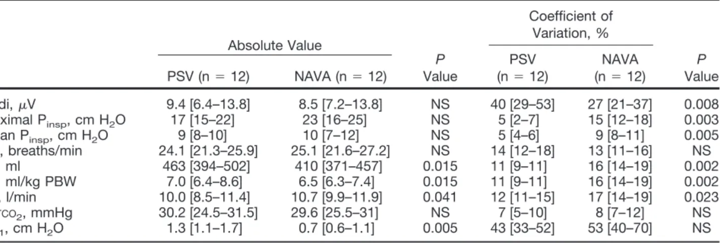
Table 4. Ventilatory Parameters Obtained during 24 h for Each Ventilatory Period in PSV and in NAVA
Absolute Value P Value Coefficient of Variation, % P Value PSV (n # 12) NAVA (n # 12) PSV (n # 12) NAVA (n # 12) EAdi, !V 9.4 [6.4–13.8] 8.5 [7.2–13.8] NS 40 [29–53] 27 [21–37] 0.008 Maximal Pinsp, cm H2O 17 [15–22] 23 [16–25] NS 5 [2–7] 15 [12–18] 0.003 Mean Pinsp, cm H2O 9 [8–10] 10 [7–12] NS 5 [4–6] 9 [8–11] 0.005 RR, breaths/min 24.1 [21.3–25.9] 25.1 [21.6–27.2] NS 14 [12–18] 13 [11–16] NS VT, ml 463 [394–502] 410 [371–457] 0.015 11 [9–11] 16 [14–19] 0.002 VT, ml/kg PBW 7.0 [6.4–8.6] 6.5 [6.3–7.4] 0.015 11 [9–11] 16 [14–19] 0.002 VE, l/min 10.0 [8.5–11.4] 10.7 [9.9–11.9] 0.041 12 [11–15] 17 [14–19] 0.023 PETCO2, mmHg 30.2 [24.5–31.5] 29.6 [25.5–31] NS 7 [5–10] 8 [7–12] NS P0.1, cm H2O 1.3 [1.1–1.7] 0.7 [0.6–1.1] 0.005 43 [33–52] 53 [40–70] NS
Data are presented as median [interquartile range].
EAdi # electrical activity of the diaphragm; NS # not significant; NAVA # neurally adjusted ventilatory assist; P0.1# occlusion pressure;
PBW # predicted body weight; Pinsp# inspiratory airway pressure; PETCO2# end-tidal partial pressure of carbon dioxide; PSV #
physicians could modify the NAVA level by steps of 0.2 cm H2O/!V if signs of respiratory distress were observed. For both modes, the clinician aimed to maintain the patient in the zone defined by the initial settings—to obtain a VT between 6 and 8 ml/kg of PBW with a RR between 20 and 30 breaths/min. Throughout the protocol, suctioning via the endotracheal tube was performed as needed.
Measurements
Standard three-lead monitoring electrodes continuously re-corded heart rate and rhythm. Oxygen saturation was con-tinuously monitored using pulse oxymetry. Systolic and di-astolic arterial blood pressures were continuously monitored
through a 20-gauge catheter inserted in a radial or femoral artery. Blood samples were obtained at baseline (in the first hour after MV for each mode) and after 24 h of MV for arterial blood gas analysis (GEM Premier 3000 analyzer; In-strumentation Laboratory, Lexington, MA) through the ar-terial catheter.
EAdi was measured with an array of electrodes mounted on a nasogastric tube. Airflow, airway pressure, VT, “esti-mated occlusion pressure” (P0.1, defined as the airway pres-sure generated 100 ms after the onset of an occluded inspi-ration, identified as an estimation of the respiratory neuromuscular drive),32,33and end-tidal partial pressure of carbon dioxide were obtained from the ventilator. From the flow signal, we obtained ventilatory rate of cycling (RR). The signals for EAdi, airflow, airway pressure, VT, RR, and P0.1 were monitored continuously online every 3 s, averaged ev-ery minute, recorded by means of a dedicated software (NAVA recording SV1.3; Maquet Critical Care), exported through a card, and analyzed using a customized software.
Every 4 h, according to our local protocol, the nurse in charge of the patient evaluated the pain and comfort using the Behavioral Pain Scale (BPS)23,34and the sedation and agitation level using the RASS.22,23 The BPS evaluates three behavioral domains (i.e., facial expression, move-ments of upper limbs, and compliance with ventilator). Each domain contains four descriptors that are rated on a 1-to-4 scale, and the total BPS value can range from 3 (no pain and excellent comfort) to 12 (most pain with
maxi-Fig. 3. Experimental records that help illustrate the effects of the two ventilatory modes during 24 h of mechanical venti-lation with pressure support ventiventi-lation (PSV; A) and with
neurally adjusted ventilatory assist (NAVA;B) in a
represen-tative patient. Note that Paw, VT, and VE are more variable in NAVA than in PSV. c/min # breaths per minute; EAdi # electrical activity of the diaphragm; P0.1# occlusion pres-sure; Paw# airway pressure; PETCO2# end-tidal partial pres-sure of carbon dioxide; RR # respiratory rate; VE # minute ventilation; VT # tidal volume.
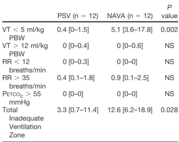
Table 5. Percentage of Time Spent in Inadequate Ventilation Zone in PSV and in NAVA
PSV (n # 12) NAVA (n # 12) P value VT " 5 ml/kg PBW 0.4 [0–1.5] 5.1 [3.6–17.8] 0.002 VT & 12 ml/kg PBW 0 [0–0.4] 0 [0–0.6] NS RR " 12 breaths/min 0 [0–0.3] 0 [0–0] NS RR & 35 breaths/min 0.4 [0.1–1.8] 0.9 [0.1–2.5] NS PETCO2& 55 mmHg 0 [0–0] 0 [0–0] NS Total Inadequate Ventilation Zone 3.3 [0.7–11.4] 12.6 [6.2–18.9] 0.028
Data are presented as median percentage time of spent in inad-equate ventilation during 24 h of the studied period. Inadinad-equate ventilation zone is defined as follows: low VT # VT " 5 ml/kg of PBW; high VT # VT & 12 ml/kg PBW; low RR # RR " 12 breaths/min), high RR # RR & 35 breaths/min): high PETCO2#
PETCO2& 55 mmHg.
NAVA # neurally adjusted ventilatory assist; NS # not significant; PBW # predicted body weight; PETCO2# end-tidal partial
pres-sure of carbon dioxide; PSV # prespres-sure support ventilation; RR # respiratory rate; VE # minute ventilation; VT # tidal volume.
Fig. 4. Contributions to inadequate ventilation of low VT (VT less than 5 ml/kg of PBW), high VT (VT higher than 12 ml/kg PBW), low RR (RR less than 12 c/min), high RR (RR higher than 35 c/min), and high PETCO2(PETCO2higher than 55 mmHg) during 24 h
of pressure support ventilation (PSV;A) and neurally adjusted ventilatory assist (NAVA; B) in the 12 studied patients. With PSV,
inadequate ventilation represented 3% [1–11%] of the total ventilation duration in this mode; with NAVA, inadequate ventilation represented 13% [6 –19%] of the total ventilation duration in this mode. c/min # breaths per minute; EAdi # electrical activity of the diaphragm; P0.1# occlusion pressure; Paw# airway pressure; PBW # predicted body weight; PETCO2# end-tidal partial
pressure of carbon dioxide; RR # respiratory rate; VE # minute ventilation; VT # tidal volume.
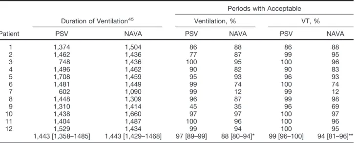
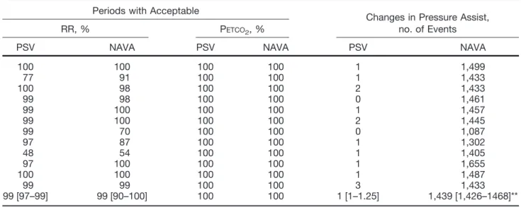
Table 6. Time Spent with an Acceptable Ventilation during PSV and NAVA
Patient
Duration of Ventilation45
Periods with Acceptable Ventilation, % VT, %
PSV NAVA PSV NAVA PSV NAVA
1 1,374 1,504 86 88 86 88 2 1,462 1,436 77 87 99 95 3 748 1,436 100 95 100 96 4 1,496 1,462 90 82 90 83 5 1,708 1,459 95 93 96 93 6 1,481 1,449 99 74 100 74 7 602 1,090 99 12 99 12 8 1,448 1,309 96 87 99 98 9 1,310 1,414 45 35 96 69 10 1,438 1,660 97 97 100 97 11 1,404 1,487 100 96 100 96 12 1,529 1,434 99 94 100 95 1,443 [1,358–1485] 1,443 [1,429–1468] 97 [89–99] 88 [80–94]* 99 [96–100] 94 [81–96]**
Acceptable ventilation is defined as VT between 5 and 12 ml/kg of predicted body weight, RR between 12 and 35 breaths/min, and PETCO2 " 55 mmHg. Periods are expressed as the percentage of the total duration of ventilation with the corresponding mode.
Summary data are expressed as median [interquartile range].
*P " 0.05 between PSV and NAVA; ** P " 0.01 between PSV and NAVA.
NAVA # neurally adjusted ventilatory assist; PETCO2# end-tidal partial pressure of carbon dioxide; PSV # pressure support ventilation;
mal discomfort). Setting changes made by the attending physician were also recorded.
Statistical Analysis
The primary endpoint was oxygenation, estimated by calcu-lation of the PaO2/FIO2ratio obtained after 24 h of MV in each mode. We used data from studies performed by our group.3,4In these studies, in the subgroup of postoperative patients, PaO2/FIO2ratio was 202 ! 48 mmHg. Assuming an "risk of 0.05 and a # risk of 0.20, we calculated that at least 12 patients would be required to identify an increase of 25% PaO2/FIO2ratio with NAVA. Therefore, we decided to in-clude 15 patients. The secondary endpoints were the vari-ability of the ventilatory parameters and the ventilatory com-fort. The variability of the ventilatory parameters was evaluated by the coefficients of variation for airway pressure, EAdi, RR, VT, VE, end-tidal partial pressure of carbon di-oxide, and P0.1, which were calculated (SD to mean ratio multiplied by 100) as described previously.10,20,35The ven-tilatory comfort of the patient was evaluated by time spent in acceptable ventilation, defined as 12 less than RR less than 35 breaths/min, 5 less than VT less than 12 ml/kg of PBW, and end-tidal partial pressure of carbon dioxide less than 55 mmHg.25,30Then, we chose to define inadequate ventilation zone as low-tidal volume (VT less than 5 ml/kg of PBW), high VT (VT more than 12 ml/kg of PBW), low RR (RR less than 12 breaths/min), high RR (RR more than 35 breaths/ min), and high end-tidal partial pressure of carbon dioxide more than 55 mmHg. Time spent with P0.1higher than 4 cm H2O was also calculated.30
Values are expressed as mean ! SD or median [interquar-tile range], according to the type of variable distribution.
Normality of the distribution was assessed with Kolmog-orov-Smirnov test. For the ventilatory variables (EAdi, air-flow, airway pressure, VT, RR, and P0.1) recorded every minute, the averaged values obtained during the 24 h of MV were used for comparisons between PSV and NAVA. Data were analyzed by paired Student t tests or Wilcoxon tests, according to their distribution. All P values were two-tailed and a P value less than 0.05 was considered significant. Sta-tistical analysis was performed using SAS/STAT software version 8.1 (SAS Institute, Cary, NC).
Results
During the 3 months of the study, we screened 55 patients and enrolled 15 consecutive postoperative patients, 3 of whom did not complete the study and could not be included in the data analysis (fig. 1). For two of the excluded patients, we could not obtain an EAdi signal despite a correct place-ment of the EAdi catheter; the third excluded patient dropped out of the study because of worsening of his sickness leading to an emergency surgery. The causes of respiratory failure of the 12 patients who concluded the study were abdominal postoperative acute respiratory failure (n # 9), cardiothoracic postoperative acute respiratory failure (n # 2), and neurosurgical postoperative acute respiratory failure (n # 1). No patients were tracheotomized. Clinical charac-teristics of the 12 patients who concluded the 48 h of the study protocol are shown in table 1. Five (42%) patients died, reflecting the selected postoperative population. No significant differences between PSV and NAVA were ob-served at baseline for all studied parameters (table 2). The main characteristics of the ventilatory settings of the two modes are summarized in table 2. Gain level changes made Table 6. Continued
Periods with Acceptable
Changes in Pressure Assist, no. of Events
RR, % PETCO2, %
PSV NAVA PSV NAVA PSV NAVA
100 100 100 100 1 1,499 77 91 100 100 1 1,433 100 98 100 100 2 1,433 99 98 100 100 0 1,461 99 100 100 100 1 1,457 99 100 100 100 2 1,445 99 70 100 100 0 1,087 97 87 100 100 1 1,302 48 54 100 100 1 1,405 97 100 100 100 1 1,655 100 100 100 100 1 1,487 99 99 100 100 3 1,433 99 [97–99] 99 [90–100] 100 100 1 [1–1.25] 1,439 [1,426–1468]**
by the attending physician in NAVA were never necessary in seven patients and were performed three, five, and nine times in three, one, and one patients, respectively. Among the 12 studied patients, 5 patients have never presented a switch from NAVA to PSV during the 24 h of NAVA ventilation period. For the remaining seven patients, NAVA was auto-matically switched (safety back-up) in PSV mode 7 [3–11] times, and the duration of each PSV use before a new automatic switch to NAVA lasted 28 [22–57] s, which corresponds to less than 0.5% of the total period of ventilation in NAVA.
Arterial blood samples were obtained in 11 of the 12 studied patients. In subject 12, arterial blood samples were not available for logistical reasons. The PaO2/FIO2ratio was similar at baseline between the two modes and it was signif-icantly higher with NAVA than with PSV after 24 h of me-chanical ventilation (table 3 and fig. 2).
Ventilatory parameters are reported in table 4. VT and P0.1were significantly lower with NAVA than with PSV. VE was significantly higher with NAVA than with PSV. Vari-ability of airway pressure, VT, and VE were all significantly higher with NAVA than with PSV. EAdi variability was sig-nificantly lower with NAVA than with PSV (table 4). Typi-cal tracing of main ventilatory parameters obtained during 24 h of MV in a patient (patient 11) with PSV and NAVA are shown in figure 3.
The time spent with inadequate ventilation was broken down into periods of low VT, high VT, low RR, high RR, and high end-tidal partial pressure of carbon dioxide, accord-ing to the definitions in the method section. The percentage of time spent with inadequate ventilation was significantly higher with NAVA than with PSV, related mainly to a low VT (table 5 and fig. 4). Table 6 shows individual time spent with an acceptable ventilation and number of changes in pressure assist during PSV and NAVA. The time spent with a P0.1higher than 4 cm H2O was significantly lower with NAVA than with PSV (1 [0 –5] min vs. 11 [1–56] min, P "
0.05). No significant differences were observed between the two modes for the BPS and RASS scores over the study period (fig. 5).
Discussion
The present study demonstrates that (1) the use of NAVA for a long period of 24 h of mechanical ventilation was feasible for selected postoperative critically ill patients; (2) oxygen-ation with NAVA was improved in comparison to PSV; and (3) variability of main ventilatory parameters (airway pres-sure, VT, and VE) was significantly higher with NAVA than with PSV, likely because of a more physiologic patient/ven-tilator adaptation.
Feasibility of Prolonged Mechanical Ventilation in NAVA
When introducing a new ventilatory mode, it is necessary to compare it with the standard of care treatment, which in our unit is PSV. To our knowledge, this is the first study to report NAVA use for 24 consecutive h in postoperative critically ill patients and to compare the ventilatory behavior with that observed with PSV. The two published studies on NAVA performed in intensive care unit patients report durations of only 20 min10and 3 h.9Moreover, our population consisted of only surgical patients (mainly abdominal surgery), whereas in the previous studies, populations were heteroge-neous (medical and surgical patients). In contrast to previous studies, which included mixed medical and surgical patients, we chose to evaluate NAVA only in postoperative patients, the majority after abdominal surgery procedures, because we also wondered if NAVA worked satisfactorily in patients at risk for postoperative diaphragmatic dysfunction. We found that with two patients, one operated on for liver transplan-tation and one for a colectomy, the EAdi signal necessary for NAVA was absent or too weak, despite the correct position-ing of the EAdi catheter. For these two patients, NAVA
A
B
PSV NAVA PSV NAVAR
A
S
S
S
P
B
5 12 Maximum +1 +4 Maximum 3 4 Minimum - 5 0 - 1 H0 H4 H8 H12 H16 H20 H24 Time (h) H0 H4 H8 H12 H16 H20 H24 Time (h) 5 MinimumFig. 5. (A) Time courses of pain and comfort evaluation performed every 4 h using the Behavioral Pain Scale (BPS) within 24 h of mechanical ventilation in pressure support ventilation (PSV) and neurally adjusted ventilatory assist (NAVA). No significant difference was observed between the two modes at any time. The total BPS value can range from 3 (no pain and excellent comfort) to 12 (most pain with maximal discomfort). (B) Time courses of sedation and agitation evaluation performed every 4 h
using the Richmond Agitation Sedation Scale (RASS) within 24 h of mechanical ventilation in PSV and NAVA. No significant difference was observed between the two modes at any time. The RASS value can range from $5 (unarousable) to %4 (combative).
allowed us to diagnose postoperative severe diaphragmatic dysfunction. The incidence of diaphragmatic dysfunction varies from 10% to 30% in postabdominal surgery.36,37This is the first study to report limitations for the use of NAVA postoperatively with patients having diaphragmatic dysfunc-tion (no EAdi signal or EAdi signal too weak to be inter-preted). Nevertheless, for these two patients, the NAVA al-gorithm immediately implemented a security process by switching to the PSV mode (safety back-up), without any complication for the patients. It is noteworthy that patients could trigger the ventilator in PSV, not with their dia-phragm, which was too weak, but with their accessory in-spiratory muscles.
Aside from these two patients and a third, who did not complete the study for independent reasons of NAVA (worsen-ing of his initial disease lead(worsen-ing to an emergency surgery with EAdi catheter withdrawal), all other patients were able to com-plete the study, confirming that prolonged use of NAVA is satisfactory and safe in critically ill postoperative patients.
Gas Exchange, Ventilatory Parameters, and Variability
In contrast to previous publications,9,10our study is the first to report oxygenation improvement with NAVA. This can be explained by the short length of NAVA trials in two stud-ies9,10and by the absence of differences in breathing pattern, ventilator assistance, and respiratory drive in two of the three sequences performed by Colombo et al.10Variable ventila-tion during 24 consecutive h can lead to oxygenaventila-tion im-provement by allowing sighs in NAVA. We can speculate that this indicates a progressive alveolar recruitment over time during ventilation with NAVA as reported in other modes, such as noisy PSV8,38or airway pressure release ven-tilation.31Several studies have reported that ventilatory vari-ability promotes improved oxygenation in healthy and in-jured lungs.7,8,20,39,40
Like Colombo et al.,10we found that variability of EAdi was higher with PSV than with NAVA (40 vs. 27% for our results, 29 vs. 22% for Colombo et al.10), whereas VT vari-ability was higher with NAVA than with PSV (16 vs. 11% for our results, 17 vs. 10% for Colombo et al.10). On the other hand, we did not find any difference in RR variability, con-trary to results shown by Colombo et al.10obtained when a high NAVA level was applied. This is probably linked to the fixed NAVA level used in our study, whereas Colombo et al.10tried three different NAVA levels. Compared with noisy PSV,8which imposes to the patient a desired variability value of pressure assist, in proportional assist ventilation2,5,13,14 and NAVA,9,10breathing pattern and pressure assist variabil-ity are imposed by the patient, which is probably a more physiologic respiratory behavior.
We found that VT was significantly higher in PSV than in NAVA, confirming that the ideal tidal volume dose and ven-tilatory support during assisted ventilation, in general, and PSV, in particular, is difficult to determine.41,42 Thille et
al.43recently reported that high VT and high PSV levels were not only associated with ineffective triggering but also with
more respiratory alkalosis, suggesting that patients with high rates of ineffective triggering received excessive pressure sup-port. Studies in animals15,16and healthy volunteers17have demonstrated that NAVA protects against excessive airway pressure and VT by a down-regulation of EAdi at high NAVA levels, unloads the respiratory muscles, and improves subject-ventilator synchrony. In our study, three patients were ventilated in NAVA with a VT less than 5 ml/kg of PBW for a period ranging from 16% to 75% (fig. 4) with no signs of discomfort or respiratory distress, which suggests that some patients need fewer VT because of lung volume reduction related to their pulmonary illness. These results suggest that, overall, compared with PSV, NAVA has the potential in some patients to limit the risk of over-assistance, as suggested by the Colombo study.10
In summary, oxygenation improvement observed with NAVA in the present study is probably due to more complex association of different features of NAVA, such as increased variability of respiratory variables, neuromechanical cou-pling improvement of the respiratory system associated with a better patient-ventilator synchronisation, presence of more alveolar auto recruitment (assimilated to more physiologic sigh), and limitation of excessive tidal volume9,44,45and/or over-assistance, which may limit ventilation-induced lung injury, especially in a nonhealthy lung.44However, the fact that mean airway pressure and PaCO2did not change signif-icantly does not mean that changes in these variables are not responsible (in some patients) for changes on oxygenation (with NAVA mean airway pressure increased by 11% and PaCO2decreased by 5%). It can be speculated that, at least in some patients, the level of assist is not comparable between the two modes.
Patients receiving MV require sedation and analgesia for anxiety and pain during the time they are intubated, but we stopped administration at the beginning of the weaning to improve it. In the Colombo study,10 patients were under light-to-moderate sedation, which may have influenced the respiratory pattern behavior. Except patient 9, who suffered from chronic kidney failure and was treated for a prolonged period with fentanyl (explaining in part the time spent with a low RR), none of our patients received either sedation drugs or morphine. In this way, their neural drive was not depressed, as shown by the variability of ventilatory param-eters. Although it is difficult to specifically evaluate the respi-ratory comfort during a prolonged period of MV, and even more so continuously, we did not observe any significant differences in BPS and RASS scores recorded every 4 h be-tween the two modes (fig. 5), suggesting that the two modes were equivalent in terms of impact on sedation and agitation adaptation.
This study has some limitations. First, we could not eval-uate all parameters of the breathing pattern (i.e., inspiratory and expiratory times, inspiratory flow) and breath-to-breath asynchrony between ventilator and patient, which should normally be uncommon with NAVA, according to previous studies.10Second, although we evaluated the agitation and
sedation-analgesia levels each 4 h (using RASS and BPS scores), there was no specific auto-evaluation of the ventila-tory comfort. Third, we did not calculate the work of breath-ing, but we evaluated the estimated P0.1 as a surrogate of inspiratory effort. Fourth, NAVA is not the only ventilatory mode that increases the variability of breathing.7,8,27,46 Studying the effects of other modalities of assisted ventila-tion, such as proportional assist ventilation or noisy PSV, based on the variability of respiratory variables, would thus provide an interesting way of comparing it with NAVA. Fi-nally, because none of the patients included in the present study was affected by moderate or severe chronic obstructive pulmonary disease, we can reasonably rule out the presence of increased levels of intrinsic positive end-expiratory pres-sure. Thus, no information on the impact of NAVA in pa-tients with chronic obstructive pulmonary disease may be drawn from our results.
In conclusion, our findings show that NAVA could be used, once there was satisfactory contact and reporting be-tween the nasogastric tube with electrodes and the ventilator. We reported that prolonged MV with NAVA in critically ill postoperative patients is satisfactory, once diaphragmatic dysfunction is eliminated. Variability of respiratory parame-ters, such as VT, VE, and airway pressure, are increased, probably participating in part to the significant improve-ment in oxygenation of patients ventilated with NAVA. Al-though the present study principally provides evidence of improved respiratory variability and oxygenation with NAVA, future studies are required to better evaluate for what duration the oxygenation may improve during the ventila-tion and better evaluate the patient/ventilator adaptaventila-tion with quantification of ineffective efforts and their effects on the length of MV and intensive care unit stay in critical ill postoperative patients ventilated with NAVA. Although NAVA is not developed to explore diaphragmatic dysfunc-tion, in the present study, it allowed diagnosis of severe dia-phragmatic dysfunction.
References
1. Esteban A, Ferguson ND, Meade MO, Frutos-Vivar F, Ape-zteguia C, Brochard L, Raymondos K, Nin N, Hurtado J, Tomicic V, Gonza´lez M, Elizalde J, Nightingale P, Abroug F, Pelosi P, Arabi Y, Moreno R, Jibaja M, D’Empaire G, Sandi F, Matamis D, Montan˜ez AM, Anzueto A, VENTILA Group: Evolution of mechanical ventilation in response to clinical research. Am J Respir Crit Care Med 2008; 177:170 –7 2. Grasso S, Puntillo F, Mascia L, Ancona G, Fiore T, Bruno F,
Slutsky AS, Ranieri VM: Compensation for increase in re-spiratory workload during mechanical ventilation. Pres-sure-support versus proportional-assist ventilation. Am J Respir Crit Care Med 2000; 161:819 –26
3. Jaber S, Delay JM, Matecki S, Sebbane M, Eledjam JJ, Brochard L: Volume-guaranteed pressure-support ventila-tion facing acute changes in ventilatory demand. Intensive Care Med 2005; 31:1181– 8
4. Jaber S, Sebbane M, Verzilli D, Matecki S, Wysocki M, Eledjam JJ, Brochard L: Adaptive support and pressure support ventilation behavior in response to increased ven-tilatory demand. ANESTHESIOLOGY2009; 110:620 –7
5. Ranieri VM, Grasso S, Mascia L, Martino S, Fiore T, Brienza
A, Giuliani R: Effects of proportional assist ventilation on inspiratory muscle effort in patients with chronic obstruc-tive pulmonary disease and acute respiratory failure. ANESTHESIOLOGY1997; 86:79 –91
6. Suki B, Alencar AM, Sujeer MK, Lutchen KR, Collins JJ, Andrade JS Jr, Ingenito EP, Zapperi S, Stanley HE: Life-support system benefits from noise. Nature 1998; 393: 127– 8
7. Gama de Abreu M, Spieth PM, Pelosi P, Carvalho AR, Walter C, Schreiber-Ferstl A, Aikele P, Neykova B, Hu¨bler M, Koch T: Noisy pressure support ventilation: A pilot study on a new assisted ventilation mode in experimental lung injury. Crit Care Med 2008; 36:818 –27
8. Spieth PM, Carvalho AR, Gu¨ldner A, Pelosi P, Kirichuk O, Koch T, de Abreu MG: Effects of different levels of pres-sure support variability in experimental lung injury. ANESTHESIOLOGY2009; 110:342–50
9. Brander L, Leong-Poi H, Beck J, Brunet F, Hutchison SJ, Slutsky AS, Sinderby C: Titration and implementation of neurally adjusted ventilatory assist in critically ill patients. Chest 2009; 135:695–703
10. Colombo D, Cammarota G, Bergamaschi V, De Lucia M, Corte FD, Navalesi P: Physiologic response to varying levels of pressure support and neurally adjusted ventila-tory assist in patients with acute respiraventila-tory failure. Inten-sive Care Med 2008; 34:2010 – 8
11. Sinderby C, Beck J, Spahija J, Weinberg J, Grassino A: Voluntary activation of the human diaphragm in health and disease. J Appl Physiol 1998; 85:2146 –58
12. Sinderby C, Navalesi P, Beck J, Skrobik Y, Comtois N, Friberg S, Gottfried SB, Lindstro¨m L: Neural control of mechanical ventilation in respiratory failure. Nat Med 1999; 5:1433– 6
13. Kondili E, Xirouchaki N, Vaporidi K, Klimathianaki M, Georgopoulos D: Short-term cardiorespiratory effects of proportional assist and pressure-support ventilation in pa-tients with acute lung injury/acute respiratory distress syndrome. ANESTHESIOLOGY2006; 105:703– 8
14. Ranieri VM, Giuliani R, Mascia L, Grasso S, Petruzzelli V, Puntillo N, Perchiazzi G, Fiore T, Brienza A: Patient-venti-lator interaction during acute hypercapnia: Pressure-sup-port vs. proPressure-sup-portional-assist ventilation. J Appl Physiol 1996; 81:426 –36
15. Allo JC, Beck JC, Brander L, Brunet F, Slutsky AS, Sinderby CA: Influence of neurally adjusted ventilatory assist and positive end-expiratory pressure on breathing pattern in rabbits with acute lung injury. Crit Care Med 2006; 34: 2997–3004
16. Beck J, Campoccia F, Allo JC, Brander L, Brunet F, Slutsky AS, Sinderby C: Improved synchrony and respiratory un-loading by neurally adjusted ventilatory assist (NAVA) in lung-injured rabbits. Pediatr Res 2007; 61:289 –94 17. Sinderby C, Beck J, Spahija J, de Marchie M, Lacroix J,
Navalesi P, Slutsky AS: Inspiratory muscle unloading by neurally adjusted ventilatory assist during maximal inspira-tory efforts in healthy subjects. Chest 2007; 131:711–7 18. Schmidt M, Demoule A, Cracco C, Gharbi A, Fiamma MN,
Straus C, Duguet A, Gottfried SB, Similowski T: Neurally adjusted ventilatory assist increases respiratory variability and complexity in acute respiratory failure. ANESTHESIOL -OGY2010; 112:670 – 81
19. Mutch WA, Eschun GM, Kowalski SE, Graham MR, Girling LG, Lefevre GR: Biologically variable ventilation prevents deterioration of gas exchange during prolonged anaesthe-sia. Br J Anaesth 2000; 84:197–203
20. Tobin MJ, Mador MJ, Guenther SM, Lodato RF, Sackner MA: Variability of resting respiratory drive and timing in healthy subjects. J Appl Physiol 1988; 65:309 –17 21. Altman DG, Schulz KF, Moher D, Egger M, Davidoff F,
Elbourne D, Gøtzsche PC, Lang T, CONSORT GROUP (Consolidated Standards of Reporting Trials): The revised
CONSORT statement for reporting randomized trials: Ex-planation and elaboration. Ann Intern Med 2001; 134: 663–94
22. Chanques G, Jaber S, Barbotte E, Verdier R, Henriette K, Lefrant JY, Eledjam JJ: [Validation of the French translated Richmond vigilance-agitation scale.] Ann Fr Anesth Re-anim 2006; 25:696 –701
23. Chanques G, Jaber S, Barbotte E, Violet S, Sebbane M, Perrigault PF, Mann C, Lefrant JY, Eledjam JJ: Impact of systematic evaluation of pain and agitation in an intensive care unit. Crit Care Med 2006; 34:1691–9
24. Sessler CN, Gosnell MS, Grap MJ, Brophy GM, O’Neal PV, Keane KA, Tesoro EP, Elswick RK: The Richmond Agita-tion-Sedation Scale: Validity and reliability in adult inten-sive care unit patients. Am J Respir Crit Care Med 2002; 166:1338 – 44
25. Ventilation with lower tidal volumes as compared with traditional tidal volumes for acute lung injury and the acute respiratory distress syndrome. The Acute Respira-tory Distress Syndrome Network. N Engl J Med 2000;342: 1301– 8
26. Bigatello LM, Pesenti A: Ventilator-induced lung injury: Less ventilation, less injury. ANESTHESIOLOGY 2009; 111: 699 –700
27. Sinderby C, Beck J: Proportional assist ventilation and neurally adjusted ventilatory assist– better approaches to patient ventilator synchrony? Clin Chest Med 2008; 29: 329 – 42, vii
28. American Thoracic Society/European Respiratory Society: ATS/ERS Statement on respiratory muscle testing. Am J Respir Crit Care Med 2002; 166(4):518 – 624
29. Navalesi P, Costa R: New modes of mechanical ventilation: Proportional assist ventilation, neurally adjusted ventila-tory assist, and fractal ventilation. Curr Opin Crit Care 2003; 9:51– 8
30. Dojat M, Harf A, Touchard D, Lemaire F, Brochard L: Clinical evaluation of a computer-controlled pressure sup-port mode. Am J Respir Crit Care Med 2000; 161:1161– 6 31. Sydow M, Burchardi H, Ephraim E, Zielmann S, Crozier TA: Long-term effects of two different ventilatory modes on oxygenation in acute lung injury. Comparison of airway pressure release ventilation and volume-controlled inverse ratio ventilation. Am J Respir Crit Care Med 1994; 149: 1550 – 6
32. Mancebo J, Albaladejo P, Touchard D, Bak E, Subirana M, Lemaire F, Harf A, Brochard L: Airway occlusion pressure to titrate positive end-expiratory pressure in patients with dynamic hyperinflation. ANESTHESIOLOGY2000; 93:81–90
33. Perrigault PF, Pouzeratte YH, Jaber S, Capdevila XJ, Hayot M, Boccara G, Ramonatxo M, Colson P: Changes in occlu-sion pressure (P0.1) and breathing pattern during pressure support ventilation. Thorax 1999; 54:119 –23
34. Chanques G, Sebbane M, Barbotte E, Viel E, Eledjam JJ, Jaber S: A prospective study of pain at rest: Incidence and characteristics of an unrecognized symptom in surgical
and trauma versus medical intensive care unit patients. ANESTHESIOLOGY2007; 107:858 – 60
35. Wysocki M, Cracco C, Teixeira A, Mercat A, Diehl JL, Lefort Y, Derenne JP, Similowski T: Reduced breathing variability as a predictor of unsuccessful patient separation from mechanical ventilation. Crit Care Med 2006; 34:2076 – 83
36. Jaber S, Sebbane M, Koechlin C, Hayot M, Capdevila X, Eledjam JJ, Prefaut C, Ramonatxo M, Matecki S: Effects of short vs. prolonged mechanical ventilation on antioxidant systems in piglet diaphragm. Intensive Care Med 2005; 31:1427–33
37. Warner DO: Preventing postoperative pulmonary compli-cations: The role of the anesthesiologist. ANESTHESIOLOGY
2000; 92:1467–72
38. Beda A, Spieth PM, Handzsuj T, Pelosi P, Carvalho NC, Koch E, Koch T, Gama de Abreu M: A novel adaptive control system for noisy pressure-controlled ventilation: A numerical simulation and bench test study. Intensive Care Med 2010; 36:164 – 8
39. Dejours P, Puccinelli R, Armand J, Dicharry M: Breath-to-breath variations of pulmonary gas exchange in resting man. Respir Physiol 1966; 1:265– 80
40. Spieth PM, Carvalho AR, Pelosi P, Hoehn C, Meissner C, Kasper M, Hu¨bler M, von Neindorff M, Dassow C, Barren-schee M, Uhlig S, Koch T, de Abreu MG: Variable tidal volumes improve lung protective ventilation strategies in experimental lung injury. Am J Respir Crit Care Med 2009; 179:684 –93
41. Villar J, Herrera-Abreu MT, Valladares F, Muros M, Pe´rez-Me´ndez L, Flores C, Kacmarek RM: Experimental ventila-tor-induced lung injury: Exacerbation by positive end-ex-piratory pressure. ANESTHESIOLOGY2009; 110:1341–7 42. Wolthuis EK, Choi G, Dessing MC, Bresser P, Lutter R,
Dzoljic M, van der Poll T, Vroom MB, Hollmann M, Schultz MJ: Mechanical ventilation with lower tidal volumes and positive end-expiratory pressure prevents pulmonary in-flammation in patients without preexisting lung injury. ANESTHESIOLOGY2008; 108:46 –54
43. Thille AW, Cabello B, Galia F, Lyazidi A, Brochard L: Reduction of patient-ventilator asynchrony by reducing tidal volume during pressure-support ventilation. Inten-sive Care Med 2008; 34:1477– 86
44. Brander L, Sinderby C, Lecomte F, Leong-Poi H, Bell D, Beck J, Tsoporis JN, Vaschetto R, Schultz MJ, Parker TG, Villar J, Zhang H, Slutsky AS: Neurally adjusted ventilatory assist decreases ventilator-induced lung injury and non-pulmonary organ dysfunction in rabbits with acute lung injury. Intensive Care Med 2009; 35:1979 – 89
45. Lecomte F, Brander L, Jalde F, Beck J, Qui H, Elie C, Slutsky AS, Brunet F, Sinderby C: Physiological response to in-creasing levels of neurally adjusted ventilatory assist (NAVA). Respir Physiol Neurobiol 2009; 166:117–24 46. Shimabukuro DW, Gropper MA: Noisy mechanical
ventila-tion: Listen to the melody. ANESTHESIOLOGY 2009; 110: