HAL Id: inserm-00851405
https://www.hal.inserm.fr/inserm-00851405
Submitted on 14 Aug 2013
HAL is a multi-disciplinary open access
archive for the deposit and dissemination of
sci-entific research documents, whether they are
pub-lished or not. The documents may come from
teaching and research institutions in France or
abroad, or from public or private research centers.
L’archive ouverte pluridisciplinaire HAL, est
destinée au dépôt et à la diffusion de documents
scientifiques de niveau recherche, publiés ou non,
émanant des établissements d’enseignement et de
recherche français ou étrangers, des laboratoires
publics ou privés.
Girerd, Gilles Garcia, Xavier Jaïs, Laurent Savale, Elise Artaud-Macari, Laura
Price, et al.
To cite this version:
David Montani, Sven Günther, Peter Dorfmüller, Frédéric Perros, Barbara Girerd, et al.. Pulmonary
arterial hypertension.. Orphanet Journal of Rare Diseases, BioMed Central, 2013, 8 (1), pp.97.
�10.1186/1750-1172-8-97�. �inserm-00851405�
R E V I E W
Open Access
Pulmonary arterial hypertension
David Montani
1,2,3†, Sven Günther
1,2,3†, Peter Dorfmüller
1,2,3, Frédéric Perros
1,2,3, Barbara Girerd
1,2,3, Gilles Garcia
1,2,3,
Xavier Jaïs
1,2,3, Laurent Savale
1,2,3, Elise Artaud-Macari
1,2,3, Laura C Price
4, Marc Humbert
1,2,3,
Gérald Simonneau
1,2,3and Olivier Sitbon
1,2,3*Abstract
Pulmonary arterial hypertension (PAH) is a chronic and progressive disease leading to right heart failure and ultimately death if untreated. The first classification of PH was proposed in 1973. In 2008, the fourth World
Symposium on PH held in Dana Point (California, USA) revised previous classifications. Currently, PH is devided into five subgroups. Group 1 includes patients suffering from idiopathic or familial PAH with or without germline mutations. Patients with a diagnosis of PAH should systematically been screened regarding to underlying mutations of BMPR2 gene (bone morphogenetic protein receptor type 2) or more rarely of ACVRL1 (activine receptor-like kinase type 1), ENG (endogline) or Smad8 genes. Pulmonary veno occusive disease and pulmonary capillary
hemagiomatosis are individualized and designated as clinical group 1'. Group 2 'Pulmonary hypertension due to left heart diseases' is divided into three sub-groups: systolic dysfonction, diastolic dysfonction and valvular dysfonction. Group 3 'Pulmonary hypertension due to respiratory diseases' includes a heterogenous subgroup of respiratory diseases like PH due to pulmonary fibrosis, COPD, lung emphysema or interstitial lung disease for exemple. Group 4 includes chronic thromboembolic pulmonary hypertension without any distinction of proximal or distal forms. Group 5 regroup PH patients with unclear multifactorial mechanisms. Invasive hemodynamic assessment with right heart catheterization is requested to confirm the definite diagnosis of PH showing a resting mean pulmonary artery pressure (mPAP) of ≥ 25 mmHg and a normal pulmonary capillary wedge pressure (PCWP) of ≤ 15 mmHg. The assessment of PCWP may allow the distinction between pre-capillary and post-capillary PH (PCWP > 15 mmHg). Echocardiography is an important tool in the management of patients with underlying suspicion of PH. The European Society of Cardiology and the European Respiratory Society (ESC-ERS) guidelines specify its role, essentially in the screening proposing criteria for estimating the presence of PH mainly based on tricuspid regurgitation peak velocity and systolic artery pressure (sPAP). The therapy of PAH consists of non-specific drugs including oral anticoagulation and diuretics as well as PAH specific therapy. Diuretics are one of the most important treatment in the setting of PH because right heart failure leads to fluid retention, hepatic congestion, ascites and peripheral edema. Current recommendations propose oral anticoagulation aiming for targeting an International Normalized Ratio (INR) between 1.5-2.5. Target INR for patients displaying chronic thromboembolic PH is between 2–3. Better understanding in pathophysiological mechanisms of PH over the past quarter of a century has led to the development of medical therapeutics, even though no cure for PAH exists. Several specific therapeutic agents were developed for the medical management of PAH including prostanoids (epoprostenol, trepoprostenil, iloprost), endothelin receptor antagonists (bosentan, ambrisentan) and phosphodiesterase type 5 inhibitors (sildenafil, tadalafil). This review discusses the current state of art regarding to epidemiologic aspects of PH, diagnostic approaches and the current classification of PH. In addition, currently available specific PAH therapy is discussed as well as future treatments.
* Correspondence:[email protected]
†
Equal contributors
1
Univ. Paris-Sud, Faculté de Médecine, Kremlin-Bicêtre F-94270, France
2AP-HP, DHU TORINO, Centre de Référence de l’Hypertension Pulmonaire
Sévère, Service de Pneumologie et Réanimation Respiratoire, Hôpital Bicêtre, Le Kremlin-Bicêtre F-94270, France
Full list of author information is available at the end of the article
© 2013 Montani et al.; licensee BioMed Central Ltd. This is an Open Access article distributed under the terms of the Creative Commons Attribution License (http://creativecommons.org/licenses/by/2.0), which permits unrestricted use, distribution, and reproduction in any medium, provided the original work is properly cited.
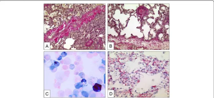
Definition and classification
Pulmonary arterial hypertension (PAH) is defined by right-heart catheterization (RHC) showing precapillary pulmonary hypertension with a mean pulmonary artery pressure (mPAP) of >25 mmHg and a normal pulmonary artery wedge pressure (PCWP) of <15 mmHg [1,2]. The classification of pulmonary hypertension (PH) has gone through a series of changes since the first classification proposed in 1973 which designated only two categories, primary pulmonary hypertension or secondary PH, de-pending on the presence or absence of identifiable causes or risk factors [3,4]. In 1998, a second World Symposium on PH was held in Evian (France) and this classification attempted to create categories of PH that shared similar pathogenesis, clinical features and thera-peutic options [5]. This classification allowed defining homogenous groups of patients to conduct clinical trials and to obtain approval for specific PAH therapies worldwide. In 2003, the third World Symposium on PH (Venice, Italy) did not propose major changes. However, the terms idiopathic PAH, familial PAH, and associated PAH were introduced. The other prominent change was to move pulmonary veno-occlusive disease (PVOD) and pulmonary capillary hemangiomatosis (PCH) from sep-arate categories into a single subcategory of PAH.
In 2008, the fourth World Symposium on PH held in Dana Point (California, USA) and the consensus of an international group of experts was to revise previous classifications in order to accurately reflect published data, as well as to clarify some areas that were unclear. In 2013, the fifth World Symposium on PH held in Nice (France) and proposed only minor modifications, how-ever, since the definite conclusions of this symposium were not yet published, we presented the Dana Point classification of PH (Table 1).
Group 1: Pulmonary arterial hypertension
The nomenclature of the subgroups and associated con-ditions has evolved since the first classification, and add-itional modifications were added in the Dana Point classification.
Group 1.1/1.2 Idiopathic and heritable PAH
Idiopathic PAH describes a sporadic disease with neither a family history of PAH nor an identified risk factor. When PAH occurs in a familial context, germline muta-tions in the bone morphogenetic protein receptor 2 (BMPR2) gene, a member of the transforming growth factor beta (TGF- ß) signaling family, can be detected in about 70% of cases [6,7]. More rarely, mutations in activin receptor like kinase type 1 (ACVRL1 or ALK1) or endoglin genes, also coding for members of the TGF-ß signaling family, have been identified in patients with PAH, predominantly with coexistent hereditary
hemorrhagic telangiectasia. Some authors suggested that mutations of genes encoding for Smads proteins (Smad8, Smad1 and Smad5), which are other members of the TGF-ß signaling pathway, or mutations in caveolin-1 gene may predispose to PAH [8-10].
BMPR2 mutations have also been detected in 11–40% of apparently idiopathic cases with no family history
Table 1 Diagnostic classification of pulmonary hypertension
1. Pulmonary arterial hypertension (PAH)
1.1 Idiopathic 1.2 Heritable
1.3 Drugs and toxins induced 1.4 Associated with (APAH):
1.4.1 Connective tissue disease 1.4.2 Infection with human
immunodeficiency virus 1.4.3 Portal hypertension 1.4.4 Congenital heart disease 1.4.5 Schistosomiasis 1.4.6 Chronic haemolytic
anaemia 1.5 Persistent pulmonary
hypertension of the newborn 2. Pulmonary hypertension with
left heart disease
2.1 Systolic dysfunction 2.2 Diastolic dysfunction 2.3 Valvular disease 3. Pulmonary hypertension due
to lung diseases and/or hypoxia
3.1 Chronic obstructive pulmonary disease
3.2 Interstitial lung disease 3.3 Other pulmonary diseases
with mixed restrictive and obstructive pattern 3.4 Sleep-disordered breathing 3.5 Alveolar hypoventilation
disorders
3.6 Chronic exposure to high altitude
3.7 Developmental abnormalities 4. Chronic thromboembolic
pulmonary hypertension 5. PH with unclear and/or multifactorial mechanisms
5.1 Haematological disorders: myeloproliferative disorders, splenectomy.
5.2 Systemic disorders: sarcoidosis, pulmonary Langerhans cell histiocytosis,
lymphangioleiomyomatosis, neurofibromatosis, vasculitis 5.3 Metabolic disorders: glycogen
storage disease, Gaucher disease, thyroid disorders 5.4 Others: tumoral obstruction,
fibrosing mediastinitis, chronic renal failure on dialysis
[11,12]. Indeed, the distinction between idiopathic and familial PAH with BMPR2 mutations is artificial, as all patients with a BMPR2 mutation have heritable disease. In addition, BMPR2 mutations were identified in only 70-80% families with PAH. Thus, it was decided to aban-don the term “familial PAH” in favor of the term “herit-able PAH”, including idiopathic PAH with germline mutations and familial cases with or without identified mutations [13,14].
Group 1.3 Drug- and toxin-induced PAH
A number of risk factors for the development of PAH have been individualized in the last European Respira-tory Society/ European Society of Cardiology (ERS/ESC) conjoint guidelines of PH [15] (Table 2).
Aminorex, fenfluramine derivatives and toxic rapeseed oil represent the only identified “definite” risk factors for PAH [5,16]. Souza et al. have demonstrated that this sub-group of PAH shares clinical, functional, hemodynamic, and genetic features with idiopathic PAH, suggesting that fenfluramine exposure represents a potential trigger for PAH without influencing its clinical course [17].
Two prospective epidemiologic investigations, the SNAP (Surveillance of North American Pulmonary Hypertension) and the SOPHIA (Surveillance of Pulmonary Hypertension
in America) study, were conducted in the USA [18,19]. These investigations included retrospectively 559 and 1335 patients with PH, respectively, and confirmed the previ-ously described association between idiopathic PAH and the use of fenfluramine. In the SNAP study, the odds ratio of developing PH was 7.5 for the use of fenfluramine more than six months of treatment [18].
The agent benfluorex is structurally and pharmaco-logical related to fenfluramine and may be also considered as an anorectic agent. Frachon and co-workers [20] showed a significantly higher prevalence of unexplained valvular heart disease in patients taking benfluorex com-pared to controls. In addition, Savale and co-workers demonstrated recently that exposure to benfluorex is sug-gested to be a trigger in the development of PAH [21]. Benfluorex was withdrawn from the market in 2009.
The SOPHIA study examined intake of a variety of nonselective monoamine reuptake inhibitors, selective serotonin reuptake inhibitors, antidepressants and anxio-lytics. No increased risk of developing PAH was ob-served [19].
Amphetamine use represents a “likely” risk factor, al-though they are frequently used in combination with fenfluramine. A recent retrospective study suggested a relationship with the use of methamphetamine (inhaled, smoked, oral, or intravenous) and the occurrence of PAH [22]. Methamphetamine use is now considered a “very likely” risk factor for the development of PAH.
Recently published data from the French registry of pulmonary hypertension suggested that dasatinib, a tyro-sine kinsase inhibitor (TKI), may induce precapillary PAH [23]. Several cases of precapillary PH in chronic myelogenous leukemia patients treated with dasatinib have been reported.
Group 1.4.1 PAH associated with connective tissue diseases
PAH associated with connective tissue diseases (CTD) represents an important clinical subgroup, in which sys-temic sclerosis represents the major cause of CTD asso-ciated PAH. The prevalence of PAH has been well established only for systemic sclerosis (SSc). Prospective studies using echocardiography as a screening method and RHC for confirmation found a prevalence of PAH between 7–12% [24,25]. PH due to lung fibrosis [26], diastolic left heart dysfunction [27] and primary cardiac involvement [28] are also frequent in the setting of pul-monary hypertension in these patients, emphasizing the importance of a systemic evaluation with RHC to accur-ately classify the underlying mechanism of PH.
Group 1.4.2 HIV infection
PAH is a rare complication of HIV infection [29,30]. HIV-associated PAH has clinical, hemodynamic, and
Table 2 Updated risk level of drugs and toxins known to be associated with PAH
1. Definite
Aminorex Fenfluramine Dexfenfluramine Toxic rapeseed oil Benfluorex 2. Likely Amphetamines L-tryptophan Metamphetamines Dasatinib 3. Possible Cocaine Phenylpropanolamine St John’s Wort Chemotherapeutic agents
Selective serotonin reuptake inhibitors Pergolide
4. Unlikely
Oral contraceptives Estrogen therapy Cigarette smoking
histologic characteristics broadly similar to those seen in idiopathic PAH. Epidemiologic data in the early 1990s, a time when therapy with highly active antiretroviral ther-apy was not yet available, indicated a prevalence of 0.5% [31]. The prevalence of HIV-associated PAH was evalu-ated more recently and showed a stable prevalence of 0.46% [32].
Group 1.4.3 Porto-pulmonary hypertension
Porto-pulmonary hypertension (POPH) is defined by the development of PAH associated with increased pressure in the portal circulation [33,34]. Prospective hemodynamic studies have shown that 2-6% of patients with portal hypertension had PH [35,36]. However, RHC is mandatory for the diagnosis of portal PH, as several mechanisms may increase pulmonary artery pressure in the setting of ad-vanced liver disease: hyperdynamic circulatory state with high cardiac output, fluid overload and diastolic dysfunc-tion. Pulmonary vascular resistance (PVR) is usually nor-mal in these cases.
Group 1.4.4 Congenital heart diseases
A significant proportion of patients with congenital heart disease (CHD), in particular those with systemic-to-pulmonary shunts, will develop PAH if left untreated. Eisenmenger's syndrome is defined as CHD with an ini-tial large systemic-to-pulmonary shunt that induces pro-gressive pulmonary vascular disease and PAH, with resultant reversal of the shunt and central cyanosis [37,38]. It represents the most advanced form of PAH associated with CHD. It has been reported that a large proportion of patients with CHD develop some degree of PAH [39-41]. The prevalence of PAH associated with congenital systemic-to-pulmonary shunts in Europe and North America has been estimated between 1.6 and 12.5 cases per million adults, with 25-50% of this population affected by Eisenmenger’s syndrome.
Group 1.4.5 Schistosomiasis
In the Dana Point classification, PH associated with schistosomiasis was included in Group 1. Recently, it has been demonstrated that PH associated with schisto-somiasis may have a similar clinical presentation and histological findings as idiopathic PAH [42,43]. The mechanism of PAH in patients with schistosomiasis is probably multifactorial including portal PH, a frequent complication of this disease [44] and local vascular inflammation, whereas mechanical obstruction by schistosoma eggs seems to play a minor role. More than 200 million people are infected and 4–8% of them will develop hepatosplenic disease. Then, PAH associated with schistosomiasis represents a frequent form of PAH in countries where the infection is endemic. Data from a recent study based on invasive hemodynamics evidenced
the prevalence of PAH in patients with hepatosplenic disease of 4.6%; also important was the prevalence of post-capillary hypertension (3%) reinforcing the need of invasive hemodynamics for the specific diagnosis of PAH in schistosomiasis [45].
Group 1.4.6 Chronic hemolytic anemia
The chronic hemolytic anemias represent a subcategory of PAH. There has been increasing evidence that PAH is a complication of chronic hereditary and acquired hemolytic anemias, including sickle cell disease [46,47], thalassemia [48], hereditary spherocytosis [49], stomatocytosis [50], and microangiopathic hemolytic anemia [51].
PH has been reported most frequently in patients with sickle cell disease, however the prevalence of PAH is not yet clearly established. The prevalence of PH in sickle cell disease is undoubtedly much lower than 32% as suggested by echocardiography [47]. Re-cently, a prospective epidemiologic studies using echocardiographic screening and direct hemodynamic confirmation were conducted in 398 outpatients with sickle cell disease at referral centers in France [52]. In this study, the prevalence of a tricuspid regurgitant jet velocity of at least 2.5 m per second measured by echocardiography was 27%. In contrast, the
preva-lence of pulmonary hypertension confirmed on
catheterization was only 6%, suggesting that echocar-diographic evaluation alone had a low positive pre-dictive value for PH in this population. Indeed, the precise mechanism of PAH in sickle cell disease re-mains uncertain.
Group 1’: Pulmonary veno-occlusive disease and/or pulmonary capillary hemangiomatosis
PVOD and PCH are uncommon conditions, but they are increasingly recognized as causes of PH [53]. A recent clinicopathologic study [54] analyzed specimens from 35 patients diagnosed as having either PVOD (n = 30) or PCH (n = 5). PCH was identified in 24 (73%) cases diag-nosed as PVOD. Indeed, venous involvement was present in 4/5 cases initially diagnosed as PCH. These findings suggest that PCH may be an angioproliferative process frequently associated with PVOD. Similarities in pathologic features and clinical presentation suggest that these disorders may be two different presentation of the same disease [54].
Although PVOD and PCH may present similarly to idiopathic PAH, there are a number of important differ-ences. These include the presence of crackles on exam-ination, radiologic abnormalities on high-resolution computed tomography of the chest (ground glass opaci-ties, septal thickening, mediastinal adenopathy) [55-58], hemosiderin-laden macrophages on bronchoalveolar lav-age [59], and a lower DLCO and PaO2in patients with
PVOD or PCH [58]. PVOD/PCH remains a difficult dis-order to categorize, as it shares characteristics with idio-pathic PAH but also has a number of distinct differences. Given the current evidence, it was decided that PVOD/PCH should be a distinct category but not completely separated from PAH and PVOD/PCH are designated as 1’ in the current classification.
Group 2: Pulmonary hypertension due to left heart disease
Left-sided ventricular or valvular diseases may produce an increase of left atrial pressure, leading to a backward transmission of the pressure and a passive increase of pulmonary arterial pressure. Left heart disease, probably represents the most frequent cause of PH [60]. In this situation, PVR is normal or near normal (<3.0 Wood units) and there is no gradient between mean PAP and pulmonary wedge pressure (transpulmonary gradient <12 mm Hg). In the Dana Point classification, the in-creasing recognition of left-sided heart dysfunction with preserved ejection fraction leads to changes in the sub-categories of Group 2 and now this group include three distinct etiologies: left heart systolic dysfunction, left heart diastolic dysfunction, and left heart valvular dis-ease. In some patients with left heart disease, the eleva-tion of pulmonary arterial pressure is out of proporeleva-tion to that expected from the elevation of left arterial pres-sure (transpulmonary gradient >12 mm Hg), and PVR is increased to >3.0 Wood units (19–35% of patients) [60]. However, there is no widely accepted hemodynamic def-inition of transpulmonary gradient, and future recom-mendations may propose new definition and threshold of this gradient. Some patients with left heart valvular disease or even left heart dysfunction can develop severe PH of the same magnitude as that seen in PAH [61-63]. The elevation of PAP and PVR may be due to either the increase of pulmonary artery vasomotor tone and/or pulmonary vascular remodeling [64,65].
Group 3: Pulmonary hypertension due to lung diseases and/or hypoxia
In this group, the predominant cause of PH is alveolar hypoxia as a result of either chronic lung disease, im-paired control of breathing, or residence at high altitude. However, the precise prevalence of PH in all these con-ditions remains largely unknown. In the revised classifi-cation, the heading has been modified to reinforce the link with the development of PH. A category of lung dis-ease characterized by a mixed obstructive and restrictive pattern was added, including chronic bronchiectasis, cystic fibrosis and the recently described syndrome of combined pulmonary fibrosis and emphysema in which the prevalence of PH is almost 50% [66,67]. In PAH as-sociated with parenchymal lung disease, the increase of
pulmonary arterial pressure is usually modest (mean PAP lower than 35 mmHg) [68]. Interestingly, in some patients, increase of PAP is out of proportion and be higher than 35 mmHg [69]. In a retrospective study of 998 patients with chronic obstructive pulmonary disease who underwent RHC, only 1% had severe PH [70]. These patients with more severe PH were characterized by mild-to-moderate airway obstruction, severe hypox-emia, hypocapnia, and a very low diffusing capacity for carbon monoxide.
Group 4: Chronic thromboembolic pulmonary hypertension
Chronic thromboembolic pulmonary hypertension
(CTEPH) was included in Group 4. The incidence of CTEPH is uncertain. CTEPH represents however a fre-quent cause of PH and occurs in up to 4% of patients after an acute pulmonary embolism [71,72]. In the clas-sification from the third World Symposium on PH, CTEPH was divided into 2 subgroups: proximal CTEPH and distal CTEPH, depending on the feasibility of performing pulmonary thromboendarterectomy. Cur-rently, there is no consensus about the definitions of proximal and distal CTEPH and the decision of surgery may vary depending on individual centers [73]. Thus, the Dana Point Classification propose only one group irrespectively of proximal or distal obstruction. Patients with suspected or confirmed CTEPH need to be referred to expert centers with experience in the management of CTEPH, to consider the feasibility of performing surgery. The indication for surgery depends on the location of the obstruction, the correlation between hemodynamics and the degree of obstruction assessed by angiography, comorbidities, the willingness of the pa-tient, and the experience of the surgeon [74,75]. Patients who are not candidates for surgery may benefit from PAH-specific medical therapy [76,77]; however, further evaluation of these therapies in randomized control trials are needed [78].
Group 5: PH with unclear or multifactorial etiologies Group 5.1 Hematologic disorders
PH has been reported in chronic myeloproliferative dis-orders including polycythemia vera, essential thrombo-cythemia, and chronic myeloid leukemia [79,80]. Several mechanisms may be implicated in PH associated with chronic myeloproliferative disorders including high car-diac output, asplenia, direct obstruction of pulmonary arteries by circulating megakaryocytes [81], CTEPH [82], portal PH, and congestive heart failure. Splenectomy as a result of trauma or as a treatment for hematologic dis-orders may increase the risk of developing PH [83]. As described above, dasatinib use may be one cause of PAH, particularly in chronic myeloid leukemia [23].
Group 5.2 Systemic disorders
The second subgroup includes systemic disorders, including sarcoidosis, pulmonary Langerhans cell histiocytosis, lymphangioleiomyomatosis, neurofibromatosis or vasculitis.
PH is a well recognized complication of sarcoidosis [84], with a reported prevalence of 1–28% [84]. PH is multifactorial and usually attributed to the destruction of capillary bed by the fibrotic process and/or the result of chronic hypoxemia [85]. However, the severity of PH is frequently out of proportion to the degree of paren-chymal lung disease and blood gas abnormalities, suggesting specific pulmonary vascular involvement [86]. Such mechanisms may include extrinsic compression of large pulmonary arteries by lymph node enlargement, and granulomatous infiltration of the pulmonary vascu-lature, especially the pulmonary veins, which sometimes mimic PVOD [87].
Pulmonary Langerhans cell histiocytosis is an uncom-mon cause of infiltrative and destructive lung disease. Severe PH is a common feature in patients with end stage disease [88] and PH in these patients is usually re-lated to chronic hypoxemia and/or abnormal pulmonary mechanics. Histopathologic examination has shown se-vere diffuse pulmonary vasculopathy involving predom-inantly intralobular pulmonary veins and, to a lesser extent, muscular pulmonary arteries [89].
Lymphangioleiomyomatosis is a rare multisystem dis-order predominantly affecting women, characterized by cystic lung destruction, lymphatic abnormalities, and ab-dominal tumors. PH is relatively uncommon in patients with lymphangioleiomyomatosis [90]. Recently, a series of 20 cases of lymphangioleiomyomatosis associated PH has been reported showing that PH is usually moderate in this setting and associated with pulmonary function impairment [91].
Neurofibromatosis type 1, also known as von Recklinghausen’s disease, is an autosomal-dominant disease. The disease is occasionally complicated by systemic vasculopathy. A series of cases of PH have been reported in the setting of Neurofibromatosis type-1 [92].
Group 5.3 Metabolic disorders
PH has been reported in a few cases of type Ia glycogen storage disease, a rare autosomal-recessive disorder caused by a deficiency of glucose-6-phosphatase [93-95]. The mechanisms of PH are uncertain but portocaval shunts, atrial septal defects, severe restrictive pulmonary function defects or thrombosis are thought to play a role. In one case, autopsy findings revealed the presence of plexiform lesions [96].
Gaucher’s disease is a rare disorder characterized by a deficiency of lysosomal B glucosidase, which results in an accumulation of glucocerebroside in reticuloendothelial
cells. In a study of 134 patients with Gaucher’s disease who were systematically screened by echocardiography, PH was not uncommon [97]. In this setting, several poten-tial mechanisms for PH have been suggested, including interstitial lung disease, chronic hypoxemia, capillary plug-ging by Gaucher cells and splenectomy [97,98].
The association between thyroid diseases and PH has been reported in some studies [99]. A prospective study of 63 consecutive adult patients with PAH found a prevalence of autoimmune thyroid disease, including both hypothyroidism and hyperthyroidism, in 49%, suggesting that these conditions may share a common immunogenetic susceptibility [100].
Group 5.4 Miscellaneous conditions
The last subgroup includes a number of miscellaneous conditions, including tumoral obstruction, fibrosing mediastinitis or chronic renal failure on dialysis.
A progressive obstruction of proximal pulmonary ar-teries leading to PH may be observed in tumor obstruc-tion when a tumor grows into the central pulmonary arteries with additional thrombosis. Such cases are due principally to pulmonary artery sarcomas, which occur rarely but are usually rapidly fatal [101-103]. The differ-ential diagnosis with CTEPH can be difficult and CT angiography may be useful to differenciate an obstruc-tion by tumor or thrombotic material. Occlusion of the microvasculature by metastatic tumor emboli represents a cause of rapidly progressive PH.
Fibrosing mediastinitis have been mainly reported in sarcoidosis, tuberculosis, histoplasmosis and after radio-therapy. Fibrosing mediastinitis may be associated with severe PH due to compression of both pulmonary arter-ies and veins.
Lastly, PH has been reported in patients with end-stage renal disease maintained on long-term hemodialysis. Based on echocardiographic studies, the prevalence of PH in this patient population is estimated up to 40% [104]. PH in these patients may be explained by high cardiac output (resulting from the arteriovenous access, anemia and fluid overload) and potential diastolic and systolic left heart dysfunctions. Furthermore, hormonal and metabolic modification associated with end-stage renal disease might lead to dysfunction of normal pulmonary vascular tone.
Epidemiology
Information relative to the natural history of PAH was derived from a national registry conducted in the USA in the early 1980s, which included 187 patients with idiopathic PAH followed for up to 5 years. This study characterized the disease and confirmed its poor prog-nosis with a median survival of 2.8 years [105,106]. Bet-ter understanding of pathophysiological mechanisms of PAH has led to the development of novel therapeutic
strategies in the last decade which improved quality of life, exercise capacity and survival of these PH patients [107].
In France, a national multicenter registry prospectively collected data from 674 adults with PAH from October 2002 to October 2003 and followed these patients during a 3-year period [108]. This prospective study has shown more up to date data on the epidemiology of PAH. This registry has confirmed the female predominance in most subtypes of PAH with a female/male sex ratio (SR) of 1.9 for all PAH patients, and particularly in idiopathic PAH (SR 1.6), familial PAH (SR 2.2) and anorexigen-associated PAH (SR 14.9) [108]. The mean age of PAH patients in this cohort was 50 years with a quarter of pa-tients older than 60 years underlining the possibility of developing PAH in all ages. In this assessed cohort, 39.2% of patients had idiopathic PAH and 3.9% familial PAH. In the subgroup of PAH, where PAH was associ-ated with other conditions, 15.3% had connective tissue disease, 11.3% congenital heart diseases, 10.4% portal hypertension, 9.5% anorexigen-related PAH and 6.2% HIV infection [108]. In France, the low estimation of the prevalence of PAH and idiopathic PAH are respectively of 15 cases and 5.9 cases/million adult inhabitants. The low estimation of PAH incidence is 2.4 cases/million adult inhabitants/year. This study conducted five years after the withdrawal of fenfluramine derivatives under-lines that anorexigen-associated PAH remains an im-portant medical problem nowadays.
Recent prospective data in consecutive patients with idiopathic, familial or anorexigen-associated PAH pa-tients followed-up for a period of three years (56 inci-dent and 298 prevalent cases) showed better survival rates than historical cohort. For incident cases, estimated survival rates were 85.7%, 69.6% and 54.5% at one, two and three year of follow-up, respectively. In combined
mixed population (incident patients and prevalent pa-tients diagnosed within three years before study entry), estimated one, two and three year survival was 82.9%, 67.1% and 58.2%, respectively. Parameters signifi-cantly associated to improved survival were female gender, preserved 6-MWD and normal cardiac output on RHC [109].
Data from the REVEAL registry (US Registry to Evalu-ate Early and Long-Term PAH Disease Management) are similar concerning the one year survival rate esti-mated to 91%. Variables independently associated with increased mortality included pulmonary vascular resist-ance >32 Wood units, NYHA functional class IV, men older than 60 years and familiy history of PAH [110].
Genetics
It is now well known that PAH can be either sporadic or clustered in families [111-114]. In 1954, Dresdale pub-lished a detailed description of a family that included three related subjects with severe PAH of unknown aeti-ology. Because of physician’s awareness of the familial occurrence of the disease, other cases of familial PAH were described. The National Institute of Health Registry (NIH Registry) provided the first estimate of this condi-tion, and evaluated that at least 6% of individuals diag-nosed with sporadic PAH have a family history of the disorder. In the French PAH registry, 26 cases of familial PAH were identified in the cohort of 674 patients, which corresponds to a prevalence of 3.9% of all cases of PAH and a proportion of 7.3% of familial PAH in the sub-group of idiopathic, familial or anorexigen-associated PAH [108]. Studies of genealogies of familial PAH ad-vanced our understanding that the disease segregates an autosomal dominant trait with a markedly reduced penetrance, since only 10-20% of necessary carriers of the mutation will develop PAH [115,116]. In 2000, link-age analysis in PAH affected families found mutations within the bone morphogenetic protein receptor type 2 gene (BMPR2) [117,118]. BMPR2 gene encodes for a type 2 receptor member of the transforming growth factor beta (TGF-β) family. Nowadays, germline BMPR2 muta-tions are detected in 58-74% of PAH patients with a family history of the disease, and in 3.5-40% of so called idiopathic PAH patients [6,11,108,119-123]. The obser-vation of the development of PAH in patients displaying hereditary hemorrhagic telangiectasia (HHT), an auto-somal dominant vascular dysplasia, allowed us to identify two other PAH predisposing genes: ACVRL1 (Activin A receptor type II-like kinase 1) and ENG (endoglin) genes. Mutations in these two genes are infre-quent in PAH but are freinfre-quently identified in HHT [120,124-130]. Moreover, recent reports described two PAH patients carriers of a mutation in Smad8 gene, one PAH patient carrier of a Smad1 mutation and one PAH
Table 3 Modified New York Heart Association (NYHA) classification for pulmonary hypertension
CLASS I Patients with pulmonary hypertension but without resulting limitation of physical activity. Ordinary physical activity does not cause undue dyspnoea or fatigue, chest pain, or near syncope.
CLASS II Patients with pulmonary hypertension resulting in slight limitation of physical activity. They are comfortable at rest. Ordinary physical activity causes undue dyspnoea or fatigue, chest pain, or near syncope.
CLASS III Patients with pulmonary hypertension resulting in marked limitation of physical activity. They are comfortable at rest. Less than ordinary activity causes undue dyspnoea or fatigue, chest pain, or near syncope.
CLASS IV Patients with pulmonary hypertension with inability to carry out any physical activity without symptoms. These patients manifest signs of right heart failure. Dyspnoea and/or fatigue may even be present at rest. Discomfort is increased by any physical activity.
patient carrier of a Smad5 mutation [8,9]. BMPR2 gene, ACVRL1, ENG, Smad8, Smad1 and Smad5 genes are en-coding proteins which are involved in the TGF-β signal-ing pathway. More recently, ussignal-ing whole exome sequencing in a large PAH family, Austin et al. demon-strated the involved of mutations in caveolin-1 gene in the development of PAH [10] They confirmed their findinds identifying a caveolin-1 mutation in an unre-lated PAH patient. Caveolin-1 protein is necessary for the formation of calveola, which are crucial for joining membrane receptors and initiating cellular signaling cas-cade such as TGF-β signalling pathway. This supports the hypothesis that mutations in genes involved in the TGF-β signaling pathway may be a trigger for pulmon-ary vascular remodeling. Moreover, this signaling path-way controls growth, differentiation and apoptosis of various cell types like pulmonary vascular endothelial cells (ECs) and smooth muscle cells (SMCs). Thereby, mutations in genes involved in the TGF-β signaling pathway may be responsible for abnormal proliferation of pulmonary vascular SMCs and may promote ECs apoptosis, which might lead to the selection of apoptosis resistant cells and formation of plexiform lesions, the hallmark of idiopathic PAH [131,132].
The analysis of clinical, functional and hemodynamic characteristics of PAH patients revealed that patients carriers of a BMPR2 or an ACVRL1 mutation are youn-ger at diagnosis than patients with idiopathic PAH. In addition, they have more severe hemodynamic parame-ters at diagnosis [114,120].
Several lines of evidence point to the potential require-ment of additional factors, either environrequire-mental or gen-etic, in the pathogenesis of the disease. As mentioned, Humbert and co-workers have shown that exposure to fenfluramine derivatives greatly increase the risk of de-veloping severe PAH in patients with BMPR2 mutations [111]. Moreover, modified genes could participate or fa-cilitate the development of PAH. In fact, recent studies have suggested the potential role of serotonin trans-porters, serotonin receptors, potassium channels, or angiopoietin-1 [133]. Finally, PAH mostly occurs in fe-males irrespective of BMPR2 mutation status [134]. To explain overrepresentation of PAH female patients, it was suggested that estrogen metabolism might partici-pate in the pathogenesis of PAH [135,136].
Pathophysiology
PAH is a disease which affects small pulmonary arteries. It is characterized by vascular obstruction leading to progressive increase in vascular resistance. This in-creases right ventricular afterload and consequently re-sults in right ventricular failure. Intima and media proliferation and its consequent pulmonary vascular ob-struction are considered to be the key element in the
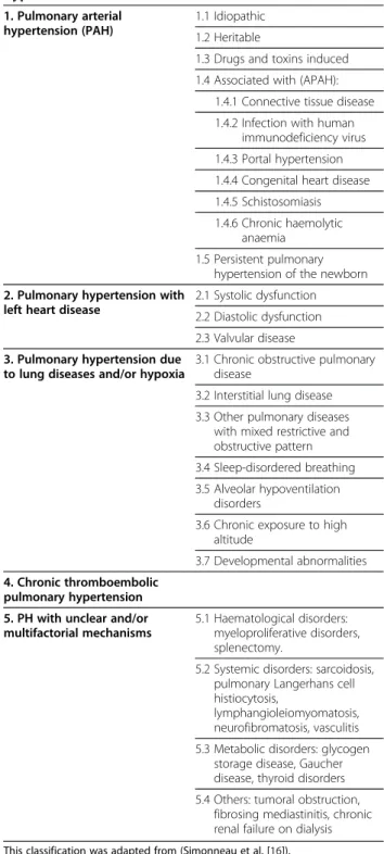
pathogenesis of PAH. Vasoconstriction, vascular remod-eling and thrombosis are factors that increase pulmonary vascular resistance in PAH [107,137]. These processes involve a multitude of cellular and molecular elements (Figure 1).
Cellular factors
Proliferation of smooth muscular cells in the small per-ipheral pulmonary arteries is a common characteristic in all forms of PAH. In hypoxic models, fibroblasts of the adventitia migrate to the media and intima, where prolif-eration and production of matrix proteins are observed [138]. Neovascularization, mainly of the adventitia, oc-curs concomitantly to the thickening of the vascular walls [139].
In response to certain stimuli, endothelial cells abnor-mally proliferate to form plexiform lesions in several forms of PAH. Plexiform lesions consist of endothelial cells, matrix proteins and fibroblasts and obliterate the vascular lumen [140]. The stimuli for endothelial prolif-eration is still unknown but several factors have been in-criminated such as hypoxia, inflammation, shear stress, drugs, viral infections and genetic susceptibility. Extrapulmonary cells may also participate in the vascular remodeling responsible for PAH. Indeed, fibrocytes and c-kit + cells are mobilized from the bone marrow, and may differentiate into vascular cells and/or produce pro-angiogenic factors to participate in the pathogenesis of PAH [141,142]. The CXCL12/CXCR4 axis may play an important role in the pulmonary recruitment of these circulating progenitors and can be therapeutically targeted [143].
Inflammatory mechanisms seems to play an important role in certain forms of PAH such as PAH associated with auto immune diseases or HIV infection [144]. In fact, in severe cases of PAH associated with systemic lupus erythematosus disease, some patients improved both clinically and hemodynamically with administrated immunosuppressant treatment. Thirty to 40% of patients with PAH have circulating auto-antibodies and elevated plasma concentrations of pro-inflammatory cytokines such as interleukin 1 (IL-1) and interleukin-6 (IL-6), and chemokines such as fractalkine and MCP-1 [145,146]. Inflammatory cells, such as lymphocytes B and T, mac-rophages, mastocytes and dendritic cells, can also be found in plexiform lesions of severe PAH [147,148]. Chemokines, like RANTES and fractalkine are also overly expressed in the pulmonary vascular endothelium of PAH patients [145].
Thrombosis and platelet dysfunction can be important in the development of PAH. Abnormalities of throm-bosis, endothelial cells or platelets can generate or aggra-vate thrombosis in situ. Eleaggra-vated plasma concentrations of D-dimers and fibrinopeptides A and B, in certain
patients with PAH, are the proof of an abnormal intravascular coagulation process. Elevated plasma concentrations of von-Willebrand factor and plasmino-gen activator inhibitor type 1 also reflect endothelial dysfunction in PAH. It has been demonstrated that shear stress creates pro-thrombotic vascular lesions in PAH that may lead to thrombosis in situ. But platelet function is not limited to coagulation. In response to certain stimuli, platelets can produce prothrombotic, vasoactive or mitogenic factors, such as thromboxane A2 (TXA2), platelet-derived growth factor (PDGF), serotonin (5-hydroxytryptamine, 5-HT), transforming growth factor beta (TGF-β) and vascular endothelial growth factor (VEGF) that participate in vasoconstriction and vascular remodeling [149,150].
Autoimmunity and PAH
The self-tolerance is controlled in the periphery by a particular population of T-lymphocytes called regulatory T-lymphocytes (Treg). The breakdown of self-tolerance can lead to the development of an autoimmune response (i.e. directed against self antigens) that can finally give rise to an autoimmune disease. Huertas et al. [151] showed that circulating Treg number was comparable in idiopathic PAH and SSc-PAH patients. However the per-centage of those expressing leptin receptors was higher in idiopathic PAH and SSc-PAH as compared to
controls, and their function was reduced in idiopathic PAH and SSc-PAH patients as compared to controls in a leptdependent manner [151]. Work on chronic in-flammatory disorders and autoimmune diseases suggest that pathogenic antibodies and T cells may be generated locally, in the targeted organ, in highly organized ectopic lymphoid follicles commonly called tertiary lymphoid tissues. Recently, Perros et al. [152] described the pres-ence of highly organized perivascular follicles in idio-pathic PAH lungs arguing for specific immune-adaptive mechanisms in the pathophysiology of the disease. One can propose that deregulated and unresolved pulmonary inflammation on the background of a genetic predispos-ition, could result in persisting vascular remodelling leading to PAH. An initial acute inflammation that is normally expected to resolve with return to homeostasis, could conduct the production of auto-antibodies against vascular wall components, and would shift to chronic persisting and chronic inflammation, endothelial barrier breakdown, infiltration by immune cells, local and chronic autoimmunity, and vascular remodeling culmin-ating in PAH.
Molecular factors
Many authors consider pulmonary vasoconstriction as an early event in the process of PAH. Vasoconstriction has been associated with an abnormal function or
Figure 1 Pathophysiology of PAH. The pulmonary vascular remodeling responsible for PAH is the consequence of closely intertwined predisposing and acquired factors. Thoses pathological elements affect all three layers of precapillary pulmonary arteries leading to intimal hyperplasia, medial thickening and adventitial remodeling/fibrosis. Intra- but also extra-pulmonary cells, such as inflammatory and progenitor cells, are suspected to play a role in this remodeling. This increases right ventricular afterload and consequently results in right ventricular failure.
expression of potassium channels and with endothelial dysfunction [107]. Endothelial dysfunction results in a decreased production of vasodilators such as nitric oxide (NO) and prostacyclin and an increased production of vasoconstrictors such as endothelin-1 [153].
Prostacyclin (prostaglandin I2) is a potent pulmon-ary vasodilator that acts via the cyclic adenosine monophosphate (cAMP) pathway. It inhibits the pro-liferation of smooth muscle cells and decreases plate-let aggregation. Production of prostacyclin is reduced in endothelial cells of patients with PAH [154]. PAH therapy based on prostacyclin and its derivates have proven efficacy both hemodynamically and in clinical trials. NO is also a pulmonary vasodilator which acts via the cyclic guanosine monophosphate (cGMP) pathway. To increase pulmonary vasodilatation de-pendant on NO, a recent therapeutic strategy has targeted type 5 phosphodiesterase which degrades cGMP. Sildenafil or tadalafil, type 5 phosphodiesterase inhibitors, have proven their efficacy in patients with PAH [155]. Vasoactive intestinal peptide (VIP) is a neurotransmitter that has systemic and pulmonary vasodilator properties. It also inhibits smooth cell proliferation and decreases platelet aggregation and acts via the activation of the cAMP and cGMP sys-tems [156]. Low plasmatic concentrations of VIP have been measured in pulmonary arteries of patients with PAH.
Endothelin-1 (ET-1) is an endothelially-derived peptide that has two receptor subtypes, designated as endothelin A (ETRA) and endothelin B (ETB), located on smooth muscle cells of pulmonary arteries. By ligating the ETRA, ET-1 intracellular calcium concentrations increase and activates the protein kinase C pathway [157]. ET-1 is a potent pul-monary vasoconstrictor and stimulates mitosis of arterial smooth muscle cells, thus contributing to pulmonary vascu-lar remodeling. Pulmonary and plasma levels of ET-1 are el-evated in human PAH and in experimental animal models of PAH [158]. The therapeutic efficacy of endothelin recep-tor antagonists (Ambrisentan, Bosentan) has been demon-strated in clinical trials in the pathophysiology of PAH.
In hypoxic models of PAH, hypoxia inhibits one or several voltage dependant potassium channels of the pulmonary arterial smooth muscle cells. This leads to membrane depolarization and opening of voltage de-pendant calcium channels with a subsequent increase of the intracellular calcium concentration and cellular contraction. Certain potassium channels are under expressed in pulmonary artery smooth muscle cells of patients with PAH [159,160]. It is still unknown whether abnormalities of the potassium channels are acquired or genetic. However, it has been demonstrated that anorexigens, such as dexfenfluramine and aminorex, dir-ectly inhibit certain potassium channel subtypes [161].
Certain medications such as dichloroacetate and sildenafil increase the expression and function of potassium channels.
In PAH, plasmatic concentrations of serotonin (5-hy-droxytryptamine, 5-HT) are elevated [150]. An association between anorexigens and serotonin was established in the 1960s. Aminorex and fenfluramin both increase plasmatic levels of serotonin. The variability of the expression and activity of the transporter of 5-HT (5-HTT) contributes to pulmonary vascular remodeling in human and experimen-tal models of PAH [162]. Some studies have shown that the serotonin selective reuptake inhibitor fluoxetin pre-vents the development of PAH in mice [163]. Some 5-HT receptor subtypes may also be implicated in the develop-ment of hypoxia induced PAH [164].
Rho proteins regulate fundamental cellular functions such as contraction, migration, proliferation and apop-tosis. Several studies have implicated Rho protein A and Rho kinases in the vasoconstriction and vascular remod-eling of PAH [165,166]. RhoA and Rho kinase activities are increased in idiopathic PAH, in association with en-hanced RhoA serotonylation. Direct involvement of the 5-HTT/RhoA/Rho kinase signaling pathway in 5-HTT-mediated pulmonary artery-smooth muscle cell (PA-SMC) proliferation and platelet activation during PH progression identify RhoA/Rho kinase signaling as a promising target for new treatments against PH [167].
Hypoxia inducible factor-1 (HIF-1) is a transcription factor that principally regulates cellular adaptation to hypoxia but also regulates several genes implicated in angiogenesis, erythropoiesis, cellular metabolism and survival [168]. In experimental mice heterozygote for the gene coding for HIF-1 alpha, hypoxia induced right ven-tricular hypertrophy, right venven-tricular pressure and med-ial thickening of pulmonary arterioles are reduced [169]. In immunohistological analysis of human plexiform le-sions of patients with severe PAH, there was an overexpression of HIF-1 alpha in proliferating endothe-lial cells [170].
In conclusion, the pathophysiology of PAH is hetero-geneous and multifactorial. The genetic mutations found in familial PAH and in a proportion of sporadic PAH are neither necessary nor sufficient for the development of PAH. Therefore, the current hypothesis is that of a gen-etic predisposition for PAH followed by a superimposed environmental factor (infection, inflammation, auto-immunity). Our understanding of the underlying patho-physiological mechanisms of PAH has lead to the development of new treatments such as prostacyclin an-alogues, endothelin receptor antagonists and type 5 phosphodiesterase inhibitors. However, future progress is still necessary in order to discover new pathophysio-logical pathways and to develop new therapeutic strat-egies in PAH.
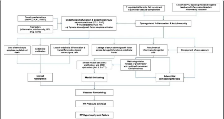
Histopathology: vascular changes
Vascular remodelling in pulmonary arterial and venous hypertension typically involves the small pulmonary ves-sels. Muscular arteries of less than 500 μm display a thickening within the intimal, medial and adventitial compartment. When involved, pulmonary veins of the interlobular septa and smallest preseptal venules show fibrous obliteration and / or muscularization. Even the smallest vascular level may be involved: patients with pulmonary venous hypertension frequently present focal proliferation of alveolar capillaries. The different histo-logical pattern is presented here below.
Arterial lesions
Isolated medial hypertrophy
This abnormality of the vessel wall can be observed in all subgroups of PAH and may even be encountered in other forms of PH, e.g. in mitral valve stenosis. The le-sion corresponds to a smooth muscle cell proliferation and / or recruitment within the tunica media; the histo-logical criterion of hypertrophy / hyperplasia is fulfilled, when the diameter of a single medial layer, delimited by its internal and external elastic lamina, exceeds 10 per cent of the arteries cross-sectional diameter (Figure 2A). Isolated hypertrophy of the medial layer may be consid-ered as an early and even reversible event as it has been shown in PH due to hypoxia in high altitude [171]. However, medial hypertrophy is usually associated with other PAH-lesions.
Concentric and eccentric non-laminar intimal fibrosis
Fibrotic lesions of the intimal layer are frequent in PAH-diseased lungs. The intima may be thickened by prolifer-ation and recruitment of fibroblasts, myofibroblasts and other connective tissue cells, and consequently by the interstitial deposition of collagen (Figure 2B, C). In a purely descriptive approach, this thickening may be uni-form and concentric, or focally predominating and ec-centric. However, the eccentric intimal thickening is frequently observed in cases with thrombotic events and could represent residues of wall-adherent, organized thrombi. Thrombotic lesions, or so called in situ throm-bosis, are a frequent pattern in different PAH-subgroups: organization and recanalization of totally occluding throm-botic material may lead to bizarre, fibrotic multi-channel lesions (so called “colander-like” lesions) which can be easily confounded with proliferative complex lesions (see below) (Figure 2D). Frequently, adventitial fibrosis is associ-ated to intimal modifications (Figure 2A, B).
Concentric laminar intimal fibrosis
This morphologically conspicuous phenotype of intimal fibrosis is also known as “onion-skin” or “onion-bulb” le-sion. Numerous concentrically arranged fibrotic layers
occlude the arterial lumen of small (diameter: 100– 200 μm) arteries (Figure 2E). The scary, cell-lacking morphology of this lesion is frequently found in patients suffering from idiopathic PAH and PAH associated to CTD [172]. Nevertheless, immunohistochemical analysis reveals fibroblasts, myofibroblasts and smooth muscle cells.
Complex lesions
The pathological classification of the World Symposium meetings in Venice and in Dana Point comprises three patterns, plexiform lesion, dilation lesion and arteritis. The plexiform lesion probably represents the most illus-trious form of vascular lesions in PAH and affects vari-ous vascular compartments: focal intimal thickening of small pulmonary arteries, preferably beyond branching points and exuberant endothelial cell proliferation, lead-ing to the formation of capillary-like, sinusoidal channels on a smooth muscle cell and collagen-rich matrix within the native arterial lumen and resulting in obstruction [173,174]. This glomeruloid-like arterial zone feeds into dilated, vein-like congestive vessels, which are perceiv-able at low magnification (Figure 2F). The latter vein-like vessels are also known as dilation lesions and may predominate the histological pattern (Figure 2G). Clas-sical arteritis with transmural inflammation and fibrinoid necrosis, as first described by Heath and Edwards for
PAH associated to congenital cardiac disease
(Eisenmenger), is not a regular finding in PAH [175]. Nevertheless, perivascular inflammatory infiltrates of diseased pulmonary arteries in PAH-patients, consisting mainly of T- and B-lymphocytes, dendritic cells,
mast-cells and macrophages can be regularly found
[141,148,176] (Figure 2H). It has not been elucidated until now, whether this inflammatory pattern is of pathogenetic importance, or if it represents a pure epi-phenomenon within disease evolution. The reported evidence of proinflammatory mediators, so called chemokines, released by altered endothelial cells of PAH-lungs strongly indicates a supporting and self-amplifying process [145,177].
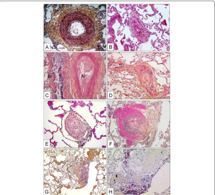
Venous and venular lesions (Pulmonary veno-occlusive disease and pulmonary hemangiomatosis)
A clear-cut differentiation between pre-and post-capillary pulmonary vascular lesions is sometimes difficult to make: lesions frequently concern veins and arteries in lungs of patients with PVOD, and vice-versa veins may be strongly involved in some subgroups of PAH. This is not contra-dictory because the clinical approach to ‘difficult-to-treat’ PAH-groups may be similar to clinical management of PVOD and PVOD-patients are frequently treated – under great precaution – with pulmonary arterial dilators, e.g. prostanoids. Recent reports, for example, indicate that
CTD associated PAH, classically being considered as pre-capillary PH, simultaneously displays a PVOD-like pattern in histology [178,179]. Like in PVOD, the observed post-capillary lesions concern septal veins and pre-septal venules and usually consist of a loose and pauci-cellular and cushion-like intimal fibrosis that may totally occlude the lumen. A muscularization of both, septal veins and pre-septal venules may be observed (Figure 3A, B).
Importantly, occult pulmonary hemorrhage regularly oc-curs in patients displaying PVOD. This particularity, which is certainly due to the post-capillary bloc, is of diag-nostic importance, as bronchio-alveolar lavage can reveal an occult hemorrhage. The degree of hemorrhage can be evaluated semi-quantitatively and qualitatively using the Golde Score, which takes number and Perls-Prussian-Blue staining-degree of intra-alveolar siderophages into
Figure 2 Pulmonary arteries of the muscular type displaying obstructive arteriopathy in lungs of patients with PAH. A Medial hypertrophy with smooth muscle cell proliferation and pronounced adventitial fibrosis. Magnification x200, Weigert-hematoxylin-phloxine-saffron staining (WHPS). B Concentric non-laminar intimal fibrosis comprising numerous myofibroblasts (arrows). C Eccentric intimal fibrosis
corresponding to organized thrombotic material. Br: bronchus, Ar: pulmonary artery. Magnification x100, HES staining. D Thrombotic lesion, so called "colander-like lesion", with partial recanalization by microvessels. Note the similarity to plexiform lesions (F). Magnification x100, HES. E Concentric laminar intimal fibrosis, so called „onion-skin lesion“. Magnification × 200, HES. F Plexiform lesion with proliferation of small sinusoid-like vessels on a fibrotic matrix. Note surrounding dilated vessels. Magnification x100, HES. G Multiple dilation lesions being the sentinel of the centrally located plexiform lesion. Magnification × 40, Elastica-van-Gieson staining (EvG). H The same plexiform lesion after immunohistochemical staining with anti-CD3, a T-lymphocytic marker. Note the perivascular distribution of the inflammatory infiltrate. Magnification x100.
consideration (Figure 3C) [180]. Pulmonary capillary hemangiomatosis has been historically described as an ag-gressive capillary proliferation with patchy distribution within the pulmonary parenchyma: alveolar septa are thickened by 3 to 4 capillary layers, and infiltration of ven-ous and bronchiolar structures with secondary occlusion may be present (Figure 3D). It is thought, that a clinically relevant post-capillary bloc is owed to this angiomatoid expansion. Occult hemorrhage or hemosiderosis, there-fore, is frequently found [181]. However, Lantuejoul and co-workers have shown in a remarkable retrospective histological analysis, that capillary hemangiomatosis-pattern is virtually always present in PVOD, and vein-remodelling is constantly observed in case with a primary diagnosis of PCH [54]. The authors suggest the possibility, that PVOD and PCH might be the same disease, with a vein- or a capillary-predominating pattern. We fully sup-port this view, and in our experience from the French Na-tional PH Reference Center, no clinical distinction is made between both conditions.
Clinical description
Symptoms and clinical signs of PH
There is no pathognomonic clinical sign of PH. Clinical presentation is related either to right heart failure or to associated diseases. Persistant dyspnea on exertion is the most frequent symptom; and it is present in almost pa-tients even in the presence of mild hemodynamic abnor-malities [1,182]. Dyspnea usually starts insiduously and is often neglected by patients which explain the delay of around two years in establishing the diagnosis of PH. The New York Heart Association (NYHA) provides a classification system for the clinicial evaluation of dys-pnoea. Four categories are proposed to classify patients in functional classes (FC) based on how much they are limited during physical activity; the limitations/symp-toms are in regard to normal breathing (Table 3).
However, at time of diagnosis, 70% of patients are in NYHA FC III or IV. Chest pain, light-headedness and syncope may occur, particularly during physical efforts and are major signs of disease severity. Palpitations are frequent during physical efforts and may reveal true car-diac arrhythmias. Other symptoms of PAH include fa-tigue and weakness. Hemoptysis may complicate PAH and could be life-threatening, justifying embolization of dilated bronchial arteries. Hoarseness of the voice may occasionally be noted and is due to compression of the left laryngeal nerve by the dilated pulmonary artery (Ortner’s syndrome).
Signs of right heart failure may be observed in the most severe patients, including venous jugular disten-sion, hepato-jugular reflux, hepatomegaly and hepatalgia. Lower limb edema, ascitis and generalized edema under-score the severity of right heart failure. Cardiac
auscultation shows usually a prominent pulmonary com-ponent of S2, a systolic murmur of tricuspid regurgita-tion and more rarely a diastolic murmur of pulmonary regurgitation. Pulmonary auscultation is usually normal and contrasts with the importance of dyspnea. History and clinical examination should also screen for manifes-tations of extra thoracic diseases, particularly Raynaud’s syndrome which can be found in PAH associated with CTD and particularly in systemic sclerosis.
Diagnostic methods
The diagnostic process of PAH requires a series of investiga-tions that are intended to make the diagnosis, to clarify the clinical class of PH and the underlying type of PAH and to evaluate the functional and hemodynamic impairment [183]. The detection of PH requires investigations including electrocardiogram, chest radiograph and trans-thoracic echocardiogram. Other conditions which can induce PH will be identified by tests such as pulmonary function tests, ar-terial blood gases, ventilation and perfusion lung scan, high resolution computed tomography (HR-CT) of the chest and pulmonary angiography. Additional investigations are re-quired for evaluation of PAH severity including exercise test-ing and hemodynamics. Additional imagtest-ing may clarify underlying lung abnormalities. Finally, right heart catheter-isation confirms the definite diagnosis.
Electrocardiogram (ECG)
The ECG may provide suggestive or supportive evidence of PH by demonstrating right ventricular hypertrophy and strain, and right atrial dilation. Right ventricular hypertrophy and right axis deviation are present in re-spectively 87% and 79% of patients with idiopathic PAH [105]. Unfortunately, the ECG has low sensitivity and specificity as a screening tool for detecting PH.
Chest radiography
In 90% of idiopathic PAH patients, chest radiography is abnormal at the time of diagnosis [105]. Findings include central pulmonary arterial dilatation which contrasts with loss of the peripheral blood vessels. Right atrial and ven-tricular enlargement may be seen in more advanced cases. Chest radiography may help to identify associated moderate-to-severe lung disease or pulmonary venous hypertension due to left heart abnormalities.
Pulmonary function test and arterial blood gases
Pulmonary function tests (PFT) will help to assess under-lying lung abnormalities. Forced expiratory volume in one second (FEV1) and total lung capacity (TLC) in idiopathic PAH are usually normal or slightly abnormal. Low diffusing capacity of the lung for carbon monoxide (DLCO) has been reported in PAH patients, but is more pronounced in PVOD patients with often severe reductions under 50% of
the predicted value [56,184]. Results of arterial blood gases usually show mild hypoxemia and hypocapnia. Severe hyp-oxemia may be a parameter of underlying PVOD or chronic lung disease.
Exercise testing
The normal physiologic response of the pulmonary vas-culature to exercise consists of distension of pulmonary arteries and arterioles as well as recruitment of previ-ously unused vascular bed. Thus, in health, pulmonary artery pressure rises minimally in response to increased blood flow and pulmonary vascular resistance decreases because of the remodeled vasculature. These mecha-nisms are impaired in the course of PH. Cardiopulmo-nary exercise testing (CPET) has been shown to be useful in assessing the severity and prognosis of PAH [15,185]. Several mechanisms are associated: (1) failure to perfuse the ventilated lung, leading to an increase of physiologic dead space and ventilatory requirement; (2) failure to increase cardiac output appropriately in re-sponse to exercise, causing an early lactic acidosis, thereby increasing acid ventilatory drive; and (3) exercise-induced hypoxemia increasing the hypoxic ven-tilatory drive. The venven-tilatory expired gas abnormalities precipitated by PH are multifactorial and associated with disease severity. CPET assesses and measure the ventila-tion–perfusion mismatch (i.e. acceptable ventilation/di-minished perfusion), reflected by an elevated VD/VT or VE/VCO2 ratio or slope and diminished partial pressure
of end-tidal carbon dioxide (PETCO2), and the de-creased peak VO2 and VO2 at the ventilatory threshold (VT). Peak VO2, VE/VCO2 ratio or slope or PETCO2, measure during CPET, all demonstrated independent and strong prognostic value as univariate markers [186].
Transthoracic doppler-echocardiography (TTE)
TTE is a non-invasive screening test for patients with suspected PH. TTE estimates pulmonary artery systolic pressure (sPAP) and may provide additional information about the cause and consequences of PH. The estima-tion of PAP is based on the peak velocity of the jet of tri-cuspid regurgitation. The simplified Bernoulli equation describes the relationship of tricuspid regurgitation vel-ocity and the peak pressure gradient of tricuspid regurgi-tation = 4 x (tricuspid regurgiregurgi-tation velocity). Estimation of PA systolic pressure require to take into account right atrial pressure (PA systolic pressure = tricuspid regurgi-tation pressure gradient + estimated right atrial pres-sure). Right atrial pressure cannot be measured and is estimated based on the diameter and respiratory vari-ation of the inferior vena cava [15]. An alternative ap-proach to echocardiographic diagnosis of PH is based on the comparison of tricuspid regurgitation velocity with values reported in a healthy population. Ideally, the in-fluence of age, sex and body mass should be taken into consideration [187]. This method avoids cumulative error but is less directly linked to the accepted hemodynamic definition of PH based on mPAP [15].
Figure 3 Pulmonary veins with obstructive venopathy in lungs of patients with PVOD and a case of pulmonary capillary hemangiomatosis. A Longitudinally dissected septal vein with asymmetric intimal and partially occlusive fibrosis. Note the intra-alveolar hemorrhage due to the post-capillary block on the upper half of the photograph. Magnification × 100, EvG. B Pre-septal venule with occlusive intimal fibrosis. Magnification × 100, EvG. C Bronchio-alveolar lavage in a PVOD-patient. Perls-Prussian-Blue staining. Note the siderophages displaying gradually different color-shades (see text). Magnification × 400. D Excessively proliferating alveolar capillaries in a patient with pulmonary capillary hemangiomatosis. Note protrusion of ectatic lumina into the alveoli. Magnification × 200, anti-CD31 staining.
Other echocardiographic variables that might raise or reinforce suspicion of PH independently of tricuspid re-gurgitation velocity should always be considered. They include an increased velocity of pulmonary valve regur-gitation and a short acceleration time of RV ejection into the PA. Increased dimensions of right heart chambers, abnormal shape and function of the interventricular septum, increased RV wall thickness, pericardial effusion and dilated main PA are also suggestive of PH, but these signs are considered to be related to the hemodynamic severity [15].
Besides identification of PH, TTE also allows a differ-ential diagnosis of the possible causes of pulmonary hypertension. TTE can recognize left heart valvular dis-eases and myocardial disdis-eases responsible for post-capillary PH, and congenital heart diseases with systemic-to-pulmonary shunts. The venous injection of agitated saline can help to identify patent foramen ovale or small sinus venosus type atrial septal defects. Transesophageal echocardiography is rarely required in the setting of PH.
Ventilation/perfusion lung scan
Ventilation/perfusion lung scan should be systematically assessed to screen for CTEPH. Indeed, V/Q lung scan is the method of choice to detect CTEPH [188] and nor-mal V/Q scan can eliminate CTEPH.
High resolution computed tomography of the chest
High resolution computed tomography of the chest (HRCT) supplies detailed information about underlying lung parenchyma disease, such as pulmonary emphy-sema or interstitial lung disease. A number of various pathologic features may be detected on chest HRCT in-cluding pericardial effusions and pulmonary artery en-largement, defined by the ratio of the diameter of main pulmonary artery to that of the thoracic aorta >1. In the setting of CTEPH, contrast HRCT of the pulmonary ar-teries may show changes like complete vessel obstruc-tion, vessel cut-offs, intimal irregularities, incorporated thrombus formations as well as bands and webs [189]. Furthermore, collaterals from bronchial arteries can be identified with this technique. Proximal pulmonary ob-struction is displaying about significant and accessible organized fibrous tissue in segmental or subsegmental arteries. If no proximal obstruction or obliteration is noted; lesions are considered to be distal, non-accessible to surgery intervention. In some cases, pulmonary angi-ography is necessary to differentiate between proximal or distal obstructions. Chest HRCT may also suggest PVOD in the presence of adenopathy mediastinal, ground glass opacities and septal lines [53].
Pulmonary angiography
In CTEPH, pulmonary angiography may be helpful to determinate surgically accessible form. Typical angio-graphic findings in CTEPH are complete obstruction, band and webs as well as intimal irregularities [74]. Pul-monary angiography may be also helpful in the setting of fibrosing mediastinitis.
Cardiac magnetic resonance imaging
Cardiac magnetic resonance imaging (MRI) allows non invasive evaluation of right ventricular size, morphology and shape. It provides information on right ventricular function and allows non-invasive assessment of blood flow including cardiac output, stroke volume, distensibil-ity of pulmonary artery and right ventricular mass [190,191]. Decreased stroke volume, an increased right ventricular right ventricular end-diastolic volume and a decreased measured at baseline are associated with poor prognosis of disease [192,193]. In addition, it has been demonstrated that deterioration of these parameters at one-year follow-up were also predictors of mortality [193]. Thus cardiac MRI could represent a non-invasive tool to evaluate severity of PAH patients at baseline and during follow-up. Further studies are needed to evaluate the precise place of cardiac MRI in the management of PAH patients.
Abdominal ultrasound scan
Abdominal ultrasound should be performed in all pa-tients if PH is suspected to exclude portal hypertension or liver disease. When portal hypertension is suspected, the diagnosis can be confirmed during RHC by measure-ment of an increased gradient between the free and oc-cluded hepatic vein pressure [194].
Blood tests
Serological tests for HIV, hepatitis B or C serology should be performed to screen for associated diseases. The thyroid hormone measurement may reveal either hyperthyroid dysfunction or autoimmune thyroiditis, fre-quently encountered in PAH.
Right heart catheterization
Invasive hemodynamic assessment with right heart catheterization is requested to confirm the diagnosis of PH showing a resting mPAP of ≥25 mmHg and a normal PCWP [2]. This value has been used for selecting pa-tients in all RCTs and registries of PAH, however normal mPAP at rest is around of 14 mmHg, with an upper limit of normal of 20 mm Hg. The significance of a mean PAP between 21 and 24 mmHg is currently un-clear. No definition of PH on exercise was currently adopted, because of the large variability of mPAP on ex-ercise in healthy individuals.