Delivering safer immunotherapies for cancer
The MIT Faculty has made this article openly available. Please share
how this access benefits you. Your story matters.
Citation
Milling, Lauren et al “Delivering Safer Immunotherapies for
Cancer.” Advanced Drug Delivery Reviews 114 (May 2017): 79–101 ©
2017 The Authors
As Published
http://dx.doi.org/10.1016/J.ADDR.2017.05.011
Publisher
Elsevier BV
Version
Final published version
Citable link
http://hdl.handle.net/1721.1/117754
Terms of Use
Creative Commons Attribution-NonCommercial-NoDerivs License
Delivering safer immunotherapies for cancer
☆
Lauren Milling
a,b, Yuan Zhang
f, Darrell J. Irvine
a,b,c,d,e,⁎
a
Koch Institute for Integrative Cancer Research, MIT, Cambridge, MA, USA b
Dept. of Biological Engineering, MIT, Cambridge, MA, USA cDept. of Materials Science & Engineering, MIT, Cambridge, MA, USA d
Ragon Institute of MGH, MIT, and Harvard, Cambridge, MA, USA e
Howard Hughes Medical Institute, Chevy Chase, MD, USA f
Dept. of Biomedical and Pharmaceutical Sciences, College of Pharmacy, University of Rhode Island, RI, USA
a b s t r a c t
a r t i c l e i n f o
Article history:
Received 18 February 2017 Received in revised form 5 May 2017 Accepted 17 May 2017
Available online 22 May 2017
Cancer immunotherapy is now a powerful clinical reality, with a steady progression of new drug approvals and a massive pipeline of additional treatments in clinical and preclinical development. However, modulation of the immune system can be a double-edged sword: Drugs that activate immune effectors are prone to serious non-specific systemic inflammation and autoimmune side effects. Drug delivery technologies have an important role to play in harnessing the power of immune therapeutics while avoiding on-target/off-tumor toxicities. Here we review mechanisms of toxicity for clinically-relevant immunotherapeutics, and discuss approaches based in drug delivery technology to enhance the safety and potency of these treatments. These include strategies to merge drug delivery with adoptive cellular therapies, targeting immunotherapies to tumors or select immune cells, and localizing therapeutics intratumorally. Rational design employing lessons learned from the drug deliv-ery and nanomedicinefields has the potential to facilitate immunotherapy reaching its full potential.
© 2017 The Authors. Published by Elsevier B.V. This is an open access article under the CC BY-NC-ND license (http://creativecommons.org/licenses/by-nc-nd/4.0/).
Keywords:
Cancer immunotherapy Checkpoint blockade Adoptive cell therapy Nanoparticles
Contents
1. Introduction . . . 80
2. Mechanisms of toxicity elicited by immunotherapy drugs. . . 80
2.1. Interleukin-2 as a paradigm for approved but toxic immunotherapy . . . 80
2.2. Checkpoint blockade . . . 81
2.3. Agonist antibodies against immune costimulatory receptors . . . 83
2.4. Tumor targeting antibodies . . . 84
2.5. Local administration of immunotherapy agents . . . 85
3. Engineering safer local therapies . . . 85
3.1. Intratumoral drug depots . . . 86
3.2. Intratumoral gene delivery . . . 88
3.3. Anchored drugs . . . 88
3.4. Tumor draining lymph node-targeted drugs . . . 88
4. Engineering safer systemic immunotherapies . . . 89
4.1. Molecularly-targeted immunotherapy . . . 89
4.2. Nanoparticle delivery of immunotherapy agents to tumors . . . 90
4.3. Systemic gene delivery of immunomodulators to tumors. . . 92
4.4. Immune cells as drug carriers . . . 92
4.5. Targeting immunotherapy to immune cells . . . 94
5. Conclusions and future perspectives . . . 95
Acknowledgments . . . 95
References . . . 95
☆ This review is part of the Advanced Drug Delivery Reviews theme issue on “Immuno-engineering”. ⁎ Corresponding author at: Dept. of Biological Engineering, MIT, Cambridge, MA, USA.
E-mail address:[email protected](D.J. Irvine).
http://dx.doi.org/10.1016/j.addr.2017.05.011
0169-409X/© 2017 The Authors. Published by Elsevier B.V. This is an open access article under the CC BY-NC-ND license (http://creativecommons.org/licenses/by-nc-nd/4.0/).
Contents lists available atScienceDirect
Advanced Drug Delivery Reviews
1. Introduction
Immunotherapies, treatments that modulate the immune system, have long been proposed as a potentially powerful approach to “func-tional” or actual cures of disease, based on the natural function of the immune system in protecting the host and its cardinal features of poten-cy, specificity, and memory[1]. Motivated by these features, immuno-therapies are now in preclinical and clinical development for treatment of diverse infectious diseases, autoimmunity, allergies, trans-plant rejection, graft vs. host disease, and cancer. Among these thera-peutic areas, cancer immunotherapy in particular has experienced dramatic recent progress in the clinic[2,3]. For many years, cancer im-munotherapies were plagued by high toxicity, low to negligible efficacy, or both. However, steady advances in fundamental cancer immunology and translational immunotherapy have now led to two classes of treat-ment with significant impact in advanced cancer patients – adoptive cell therapy (ACT), based on the injection of autologous tumor-directed T cells[4,5]; and checkpoint blockade, treatment with antibodies that block the inhibitory receptors cytotoxic T lymphocyte antigen-4 (CTLA-4) or programmed death-1 (PD-1, or its counter-receptors PD-L1/PD-L2)[6,7]. ACT therapy in patients with advanced metastatic melanoma and several hematologic cancers has shown a high proportion of com-plete responses (comcom-plete elimination of detectable tumor burden), some of which are durable responses lasting many years[8]. Treatment with ipilimumab, a fully human anti-CTLA-4 antibody, has led to com-plete responses in approximately 20% of advanced melanoma patients, with durations lastingN10 years[9]. Treatment with PD-1 blocking anti-bodies has elicited objective responses in a variety of solid tumors includ-ing melanoma, lung cancer, prostate cancer, breast cancer, ovarian cancer, head and neck cancer, and a subset of colorectal cancers[6]. Reflecting their complementary modes of action, combination therapy with anti-CTLA-4 and anti-PD1 has led to even greater response rates in melanoma patients, where a significant fraction of patients exhibit complete tumor regressions in a space of ~10 weeks[10,11].
Thesefindings have energized the field and motivated a massive ef-fort to further explore combination immunotherapies that optimally arm the immune system against metastatic disease, but the power of the immune system creates the potential for not only a dramatic attack on tumors but also a significant danger to healthy tissues. For example, monotherapy with anti-CTLA-4, which both blocks a negative regulato-ry signal during T cell activation and inhibits the function of regulatoregulato-ry T cells, leads to a series of autoimmune side effects, including gastrointes-tinal toxicity, pruritis, and fatigue, side effects which become grade 3 or 4 serious adverse events in ~23% of patients[12]. When anti-CTLA-4 is combined with anti-PD-1, enhanced anti-tumor activity comes at the cost of synergistically exacerbated toxicity; ~55% of previously untreat-ed melanoma patients given the combination experiencuntreat-ed grade 3 or 4 adverse events[11,12]. As discussed in detail in this review, serious tox-icities are characteristic of a broad range of immunomodulatory drugs. Thus, a looming challenge in thefield is the development of effective strategies to harness the potential of combination treatments while avoiding debilitating toxicities that prevent immunotherapies from reaching their full curative potential. Clinical studies are already under-way seeking to optimize timing and dosing to limit the toxicity of these promising immunotherapy drugs, but in the setting of intravenous ad-ministration– believed to be key for systemically modulating the im-mune response against disseminated tumors– dosing schedules with high safety and high efficacy are often diametrically opposed.
In this review, we discuss the potential for drug delivery technolo-gies spanning a range of approaches to enhance immunotherapies, with a particular emphasis on the potential for enhancing the safety of immunomodulatory drugs. Wefirst review representative mechanisms of immune toxicity from immunotherapy agents of both clinical and preclinical interest, separating systemic and local (i.e. intratumoral) drug administration issues. We then discuss approaches to ameliorate these toxicities based in concepts from thefield of drug delivery,
employing technologies ranging from nanoparticles to synthetic biolo-gy. The immune system as a target for therapy presents several chal-lenges and opportunities relative to somatic tissues: Immune cells circulate through the blood, creating the potential for efficient direct targeting of therapeutics to these cells (relative to, for example, targeting drugs to tumor cells); and immune cells proliferate, providing a source for self-amplification of small doses of appropriately-targeted drugs. However, there is a need to direct immunomodulatory drugs to tumor-specific cells rather than stimulating the entire leukocyte com-partment non-specifically, and these cells may be preferentially enriched at tumor sites and tumor-draining lymph nodes. There are thus both challenges and opportunities for thefield of drug delivery to impact cancer immunotherapy.
2. Mechanisms of toxicity elicited by immunotherapy drugs To rationally approach strategies for increasing the safety of system-ic immunotherapies, an understanding of mechanisms underlying the toxicity of systemically-administered immunoregulatory drugs is need-ed. In this section, we review the mechanisms of toxicity underlying several important classes of immunotherapy agents: interleukin-2, rep-resentative of several importantγ-chain cytokines that promote lym-phocyte proliferation and effector function; agonistic antibodies against the costimulatory receptors CD137 (also known as 41BB) and CD28, representative of agonistic antibodies against lymphocyte co-stimulatory molecules; and the checkpoint blockade agents anti-CTLA-4 and anti-PD-1. A discussion of all immunoregulatory agents in preclin-ical and clinpreclin-ical testing for cancer immunotherapy is beyond the scope of any single review, but these example biologics represent 3 important distinct mechanisms of immunomodulation relevant to much of the on-going clinical development of immunotherapy.
2.1. Interleukin-2 as a paradigm for approved but toxic immunotherapy Systemic high-dose interleukin-2 (IL-2) was one of thefirst immuno-therapy agents to be licensed for cancer immuno-therapy, approved by the FDA for metastatic melanoma and renal cell carcinoma (RCC) treatment in 1992. IL-2 wasfirst isolated as a factor promoting the growth of activated T cells, but also stimulates natural killer (NK) cells, both of which motivated its use as a cancer therapeutic. However, it is now also conversely known to also promote activation-induced cell death of stimulated T cells and maintains the survival and function of regulatory T-cells, which restrain the effector arms of the immune system to maintain tolerance and protect healthy tissues from autoimmune attack[13]. Interleukin-2 biology is fur-ther complicated by the nature of its tripartite receptor, which is com-prised of the IL-2Rα chain (CD25), β chain (CD122), and common γ chain (CD132)[13]. Differential expression of the three components of the IL-2R leads to different signaling and functional outcomes on different cell types at different stages of activation.
Based on dosing schedules established clinically in the 1980s, IL-2 is approved as a“high dose” (HD) IL-2 therapy for melanoma and RCC ad-ministered intravenously every 8 h for up to 14 total doses[14]. Although much is made in the current renaissance of cancer immunotherapy around the“tail of the curve” effect, where a small proportion of patients treated with checkpoint blockade become long-term survivors[9], such durable increases in survival were already seen in the early 1990s in pa-tients treated with IL-2, where ~12% of papa-tients treated with HDIL-2 at the National Cancer Institute had survival of at least 10 years[14]. Al-though HDIL-2 elicits objective responses in ~16% of patients, it is also ex-tremely toxic. The very short half-life of IL-2 (~12 min[15]) leads to a requirement for high doses to be administered in order for functional levels to be maintained for a sufficient timespan. High level dosing in turn leads to dose-related toxicities including vascular leak syndrome (VLS) and cytokine release syndrome, a massive systemic cytokine release and inflammatory reaction caused by IL-2 immune stimulation[16]; le-thal adverse events were found in 2% of patients[14]. These issues
mean that IL-2 therapy requires careful selection of patients with analyses of baseline cardiac risk factors, performance status, and biomarker analy-sis. IL-2's modes of action in expanding T-cells and NK cells make it a log-ical candidate for combination immunotherapies, but lethal toxicities were seen when HDIL-2 was administered after checkpoint blockade
[17]or tyrosine kinase inhibitor (TKI) therapy[18].
The toxicity of IL-2 is multifactorial and involves a complex set of in-teractions, most notably between immune cells and the vascular endo-thelium. IL-2 therapy has an acute impact on circulating effector lymphocytes, with a rapid transient induction of lymphopenia. IL-2-acti-vated cells strongly bind to endothelial cells (ECs), leading to endothelial cell cytotoxicity by NK cells and granulocytes[19–21]. In addition, IL-2-in-duced pulmonary edema is promoted by the interaction of IL-2 with func-tional IL-2 receptors (IL-2R) expressed on lung endothelial cells; blocking IL-2 interactions with IL-2Rα (CD25) abrogates IL-2-mediated pulmonary edema[22–24]. The endothelial cell damage caused by IL-2-activated host effector cells and/or cytokines and chemokines released in response to IL-2 (e.g., IFN-γ and TNF-α) contribute to VLS and systemic toxicity[25–30]. The role of cytokines in the development of VLS is related to both their di-rect effect on increasing the permeability of the vascular endothelium and their effects on leukocyte and EC activation[31]. Activation of both ECs and monocytes/neutrophils by cytokines results in the release of large amounts of nitric oxide (NO)[32,33], which further damage ECs and en-hance the adhesion of neutrophils through upregulation of adhesion receptors[34]. Administration of NO inhibitors has been reported to de-crease IL-2-induced vascular toxicity in mice[30], and it has been shown that NK cell depletion protects mice from IL-2-induced VLS[35, 36]. Neutrophils also play a critical role in VLS by adhering to ECs and inducing lysis via reactive oxygen intermediates (ROI) and proteases
[32,37,38]. IL-2 also leads to complement activation which induces mast
cell degranulation, resulting in the release of vasoactive mediators and an increase in vascular permeability[39].
The efficacy of IL-2 has fueled much interest in strategies to mitigate its toxicity, as discussed in the subsequent sections below. Continuous intravenous infusion of low dose IL-2 is less toxic than bolus injection of high dose IL-2, but the therapeutic efficacy is also compromised[40, 41]. Strategies strictly localizing IL-2 to tumors or tumor-draining lymph nodes (discussed further below) lack the toxicity of i.v. adminis-tration[42–44], suggesting that IL-2's toxicity is linked to systemic ex-posure. Thus, approaches to target IL-2 to specific cell types, engineer its interaction with different cell subsets, or localize it to tumors are all strategies of interest to improve its therapeutic index.
2.2. Checkpoint blockade
For many in the general clinical oncology world, the real break-through in cancer immunotherapy came with the results from recent clinical trials of so-called“checkpoint blockade” antibodies – antibodies that block negative regulatory signals restraining the host T-cell re-sponse against tumors. Two well studied checkpoint receptors, cytotox-ic T lymphocyte antigen-4 (CTLA-4) and programmed death-1 (PD-1), are expressed on T-cells and act to diminish responses in early activated and mature peripherally disseminated T-cells, respectively. T-cells are activated when the T-cell receptor binds peptide presented in the major histocompatibility complex on antigen presenting cells (APCs, es-pecially dendritic cells) and costimulatory receptors on the T-cell and APC are co-ligated (e.g., CD28 on the T-cell binding to CD80 and CD86 on APCs). CTLA-4 is upregulated on T-cells during early stages of activa-tion, with expression increasing around 2 days after encounter with cognate peptide presented on APCs in lymphoid organs[12]. As a high
CD80/86 CD28 CTLA-4 CD80/86 αCTLA-4 TCR MHC Peptide T cell APC CD137 CD137L Tumor cell T cell TCR MHC Peptide PD-L1 PD-1 αPD-1 CD137 αCD137 CD137 αCD137 FcγR APC
(A)
(B)
Treg αCTLA-4 CTLA-4 Tumor associated macrophage NK(C)
Fig. 1. Mechanisms of action for immunomodulatory antibodies. (A) T-cells are activated by APCs presenting specific peptide-MHC complexes in tandem with signals from both positive (e.g., CD28, CD137) and negative (e.g., CTLA-4) costimulatory receptors binding cognate partners on the APC surface. Anti-CTLA-4 blocks receipt of negative regulatory signals from CTLA-4 engagement to boost T-cell priming by APCs. (B) Anti-PD-1 augments T-cell function in the effector phase by blocking negative regulatory signals delivered by PD-L1 expressed on tumor cells or PD-L2 expressed by APCs and other cells. Anti-CD137 can also boost T-cell effector function through crosslinking of the CD137 costimulatory receptor and/or clustering receptors by antibody displayed on APCs through their Fcγ receptors. (C) In murine models antibodies have been utilized to bind cells and initiate their depletion/killing through complement mediated cytotoxicity and antibody dependent cellular cytotoxicity (ADCC). This mechanism can be applied directly to tumor cells. Anti-CTLA-4 antibodies also act to boost the immune response by triggering ADCC-mediated depletion of intratumoral Tregs that express high levels of CTLA-4 receptor.
affinity homologue of CD28, CTLA-4 serves to interrupt costimulation signals from CD28 to the newly activated T-cells by competition for CD28's ligands, and thereby dampens the immune response (Fig. 1A)
[45]. Inhibition of CTLA-4 using monoclonal antibodies lifts the inhibito-ry effects of CTLA-4, but only on activated T-cells[46]. In addition, CTLA-4 is highly expressed on regulatory T cells (Tregs); therefore, in mice, administration of anti-CTLA-4 antibodies has been shown to deplete Tregs specifically from the tumor microenvironment where CTLA-4 is most highly expressed by these cells ([47–49]. In contrast to CTLA-4, PD-1 is primarily expressed on T cells in the periphery and in the tumor microenvironment (Fig. 1B)[12]. Interaction with one of its two ligands (PD-L1 or PD-L2) downregulates antigen receptor signaling in mature T-cells and decreases the expression of pro-inflammatory cy-tokines. PD-1 expression is present during T-cell effector phases and on re-exposure to antigen[50]. Blockade of the PD-1 interaction with its li-gand can transiently reverse exhaustion of T-cells in the periphery[51]. These distinct mechanisms of action have led to therapeutic blocking antibodies targeting CTLA-4, PD-1, or PD-1's ligands. Ipilimumab, an an-tibody that blocks CTLA-4 was approved in 2011 following a pivotal trial demonstrating its ability to improve long term overall survival in ad-vanced metastatic melanoma - thefirst new drug for advanced melano-ma approved inN30 years[52]. Ipilimumab therapy leads to complete tumor regressions lasting at least 10 years in ~20% of treated melanoma patients[9]. Following on the heels of ipilimumab's approval, several antibodies blocking PD-1 (expressed by T-cells) binding to its ligands PD-L1 or PD-L2 (expressed by many cells, including tumor cells)– en-tered the clinic and have shown even more impressive initial responses, with 30–50% of patients in diverse diseases ranging from melanoma to lung cancer exhibiting objective tumor regressions[53–55]. The ability of PD-1 blockade to elicit responses in solid tumors not previously viewed as“immunogenic” (e.g., lung cancer) heralded a new era of promise for immunotherapies. However,“taking the brakes off” of the immune system with systemic checkpoint blockade, leads (not unex-pectedly) to toxicities, which are amplified when these drugs are employed in combination.
Ipilimumab therapy elicits adverse events, with 60–65% of patients experiencing immune related adverse events (irAEs) at a moderate dose of 3 mg/kg every three weeks[52,56]. In two large clinical trials, the most common adverse events included pruritus (25–35%), diarrhea (23–33%), rash (15–33%), and fatigue (15–28%)[11,57]. Serious Grade 3 and 4 adverse events occur in 20–27% of patients, the most frequent being gastrointestinal toxicities resulting in enterocolitis and diarrhea (6–8% of grade 3–4 events)[11]. Of the 14 patients who died in the phase III clinical trial mentioned above, 7 died due to immune-related adverse events[11,52,56,57]. Overall, ipilimumab elicits broad irAEs of the skin, gastrointestinal tract, and endocrine system.
Anti-CTLA-4 irAEs may manifest from depletion of regulatory T cells, as evidenced by the need for depleting antibody isotypes (such as IgG2a) capable of engaging Fcγ receptors to mediate antitumor activity in mouse models[48,58,59]. CTLA-4 blocking antibodies of IgG2a isotype yielded depletion of regulatory T-cells at tumor sites (Fig. 1C) and an increase in CD8+T effector cells in the periphery, while other
isotypes expanded both regulatory and effector T-cells in the periphery
[59]. Some evidence however demonstrates that the depletion of regu-latory T-cells is context specific and limited to the tumor microenviron-ment, due to the high frequency of Fcγ receptor-expressing tumor associated macrophages, though depletion of regulatory T-cells in the periphery remains conceivable[48]. Regulatory T-cells are known to maintain tolerance and restrict lymphocytic infiltration to mucosal lin-ings of organs including the lungs and gastrointestinal tract[60]. Deple-tion of such regulatory T-cells in the intestines may account for increased intraepithelial lymphocytes and leukocyte infiltration in the lamina propria of ipilimumab-treated patients as revealed by endoscop-ic analysis, thus causing irAEs such as diarrhea[61,62].
PD-1 blockade antibodies have in general shown less serious side ef-fects compared to anti-CTLA-4 in humans, with only ~15% of patients
experiencing serious adverse events, primarily pneumonitis. For com-parison, in phase I dose escalation studies, administration of nivolumab (one of the approved anti-PD-1 antibodies) resulted in grade 3–4 ad-verse events in only 14% of patients compared to the 20–27% in early ipilimumab trials. Lastly, irAEs of any grade affected only 41% of patients compared to ipilimumab's 60–65%[63]. Consistent with their distinct mechanisms of action, the immune related adverse events differ be-tween anti-CTLA-4 and anti-PD-1/PD-L1 therapeutics: anti-PD-1 irAEs include rash, hypothyroidism, hyperthyroidism, pneumonitis, diarrhea, and elevated aminotransferase levels[12]. A large phase III trial con-firmed that both nivolumab and pembrolizumab (both now clinically approved anti-PD-1 antibodies) induce fewer adverse events than ipilimumab[64].
Mechanistic differences between PD-1 and CTLA-4 receptors are likely responsible for the differences in adverse outcomes and better tol-erability of PD-1 checkpoint blockade. Given that CTLA-4 is expressed on all T cells around the time of activation in lymphoid tissue, broad spectrum autoimmunity can result from blockade of CTLA-4. Addition-ally, as noted above administration of anti-CTLA-4 antibodies in mice depletes regulatory T cells, which play an important role in restraining autoimmune attack of host tissue[12], and systemic depletion of Tregs leads to fatal autoimmune pathology. Such a mechanism would moti-vate strategies to localize the action of anti-CTLA-4 in vivo, but this phe-nomenon remains unconfirmed in humans so far. Unlike CTLA-4, the PD-1 receptor is largely expressed on antigen re-exposed cells in the pe-riphery; therefore, a smaller pool of T-cells are likely affected by treat-ment with anti-PD-1 antibodies. Because expression of CTLA-4 is dependent on initial priming of T-cells, the onset of irAEs and therapeu-tic efficacy from anti-CTLA-4 antibodies is often delayed compared to PD-1 blockade, which reactivates T-cells already present in the tumor microenvironment[12]. The varying populations of T-cells targeted by checkpoint blockade antibodies may explain the severity and time to onset of immune-related adverse events.
CTLA-4 and PD-1 are just thefirst successfully targeted inhibitory re-ceptors on lymphocytes, two members of a large collection of negative regulatory receptors expressed by cells. A non-exhaustive list of T-cell-expressed negative regulatory receptors explored in preclinical studies and clinical trials includes Lymphocyte activation gene 3 (LAG-3), T cell membrane protein 3 (TIM-(LAG-3), T-cell immunoreceptor with im-munoglobulin and ITIM domains (TIGIT), V-domain Ig suppressor of T-cell activation (VISTA), adenosine A2a receptor (A2aR), and B and T lymphocyte attenuator (BTLA)[65–68]. In addition, because of their dis-tinct mechanisms of action (despite their classification together in the immune-oncology world as checkpoint blockade agents), therapies combining anti-CTLA-4 and anti-PD-1 were obvious to explore, and showed promising synergy in preclinical mouse models[69]. Recently, thefirst trials of ipilimumab combined with the PD-1 blocking antibody nivolumab in melanoma were completed, and showed that while the combination achieves a striking increase in efficacy – with 75% of pa-tients experiencing objective responses– this enhanced efficacy was ac-companied by a concomitant increase in serious toxicities, with 53% of patients experiencing grade 3 or 4 serious adverse events[10,11,70, 71]. Grade 3–4 toxicities were commonly characterized by elevated li-pase, aspartate aminotransferase, and alanine aminotransferase levels, indicating pancreas and liver toxicities. Unexpected side effects includ-ing acute onset diabetes and diabetic ketoacidosis were noted in 17 pa-tients co-treated with ipilimumab and nivolumab[72]. Often these autoimmune adverse effects do not present until weeks to months after treatment. Similar toxicities appear to be prevalent in a phase I study of a different anti-CTLA-4 antibody (tremelimumab) in combina-tion with an anti-PD-L1 antibody (durvalumab) tested in NSCLC pa-tients[73]. Recently reported interim results of an ongoing phase II trial of ipilimumab combined with nivolumab in recurrent small cell lung cancer reported a lower incidence rate of serious side effects but also lower levels of overall response[74]. Adjustment of dosing sched-ules in a phase I study in non-small cell lung cancer patients (nivolumab
given every 2 weeks but ipilimumab given every 6 or 12 weeks) showed some reduction in toxicity compared to the initial melanoma trials, with only 33–37% grade 3–4 adverse events[75].
Combination treatment with checkpoint blockade antibodies epito-mizes the promise and the challenge of immunotherapy– co-adminis-tration of these agents systemically leads to significant increases in anti-tumor efficacy, but also synergistic amplification of toxicity. Achieving the goal of long term durable remissions in a majority of pa-tients is unlikely to be achieved by single drugs in difficult-to-treat solid tumors, and this has led much of thefield to be convinced of the need for combination immunotherapy strategies combining multiple drugs act-ing via complementary mechanisms[76]. Solutions from thefield of drug delivery may be critical to achieve this goal while avoiding life-threatening toxicities that plague many immunotherapy drugs adminis-tered using the systemic dosing strategies traditionally employed in oncology.
2.3. Agonist antibodies against immune costimulatory receptors
CTLA-4 and PD-1 represent important inhibitory receptors that re-strain T-cell priming and effector functions. These negative regulatory receptors are counter-balanced by a suite of positive costimulatory re-ceptors that support T-cell activation. Canonical examples include CD28, CD137 (also known as 4-1BB), and CD134 (also known as OX40 or Tumor necrosis factor receptor (TNFR) superfamily member 4), but like the negative regulatory receptors, a large collection of these recep-tors (the TNFR superfamily) has been discovered. These proteins are expressed by T-cells during activation by antigen presenting cells, and bind to counter receptors expressed on the APC surface, providing sig-naling synergistic to triggering of the T-cell receptor promoting T-cell expansion, survival, and effector functions[77]. These receptors are also expressed by other immune cells, such as natural killer cells. The natural mode of costimulation from these receptors occurs at cell-cell contacts, but this signaling can alternatively be induced by cross-linking of costimulatory receptors on T cells by agonistic antibodies. This repre-sents an additional strategy for immunomodulation, and antibodies di-rected against costimulatory molecules such as CD137, OX40, and CD28, represent another class of promising cancer immunotherapies limited clinically by systemic toxicity. Though capable of amplifying tumor-spe-cific cytotoxic T cells, agonistic antibodies to T-cell costimulatory mole-cules have on-target off-tumor effects due to the presence of their ligands on non-tumor-specific T-cells and other immune subtypes, as well as in some cases expression by other non-immune cell populations. Off-tumor effects and deregulated production of proinflammatory cyto-kines have led to dose limiting toxicities of both CD137 and anti-CD28 antibodies. Their mode of action- actively triggering intracellular signaling rather than simply blocking a ligand-receptor interaction, means that such antibodies function distinctly from the checkpoint blockade antibodies described above. Antibodies are comprised of two antigen-binding Fab domains and a rear Fc domain that can be bound by Fc receptors (FcRs). Agonist antibodies against the TNFR superfamily member CD40 have formally been shown to require an ability for their Fc regions to bind Fcγ receptors for their activity in mice[78,79], sug-gesting that the antibodies are“presented” from the surface of FcR-ex-pressing cells and crosslink CD40 receptors on a neighboring cell in a manner mimicking the natural cell-cell engagement in T-cell/APC con-tacts (Fig. 1B). Similarly, agonist antibodies against OX40, CD137, and CD27 require or show greatly enhanced activity if they are competent for FcR binding[80,81].
CD137-targeting antibodies provide a useful case study of this class of agonist antibody therapeutics. CD137 is expressed on the surface of activated CD8+T cells, and to a lesser extent on CD4+T cells, natural
killer (NK) cells, NKT cells, regulatory T cells, dendritic cells, macro-phages, neutrophils, and eosinophils[82]. In addition, this receptor has been found to be expressed on vascular and lymphatic endothelial cells at sites of inflammation and tumor vasculature[83–85]. Its natural
ligand, CD137L, is present on antigen presenting cells. As described above, engagement of CD137 on T-cells by CD137L results in enhanced T-cell proliferation, production of proinflammatory cytokines, and pro-tection from activation-induced apoptosis[86]. Due to expansion of memory T-cells directed against tumor antigen, administration of anti-CD137 agonistic antibodies in preclinical mouse models has significant anti-tumor activity[87,88]. For example, mice treated solely with anti-CD137 showed complete regression of tumors in a mastocytoma model [89]. As a monotherapy anti-CD137 activates endogenous tumor-reactive T-cells, but in combination with adoptive T-cell transfer yielded 80% survival in a thymoma model[87]. Similarly, combinations of anti-CD137 with chemotherapy, irradiation, and tumor lysate pulsed dendritic cells are also effective[90–92]. NK cell function is also en-hanced by anti-CD137 therapy, though NK cells play an auxiliary role to T-cells in the anti-tumor activity of anti-CD137 agonists[86,93]. Ef fi-cacy in most examined syngeneic mouse models is dependent on CD8+
T-cells though CD4+T-cells, NK cells, cross-presenting dendritic cells,
and IFN-γ production were proven to contribute in some tumor models
[87,88,94].
Despite these promising therapeutic outcomes, anti-CD137 treat-ment was noted to induce dysregulated hematopoiesis and liver dys-function in preclinical murine studies. Increased T-cell infiltrates in the liver, hepatitis, lymphopenia, thrombocytopenia, splenomegaly, hepa-tomegaly, and lymphadenopathy have been reported[94]. Additional side effects– namely alopecia, scaly skin, and increased AST and ALT levels– appear more characteristic of systemic inflammation[95]. Sys-temic cytokine release syndrome is also observed with elevated system-ic levels of proinflammatory cytokines such as IFN-γ, TNF-α, IL-12, and type I interferons in mice treated with anti-CD137 monotherapy[93, 96]. TNF-α produced by CD8+T-cells was shown to be critical in the
de-velopment of splenomegaly, lymphadenopathy, hepatomegaly, and hepatitis. Conversely, IFN-γ and type I interferons contributed to the ex-pansion of blood cells and mislocalization of T-cells, but were non-es-sential to the overall development of toxicity. These toxicities are T-cell- and CD137-dependent, as they are alleviated in Rag−/−and CD137−/−mice[94]. Of note, the increased mononuclear cell accumula-tion in the portal areas of the liver was dependent on polyclonal T-cell expansion: Due to the lack of oligoclonal T-cells in the liver, it is pre-sumed that intra-liver T-cells are not directed against self-antigens
[96]. Localization of these cytotoxic cells to the liver contributes to apo-ptosis of hepatocytes and subsequent hepatitis[94,96]. The mechanisms governing anti-CD137 induced hepatotoxicity remain ill defined; however, preliminary evidence suggests IL-27 produced by anti-CD137 stimulated myeloid subsets mediates the recruitment and acti-vation of liver damaging T cells. Additionally, depletion of FoxP3 + reg-ulatory T cells aggravates liver toxicity, suggesting a role for Tregs in restraint of anti-CD137 initiated immune responses[97].
In spite of the adverse events in mice, studies by Bristol Myers Squibb in cynomolgus monkeys demonstrated tolerability of the anti-CD137 agonistic antibody urelumab, with no hepatic side effects at doses up to 100 mg/kg, warranting a transition to human clinical trials
[98]. Urelumab wasfirst tested in melanoma, non-small cell lung cancer, and other advanced solid tumors [99]. During dose escalation of urelumab, dose limiting grades 3 and 4 neutropenia were accompanied by frequent yet mild adverse outcomes including leukopenia, thrombo-cytopenia, and hyperbilirubinemia, mirroring toxicology results in mice. In this study, partial remissions and stabilized tumor growth justified further study of urelumab. Several clinical studies of urelumab in com-bination with checkpoint blockade and other cancer treatments such as chemotherapy and adoptive cell transfer have subsequently moved forward, despite urelumab toxicities resulting from broad expression of CD137 on many leukocyte populations (seeclinicaltrials.gov). Most adverse events from urelumab have been managed with corticosteroids and anti-TNF-α antibodies; however, further clinical progress with anti-CD137 will require a deeper understanding of the mechanisms of toxic-ity[100].
A more dramatic cautionary tale for agonistic antibodies can be found in CD28 superagonists (CD28SAs). Preclinical studies in rats, non-human primates, and cultures of human cells failed to predict nearly lethal cyto-kine release syndrome in the six healthy volunteersfirst dosed with TeGenero's CD28SA TGN1412[101]. Anti-CD28 superagonist antibodies are capable of activating T-cells through the CD28 costimulatory receptor in the absence of the classical“signal one” stimulus from peptide-MHC binding the T cell receptor[102]. Crosslinking of CD28 using superagonist antibodies led to proliferation of all subsets T-cells in mouse and rat models[103]. In rodents CD28SAs also trigger the rapid expansion of Tregs, allowing for potential applications in autoimmune disease in addi-tion to cancer[104]. However, when applied in healthy human volun-teers, the release of proinflammatory cytokines was so significant that all six patients were hospitalized, indicating major shortcomings in the extensive preclinical research[105].
Six years of investigation were required to uncover the underlying causes of the toxicity and why the adverse outcomes weren't predicted preclinically. Ultimately, discrepancies between the clinical and preclin-ical data were ascribed to 1) differences in the balance of Treg and T ef-fector memory cells in humans and rodents, 2) loss of CD28 in T efef-fector memory cells during CD4+cell differentiation in primates but not
humans, and 3) failure of human PBMC culture conditions to adequately recapitulate the tonic TCR signals found when T-cells are present at high densities promoting extensive cell-cell contacts as in lymphoid tissues
[105]. In mice, two waves of T-cell expansion arise after administration of CD28SAs– the first a rise in conventional T cells and Tregs, the second a Treg-exclusive expansion[106]. The limited number of expanded con-ventional CD4+T-cells and high percentage of Treg cells restrains an
in-flammatory immune response in rodents. The two wave model does not hold true in humans[107–109]. In humans, primarily CD4+T effector
memory cells are activated upon CD28SA administration[110]. These cells are tissue resident T-cells which accumulate over time to quickly respond to antigen rechallenge. The activation of T effector memory cells in humans without suppression by Tregs was one leading cause of cytokine release syndrome. Accumulation of T effector memory cells is common in humans which are constantly subjected to antigen exposure whereas they are less prominent in mice housed in clean cag-ing, thus explaining one difference between the preclinical and clinical responses[110]. Eastwood et al. showed that CD4+T-cell differentiation
into T effector memory cells causes loss of CD28 expression in macaque but not humans, explaining the inadequacy of the non-human primate studies[110]. Lastly, testing of CD28SAs on human PBMC in culture did not predict cytokine storm because PBMC grown in low density non-ad-herent culture fail to recapitulate the tonic TCR signal present in T-cell populations[111]. Treatment of high density human PBMC culture with CD28SAs revealed enhanced proliferation of T-cells and proin flam-matory cytokine production lacking in earlier studies. Cell-cell contacts in these altered culture conditions allowed for minimal residual TCR ac-tivation in T-cells required for the function of CD28 superagonists[111]. The development of CD28SAs demonstrates the difficulty is translating safe immunotherapies from preclinical models to human trials.
Currently CD28SAs are not being clinically explored for cancer treat-ment, however, the agonist described above (TGN1412) is undergoing testing at lower doses for use in autoimmune diseases[112]. Newly de-veloped human PBMC culture assays are utilized to test the compound, now named TAB08. At 1000-fold lower concentrations than used in the disastrous phase I clinical trial in 2006, TAB08 was well tolerated. At these low doses, TAB08 is expected to induce expansion of Treg popula-tions and production of anti-inflammatory cytokines like IL-10 for use in autoimmune disease indications[112].
Altogether, similar to the checkpoint blockade antibodies, a general conclusion in the development of agonist immunostimulatory antibod-ies (and related recombinant agonist ligands) is that broad nonspecific stimulation of all leukocytes (or other cell types) expressing any given regulatory receptor is liable to be fraught with systemic toxicities arising from on-target, off-tumor stimulation of cells in the blood or healthy
tissues. These observations motivate strategies to engineer control over multiple facets of immune stimulation: what cellular subsets are stimulated, where are they stimulated, and for what duration does stim-ulation last.
2.4. Tumor targeting antibodies
Antibodies recognizing tumor antigens can also be utilized as immu-nologic agents to promote tumor cell death. When directed against tu-mors, antibodies can facilitate a host of effects both immune system-dependent and–independent, including direct blockade of intracellular signaling, induction of signaling-based apoptosis, enhanced sensitivity to chemotherapy, complement mediated cytotoxicity (CMC), and anti-body-dependent cellular cytotoxicity (ADCC) often mediated by NK cells[113]. In many cases, evidence suggests antibodies originally devel-oped to block oncogenic receptor signaling also act through immune-dependent mechanisms. For example, trastuzumab is an approved anti-human epidermal growth factor receptor type 2 (HER2) monoclo-nal antibody; adjuvant administration of trastuzumab in breast cancer patients results in a 23–35% increase overall survival[119]. HER2 is a transmembrane tyrosine kinase receptor promoting a host of cellular functions including proliferation through activation of the MAPK path-way[114]. HER2 amplification is detectible in approximately 30% of human breast cancers; overexpression and mutations induce receptor dimerization and near-constitutive activation of proliferation and anti-apoptotic pathways[115,116]. Trastuzumab binds to the extracellular portion of HER2, decreasing receptor dimerization and therefore intra-cellular signaling, increasing endocytosis of HER2, and inhibiting shed-ding of the extracellular domain of HER2[117]. However, studies regarding Fc receptor polymorphisms in cancer patients also suggest that trastuzumab therapy may rely on immune effectors and ADCC by NK cells and monocytes for efficacy[118].
Many tumor targeting antibodies, trastuzumab included, have quite favorable safety profiles, though rare incidences of grade 3 and 4 toxic-ities have been noted. Trastuzumab, for example, is well-tolerated; however, cardiotoxicity remains a concern due to on-target off-tumor effects on HER2 expressing cardiomyocytes and cardiac stem cells
[120]. Cardiac dysfunction is prevalent in 8% of patients treated with trastuzumab alone and increases to 30% in patients on concurrent anthracyclines [121,122]. HER2 has been implicated in repair of cardiomyocytes following anthracycline-induced and reactive oxygen species induced damage, suggesting that combination of trastuzumab with anthracyclines leads to on-target cardiac damage[123,124]. Cardiotoxicity ranges from decreased left ventricular ejection fraction (LVEF) to congestive heart failure. Comparison of a short 6 month trastuzumab regimen to a 12 month regimen reveals increased risk of LVEF decline with longer exposure to trastuzumab, though more study is needed to confirm these results[125]. Meta-analysis of 10,000 pa-tients determined a risk ratio of 5.11 (p≤ 0.0001) for congestive heart failure with trastuzumab compared to control populations [125]. While cardiotoxicity poses a serious threat to trastuzumab treated pa-tients, identification of risk factors and patient surveillance are likely to limit treatment related deaths and hospitalizations[120].
Similarly, in the case of rituximab, an anti-CD20 human-mouse chi-meric antibody developed for its ability to deplete CD20-expressing B cells via CMC and ADCC in patients with B-cell lymphoma, toxicities are typically mild, though serious adverse events are noted in a portion of the patient population. Common grade 1 and 2 events include pruri-tus, nausea, dizziness, and fevers. Serious infusion-related events such as anaphylaxis and myocardial infarction have occurred after initial ri-tuximab administration, though these side effects are rarely fatal and can be managed with acetaminophen and antihistamines[126]. Addi-tionally, increased rates of infection (8.1% of patients receiving rituxi-mab, 3.9% in control arm) and neutropenia (13.4% of patients receiving rituximab, 6.3% in control arm) were noted[127], with the for-mer likely related to loss of normal B cells during rituximab treatment.
Patients on a rituximab maintenance regimen also experienced more infection-related adverse events compared to patients on observation alone (Risk ratio 1.99)[128]. Grade 3 and 4 infection-related adverse events all required hospitalization; patients on rituximab had more of these severe events than the control population (Risk ratio 2.90)
[128]. Non-infection related respiratory adverse events including cough, dyspnea, and sinusitis afflict 38% of patients receiving rituximab. Meta-analysis of clinical studies up to June 2010 report 121 cases of ri-tuximab-associated interstitial lung disease (ILD) characterized by dif-fuse bilateral lung infiltrates and hypoxaemia with ILD fatalities occurring in 18 patients[129]. Altogether, the side effects from tumor targeting antibodies are generally mild and grade 3/4 adverse events are rare, but the ability of antibodies to engage cellular components of the immune system remains an issue that requires careful consideration during antibody development, especially when overexpressed self-anti-gens present in healthy tissues are targeted.
2.5. Local administration of immunotherapy agents
One simple approach to mitigate immune toxicity has been the local injection of immunomodulatory drugs directly into accessible lesions, either primary tumors or metastases. Such an approach is predicated on the expectation that locally-administered drugs will be preferentially retained at the injected tumor site, and that such retention might be fa-vored if concentrated local delivery allows the drug to be given at lower doses than used systemically. Local injections have thus been explored both preclinically and clinically for a variety of immunotherapy drugs. Local therapy can be considered in any cancer where primary or meta-static lesions are accessible either directly or through surgery, and thus a great variety of tumors have been treated clinically through local thera-py administration, for example melanoma, breast cancer, ovarian can-cer, bladder cancan-cer, lymphoma, and lung metastases in multiple diseases[130–135].
Local administration of immunotherapy is motivated by the hypoth-esis that the immune system, if stimulated locally, can disseminate from the treatment site to attack other tumors which did not directly receive any of the immunotherapy drug– if correct, this idea formalizes one of the cardinal distinctions between immunotherapy and traditional tumor-directed chemotherapy. Presently, several clinical and preclinical studies provide evidence in favor of this hypothesis. For many years, it has been known that some patients who receive radiation therapy at one selected tumor exhibit regressions of distal untreated lesions; this phenomenon was termed the abscopal effect in the 1950s by Robert Mole[136]. Only recently, as the role of the immune system in the re-sponse to many traditional cancer therapies has become more clear, was it demonstrated in preclinical mouse models that the abscopal ef-fect is dependent on the host immune system[137], and that in fact ra-diation treatment of tumors acts through the innate immune system to amplify the adaptive immune response[138–140]. Similar abscopal-like responses have now been reported in preclinical studies and clinical tri-als of several types of immunotherapy: For example, local intratumoral injection of an oncolytic virus combined with systemic anti-CTLA-4 led to rejection of both treated and untreated tumors[141]. Intratumoral injection of anti-CTLA-4 with the immune-agonist antibody anti-OX40 and the Toll like receptor agonist CpG led to depletion of regulatory T-cells in the injected tumors, followed by systemic tumor regression
[142]. Phase I studies of intratumoral CpG combined with local radiation therapy elicited partial responses in uninjected lesions of both lympho-ma and mycosis fungoides patients[134,143]. Seung et al. found that a combination of localized radiation treatment with systemic IL-2 in mel-anoma patients led to a high proportion of complete or partial responses (74%), correlating with expanded CD4+T-cell responses in the
periph-eral blood[144]. These are just a few examples of systemic responses to local immunotherapy, which has also been termed intratumoral vacci-nation or in situ vaccivacci-nation based on the concept of the treated
tumor itself serving as a source of antigens for priming new T-cell re-sponses in draining lymph nodes[145–148].
Local immunotherapy thus has clinical relevancy for both primary and metastatic disease; however, intratumoral injection of free thera-peutics does not necessarily limit systemic exposure to toxic immuno-therapies. Compounds injected into the intratumoral/peritumoral space may reach systemic circulation via lymphatic drainage or by di-rect access through leaky tumor vasculature. By definition, such system-ic dissemination raises the potential for systemsystem-ic toxsystem-icity mirroring direct intravenous administration. For example, intratumoral injections of agonist antibodies or cytokines in mouse models of solid tumors has resulted in the rapid appearance of high serum concentrations of these agents[43,149,150]. The dissemination of these compounds into the systemic circulation can result in significant weight loss, systemic cyto-kine storms, and even lethality from systemic immunotoxicity[43]. Intratumoral administration also does not provide persistent stimula-tion at the tumor site; for example 48 h after intratumoral injecstimula-tion of an agonistic anti-CD40, the antibody was nearly undetectable in tumors by immunohistochemistry[149]. Similarly, intratumoral or peritumoral injections of other cytokines, antibodies, and TLR agonists have all been shown to lead to systemic dissemination of these agents and often, sys-temic toxicity in mouse models[149–152]. These preclinical results echofindings in the clinic: In phase I studies of recombinant IL-12 and TNF-α, patients receiving intratumoral injections showed the recombi-nant cytokines at high levels in plasma within 30 min after injection, in-dicating a lack of local retention[153,154]; systemic levels of IFN-γ and IL-10 and fever-like systems were elevated within 4–8 hours post-injec-tion and did not return to background levels for 48 h[154]. Other stud-ies of intratumorally-injected cytokines where dissemination of the drug was not characterized reported toxicities equivalent to systemic injections, suggesting systemic exposure[155]. Trials of low doses of IFN-γ injected intratumorally have shown good safety profiles, but also lacked efficacy, which may reflect the low doses and/or poor reten-tion of the therapeutic in the injected lesions[156]. Thus, local injection is a well characterized strategy to alter the pharmacokinetics of drug treatments, but this simple approach does not fully isolate immunother-apies from the systemic circulation. Taking full advantage of abscopal-like effects of immunotherapies while mitigating systemic toxicities re-quires strategies to locally target and retain drugs in the tumor microenvironment.
3. Engineering safer local therapies
The previous two sections highlight a variety of challenges associat-ed with the yin and yang of efficacy vs. toxicity in both systemic and local immunotherapy. Though it is clear that dosing parameters have a significant impact on safety and therapeutic outcome[157], these chal-lenges often cannot be solved by optimizing dosing and timing alone (e.g., lowering dose increases safety but lowers efficacy). Drug delivery technologies provide many potential solutions to these issues. While enhancing the safety of systemic immunotherapies is important, we first discuss the conceptually simpler problem of enhancing the safety and efficacy of local immunotherapy. A key objective is promoting bet-ter local retention of immunotherapeutics and blocking their dissemi-nation into the circulation. Approaches include the use of local drug depots that match release rates of drugs to their uptake by target im-mune/tumor cells, blocking therapeutic diffusion through locally-injected biomaterial anchors, and confining therapeutics to tumors through localized intratumoral gene delivery (e.g., using oncolytic viral vectors). We discuss in turn examples of each of these approaches applied to immunotherapy. The use of drug delivery technologies to en-hance the safety of cancer vaccine formulations, e.g., through enen-hanced delivery to lymph nodes or targeting of specific APC populations, is a subject of much research effort, but as this topic couldfill a review on its own, we have chosen to focus on treatments focused on the tumor
or tumor-draining lymph nodes, and refer interested readers to other recent reviews on the subject of vaccine technologies[158–162].
3.1. Intratumoral drug depots
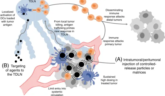
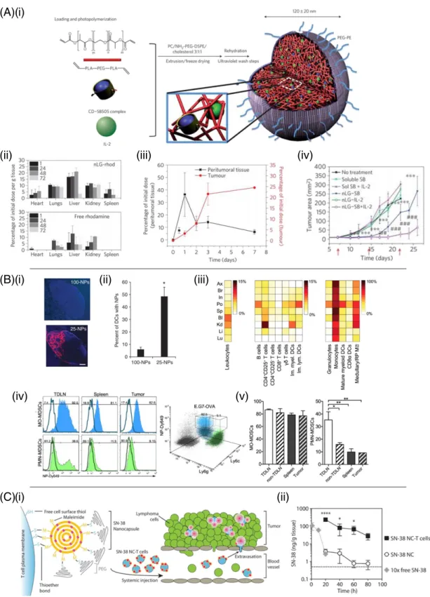
Because many immunotherapy toxicities (e.g., vascular leak syn-drome) are linked to systemic stimulation of circulating leukocytes and/or direct action of immunomodulators on endothelial cells, an obvi-ous strategy to enhance local therapy is to better confine therapeutics to a chosen target lesion. One way to achieve this is through local con-trolled release of drug from intra- or peritumorally injected drug deliv-ery matrices (Fig. 2A). One of thefirst demonstrations of this approach was work by Egilmez et al. seeking to improve on the safety and efficacy of IL-12 as an immunotherapeutic[163,164]. IL-12's potent anticancer effects are limited by dose- and temporal regimen-dependent toxicity when administered systemically[165,166]. Egilmez showed that a sin-gle intratumoral (i.t.) injection of biodegradable polylactic acid micro-spheres exhibiting controlled release of IL-12 was safe and led to complete regression of transplanted lung tumors, prevented metastatic spread to the lung, and enabled animals to reject a subsequent re-chal-lenge with live tumor cells, indicating the development of systemic an-titumor immunity[163]. More recently, chitosan matrices have been similarly used to provide sustained localized dosing of IL-12 in several tumor models, including as a neoadjuvant treatment in a breast cancer model prior to surgical resection of the primary tumor[167–169]. Hanes et al. used crosslinked gelatin/chondroitin sulfate microspheres as delivery vehicles for intratumoral delivery of IL-2 in models of brain or hepatic melanoma metastases[151]. IL-2 delivered by these micro-spheres persisted in tumors for 3 weeks, compared to only 24–48 h for bolus-injected drug. At the same time, bolus-injected IL-2 was found in the blood, spleen, and other organs minutes after bolus i.t. in-jection of IL-2, while microsphere-delivered cytokine led to very low or undetectable cytokine outside of the tumor microenvironment. Hori et al. designed self-crosslinking alginate gels[170]and used these to provide sustained local release of IL-15 following peritumoral injec-tion[150]. This strategy lowered the systemic exposure to the cytokine by ~2-fold and increased the dosing within the tumor 40-fold relative to a bolus injection of IL-15.
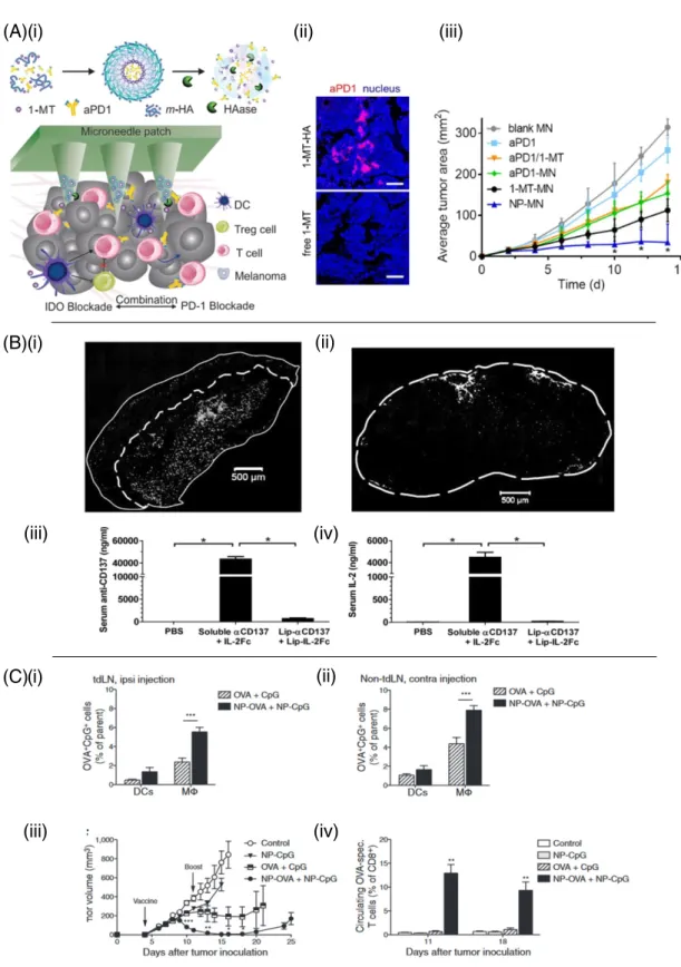
Checkpoint blockade antibodies have also been shown to benefit from localized slow-release delivery at tumors. Peritumoral injection of water-in-oil emulsions (Montanide) containing anti-CTLA-4 allowed a low dose of the checkpoint blockade antibody to effectively drive anti-tumor immunity while providing greatly decreased systemic exposure and reduced liver toxicity compared to traditional systemic anti-CTLA-4 dosing[152]. Recently, Wang et al. developed dissolving microneedles that deposited slow-release dextran nanoparticles into melanoma le-sions in the skin[171]. These particles dissolved in response to local tis-sue glucose mediated by incorporated glucose oxidase, releasing anti-PD-1 in a sustained manner that enhanced survival in treatment of B16F10 tumors. A similar microneedle-based transdermal patch was applied to synergistically co-deliver anti-PD-1 and an inhibitor of the immunosuppressive enzyme indoleamine 2,3-dioxygenase (IDO) to mouse melanoma, achieving effective T cell immunity and reduced im-munosuppression in the local site (Fig. 3A i–iii)[172].
Intratumoral depots of cytokines or checkpoint blockade antibodies primarily act to enhance the action of a pre-existing immune response against tumors. Another approach is to create intratumoral depots of agents aiming to promote de novo tumor killing, antigen capture and priming of new T-cell responses by tumor-localized immune cells using innate immune stimulating“danger signals”. Imidazoquinolines are a class of small molecule drugs that bind to Toll like receptors (TLRs) 7 and 8, promoting dendritic cell and macrophage activation. In-jection of unformulated forms of these compounds leads to rapid dis-semination into the blood and systemic cytokine storm signatures
[173,174]. However, acylated forms of these TLR agonists can be formu-lated into liposomes or oil/water emulsions for localized retention in tis-sue[173,175]. Singh et al. demonstrated that an acylated TLR7 agonist delivered in an oil emulsion into solid tumors could both arrest the growth of the treated tumor and prime a disseminated immune re-sponse that attacked distal, untreated tumors[175].
Related to these intratumoral treatments, another strategy is to in-troduce controlled-release biomaterials into a tumor resection site, with the goal of stimulating local immunity to residual tumor cells in the absence of the bulk immunosuppressive factors derived from a large primary tumor: Stephan et al. used alginate gels as a matrix for co-delivery of tumor-specific T-cells and mesoporous silicon micropar-ticles to resection sites of primary breast tumors. The microparmicropar-ticles
TDLN
(B)
Targeting of agents to the TDLN Sustained high dosing in treated tumor Limit entry intosystemic circulation Disseminating immune response attacks distal tumors From local tumor
killing, antigen trafficking primes new response in TDLN T Localized activation of DCs loaded with tumor antigen Immune response attacks primary tumor
(A)
Intratumoral/peritumoral injection of controlled-release particles or matricesFig. 2. Strategies for enhancing localized immunotherapy. (A) Synthetic particles/matrices administered directly to accessible lesions can provide sustained local dosing of immunomodulatory drugs with greatly lowered systemic exposure. Local immune activation leads to tumor cell death in the treated tumor and tumor antigen delivery to the TDLN, priming new T-cell responses that return to the treated tumor as well as disseminate to attack other metastases that were not directly treated. (B) Nanoparticles can be used to safely target immunomodulators to the TDLN, activating tumor antigen-loaded dendritic cells to prime T-cell responses that disseminate to attack tumors systemically.
(A)(i)
(ii)
(iii)
(B)(i)
(C)(i)
(ii)
(iii)
(iv)
(ii)
(iii)
(iv)
Fig. 3. Strategies for enhancing local delivery of immunotherapy. (A)(i) Microneedle-based delivery platform containing self-assembled nanocarriers (m-HA or NP) loaded with IDO inhibitor 1-MT and anti-PD-1. (ii) Immunofluorescence comparison of anti-PD-1/1-MT nanocarrier loaded microneedles and free anti-PD-1/1-MT loaded microneedles in tumors days post drug administration. (iii) B16F10 tumor growth versus time for 1-MT and anti-PD-1 delivered from microneedles (MN) as free drugs or encapsulated within nanocarriers (NP). Reproduced with permission from[172]. (B)(i) Retention of anti-CD137 and IL-2Fc-conjugated liposomes (white) within subcutaneous B16F10 tumors 24 h after intratumoral injection. (ii) Cryosection offluorescently labeled immuno-liposomes in the TDLN. (iii–iv) Serum levels of anti-CD137 and IL-2Fc 18 h following intratumoral injection in soluble or liposome-bound form. Reproduced from[43]with permission. (C) Conjugation of CpG and antigen to PPS nanoparticles improves the efficacy of a TDLN-targeted cancer vaccine. (i) 7 days after E.G7-OVA tumor inoculation,fluorescently labeled nanoparticle-conjugated OVA and CpG were delivered intradermally ipsilateral or contralateral (ii) to the tumor and presence of signal was analyzed 24 h later in the brachial lymph node. (iii) Tumor volume versus time with nanoparticle delivery of antigen and adjuvant to the tumor draining lymph node. (iv) Resulting antigen specific T-cell frequency following lymph node draining vaccine. Reproduced from[198]with permission.
carried anti-CD3/CD28 stimulators for T-cells and slowly released IL-15, providing localized TCR and cytokine support for the T-cells and leading to elimination of tumor recurrence that was not achieved if T-cells were administered systemically or lacking the matrix of supporting factors
[176]. Thus, a number of approaches can be used to augment local im-munotherapy while improving safety profiles of immunoregulatory drugs through local controlled release materials.
3.2. Intratumoral gene delivery
An alternative approach to slow-release depots of immunotherapy drugs is to locally produce the agent of interest through gene transfec-tion in the tumor microenvironment. This is perhaps best exemplified by oncolytic viral vectors, viruses which specifically replicate within tumor cells and promote tumor cell death, often in tandem with expres-sion of immunomodulatory proteins delivered in the viral genome. Talimogene Laherparepvec (T-Vec), a granulocyte-macrophage colo-ny-stimulating factor (GM-CSF)-encoding oncolytic herpes simplex virus, is thefirst example of this strategy to receive FDA approval, in the setting of metastatic melanoma. T-vec is administered by direct intratumoral injection, and was shown to trigger complete regression of both injected and uninjected lesions in 16% of treated patients, sug-gesting intratumoral administration of an oncolytic virus can effectively cross-prime and amplify antitumor immunity[177,178]. In this setting, GM-CSF expression from infected tumor cells is thought to promote the chemoattraction and differentiation of dendritic cell precursors to the tumor site, combined with immunogenic tumor cell death leading to enhanced presentation of antigen to prime new T-cell responses against the tumor. Similarly, intratumoral injections of escalating doses of a GM-CSF-expressing vaccinia virus to patients with cutaneous melano-ma or non-hepatocellular carcinomelano-ma, result in favorable immune re-sponses and tumor regression [179,180]. These intratumorally-injected viruses only spread locally within the tumor microenviron-ment due to their large size, and do not spread systemically to distant sites of tumor growth. Thus, the systemic toxicity observed has been in-frequent and rapidly resolving[179].
Therapeutic efficacy can be achieved through intratumoral injection of viruses, DNA, or RNA expressing immunoregulatory factors even without direct oncolytic activity of the nucleic acid vector. For example, i.t. injection of an adenovirus or alphavirus expressing IL-12 induced highly localized cytokine expression in tumors, leading to tumor regres-sion and long-term immunity[181,182]. Intralesional injection of adeno-virus encoding human CC chemokine ligand (CCL) 16 inhibited mammary tumor growth and prevented metastatic spread in mice bear-ing 4T1 mammary adenocarcinoma[183]. Polyplexes of DNA plasmids encoding IL-2 and folate-targeted polyethyleneimine-cyclodextrin were shown to be an effective and safe therapy for melanoma in mice, with an efficacy comparable to that of recombinant adenoviruses expressing IL-2 (rAdv-IL2)[184]. In clinical studies, intratumoral injection of naked DNA encoding cytokines (IL-2, IL-12) has shown some clinical benefits for melanoma patients[185,186]. A particularly promising approach is to combine localized tumor microenvironment modulation through intralesional gene or oncolytic virus delivery with systemic administra-tion of checkpoint blockade antibodies or other immune modulators with known/acceptable systemic toxicities, enabling an immune response primed by local therapy to be protected as it disseminates to attack un-treated lesions[141]. Thus, a variety of vectors and approaches are being explored for local expression of immunostimulatory cytokines and chemokines in tumors.
3.3. Anchored drugs
The examples above are based on releasing immunomodulators at controlled rates within tumor sites, which can only avoid systemic ex-posure if release rates are in careful balance with drug consumption/ degradation rates in the tumor. An alternative is to deliver
immunotherapy agents into the tumor microenvironment bound to particles, synthetic matrices, or extracellular components of the tumor microenvironment itself that present these molecules to surrounding immune cells but physically prevent their free diffusion out of the tumor site. An early example of this strategy utilized the injection of lipidated recombinant costimulatory receptors to“paint” tumors with a costimulatory ligand that would promote T-cell recognition of tumor cells[187,188]. Insertion of the lipid tail of these recombi-nant proteins into the membranes of tumor cells following intratumoral administration led to retention of these proteins at the tumor site, enabling engineering of tumor cell recognition by immune cells or induction of chemotactic signals to recruit more immune effectors to the tumor microenvironment. Lipid conjugation to DNA oligonucleotides has similarly been used to anchor immunostimulatory CpG DNA (a TLR9 agonist) in tumors, promoting retention in the microenvironment that improved the safety profile and efficacy of intratumoral CpG therapy[189].
A second approach to“anchoring” therapeutics in the tumor is to uti-lize nanoparticles not for their capacity to home to tumors spontane-ously but rather for their tendency to become entrapped in the tumor ECM following intratumoral injection. This approach has been demon-strated with combinations of potent cytokines and innate immune stim-ulators. As noted inSection 2, both IL-2 and anti-CD137 elicit potent antitumor immune responses, but their clinical use is limited by in flam-matory toxicities upon systemic administration. These toxicities are fur-ther amplified in combination treatment with these drugs, which elicits lethal systemic toxicities even following intratumoral administration at therapeutic doses[43]. To block the systemic dissemination that drives this toxicity, Kwong et al. conjugated anti-CD137 and an engineered IL-2-Fc fusion protein to the surface of PEGylated liposomes. Intratumoral injection of these immunoliposomes restricted the immunotherapeu-tics to the tumor and tumor-draining lymph nodes, but completely blocked their entry into the systemic circulation by virtue of physical trapping of the liposomes in the tumor extracellular matrix (Fig. 3B i– iv). Treatment with these particles eliminated injected primary tumors, elicited systemic antitumor immunity, and eliminated systemic in flam-matory toxicity compared to equivalent intratumoral doses of soluble immunotherapy. A similar approach was used to deliver liposome-an-chored anti-CD40 and CpG intratumorally, leading to significant tumor growth inhibition and enhanced survival similar to their soluble coun-terparts after intratumoral injection, while avoiding systemic exposure
[149]. Immunotherapy agents may also be intrinsically nanoparticulate in nature, promoting their local retention following intratumoral injec-tion. For example, Lizotte et al. used cowpea mosaic viruses for in situ vaccination of tumors[190]. Although the precise mechanism remains to be defined, these plant virus-derived nanoparticles stimulated intratumoral inflammation that led to a systemic anti-tumor immune response. Thus, particulate immunostimulatory therapies administered intratumorally can significantly decrease or eliminate systemic inflam-matory side effects, while retaining the anti-tumor efficacy of free solu-ble drugs.
3.4. Tumor draining lymph node-targeted drugs
Besides the tumor itself, tumor-draining lymph nodes (TDLNs, or sentinel lymph nodes) are of interest as a target for localized immuno-modulatory drugs, because despite evidence for lymphatic dysfunction in some tumor models, TDLNs are known to accumulate antigens from dying tumor cells that could be used to prime de novo anti-tumor T-cell responses. However, tumor-induced dendritic T-cell dysfunction within TDLNs is a known mechanism of immune evasion[191,192]. Concentration of innate immune-stimulatory adjuvants within sentinel lymph nodes allows for the activation and maturation of dendritic cells exposed to tumor-associated antigen while preventing cytokine storm-like symptoms which occurs from systemic administration of these agents[193,194]. Local injections near a tumor can be used to target
TDLNs through the local lymphatic tree (Fig. 2B). Lymphatic drainage from the interstitial space is highly dependent on molecule or particle size. Small particlesb30 nm diam. injected intradermally can be found within 50% of lymph node-resident dendritic cells, while larger 100 nm particles only reach 6% of the same population, suggesting that larger particles may be engulfed by phagocytic cells prior to lymph localization and/or impeded in convection through the ECM
[195,196]. Conversely, while small particles can traverse through dense interstitial matrix to directly reach the lymph nodes, they may not be retained within lymphoid organs– as evidenced by significant blood concentrations of 30 nm sized particles 12 hours post-intrader-mal injection[197]. Therefore, favorable lymphatic localization is reliant upon a balance of drainage from the injection site and capture within local lymph nodes.
Exploiting these principles, Jeanbart et al. synthesized 25 nm diam. pluronic-stabilized poly(propylene sulfide) (PPS) nanoparticles capable of concentrating within TDLNs following intradermal injection[198]. These nanoparticles are carried via interstitialflow from the injection site into lymphatic capillary beds and from there to TDLN-resident den-dritic cells. Conjugation of the TLR9 agonist CpG DNA to these particles elicited activation of dendritic cells in tumor draining lymph nodes, and primed anti-tumor adaptive immune responses in murine thymoma and melanoma models, while limiting systemic pro-in flam-matory responses (Fig. 3C i–iv)[198]. CpG has also been targeted to tumor draining lymph nodes through association with cationic gelatin nanoparticles and cationic polyethylenimine (PEI) coated poly(lactic-co-glycolic acid) (PLGA) nanoparticles, alone and in combination with IL-10 siRNA[199–201]. TLR7/8 agonists have also been successfully de-livered to tumor draining lymph nodes in nanoparticle format to focus the production of pro-inflammatory cytokines within the site of T cell priming and prevent systemic inflammation[173,202].
In addition to size-dependent trafficking of drug through lym-phatics, molecular targeting to dendritic cells which migrate to and re-side within lymph nodes has improved the safety and efficacy of immunotherapies. Cruz et al. describe in detail dendritic cell targeting via DEC205, CD11c, and CD40 dendritic cell receptors[162]. Lymph node targeting by physical, chemical, and molecular properties has been extensively studied for applications in both prophylactic and
therapeutic vaccines as vaccination depends on the delivery of antigen and adjuvant to these sites of immune cell education[161].
4. Engineering safer systemic immunotherapies
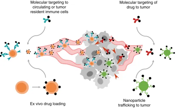
The administration of immunotherapy agents systemically is de-sired for treatment of metastatic disease, but faces limitations due to non-tumor-targeted stimulation of leukocytes and other cell types expressing immunoregulatory receptors. An ongoing challenge is the design of strategies to deliver immune-modulating drugs to appropriate immune cells in target tissue sites (e.g., tumors and tumor-draining lymph nodes) while minimizing non-specific sys-temic stimulation.
4.1. Molecularly-targeted immunotherapy
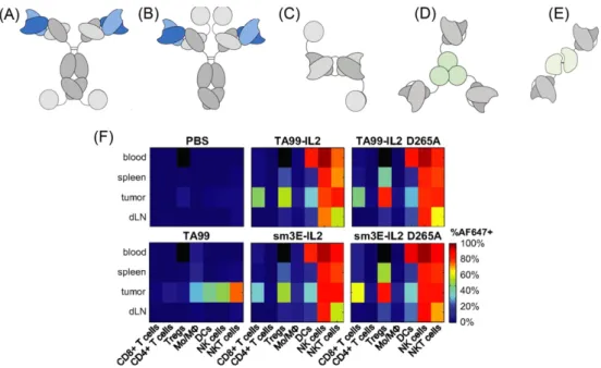
A common strategy to target therapeutics to tumors employs conju-gation of drug to a tumor-antigen specific ligand, antibody, or other engineered binding molecule to achieve local accumulation of the drug following systemic delivery. The fusion of pro-inflammatory cyto-kines to tumor-associated antigen specific antibodies, known as immunocytokines, represents a common approach to direct the deliv-ery of cytokines to the tumor microenvironment. Cytokines can be fused to either the N or C termini of heavy or light chains of IgG mole-cules (Fig. 4A and B)[203]. In these formats, functions of the antibody such as antigen binding, interaction with Fc receptors, and participation in the complement cascade can be maintained. Alternatively, cytokines can be fused to diabodies or single chain variable fragment (scFv) anti-bodies to solely maintain the antigen binding property of the antibody (Fig. 4C–E). One proposed mechanism of action of immunocytokines is the bridging of tumor cells to leukocytes[204–207]. In the case of IL-2 immunocytokines, the antibody interacts with tumor surface anti-gens while the IL-2 binds to the IL-2 receptor (IL-2R) on T-cells and NK cells, thereby promoting their proliferation and effector function in the tumor microenvironment. Antibody-dependent cellular cytotoxicity (ADCC) afforded by interaction with the Fc domain of the antibody com-ponent has also been shown to be important for the efficacy of immunocytokines[208]. Finally, immunocytokines have longer blood
Fig. 4. Tumor targeting with immunocytokines. (A–E) Immunocytokine formats based on IL-2, IL-12, and TNF-α. (A) IgG format with IL-2 cytokine covalently linked at the c-terminus of the heavy or (B) light chains. (C) Diabody fusion protein featuring IL-2. (D) Homotrimeric scFv-TNF fusion protein. (E) Heterodimer featuring scFv fused to p40 and p35 subunits of IL-12. Reproduced from[203]with permission. (F) Biodistribution of TA99-IL-2 immunocytokine of format (A) targeting Trp-1 melanoma antigen in mice bearing subcutaneous B16F10 tumors. 24 hours post injection of Alexa Fluor 647-labeled proteins, organs were dissociated into single cell suspensions and stained for immune lineage markers. D265A indicates a mutation in the Fc portion of TA99 abrogating interaction with Fc receptors. The sm3E antibody targeting carcinoembryonic antigen is an irrelevant antibody in model. This irrelevant immunocytokine features similar biodistribution to the melanoma-targeted TA99 immunocytokine. Used with permission from[210].