Alcohol & Alcoholism Vol. 41, No. 6, pp. 678–680, 2006 doi:10.1093/alcalc/agl054 Advance Access publication 15 August 2006
CASE REPORT
MULTIPLE CEREBRAL METASTASES MIMICKING WERNICKE’S ENCEPHALOPATHY
IN A CHRONIC ALCOHOLIC
OZGUR YALDIZLI1, FRIEDRICH M. WURST1*, SEBASTIAN EULER1,
BERENIKA WILLI2and GERHARD WIESBECK1
1Psychiatric University Clinic and2Department of Radiology, University of Basel, Switzerland
(Received 27 February 2006; first review notified 9 March 2006; in revised form 15 June 2006; accepted 15 June 2006; advance access publication 15 August 2006)
Abstract— Aims: Alcohol dependent patients in withdrawal display a wide spectrum of neurological and neuropsychological symp-toms that complicate diagnosis. We report the case of a 53-year-old male alcoholic with disorientation, ataxia and nystagmus in alcohol withdrawal probably due not to initial supposed Wernicke’s encephalopathy (WE) but rather due to multiple cerebral metastases of a non-small cell cancer of the lung. Results: The findings illustrate the importance of initially maintaining a tentative attitude toward causation of symptoms and the role of brain imaging in formulating an accurate diagnosis.
Alcohol dependent patients in withdrawal can demonstrate a wide spectrum of neurological and neuropsychological symptoms and sometimes pose great challenge to physicians. Mr A was a 53-year-old undernourished, unemployed, cook who had been transferred to our clinic by a general practitioner due to confusion and disorientation with the onset of acute symptoms a few days before admission. A diagnosis of intox-ication due to inadvertent high intake of prescribed medintox-ication (ranitidine, lansoprazole, propyphenazone and drofenin) and ethanol had been made.
Mr A had a history of alcohol dependence of 30 years duration with three previous treatments for alcohol with-drawal. He had been admitted to our clinic 3 years before, when oxazepam was prescribed for alcohol withdrawal symp-toms. Due to poor compliance, he only stayed 3 days. He did not agree with a rehabilitation program for maintenance of abstinence. After discharge he recommenced consuming alco-hol in large quantity.
The first time Mr A was admitted to our clinic, by his general practitioner because of uncontrolled alcohol con-sumption, in the same year the detoxification treatment with a rehabilitation program (55 days) was complicated due to
a general tonic–clonic epileptic seizure. Along with
oxazepam, he received antiepileptic therapy with carba-mazepine. The third hospitalization was one year before the present admission and lasted for 56 days followed by four months of intensive rehabilitation program in a clinic outside. Mr A had also been dependent on benzodiazepines (75 mg oxazepam/day and an unknown dose of bromazepam) for the past 3 years and nicotine for 35 years at a level of 70–100 pack years (Fagerstrom score: 9). Six years prior to the final admission he had undergone excision of pancreatic pseudo cysts as complication of a chronic pancreatitis.
We usually recommend all alcohol-dependent patients
600 mg thiamine, 30 mg riboflavin, 20 mg pyridoxine, 20mg
cobalamine, 0.3 mg biotine, 50 mg panthotenacid, and
100 mg nicotinamid orally per day for vitamin supplementa-tion, but Mr A did not agree to vitamin therapy.
During the three past hospitalization periods of overall 114 days Mr A received orally only on 19 days, 600 mg thiamine/day for vitamin supplementation. The patient had also not taken vitamin supplements outside of our clinic.
At admission, his height was 173 cm and his weight 52 kg,
corresponding with a body mass index of 17 kg/m2
(age-adjusted reference of the body mass index 22–27). Clinical signs of vitamin deficiency such as disturbance of nails or hair growth were not detectable. His wife reported that he had consumed alcohol daily during the four months before admission.
Mr A was awake but disorientated to person, location, situ-ation, and time. Reduced attention and short memory deficit were readily observable. Thought disturbance, phobia, com-pulsion, delusions, hallucinations, and depersonalisation were not detectable. His affect was slightly dysphoric and he was restless as well as impulsive. He denied suicidal ideation. In the Mini-Mental-State test (MMS) (Folstein et al., 1975) he achieved a score of 15 out of 30 points.
Neurological examination revealed gaze-evoked nystagmus in all directions. Meningism was not found. No abnormal functioning of the other cranial nerves, paralysis or sense disturbances were detected. All deep tendon reflexes were symmetrically elevated. Pathological reflexes were not detect-able. The finger–nose test was atactic. Standing and gait with open eyes evidenced a distinct non-directional ataxia with tre-mors of the upper extremity. The Romberg sign was positive. Apart from a slightly elevated gammaglutamyl transferase (GGT) (94 U/l; reference 11–66 U/l) and mean corpuscular hemoglobin (MCH) (32 pg; reference 27–31 pg), routine blood parameters including hemogram, chemogram, coagula-tion, C-reactive protein, and ammoniac were normal.
Confusion, ataxia, and nystagmus suggested acute
Wernicke’s encephalopathy (WE). He received 100 mg thiam-ine intramuscular directly after neurological examination (Day 0). Simultanously, we applied for neuro-imaging for exclusion of other possible causes of the very distinctive disorientation and ataxia. On the next day his status had not improved. Cranial computed tomography revealed multiple
*Author to whom correspondence should be addressed at: Psychiatric University Clinic, University of Basel, Wilhelm-Klein-Strasse 27, CH-4025 Basel, Switzerland. Tel.: +41 61 325 51 12; Fax: +41 61 325 55 83; E-mail: friedrich.wurst@upkbs.ch
678
infra- and supra-tentorial, hyperdense, space-occupying, maximally sized 4.5 cm lesions with distinctive perifocal oedema, central hypodensity, and compression of the side ven-tricle due to elevated intracerebral pressure (Fig. 1), indicating possible metastases. The corpora mamillaria were not hypo-trophic. Magnetic resonance tomography imaging was not possible due to the patient’s poor compliance.
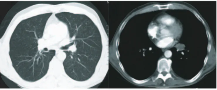
To locate the primary tumor, computed tomography of thorax was performed and revealed a space occupying lesion in the left pulmonal hilus between the bronchus of the lingula and lower lobe with mass effect and dorsal replacement of the vessels without hints of angio-invasion (Fig. 2).
Skeletal scintigraphy showed multiple diffused elevated enhancements particularly in the ribs suggesting multiple bone metastases. The diagnosis of a less differentiated malig-nant non-small cell bronchial carcinoma was corroborated by bronchoscopy and transbronchial biopsy.
Multidisciplinary therapy was planned with radiotherapy, oncology, neurology, and psychiatry. After beginning pallia-tive radiation treatment on Day 15, disorientation and gait disturbances initially became much worse. We used repeated doses of 10–20 mg haloperidol as reserve medication for agitation and 175 mg fentanyl for skeletal pain. The patient declined further vitamin supplementation. From Day 46, Mr A received prednisone 100 mg/day to reduce brain edema. In the following days, the neurological and neuropsy-chological symptoms improved markedly. On Day 61, the patient was discharged with significantly improved disorienta-tion, pain, and gait ataxia, receiving palliative medicadisorienta-tion, prednisone 100 mg/day and fentanyl 175 mg/day. He was
orientated to person, location, and situation, but not to time. He was able to stand alone and walk with help. He died about 2 months after his discharge in the circle of his family.
DISCUSSION
To the best of our knowledge, this is the first case reporting multiple intracerebral metastases in a chronic alcoholic with observable signs of WE.
WE is an acute, preventable, life-threatening metabolic disease of the central nervous system caused by thiamine deficiency. Thiamine dependent enzymes such as transketo-lase and pyruvate dehydrogenase are essential for cerebral myelinization. WE is probably caused by decreased energy metabolism caused by citric acid cycle disturbance, and decreased synthesis of neurotransmitter such as acetylcholine (McCandless et al., 1969). Thiamine deficiency and WE may occur in anorexia nervosa, hyperemesis gravidarum, small-bowel obstruction, AIDS, dialysis, prolonged intra-venous feeding, or other conditions associated with prolonged poor intake (Reuler et al., 1985; Homewood et al., 1999; Togay-Isikay et al., 2001; Ogershok et al., 2002). However, malnourishment is the most common basis for WE in alco-holics. The daily thiamine requirement for healthy individuals is between 1 and 2 mg/day but both alcohol and malnutrition may interfere with the absorption of thiamine (Thomson et al., 2006a). Already 17 years ago, Gastaldi et al. (1989) found that ethanol can damage the intestinal mucosa of rats with consecutive thiamine malabsorption. Low plasma levels of thiamine have been reported in up to 80% of alcoholic patients (Cook et al., 1998).
Autopsy studies reveal that WE is often under-diagnosed: Naidoo et al. (1991) found histological changes of WE in 17 out of 29 consecutive alcohol-related deaths. The classical clinical triad of WE are oculomotor findings, ataxia, and mental confusion (Reuler et al., 1985). Typically, oculomotor
deficits include weakness of abduction, gaze-evoked
nystagmus, internuclear ophthalmoplegia, vertical nystagmus in primary position, and decreased vestibule-ocular-reflex. The oculomotor finding in Mr A was an omni-directional gaze-evoked nystagmus.
It is difficult to diagnose WE clinically since only a third of the cases display the classical triad symptoms. Clinical signs can be masked by level of consciousness or other neurological symptoms (Harper, 1983; Reuler et al., 1985). Due to the variable symptom-pattern of WE, the possibility of thiamine deficiency should be considered in any patient with docu-mented or suspicious history of alcoholism showing any neurological features including even coma (Wallis et al., 1978). Thiamine should also be supplemented fully in mal-nourished alcoholics. The administration of intravenous fluids containing glucose without adequate thiamine supplementa-tion in alcoholics could aggravate the thiamine deficiency leading to irreversible cerebral lesions and death (Yokote et al., 1992; Koguchi et al., 2004).
In 1997 Caine et al. (1997) developed improved operational criteria for the diagnosis of WE in alcoholics: WE can be identified by the presence of malnourishment, oculomotor abnormalities, cerebellar dysfunction, memory impairment, or altered mentation. Mr A demonstrated all of these criteria.
Fig. 1. Axial cranial computed tomography with contrast injection (slide thickness 3 mm): multiple round, cystic space occupying lesions with central hypodensity, perifocal edema, mass effect and marginal contrast enhancement
frontal, fronto-temporal and parietal. Maximal size: 4.5 cm.
Fig. 2. Thoracal computed tomography with contrast injection (slide thick-ness 7 mm): space occupying lesion in the left hilus, central discrete dorsal
replacement of the vessels.
During recent years, brain imaging has proved useful in confirming the diagnosis of WE and in contributing to earlier detection (Antunez et al., 1998). The usual findings at MR imaging in patients with WE include high signal intensities in the mamillary bodies, medial thalami, tectum of the midbrain, and the periaqueductal region. The chronic stage may show atrophy of of the mamillary bodies and midbrain tegmentum, as well as dilatation of the third ventricle (Yokote et al., 1991; Doraiswamy et al., 1994). Unfortunately, we could not perform MRI in our patient due to poor compliance.
Although we have not determined plasma thiamine level as it is not a routine parameter on admission we believed that Mr A’s initial symptoms were not due to WE but due to the multiple brain metastases. There are some hints that support this hypothesis. First, due to poor compliance Mr A got only one administration of 100 mg thiamine intramuscular. Intramuscular doses of >200 mg daily are believed to be required to show improvement in disorientated WE-patients (Ambrose et al., 2001; Thomson et al., 2002). Second, the neuropsychological improvement of Mr A occurred too late to attribute it to the administration of thiamine: Mr A received a singular dose of 100 mg thiamine on admission day and improved after the Day 46. We attributed his eventual neuro-logical and neuropsychoneuro-logical improvement to the palliative whole brain radiation therapy (WBRT) and prednisone treatment. The initial worsening of neurological symptoms is typical for WBRT. WBRT is the treatment of choice for brain metastases because it prolongs the mean survival rate from 1 to 6 months (Zabel et al., 2004), affords a good local control, eliminates any micrometastases, reduces the risk of recurrent brain metastases, improves overall survival and quality of life as in our case, and can prevent death due to brain compres-sion syndrome (Ellis et al., 1998).
Mr A’s case demonstrates that the clinical criteria for WE are not pathognomic. The spectrum of differential diagnoses is quite broad and includes intracranial haemorrhage, stroke, brain tumor, intracerebral metastases, hepatic failure, central pontine myelinolysis, as well as, cerebral infections such as meningitis. According to recent published recommendations it is good clinical practice that patients at risk of WE receive 250 mg thiamine intramuscular for 3–5 days (Thomson et al., 2006b) but if there is no rapid response, further neurological examination are required in order to establish the correct diag-nosis and serve optimal medical treatment.
REFERENCES
Ambrose, M. L., Bowden, S. C. and Whelan, G. (2001) Thiamine treatment and working memory function of alcohol-dependent people: preliminary findings. Alcoholism: Clinical and Experimental Research 25, 112–116.
Antunez, E., Estruch, R., Cardenal, C. et al. (1998) Usefulness of CT and MR imaging in the diagnosis of acute Wernicke’s encephalopathy. American Journal of Roentgenology 71, 1131–1137.
Caine, D., Halliday, G. M., Kril, J. J. et al. (1997) Operational criteria for the classification of chronic alcoholics: identification of Wernicke’s encephalopathy. Journal of Neurology, Neuro-surgery, and Psychiatry 62, 51–60.
Cook, C. C., Hallwood, P. M. and Thomson, A. D. (1998) B-vitamin deficiency and neuro-psychiatric syndromes in alcohol misuse. Alcohol and Alcoholism 33, 317–336.
Doraiswamy, P. M., Massey, E. W., Enright, K. et al. (1994) Wernicke-Korsakoff syndrome caused by psychogenic food refusal: MR findings. American Journal of Neuroradiology 15, 594–596.
Ellis R. and Gregor A. (1998) The treatment of brain metastases from lung cancer. Lung Cancer 20, 81–84.
Folstein, M. F., Folstein, S. E. and McHugh, P. R. (1975) Mini-mental-state: a practical method for grading the cognitive state of patients for the clinician. Journal of Psychiatric Research 12, 189–198.
Gastaldi, G., Casirola, D., Ferrari, G. et al. (1989) Effect of chronic ethanol administration on thiamine transport in microvil-lous vesicles of rat small intestine. Alcohol and Alcoholism 24, 83–89.
Harper, C. (1983) The incidence of Wernicke’s encephalopathy in Australia—a neuropathological study of 131 cases. Journal of Neurology, Neurosurgery, and Psychiatry 46, 593–598.
Homewood, J. and Bond, N. W. (1999) Thiamin deficiency and Korsakoff’s syndrome: failure to find memory impairments following nonalcoholic Wernicke’s encephalopathy. Alcohol 19, 75–84.
Koguchi, K., Nakatsuji, Y., Abe, K. et al. (2004) Wernicke’s encephalopathy after glucose infusion. Neurology 62, 512. McCandless, D. W. and Schenker, S. (1969) Neurologic disorder of
thiamine deficiency. Nutrition Reviews 27, 213–215.
Naidoo, D. P., Bramdev, A. and Cooper, K. (1991) Wernicke’s encephalopathy and alcohol-related disease. Postgraduate Medical Journal 67, 978–981.
Ogershok, P. R., Rahman, A., Nestor, S. et al. (2002) Wernicke’s encephalopathy in nonalcoholic patients. The American Journal of Medical Sciences 323, 107–111.
Reuler, J. B., Girard, D. E. and Cooney, T. G. (1985) Current concepts. Wernicke’s encephalopathy. New England Journal of Medicine 312, 1035–1039.
Thomson, A. D. and Marshall, E. J. (2006a) The natural history and pathophysiology of Wernicke’s encephalopathy and Korsakoff’s Psychosis. Alcohol and Alcoholism 41, 151–158.
Thomson, A. D. and Marshall, E. J. (2006b) The treatment of patients at risk of developing Wernicke’s encephalopathy in the community. Alcohol and Alcoholism 41, 159–167.
Thomson, A. D., Cook, C. C., Touquet, R. et al. (2002) The Royal College of Physicians report on alcohol: guidelines for managing Wernicke’s encephalopathy in the accident and Emergency Department. Alcohol and Alcoholism 37, 513–521.
Togay-Isikay, C., Yigit, A. and Mutluer, N. (2001) Wernicke’s encephalopathy due to hyperemesis gravidarum: an under-recognised condition. The Australian and New Zealand Journal of Obstetrics and Gynaecology 41, 453–456.
Wallis, W. E., Willoughby, E. and Baker, P. (1978) Coma in the Wernicke-Korsakoff syndrome. Lancet 2, 400–401.
Yokote, K., Miyagi, K., Kuzuhara, S. et al. (1991) Wernicke encephalopathy: follow-up study by CT and MR. Journal of Computer Assisted Tomography 15, 835–838.
Yokote, K., Yamanouchi, H., Mizutani, T. et al. (1992) Clinical characteristics of Wernicke’s encephalopathy in the elderly. Nippon Ronen Igakkai Zasshi 29, 35–40.
Zabel, A. and Debus, J. (2004) Treatment of brain metastases from non-small cell lung cancer (NSCLC): radiotherapy. Lung Cancer 45, 247–252.