Letters
HCV-RNA, is not yet routinely available and is limited by false-positive or false-negative results.
Meanwhile, in order to reduce the incidence of HCV infection, strict adherence to the universal precautions (CDC, Atlanta) must be carried out. According to current knowledge the application of these measures of hygiene is probably sufficient to prevent HCV spread in the dialysis setting, and the isolation of positive patients is not warranted.
Department of Nephrology and M. Beccari Dialysis,
Fatebenefratelli and Oftalmico Hospital,
Milan, Italy
1. Sungur C. Is HCV a nosocomial infection in haemodialysis patients? Nephrol Dial Transplant 1994; 9(7): 875-876 2. Mclntyre PG, McCruden EAB, Dow BC et al. Hepatitis C virus
infection in renal dialysis patients in Glasgow. Nephrol Dial Transplant 1994; 9(3): 291-295
3. Jadoul M, Cornu C, van Ypersele de Strihou C, UCL Collaborative Group. Incidence and risk factors for hepatitis C seroconversion in hemodialysis: a prospective study. Kidney Int 1993; 44: 1322-1326
4. Allander T, Medin C, Jacobson SH, Grillner L, Persson MAA. Hepatitis C transmission in a hemodialysis unit: molecular evid-ence for spread of virus among patients not sharing equipment. J Med Virol 1993; 43: 415-419
5. Caramelo C, Navas S, Alberola ML, Bermejillo T, Reyero A, Carrefio V. Evidence against transmission of hepatitis C virus through hemodialysis ultrafiltrate and peritoneal fluid. Nephron 1994; 66: 470-473
Sir,
I read Dr Beccari's contribution on the issue of nosocomial spread of hepatitis C virus (HCV) infection among haemodialysis patients with interest. He stresses another controversial aspect of the problem; the isolation policy for HCV-infected patients in dialysis units, and concludes that if universal precautions are followed strictly, there is no need for such an isolation policy.
I agree with Dr Beccari that adherence to universal precau-tions is an essential measure in the prevention of nosocomial blood-borne viral infections, but on the other hand I think it is hard to draw a concrete conclusion about isolation policies for the following reasons:
1. Molecular biological studies which show the presence of viral genetic material in the haemodialysis ultrafiltrate rather than the ones which fail to detect the viral RNA should be regarded as important microbiological find-ings. Apart from methodological differences and errors, the degree of viraemia of a patient is probably an important factor in the contamination of the ultrafiltrate.
2. Haemodialysis treatment itself is a well-established risk factor for acquiring HCV infection [1], especially if the same machine is shared by more than four patients [2]. 3. In the previous decade isolation policies for hepatitis B virus (HBV) infection, which shares common routes of transmission with HCV, has been successful in reducing the nosocomial transmission of HBV infection in dialysis units, and isolation policies for HBV-infected dialysis patients are mandatory.
4. The risk of acquiring nosocomial HCV infection is strikingly different in units with a low rate of infection from those in which 50-70% of chronic dialysis patients are infected.
1475 5. The issue becomes more complex when one remembers the lack of protective effect of anti-HCV antibodies, different infectivity and pathogenicity potentials of dis-tinct HCV strains, and studies showing dialysis patients infected with more than one strain of HCV [3,4]. The answer to this controversy probably resides in the data which will be derived from the molecular epidemiolog-ical studies. Demonstration of a particular strain of HCV in infected patients sharing the same machine and/or dialysis unit will be an invaluable additional finding in favour of nosocomial transmission. Unfortunately, as Dr Beccari points out most of these molecular diagnostic tools are inaccessible to the majority of physicians taking care of dialysis patients.
I believe that isolation policies for HCV-infected dialysis patients should be developed according to the prevalence and rate of infection in a particular unit, and every effort should be undertaken to prevent spread of infection to newly admitted cases.
Bayindir Medical Center, Cem Sungur TR-06520 SQgutozu,
Ankara, Turkey
1. Jadoul M, Cornu C, van Ypersele de Strihou C. UCL Collaborative Study Group. Incidence and risk factors for hepat-itis C seroconversion in hemodialysis: a prospective study. Kidney Int 1993; 44: 1322-1326
2. Wagner A, Kocaman A, Gessman M, Debusman E, Philipp T, Traenhart O. Epidemiology of HCV-infection in patients on chronic hemodialysis (abstract). J Am Soc Nephrol 1993; 4: 394A 3. Yoshida C, Vanderborght B, Stuyver L, Rouzere C, Maertens G.
Tracing non-transfusional transmission of HCV infection through genomic typing of HCV in hemodialysis unit (abstract). In: Xllth International Congress of Nephrology, 1993, Jerusalem, 394 4. Martin P. Hepatitis C genotypes: the key to pathogenicity? Ann
Intern Med 1995; 122: 227-228
Complement activation by LDL-apheresis using dextran sulphate
Sir,
LDL-apheresis (LDL-aph) using dextran sulphate (DS) adsorption has been used successfully as a therapeutic tool to reduce LDL-cholesterol in familial hypercholesterolaemia [1]. Anaphylactoid reactions have been reported in patients treated with LDL-aph using DS columns [2]. These reactions have been attributed to the release of bradykinin, a potent vasodilator. However, artificial biomaterials can also activate the complement system, which may play a major role in such anaphylactoid reactions. We studied the effects of LDL-aph with DS (Kaneka LA 15 Liposorber) on the complement system during three apheresis sessions, in a 25-year-old patient with familial hypercholesterolaemia.
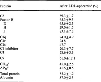
Plasma samples were collected in EDTA before, during, and after each procedure. Complement activation products C4d, Bb, and SC5b-9 were measured with commercial kits (Quidel). The concentrations of several proteins, CH50 and AP50 were reduced after LDL-aph (see Table 1). As shown in Figure 1, complement activation products Bb and SC5b-9 significantly increased at the end of LDL-aph (Bb, in ug/ml, from 0.95 + 0.05 to 34.4+1.1; SC5b-9, inng/ml, from 25.0 + 4.5 to 830 + 334); however, there was no significant increase in C4d. Of note, plasma samples coming out of the DS column were practically devoid of factor H, and had increased concentrations of Bb and SC5b-9. There was also
1476 Letters
Table 1. C o m p l e m e n t m e a s u r e m e n t s after L D L - a p h e r e s i s in a patient with familial hypercholesterolaemia"
Protein C3 F a c t o r B D H I C l q C l r Cls C l inhibitor C4 C9 CH5 0< AP5 OC Total protein A l b u m i n After L D L - a p h e r e s i sb (%) 69.3 + 1.7 65.3 + 9.3 45.6 + 2.3 29.0+1.5 83.1 ±7.3 24.0 + 4.9 24.8 47.7 70.3 + 7.7 78.6±3.3 61.0+12.1 45.0 + 2.5 41.5±0.5 85.2 + 1.2 87.0±2.3
"Antigenic concentrations, except for factor D, CH50, AP50
(haemolytic assays). All values were in the normal range before apheresis. Total protein and albumin are used as controls.
b
Values are expressed as the percentage of the pre-apheresis levels. Each value represents the mean + SEM from three different apheresis sessions, except for Clr and Cls (single values).
c
Standard haemolytic assays assessing classical pathway (CH50) and
alternative pathway (AP50) of complement.
SC5b-9 (ng/ml) 1800 1600 1200 B X i 100. 80. 60. 40. 20. 0. factor H
h
SCSb-9/ / / / Q Bb fragment \ \ ,\
y ^
Bb 50 ' 40 . « 30 ' 20 • 10 . ^ o— 1 2 3 4 5 6 plasma samplesFig. 1. Changes in the concentration of complement activation
prod-ucts Bb and SC5b-9, and of complement inhibitory protein factor H, during LDL-apheresis with DS. Each value represents the mean from two different apheresis sessions. Plasma samples: 1. Before starting LDL-aph. 2. After separation of plasma from whole blood with filter made of polysulphone, before DS column. 3. Plasma coming out of DS column. 4. At the end of LDL-aph. 5. Two hours after the end of LDL-aph. 6. Twenty-four hours after the end of LDL-aph.
a marked C3c formation (C3 conversion to C3c) as assessed by double immunodiffusion (from < 1 % to 28.6 + 3.5%, at the end of LDL-aph). Twenty-four hours after a normal LDL-aph session, no C3c was detectable in plasma, and CH50, AP50 had increased to 72 and 86%, respectively. In two sessions of LDL-aph, the neutrophil count (per microlitre) declined from 3875 + 31 to 1645±11 after 2 h of LDL-aph.
The main finding of this study was the strong activation of the alternative pathway (AP) of complement during LDL-aph using DS, probably due to its deregulation in the fluid phase because of the reduction of factor H. We could not
demonstrate activation of the classical pathway. The signi-ficant reduction in factor H (and Clq) was due to adsorption, since more than 90% of these proteins were recovered in the eluates from the DS columns. In vitro, previous work has shown that the interaction of human serum with negatively charged polystyrene sulphonate results in massive comple-ment activation by the AP due to an immediate adsorption of factor H [3]. Factor H is an essential inhibitory protein of the AP under normal conditions; thus its adsorption or
in-vivo blockade results in hypercatabolism of the AP [3,4].
The resulting strong complement activation by the AP may cause anaphylactoid reactions. Nafamostat mesilate, which inhibits both complement activation and the release of brady-kinin, may be the anticoagulant of choice for LDL-aph using DS, in order to avoid potential life-threatening reactions [5]. Renal Unit, Massachusetts General M. Pascual Hospital, E. Blanc Boston, USA J.A. Schifferli Hospital of Sion, Sion,
Switzerland
Department of Medicine, Kantonsspital,
Basel, Switzerland
1. Mabuchi H, Michishita I, Takeda M et al. A new low density lipoprotein apheresis system using two dextran sulfate cellulose columns in an automated column regenerating unit (LDL continuous apheresis). Atherosclerosis 1987; 68: 19-25 2. Keller C, Grutzmacher P, Bahr F et al. LDL-apheresis with
dextran sulphate and anaphylactoid reactions to ACE inhibitors. Lancet 1993; 341: 60-61
3. Pascual M, Piastre O, Montdargent B, Labarre D, Schifferli JA. Specific interactions of polystyrene biomaterials with factor D of human complement. Biomaterials 1993; 14: 665-70
4. Veerhuis R, Van Es LA, Daha MR. In vivo modulation of rat complement activities by infusion of anti-H antibodies. Immunobiology 1985; 170: 133-145
5. ICojima S, Harada-Shiba M, Nomura S et al. Effect of nafamostat mesilate on bradykinin generation during low-density lipoprotein apheresis using a dextran sulfate cellulose column. ASAIO Trans 1991; 37: 644-648
ACE inhibitors do not decrease rHuEpo response in patients with end-stage renal failure
Sir,
Angiotensin-converting enzyme (ACE) inhibitors have been successfully utilized to reduce haemoglobin concentrations in patients with post-transplant erythrocytosis [1,2]. Some authors have suggested that ACE inhibitors could decrease serum erythropoietin levels [3] and bone-marrow erythro-poiesis in ESRD [4]. In this way Walter [5] has suggested that ACE inhibitors could reduce the efficacy of rHuEpo in ESRD patients. However, other studies were unable to find any effect of ACE inhibitors on haematocrit or rHuEpo dose [6].
We have studied 252 patients on haemodialysis from 17 units; 48 of them were treated with ACE inhibitors. All patients had been treated with rHuEpo for at least 6 months, and to be included in the study a patient's haemoglobin had to be over 9 g/dl in the 3 previous months. Age, sex, months on rHuEpo treatment, months on HD, haemoglobin, haem-atocrit, iron metabolism, iPTH, aluminium, B2-microglobulin
and Ul/kg/week of rHuEpo were monitored. The group treated with ACE inhibitors was younger (51 + 14 versus 57+16 years, /><0.05), but both groups were similar in