HAL Id: tel-01865832
https://tel.archives-ouvertes.fr/tel-01865832
Submitted on 2 Sep 2018HAL is a multi-disciplinary open access archive for the deposit and dissemination of sci-entific research documents, whether they are pub-lished or not. The documents may come from teaching and research institutions in France or abroad, or from public or private research centers.
L’archive ouverte pluridisciplinaire HAL, est destinée au dépôt et à la diffusion de documents scientifiques de niveau recherche, publiés ou non, émanant des établissements d’enseignement et de recherche français ou étrangers, des laboratoires publics ou privés.
Specific activation of the alternative cardiac promoter of
Cacna1c by the mineralocorticoid receptor
Thassio Ricardo Ribeiro Mesquita
To cite this version:
Thassio Ricardo Ribeiro Mesquita. Specific activation of the alternative cardiac promoter of Cacna1c by the mineralocorticoid receptor. Cardiology and cardiovascular system. Université Paris Saclay (COmUE); Universidade Federal de Sergipe, 2017. English. �NNT : 2017SACLS530�. �tel-01865832�
Specific Activation of the
Alternative Cardiac Promoter of
Cacna1c by the Mineralocorticoid
Receptor
Thèse de doctorat de l'Université Paris-Saclay et de Federal University of Sergipe, préparée à l’Université Paris-Sud XI en Châtenay-Malabry École doctorale n°569: innovation thérapeutique:
du fondamental à l'appliqué (ITFA) Graduate School in Physiological Sciences
Spécialité de doctorat: Physiologie et Physiopathologie
Thèse présentée et soutenue à São Cristõvão (Brésil) de soutenance, le 13 Décembre 2017, par
M Thássio Ricardo Ribeiro Mesquita
Composition du Jury :
M Daniel Badauê Passos JÚNIOR
Professeur, Federal University of Sergipe, Brésil Président M José Manuel CANCELA
Directeur de Recherche, CNRS, France (UMR 9197) Rapporteur M Jader Santos CRUZ
Professeur, Federal University of Minas Gerais, Brésil Rapporteur M Mark William CHAPLEAU
Professeur, The University of Iowa, États-Unis Rapporteur M Luciano dos Santos Aggum CAPETTINI
Professeur, Federal University of Minas Gerais, Brésil Examinateur M Jean Pierre BENITAH
Directeur de Recherche, Inserm, France (UMR-S 1180) Directeur de thèse Mme Sandra Lauton SANTOS
Professeur, Federal University of Sergipe, Brésil Co-Directrice de thèse
NNT : 2 0 1 7 SAC L S5 3 0
Université Paris-Saclay Espace Technologique / Immeuble Discovery
Route de l’Orme aux Merisiers RD 128 / 91190 Saint-Aubin, France
Titre : Activation spécifique du promoteur cardiaque alternatif du Cacna1c par le récepteur aux minéralocorticoïdes Mots clés : Canaux Ca2+ de type L, aldostérone, récepteur aux minéralocorticoïde, Cœur, muscle lisse vasculaire.
Résumé :
Les antagonistes des récepteurs aux minéralocorticoïdes (MR) appartiennent à l'arsenal thérapeutique pour le traitement de diverses maladies cardiovasculaires, mais les mécanismes conférant leurs effets bénéfiques sont encore mal compris. Une partie de ces effets peuvent être liée à la régulation de l'expression du canal Ca2+ de
type L Cav1.2, largement impliqué dans l'insuffisance
cardiaque et l'hypertension. Nous montrons que MR fonctionne comme un facteur de transcription transformant le signal de l'aldostérone dans l'utilisation du 'cardiaque' promoteur alternatif P1, dirigeant l'expression du long N-terminal transcrit (Cav1.2-LNT)
L'aldostérone augmente de façon concentration- et de temps dépendente l'expression de Cav1.2-LNT dans les
cardiomyocytes en raison de l'activation du promoteur P1, par interactions des MR avec des séquences spécifiques de l'ADN sur le promoter P1. Ce mécanisme de cis-régulation induit l'activation de promoteur P1 dans les cellules vasculaires conduisant à une nouvelle signature moléculaire de Cav1.2-LNT
associé à une sensibilité réduite aux bloqueurs des canaux Ca2+. Ces résultats révèlent Ca
v1.2-LNT comme
une cible minéralocorticoïde spécifique qui pourrait influencer sur l'éfficacité thérapeutique dans les maladies cardiovasculaires.
Title : Specific activation of the alternative cardiac promoter of Cacna1c by the mineralocorticoid receptor Keywords : L-type Ca2+ channel; aldosterone; mineralocorticoid receptor; heart; vascular smooth muscle cells.
Abstract :
The mineralocorticoid receptor (MR) antagonists belong to the current therapeutic armamentarium for the management of cardiovascular diseases, but the mechanisms conferring their beneficial effects are still poorly understood. Part of these MR effects might be related to the L-type Cav1.2 Ca2+ channel expression
regulation, critically involved in heart failure and hypertension. Here, we show that MR acts as a transcription factor triggering aldosterone signal into specific alternative 'cardiac' P1-promoter usage, given rise to long (Cav1.2-LNT) N-terminal transcripts.
Aldosterone increases Cav1.2-LNT expression in
cardiomyocytes in a time- and dose-dependent manner due to MR-dependent P1-promoter activity, through specific DNA sequence-MR interactions. This cis-regulatory mechanism induced a MR-dependent P1-promoter switch in vascular cells leading to a new Cav1.2-LNT molecular signature with reduced Ca2+
channel blocker sensitivity. These findings uncover Cav1.2-LNT as a specific mineralocorticoid target that
might influence the therapeutic outcome of cardiovascular diseases.
Título : Ativação específica do promotor cardíaco alternativo do Cacna1c pelo receptor de mineralocorticóide Palavras-chave : Canal para Ca2+ do tipo L; aldosterono; receptor de mineralocorticoide; coração; músculo liso
vascular.
Resumo:
Atualmente, os antagonistas do receptor de mineralocorticóide (MR) pertencem ao arsenal terapêutico para o tratamento de diversas doenças cardiovasculares. No entanto, os mecanismos pelos quais essa classe de medicamentos exercem seus efeitos benéficos ainda não estão totalmente compreendidos. Parte desses efeitos estão relacionados à regulação da expressão do canal para Ca2+ do tipo L Ca
v1.2,
amplamente envolvido na insuficiência cardíaca e hipertensão. No presente estudo, demonstramos que o MR atua como um fator de transcrição regulando a utilização alternativo do promotor P1 cardíaco, aumentando a transcrição da forma longa da região N-terminal (Cav1.2-LNT).
A aldosterona aumenta a expressão de Cav1.2-LNT em
cardiomiócitos de maneira dependente do tempo e da concentração via aumento na atividade do promotor P1, através de interações específicas do MR com sequências no DNA. Este mecanismo cis-regulador também induziu aumento na atividade do promotor P1 em células vasculares, levando à uma nova assinatura molecular do Cav1.2-LNT, estando associado com reduzida
sensibilidade aos bloqueadores de canais para Ca2+.
Portanto, esses achados revelam o Cav1.2-LNT como
um novo e específico alvo do MR, podendo também influenciar a eficiência terapêutica no tratamento das doenças cardiovasculares.
3
Acknowledgements
It is a pleasure to thank many people, who made this thesis possible, as well as the institutions (UFS-PROCFIS and UPSUD/UPSaclay- École Doctorale n°569) and animals used in the experiments.
My first and sincere thank goes to my supervisor, Dr. Jean Pierre Benitah. His scientific rigor and careful guidance were inspirational throughout my journey along this path. As a true scientific mentor, his wisdom and respect towards science are contagious.
I am grateful to all UMR-S 1180 members (ex-U769), in special to the Team 3, for their warmed reception that certainly made my daily work easy. My special gratitude to Dra Jessica Sabourin and Dra. Ana Maria Gomez for the guidance and constant encouragement. I would like to thanks, Florence (Flo), Patrick and Sophie for the valuable technical support. I also thank Francoise Boussac for administrative assistance. My unforgettable labmates Sarah I, YueYi, Bibi, Liang, Jean-Pierro, Riccardo, Debora, Fiona, Delphine, Dawei, Max and many others that made my life happy in the lab. Merci!
I owe my thank to Dra Sandra Lauton Santos, my supervisor, and all members of the lab for the scientific support and personal cheering. Undoubtedly, the experience acquired in the lab gave me the necessary knowledge to successful reach higher scientific achievements.
I would like to thanks, Dra Angélica Rueda y Sánchez de la Veja (Cinvestav-Mexico) and the members of her lab, Carlos (carnalito), Martha, Rogelio, Nohemi, Juan Carlos, Huguet, Tati and Tamara for having me there. Gracias!
My thanks also extend to the members of my thesis committee for the insightful comments and constructive criticism.
I sincerely thank my family, in special my sister (Taty) for her love, patience and unmeasurable support.
None of my acknowledgments would be complete without giving thanks to my wife, July. I know I am not alone on any path. She is always on my side to overcome my frustrations and celebrate good moments. Her emotional and spiritual support was indispensable to sustain me and to complete this thesis. A simple "thank you" does not express how much she means to me.
Finally, I dedicate this thesis to my mother, who gave me the bless of life and always teach me how to be a good human being. Te Amo!
4
Résumé Étendu
Activation spécifique du promoteur cardiaque alternatif du Cacna1c par le
récepteur aux minéralocorticoïdes
Actuellement, les antagonistes des récepteurs aux minéralocorticoïdes (MR) appartiennent à l'arsenal thérapeutique pour le traitement de diverses maladies cardiovasculaires. Cependant, les mécanismes par lesquels cette classe de médicaments exercent leurs effets bénéfiques ne sont pas encore entièrement compris. Les recherches menées par notre groupe ont démontré à travers les expériences in vitro, ex vivo et in vivo, qu'une partie des effets délétères de l'aldostérone par le biais d'activation du MR implique l’augmentation de l'expression et de l'activité du Cav1.2, agissant comme
un déclencheur pour le développement de pathologies cardiaques. Donc, l'objectif central de cette étude a été d'évaluer les mécanismes génomiques impliqués dans la modulation de l'expression du Cav1.2 par le MR.
Dans cette étude, nous avons démontré dans les cultures primaires de myocytes ventriculaires de rat nouveau-né (NRVM) que le traitement à l’aldostérone durant 24 h augmente de façon concentration dépendente (10-10 à 10-9 M) l’expression du Cav1.2. Il est important souligner que les
sous-unités auxiliaires du Cav1.2 (β2 et α2δ2) présentent également une augmentation de l'expression
de leurs messagers respectifs , suggérant ainsi une activité transcriptionnelle précisément coordonnée. Faisant partie de la famille de récepteurs nucléaires, la translocation cytosol-noyau du MR a été évaluée dans les NRVM à différentes concentrations d'aldostérone (10-9 à 10-7 M) et à différents temps de traitement (30-90 min). Nous avons observé l’augmentation de la translocation nucléaire du MR à de faibles concentrations d'aldostérone (10-9 M) et après une courte péiode de temps (30 min) de traitement. En outre, nous avons validé le trafic rapide intracellulaire du MR au noyau (visible à partir de 10 min) dans les NRVMs transfectées par un plasmide contenant le hMR-GPF a , sur . Il est important souligner que de hautes concentrations de dexaméthasone (10-8 M) – une puissante hormone glucocorticoïde synthétique – ne modifie pas la localisation cellulaire du MR.
L'expression du Cav1.2 est régulée par deux promoteurs distincts et, même si elle n’est pas
totalement comprise, sa régulation se produit de manière spécifique sur certains types tissulaires. Par exemple, le promoteur "cardiaque" (P1) régule l’expréssion de la forme longue de la région N-terminaledu Cav1.2 (Cav1.2-LNT), tandis que le promoteur "vasculaire/cérébral" (P2) régule la forme
courte de ce canal (Cav1. 2-SNT). Nous avons observé que l’aldostérone augmente la transcription
du l'exon 1a (Cav1.2-LNT) dans les NRVMs, sans altérer l’exon 1b (Cav1.2-SNT). Ces données sont
liées à l'activitation du promoteur P1 de façon concentration et de temps (6-24h) dépendantes. La spécificité de ces effets, qui implique l’activité transcriptionnele du MR, ont été évaluées dans la culture cellulaire par le traitement simultané avec l’inhibiteur sélectif par le MR (RU28318,
5 concentration 100x supérieure par rapport à aldostérone) ou à travers l’inactivation (siRNA) du MR. Ainsi, nous démontrons par ces deux démarches expérimentales, que les effets de l'aldostérone sur l’augmentation de l’expression du Cav1.2 exon 1a et l'activité du promoteur P1 sont complètement
abolis. Afin de confirmer ces résultats dans les cellules adultes, nous avons généré une souris génétiquement modifiée par l’expression du gène reporter luciférase sous le contrôle du promoteur P1 du Cacna1c (PCa.luc). De façon similaire à ce que nous observons sur les NRVMs, les cardiomyocytes ventriculaires adultes des souris PCa.luc, traités durant 24 h à l'aldostérone montrent une augmentation significative de façon concentration dépendante de l'activité de la luciférase. Ces résultats sont confirmé in vivo après traitement des souris PCa.luc durant 3 semaines avec de l'aldostérone. Nos données indiquent clairement que l'activation du MR provoque la transactivation du promoteur P1 et, par conséquent, la transcription Cav1.2-LNT.
À l'aide d'analyses in silico et par mutagenèse, nous avons identifié que le MR interagit avec des éléments spécifiques activateurs et répresseurs de liaison à l'ADN du promoteur P1. Nous avons identifié 5 éléments de réponse aux glucocorticoïdes (GRE), dont 1 est «négatif», proche de la région de départ de la transcription du exon 1a du gène Cacna1c, chevauchant donc la séquence du promoteur P1 que nous utilisons. Pour évaluer la fonction de ces importantes régions, hautement spécialisées dans la régulation de l'activité du promoteur P1, chaque GRE préalablement identifiée ont été remplacées par une autre séquence (scrambled). Dans les NRVMs transfectés par le plasmide contenant le variant d'épissage hMRΔ5,6 qui ne posséde pas le site de liaison au ligant, étant donc
constitutivement actif, l'activité de la luciférase est augmenté de 2.5 fois. Si la mutation de la région plus distale (GRE-1) ne modifie pas l'activité, la mutation des GRE-2, GRE-3 ou nGRE, diminuée la réponse au hMRΔ5,6; restant cependant plus élevée par rapport aux cellules non transfectées par le
hMRΔ5,6. En revanche, la mutation de la région plus proximale (GRE-4) augmente de 400% l'activité
du promoteur P1. Ces données ont été corroborées par des expériences similaires réalisées avec l'aldostérone et le MR endogène dans le NRVM.
Étant donné que les glucocorticoïdes et les minéralacorticoïdes partagent plusieurs mécanismes moléculaires, nous avons évalué la contribution des GREs dans le contrôle du promoteur P1 en réponse à la dexaméthasone. Nous montrons que la mutation du GRE-1 ou -2 ne modifie pas la sensibilité du promoteur P1 à la dexaméthasone, alors que les mutations du GRE-3, -4 ou nGRE entraînent une augmentation de la sensibilité. Conjointement, ces essais montrent que l'aldostérone, via l’action spécifique du MR, agit en activant le promoteur P1 par la transactivation du GRE-2, -3 et nGRE et la répression du GRE-4. En outre, ces résultats mettent en évidence la différence moléculaire d'utilisation des GREs par les MRs et GRs.
Nous nous sommes ensuite demandé si cette régulation pouvait être étendue à d'autre tissus. Le MR est exprimé dans les cellules endothéliales et le muscle lisse artériel, participant au controle de la pression artérielle. Si des études se sont concentrée sur la régulation hémodynamique dépendante de l'endothélium (vasodilatation), les recherches récentes ont démontré la contribution du RM, situé dans le muscle lisse, dans la régulation de la pression artérielle. Dans notre étude, nous
6 montrons que le traitement des artères coronaires (LADCA), mésentériques et de l'aorte par l'aldostérone (10-8 M durant 24 h) augmente l'expression protéique du Cav1.2. Ces changements sont
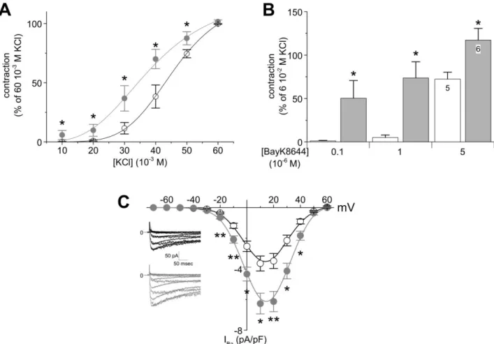
accompagnés de l'augmentation de la contractilité vasculaire induite par la dépolarisation ou par l'agoniste sélectif du Cav1.2 (Bay K 8644), ces réponses étant indépendantes de l'endothélium.
Comme prévu, ces réponses ont été accompagnées d'une diminution de la relaxation vasculaire induite par l'acétylcholine en LADCA. De plus, nous montrons que les cellules musculaires lisses de LADCA traitées à l’aldostérone présentent une augmentation du courant Ca2+ s, sans changements
dans leurs propriétés biophysiques.
De même que sur les cardiomyocytes, nous montorns que l'aldostérone augmente l'activité du promoteur P1, conduisant à une augmentation de la transcription du l'exon 1a et du l'exon 8a dans le tissu vasculaire. Par contre, ce traitement ne modifie pas l'expression du microARN-155 impliqué dans la régulation MR-dépendant (mais indépendamment de l'aldostérone) de l'expression du Cav
1.2-SNT dans le tissu vasculaire des animaux âgés. L'une des caractéristiques plus marquantes qui différencient le Cav1.2-SNT et le Cav1.2-LNT est la grande sensibilité à la nifédipine dans le tissu
vasculaire (Cav1.2-SNT). Ainsi, en confirmant nos résultats, nous montrons une perte significative
de la sensibilité à la nifédipine en LADCA traitées à l'aldostérone.
Conjointement les résultats obtenus dans cette étude indiquent que l'activation du MR dans les cellules musculaires lisses – principales responsables par le maintien du tonus myogénique et le précis réglage de la pression artérielle – subissent un mécanisme sophistiquée de switch moléculaire d'utilisation alternative du promoteur du gène Cacna1c (du promoteur P2 à P1), générant donc l'apparition d'une nouvelle sous-population de Cav1.2-LNT dans le tissu vasculaire qui se répercute
sur la dysfonction vasculaire. Par ailleurs, les animaux traités à l'aldostérone/sel (0,9% NaCl pendant 3 semaines) présentent une augmentation de la pression artérielle et de l'activité du promoteur P1 cardiaque du gène Cacna1c dans le tissu vasculaire.
Les résultats de cette étude identifie le Cav1.2-LNT comme une nouvelle cible spécifique du
MR. Au-delàs du nouveau concept mécanistique, nos résultats fournissent des évidences que les actions spécifiques d'éléments cis-régulateurs en réponse à l'aldostérone peuvent conduire à l’utilisation alternative du promoteur Cacna1c 'cardiaque" dans le tissu vasculaire et, par conséquent, étendre la diversité Cav1.2 dans ce tissu. Cette étude apporte de nouvelles perspectives sur la
régulation de ce mécanisme génomique dans le développement de maladies cardiovasculaires, dans lequel l'aldostérone à travers le MR joue un rôle important.
Mots-clés: Canaux Ca2+ de type L, aldostérone, récepteur aux minéralocorticoïde, Cœur, muscle lisse vasculaire.
7
Resumo Expandido
Ativação específica do promotor cardíaco alternativo do Cacna1c pelo receptor
de mineralocorticóide
Atualmente, os antagonistas do receptor de mineralocorticóide (MR) pertencem ao arsenal terapêutico para o tratamento de diversas doenças cardiovasculares. No entanto, os mecanismos pelos quais essa classe de medicamentos exercem seus efeitos benéficos ainda não estão totalmente compreendidos. Evidências experimentais do nosso grupo têm demonstrado através de experimentos
in vitro, ex vivo e in vivo que, parte dos efeitos deletérios da aldosterona, via ativação do MR,
envolvem o aumento da expressão e atividade do Cav1.2, agindo como um importante gatilho para o
desenvolvimento de patologias cardíacas. Portanto, o objetivo central deste estudo foi avaliar os mecanismos genômicos envolvidos na modulação da expressão do Cav1.2 pelo MR.
No presente estudo, demonstramos em cultura primária de miócitos ventriculares de ratos neonatais (NRVM), que o tratamento com aldosterona durante 24 h aumentou, de maneira concentração dependente (10-10 à 10-9 M) a expressão do Cav1.2. Importante destacar que as
subunidades auxiliares do Cav1.2 (β2 e α2δ2) também apresentaram aumento na expressão de seus
respectivos mensageiros, sugerindo assim, uma atividade transcricional finamente coordenada. Compondo parte da família de receptores nucleares, a translocação citosol-núcleo do MR foi avaliada em NRVM sob diferentes concentrações de aldosterona (10-9 à 10-7 M) e tempos de tratamento (30 – 90 min). Observamos aumento na translocação nuclear do MR em baixas concentrações de aldosterona (10-9 M) e curto período de tempo (30 min) de tratamento. Além disto, em NRVM transfectados com um plasmídeo contendo hMR-GFP, foi possível validar o rápido tráfico intracelular do MR para o núcleo (visível a partir de 10 min) em células estimuladas com aldosterona (10-9 M). Importante ressaltar que altas concentrações de dexametasona (10-8 M), um potente hormônio glicocorticóide sintético, não alterou a translocação nuclear do MR.
A expressão do Cav1.2 é regulada por dois promotores distintos e, apesar de não ser totalmente
compreendida, sua regulação ocorre de maneira específica em determinados tipos celulares. Por exemplo, o promotor “cardíaco” (P1) regula a expressão da forma longa do Cav1.2 (Cav1.2-LNT),
enquanto que o promotor “vascular/cerebral” (P2) regula a expressão da forma curta deste canal (Cav1.2-SNT), ambos localizados na região N-terminal. Interessantemente, a aldosterona aumentou
a transcrição do exon 1a (Cav1.2-LNT) em NRVM, sem alterar a transcrição do exon 1b (Cav
1.2-SNT). Estes dados se correlacionam com a atividade do promotor P1 de maneira concentração e tempo (6 – 24 h) dependentes.
A especificidade destes efeitos que, possivelmente, envolvem a atividade transcricional do MR, foram avaliados em cultura celular através do tratamento simultâneo com o inibidor seletivo
8 para o MR (RU28318, concentração 100x superior em relação à aldosterona) ou através do silenciamento (siRNA) do MR. Deste modo, demonstramos que em ambas abordagens experimentais, os efeitos da aldosterona sobre o aumento da expressão do Cav1.2, exon 1a e atividade do promotor
P1 foram completamente abolidos. Afim de explorar o aumento da expressão do Cav1.2-LNT
induzido pela aldosterona em células de animais adultos, geramos um camundongo transgênico expressando gene repórter luciferase sob o controle do promotor P1 do Cacna1c (PCa.luc). De forma similar ao encontrado em NRVM, cardiomiócitos ventriculares adultos de animais PCa.luc tratados por 24 h com aldosterona, demonstraram um aumento significativo, de modo concentração dependente, na atividade da luciferase. Coletivamente, nossos dados claramente indicam que a ativação do MR causa transativação do promotor P1 e, consequentemente, transcrição do Cav
1.2-LNT.
Através da análise in silico e mutagênese, identificamos que o MR interage com específicos elementos ativadores e repressores de ligação ao DNA do promotor P1. Foram identificados 5 elementos de resposta aos glicocorticóides (GRE), sendo um “negativo”, próximos à região de início da transcrição do exon 1a do gene Cacna1c, portanto, sobrepondo-se a sequência do promotor P1. Para avaliar a função dessas importantes regiões altamente especializadas na regulação da atividade do promotor P1, cada GRE previamente identificada foi individualmente substituído por uma outra sequência (scrambled). O distúrbio causado pleas sequências mutadas foram confirmadas através do software Genomatix. Em NRVM transfectadas com o plasmídeo contendo o splice variante do hMR∆5,6, que não possui o domínio de ligação ao ligante, portanto, estando constitutivamente ativo,
a atividade da luciferase aumentou 2,5 vezes em relação ao controle (não transfectado com splice). Apesar da mutação da região mais distal (1) não alterar a atividade, mutações no 2, GRE-3 ou nGRE diminuiu a resposta induzida pelo hMR∆5,6, embora ainda tenham permanecido elevadas
em relação às células não transfectadas com hMR∆5,6. Em contraste, a mutação da região mais
proximal (GRE-4), aumentou em 400% a atividade do promotor P1. Estes dados foram corroborados por experimentos realizados com aldosterona e o MR endógeno em NRVM.
Devido os glicocorticóides e mineralacorticóides compartilharem diversos mecanismos moleculares, avaliamos a contribuição dos GREs no controle do promotor P1 em resposta à dexametasona. Demonstramos que a mutação do GRE-1 ou -2 não modificou a sensibilidade do promotor P1 à dexametasona, enquanto que a mutação do GRE-3, -4 ou nGRE conduziu à um aumento na sensibilidade. Em conjunto, estes ensaios mostram que a aldosterona, via ação específica do MR, age ativando o promotor P1 através da transactivação do GRE-2, -3 e nGRE e repressão do GRE-4. Além disto, os resultados demonstam uma evidente diferença molecular de utilização dos GREs pelos MRs e GRs.
Expresso em células endoteliais e músculo liso arterial, o MR tem ganhado grande atenção em estudos experimentais e clínicos. Contudo, apesar de grande parte dos estudos concentrarem-se na regulação hemodinâmica dependente do endotélio, recentes estudos têm demonstrado a contribuição do MR localizado no músculo liso vascular na regulação da pressão arterial. No presente
9 estudo, demonstramos que o tratamento de artérias coronárias (LADCA), mesentéricas e aorta com aldosterona (10-9 M por 24 h) induziu aumento da expressão proteica do Cav1.2. Essa alteração foi
acompanhada pelo aumento na contratilidade vascular induzida por despolarização ou pelo agonista seletivo do Cav1.2 (Bay K 8644), sendo essas respostas, independentes do endotélio. Como esperado,
essas respostam foram acompanhadas por uma diminuição do relaxamento vascular induzido pela acetilcolina em LADCA. Além disso, demonstramos que LADCA tratadas com aldosterona apresentaram aumento na corrente iônica de Ca2+ através dos canais para Ca2+ do tipo L em células musculares lisas, sem alterações em suas propriedades biofísicas.
Supreendentemente, assim como NRVM, demonstramos que a aldosterona também aumentou a atividade do promotor P1, levando a um aumento da transcrição do exons 1a e 8a em tecido vascular. Embora tenha sido recentemente demonstrado o envolvimento do mir-155 na regulação da expressão do Cav1.2-SNT mediada pelo MR em tecido vascular de animais idosos, não verificamos alteração
de sua expressão em aortas de animais jovens tratadas com aldosterona (10-9 M por 24 h). Uma das
marcantes características que diferenciam o Cav1.2-SNT do Cav1.2-LNT é a notável sensibilidade à
nifedipina em tecido vascular (Cav1.2-SNT). Assim, corroborando nossos resultados, demonstramos
uma significante perda da sensibilidade à nifedipina em LADCA tratadas com aldosterona. Em conjunto, os resultados obtidos nesse estudo indicam que a ativação do MR em células musculares lisas, principais responsáveis pela manutenção do tono miogênico e, o fino ajuste da pressão arterial, sofrem um sofisticado mecanismo de switch molecular de utilização alternativa do promotor do gene
Cacna1c (do promotor P2 para o P1) gerando o aumento de uma nova subpopulação de Cav1.2-LNT
em tecido vascular, que repercutem no desenvolvimento da disfunção vascular aqui demonstrada. Além disto, animais tratados com aldosterona/sal (0,9% NaCl, por 3 semanas) apresentaram aumento na atividade do promotor P1 do gene Cacna1c no coração, associado com função ventricular melhorada e ausência de anormalidades estruturais. Finalmente, também demonstramos através da abordagem in vivo, aumento da pressão arterial e utilização alternativa do promotor do gene Cacna1c em tecido vascular.
Em conjunto, os resultados deste estudo identificaram o Cav1.2-LNT como um novo alvo
específico do MR, onde deciframos o mecanismo molecular implicado na regulação do promotor P1 do gene Cacna1c. Além do novo conceito mecanístico, nossos resultados fornecem evidências de que as específicas ações sob os elementos cis-regulatórios em resposta à aldosterona podem conduzir na utilização alternativa do promotor Cacna1c no tecido vascular e, consequentemente, ampliando a diversidade Cav1.2 neste tecido. Portanto, este estudo abre novas e interessantes perspectivas sobre a
regulação deste mecanismo genômico no desenvolvimento de doenças cardiovasculares nas quais, a aldosterona através do MR, desempenha um importante papel.
Palavras-chave: Canal para Ca2+ do tipo L, aldosterona, receptor de mineralocorticóide, coração, músculo liso vascular.
10
TABLE OF CONTENTS
Chapter 1 Introduction ... 11
1.1 MR transcriptional factor ... 12
1.1.1 Specificity of MR in target tissues ... 16
1.2 New pathological role of MR in non-classical target tissue ... 18
1.2.1 Pathological role of MR in the heart ... 20
1.3 L-type Ca2+ channel ... 27
1.3.1 Regulation of Cav1.2 expression ... 31
1.3.2 Cav1.2 promoters and alternative splice variants ... 34
1.3.3 Tissue-specific regulation of Cav1.2-LNT and Cav1.2-SNT ... 38
1.4 Rationale and Aim ... 42
Chapter 2 Materials and Methods ... 44
2.1 Neonatal Rat Ventricular Myocytes (NRVM) primary cultures ... 44
2.2 Cell transfection... 44 2.3 Western Blot ... 45 2.4 Quantitative RT-PCR ... 45 2.5 Luciferase Assays ... 47 2.6 Promoter analysis ... 47 2.7 Mutagenesis... 47
2.8 Immunostaining and fluorescence microscopy... 47
2.9 PCa.luc transgenic mice ... 48
2.10 Artery harvest and organ culture ... 49
2.11 Vascular reactivity ... 49
2.12 Rat coronary artery SMC isolation and electrophysiological recordings ... 49
2.13 Aldosterone-salt model ... 50
2.14 Data analysis ... 51
Chapter 3 Results ... 52
3.1 MR mediates aldosterone-induced expression of Cav1.2 ... 52
3.2 Aldosterone increases Cav1.2-LNT expression through MR-dependent activation of the 'cardiac' Cacna1c P1-promoter ... 54
3.3 Aldosterone induced a MR-dependent 'cardiac' Cacna1c P1-promoter switch in vascular cells ... 60 Chapter 4 Discussion... 67 Chapter 5 Conclusion ... 70 Perspectives ... 70 References ... 72 Appendix ... 100
11
Chapter 1 Introduction
The mineralocorticoid receptor (MR), a ligand-dependent transcription factor belonging to the nuclear receptor superfamily, is the primary mediator of aldosterone. Classically, this steroid hormone, mainly but not exclusively produced and secreted by the adrenal cortex (MacKenzie et al., 2012), binds and activates the MR located in the cytosol of epithelial target cells. The steroid receptor (SR) complex translocates to the nucleus where it modulates gene expression and translation of specific aldosterone-induced proteins that regulate electrolytes and fluid balance and subsequent blood pressure (BP) homeostasis.
New paradigms indicate the pivotal importance of aldosterone and MR as regulators of cellular and organ function, far beyond their effects on the kidney. The MR is also expressed in non-epithelial tissues turning them sensitive to aldosterone stimulation, such as heart, blood vessels, adipose tissues, hippocampus, and cells of the immune system (Viengchareun et al., 2007). Independent of its effects on BP, aldosterone excess leads to adverse cardiovascular, renal, central nervous, and psychological effects, raising clinical and research interest (Jaisser and Farman, 2016). Collectively, experimental studies and clinical trials, suggest a detrimental association between aldosterone and life-threatening arrhythmias in heart failure (HF) patients that may be prevented by MR blockade (Pitt et al., 1999, 2003; Zannad et al., 2011). In addition, aldosterone plays a wide range of direct vascular effects, including those regulating vascular reactivity and growth or cell death. Notably, aldosterone regulates BP and exerts influence on end-organ damage directly via MR of vascular smooth muscle cells (VSMC) and endothelial cells (DuPont et al., 2014; Leopold et al., 2007; McCurley and Jaffe, 2012; Romagni et al., 2003).
The impressive therapeutic benefit with MR antagonists, notably in the reduction of sudden cardiac death in HF patients and the control of BP in patients with resistant hypertension (DuPont et al., 2014; Leopold et al., 2007; McCurley and Jaffe, 2012; Romagni et al., 2003; Williams et al., 2015), is however clinically limited by the concomitant blockade of the epithelial MR with the consequent development of significant hyperkalemia. There is thus clearly a need for the development of tissue-specific MR antagonists. However, signaling pathways and underlying mechanisms participating to these cardiovascular effects remain elusive. Research efforts on MR signaling have revealed a host of new pathophysiological mechanisms, both by acting on the heart and by affecting factors extrinsic to the heart, which could contribute to lethal arrhythmias (Dawson et al., 2004), in which aberrant Ca2+ fluxes are a recurrent theme (Benitah et al., 2010). The main mediator of cardiac Ca2+ influx is the L-type Ca2+ channel (LTCC), whose main pore-forming subunit is Cav1.2 (encoded
by Cacna1c). LTCC plays a pivotal role in function and dysfunction of the ventricular cardiac myocytes, not only because it controls contraction, but also it contributes to rhythmogenecity, cellular
12 metabolism and gene transcription (Benitah et al., 2010). The cardiac MR activation by aldosterone upregulates LTCC and might be involved in triggering after depolarization-related fatal ventricular tachyarrhythmia (Bénitah and Vassort, 1999; Ouvrard-Pascaud et al., 2005; Perrier et al., 2004, 2005). Moreover, several studies have suggested effects of aldosterone on VSMC LTCCs (DuPont et al., 2014), one of the main determinants of vascular tone (Joseph et al., 2013). Indeed, VSMC-specific MR knockout in mice decreased Cav1.2 expression and activity contributing to attenuate age-related
hypertension (DuPont et al., 2016; McCurley et al., 2012).
However, the molecular mechanisms and signaling pathways whereby aldosterone and MR may directly modulate Cav1.2 expression and function have yet to be identified. Cav1.2 channels are
expressed as two distinct tissue-specific transcripts of Cacna1c, which encode, respectively, a long 'cardiac' (Cav1.2-LNT) and a short 'vascular/brain' (Cav1.2-SNT) N-terminal region, regulated by two
alternative promoters (P1 and P2) (Hofmann et al., 2014). Here, we show that MR acts as a transcription factor to transduce aldosterone signal into specific Cacna1c 'cardiac' P1-promoter activation in cardiac cells but also in VSMC, in which the major Cav1.2 transcript initiation is
normally controlled by the canonical P2-promoter, conferring thereby a new molecular signature to Cav1.2 in this tissue.
1.1 MR transcriptional factor
The biological effects of aldosterone are mainly mediated by the activation of the cytosolic MR (encoded by the gene NR3C2). Belonging to the nuclear receptor subfamily, MR is a member of the steroid-thyroid-retinoid superfamily of ligand-dependent transcription factors (Rogerson et al., 2004), being expressed in many cell types at different levels (Arriza et al., 1987; Gomez-Sanchez and Gomez-Sanchez, 2014; Gomez-Sanchez et al., 2006).
MR is composed of 984 amino acids distributed in three major domains, an N-terminal domain, a highly conserved DNA-binding domain (DBD), and a ligand-binding domain (LBD) (Arriza et al., 1987), displaying ∼57% amino acid identity with GR in the LBD, and ∼94% in the DBD. Moreover, progesterone receptors (PR) and androgen receptors (AR) also show significant amino acid homology (LBD: ∼50%; DBD: ∼90%) with MR and GR (Funder, 1997; Viengchareun et al., 2007). The DNA-binding domain contains sites for the consensus sequences, known as hormone-responsive elements (HRE), on the promoters/enhancers regions of targets genes. In common, during unliganded state MR/GR/PR/AR are associated with a complex of chaperone proteins, including the heat shock proteins (HSP), which maintain the receptors in an inactive form with high affinity for the hormone. In the cytosol, MR forms a direct complex with chaperones, mainly HSP90, but indirectly interacts with HSP70, p23, p48, FKBP-51 immunophilins and CYP40 cyclophilin (Bruner et al., 1997; Pratt
13 and Toft, 1997; Viengchareun et al., 2007). These pivotal chaperones interactions play a role in maintaining MR in an appropriate conformation for ligand binding. Upon ligand binding, MR dissociates from chaperone proteins, and FKBP51 is replaced by FKBP52, enabling the complex to interact with the motor protein dynein. The ligand–MR–dynein complex then translocates along microtubules from the cytosol to the nucleus supported by numerous molecular partners in a coordinated and sequential manner to ensure appropriate transcriptional regulation (Bruner et al., 1997; Nagase and Fujita, 2013; Pratt and Toft, 1997; Rogerson et al., 2004; Viengchareun et al., 2007). Once receptor/hormone complex is active, it binds to HRE in the 5'-untranslated region of aldosterone-responsive target genes to activate or repress the gene transcription (Figure 1). The control of gene transcription may also occur via other transcription factors, in which allow transcriptional control without direct interactions between receptor–steroid complex and DNA-binding sites (Karin, 1998).
Figure 1. Mechanisms of MR activation. Upon ligand binding, aldosterone acts as a ligand-activated transcription factor translocating along microtubules from the cytosol to the nucleus, where binds to hormone response elements in the promoter regions of target genes. For the efficient initiation of gene transcription, MR
14
regulates transcription by controlling chromatin remodeling and the recruitment of co-activators (Nagase and Fujita, 2013).
As the result of high level of structural and functional homology, MR and GR bind to common nuclear HREs, with a consensus of 15 nucleotides sequence (AGAACAnnnTGTTCT), which may vary despite remaining fairly conserved. These regions are referred as Glucocorticoid Responsive Elements (GRE), being composed of palindromic sequences centrally separated by three nucleotides (Arriza et al., 1988; Clyne et al., 2009; Lombès et al., 1993). This mechanism involves an orchestrate recruitment of co-regulators, known as transcriptional cis-activation to denote direct DNA binding to canonical HREs (Mangelsdorf et al., 1995).
MR and GR act regulating gene expression by binding to specific DNA sequences in the form of monomer or heterodimers. The binding to these regulatory elements activate or repress the transactivation. Even though that GRE operates individually, synergism between GREs has been reported (Jantzen et al., 1987; Liu et al., 1996; O’Hara et al., 2014; So et al., 2007). Moreover, MR arrangement on the DNA differs from that one displayed by GR. A single amino acid difference, at tyrosine 478 in the DBD, is responsible for the distinct structural binding conformation of MR and GR to DNA sequences. GR stimulation is also able to activate (Hollenberg et al., 1987; Webster et al., 1988) or repress (Schüle et al., 1990; Surjit et al., 2011; Yang-Yen et al., 1990) the transcription of several genes. To activate gene transcription, GR DBD cooperatively dimerizes on inverted repeat activating GREs (Hudson et al., 2014; Luisi et al., 1991).
Although the structural basis of GR-mediated transcriptional repression is still not clear, there is evidence that negative GRE (nGRE) plays a role in GR-mediated transcriptional repression (Surjit et al., 2011) by monomeric GR DBD binds to an inverted repeat with negative cooperativity (Hudson et al., 2013). Accordingly, it has been shown that GR activation downregulates the transcription of numerous anti-inflammatory genes (Kadmiel and Cidlowski, 2013). In contrast, MR activation causes proinflammatory transcriptional state (Chantong et al., 2012). Despite the opposing effects on inflammation status, GR agonists prolong the lifespan of MRnull mice, indicating that GR and MR share some transcriptional sites (Berger et al., 1998). Furthermore, GR binds, with distinct conformations, to both activating GREs and nGREs within the TSLP promoter. Interestingly, MR was not able to repress transcription by TSLP nGRE, thereby confirming that GR and MR show divergence function at nGREs, which is quite remarkable considering the high sequence identity of GR and MR within the DBDs as well as the small size of the domain (75 aa) (Hudson et al., 2014). Overall, these discrepancies may explain the differential ability of MR and GR to modulate gene expression despite sharing overlapping DNA sites and ligand binding properties (Hudson et al., 2014).
In addition to the DNA sequence, the localization of GRE towards of the transcriptional start site is also critically important for the transcription initiation. GREs may be located upstream or
15 downstream of the transcription-initiating site, being found closely (2.5kb) or more remotely (10kb) from the promoter (Jantzen et al., 1987; Luecke and Yamamoto, 2005). In accordance, in A549 human lung cells it has been show (So et al., 2007) that GREs are equally distributed upstream and downstream of the transcription start sites, with 63% of them >10 kb from those sites. More dramatic examples have been reported, such as an estrogen response element 144 kb upstream of the promoter of the NRIP gene (Carroll et al., 2005), and an intragenic region 65 kb downstream from the Fkbp5 promoter that appears to serve as an androgen response element (Magee et al., 2006).
Regulation of transcription is not only dependent on direct interaction with HRE, but also through other transcription factors, which may act as bridge factors between the activated receptor and transcription initiation complex (Halachmi et al., 1994). For instance, GR trans-represses NFκB, AP-1 and other inflammatory transcription factor complexes without direct binding to DNA but via protein-protein tethering. These “trans” mechanisms are recognized as the primary way by which GC affects a large repertoire of inflammatory gene targets including cytokines, chemokines and adhesion molecules (Dougherty et al., 2016; Glass and Saijo, 2010; Reichardt et al., 2001; Scheinman et al., 1995). In contrast to the anti-inflammatory effects of GR stimulation, activation of MR promotes cardiovascular inflammation (Brown, 2008; Fiebeler et al., 2007; Funder, 2004). Moreover, NFκB and AP-1 are activated in hypertensive rats treated with aldosterone and high-salt diet (Fukuda et al., 2011; Tostes et al., 2002).
The MR-mediated aldosterone actions can be divided into nongenomic (few minutes) and genomic (>6 h) responses in epithelial and non-epithelial cells (Connell and Davies, 2005). About two decades ago, it was demonstrated that aldosterone, at nanomolar concentrations, produces a rapid, nongenomic effects in a variety of tissues (Christ et al., 1999; Wehling, 1995). Subsequent studies have further investigated the mechanism of rapid nongenomic effects of aldosterone on VSMC (Alzamora et al., 2000) and cardiomyocytes (Mihailidou et al., 2004). It has been reported that the rapid aldosterone action may involve the activation of protein kinase C (PKC) (Wehling et al., 1995). In human VSMC, aldosterone induces a concentration-dependent rise of intracellular [Ca2+] reaching plateau phase within 2–3 min (Wehling et al., 1995). Aldosterone (100 nM) has also been reported to activate αPKC in VSMC, which stimulates its translocation from the cytosol to the membrane within 5 min (Christ et al., 1995). In contrast, aldosterone decreased PKC activity in myocyte-enriched cultures from neonatal Sprague-Dawley rat hearts (Sato et al., 1997). This rapid effect was observed after 1 min of exposure as little as 1 nM of aldosterone. However, in adult ventricular myocytes Na+/K+/2Cl− cotransporter and the Na+-K+ pump are acutely regulated by aldosterone, suggesting that aldosterone regulates cotransporters or pumps by direct activation of εPKC (Mihailidou et al., 2004). Furthermore, an increase in PKC activity may also result in activation of transcription factors, and thus it is suggestive to propose that the nongenomic actions of aldosterone may influences gene
16 expression via aldosterone-activated PKC (Chun and Pratt, 2004).
1.1.1 Specificity of MR in target tissues
The discussion of MR specificity arises from the equal binding affinity of the MR for aldosterone and glucocorticoids (Connell and Davies, 2005). Indeed, plasmatic levels of glucocorticoids (GC) are 100- to 1000-fold higher than aldosterone. Thus, in theory, GCs should occupy more MR than aldosterone (Funder, 2009). However, there is a key mechanism to regulate MR specificity to counter-react cortisol occupation of the MR (Ferrari and Krozowski, 2000; Lombes et al., 1994). Firstly described in epithelial tissues, MR specificity is regulated by the action of the type 2 isoform of 11-hydroxysteroid dehydrogenase enzyme (11β-HSD2) that inactivates cortisol into 11-cortisone, which cannot binds to the MR, thereby protecting MR from inappropriate activation by GC (White et al., 1997). Although this mechanism explains aldosterone specificity at epithelial tissues, MR has also been shown to be expressed in non-epithelial cells such as, in the heart, brain and blood vessels (Freel and Connell, 2004; Marney and Brown, 2007).
In the heart, MR is expressed in cardiomyocytes (Lijnen and Petrov, 2003; Lombès et al., 1995) and cardiac fibroblasts (Rombouts et al., 2001; Stockand and Meszaros, 2003). Furthermore, the expression of 11β-HSD2 has been demonstrated in the heart, although some researchers failed to show similar results (Kayes-Wandover and White, 2000; Lombès et al., 1995; Sheppard and Autelitano, 2002; Slight et al., 1996). However, it is generally accepted that 11β-HSD2 expression levels are much lower levels than those found in epithelial tissue (Funder, 2004; Slight et al., 1993). Therefore, it has been proposed that cardiac MR is mainly occupied by cortisol rather than aldosterone (Chai and Danser, 2006). Additionally, this hypothesis is further supported by the notion that the stoichiometric efficiency of 11β-HSD2 to metabolize cortisol cannot be achievable to allow a comparable MR competition between GC and mineralocorticoid (Funder, 2007).
17
Interconversion of cortisol and cortisone by 11β-HSD1 and 11β-HSD2, respectively. Under certain circumstances and in cell types, 11β-HSD1 may acts as an NADP-dependent dehydrogenase, inactivating cortisol, while 11β-HSD2 catalyzes the NAD+-dependent inactivation of cortisol, converting it to cortisone
(Chapman et al., 2013).
Even despite the avid support of the eminent researcher Dr. John W. Funder advocating the MR promiscuity in cardiomyocytes, a bulk of evidence has shown aldosterone binding to cardiac MR, even from Pr. Funder (Arriza et al., 1987; Barnett and Pritchett, 1988; Pearce and Funder, 1987; Yang and Young, 2009). For instance, in vivo injection of [3H]aldosterone in adrenalectomized rats demonstrated that aldosterone binds to MR in epithelial and non-epithelial tissue. In the heart, aldosterone is a 3-fold better binder to MR than a GC (Funder and Myles, 1996).
Figure 3. Binding of tracer [3H]aldosterone in kidney, colon, heart and hippocampal extracts 15 min after
injection of tracer alone, or with half-logarithmic increasing doses of nonradioactive aldosterone (○) or corticosterone (□) (Funder and Myles, 1996).
Even though that mineralocorticoids and GC bind with equal affinity, there are compelling evidence showing that the off-rate of aldosterone from MR is 5 times lower than GCs (Hellal-Levy et al., 1999; Lombes et al., 1994) and, the MR activation by aldosterone reaches greater
18 transactivation compared to GCs (Farman and Rafestin-Oblin, 2001). Taken together, it has now become clear that distinct ligands may produce differential effects on gene transcription probably, due to conformational differences in the MR-ligand complex, which may lead to variable degrees of stability and DNA interactions (Farman and Rafestin-Oblin, 2001; Hellal-Levy et al., 2000; Huyet et al., 2012).
Figure 4. MR transcriptional activity in response to different ligands and MR-ligand complex stability. A, Transactivation activity of the human MR (hMR) in response to aldosterone (Aldo), deoxycorticosterone (DOC) and cortisol in COS-7 cells transiently and treated for 24 h with agonist. B, Kinetic dissociation of aldosterone and cortisol from hMR in response to 10−6 M aldosterone and cortisol (Huyet et al., 2012).
1.2 New pathological role of MR in non-classical target tissue
Aldosterone has been viewed as a hormone involved in the regulation of physiological responses such as ions transport, fluid homeostasis, and BP control (Booth et al., 2002; Chai and Danser, 2006). However, this traditional concept has been updated in the past decades. Experimental and clinical data indicate that aldosterone and its receptor, MR, play an important direct or indirect role in cardiovascular diseases. Notably, these findings suggest a detrimental association between high aldosterone levels and great incidence of cardiovascular events such as, inflammation, oxidative stress, endothelial dysfunction, hypertension, fibrosis, arrhythmias and HF, being some of them a salt-dependent dysfunction (Brilla and Weber, 1992; Funder, 2007; Marney and Brown, 2007; Pimenta et al., 2008; Rocha et al., 2002; Sato and Saruta, 2004). In accordance with the deleterious correlation involving plasmatic aldosterone levels, several studies have shown elevated plasma aldosterone concentration in patients with HF, even despite maximal renin-angiotensin system
19 blockade, which is associated with disease progression (Swedberg and Kjekshus, 1990), and higher prediction of mortality (Güder et al., 2007). However, in spite of effective blockade of ACE activity and beneficial effects, some biochemical anomalies still persistent. In particular case, plasma aldosterone levels remained elevated in 40% of patients with symptomatic HF (MacFadyen et al., 1999), in 50% of patients with left ventricular hypertrophy (Sato and Saruta, 2001) and still progressively increasing in patients after-MI (Borghi et al., 1993) despite treatment with ACE inhibitor. This phenomenon has been termed as "aldosterone escape" (Struthers, 2004), being also found uncontrolled aldosterone levels even in a combination of ACE inhibitor plus angiotensin type-1 receptor antagonist (McKelvie et al., type-1999). Therefore, this inability to decrease aldosterone concentrations may contribute to aggravate the disease and, might explain the poor outcomes obtained from the current and conventional therapies in patients with cardiovascular disease.
Figure 5. Inappropriate MR activation is linked with several physiological processes in various tissues (Zennaro et al., 2009).
20
1.2.1 Pathological role of MR in the heart
Since the pioneering work on the development of cardiac fibrosis induced by aldosterone in combination with high dietary sodium intake (Brilla and Weber, 1992), great attention has been given to aldosterone as an independent risk factor for the onset and /or progression of cardiovascular disease. Undoubtedly, aldosterone stimulates the expression of several pro-fibrotic factors that may contribute to the pathological cardiac remodeling. Later studies validated the findings of Weber, suggesting that cardiac fibrosis is an independent event of hypokalemia and hypertension (Young et al., 1995), being those deleterious effects blocked by MR antagonists (Brown et al., 1999; Zannad and Radauceanu, 2005). Altogether, these studies suggest that aldosterone is not simply a biomarker of disease, but a potent mediator of cardiovascular disease.
Intriguingly, a transgenic cardiac-specific overexpression of aldosterone synthase mice showed coronary endothelial dysfunction but not cardiac fibrosis (Garnier et al., 2004). Moreover, human MR overexpression mice in cardiomyocytes lead to cardiac arrhythmias but not cardiac fibrosis (Ouvrard-Pascaud et al., 2005). In contrast, transgenic cardiac-specific overexpression of 11β-HSD2 mice displayed cardiac hypertrophy, fibrosis, and HF under normal salt diet, while eplerenone, a MR antagonist, successfully reversed these effects (Qin et al., 2003). It has been shown that high aldosterone levels, when at appropriate physiological conditions such as during low-sodium intake, are not by themselves detrimental to the heart nor to develop evident cardiac hypertrophy and fibrosis. However, high plasmatic aldosterone concentrations associated with excessive salt loading leads to pathological cardiac remodeling (Wang et al., 2004). Altogether, these studies suggest that the cardiac hypertrophy and fibrosis as seen in DOCA- or aldosterone/salt-treated animal models may be due to a synergistic effect of salt loading and mineralocorticoid rather than to the elevated circulating aldosterone levels alone. Thus, whether aldosterone/salt loading induces cardiac fibrosis by directly increasing the expression of extracellular matrix component (Brilla et al., 1993, 1994; Robert et al., 1994; Sun et al., 2004; Wahed et al., 2005; Zhou et al., 1996) or, indirectly, by cross-talk with other signaling pathways it remains to be elucidated (Chander et al., 2003; Guarda et al., 1993; Rude et al., 2005; Tostes et al., 2002; Wong et al., 2007).
Severe vascular inflammation and augmented macrophage infiltration have been identified in the myocardium before the onset of fibrosis, suggesting macrophage as a key player in the initiation and progression of MR-mediated cardiac fibrosis (Fujisawa et al., 2001; Rocha et al., 2002; Young et al., 2003). It is worth to note that macrophages contain MR and GR but not 11β-HSD2, thereby the MR, in macrophage cell, is mainly occupied by GCs (Bienvenu et al., 2012; Lim et al., 2007). However, the relative contribution of activating MR- and GR-expressing macrophages in the context of cardiovascular disease is not fully understood.
21 Interestingly, it has been proposed that the macrophage MR signaling plays a critical role in the development of cardiac fibrosis under inappropriate mineralocorticoid-salt status (Rickard et al., 2009). This study showed that selective deletion of the MR in the monocyte/macrophage cells is protective against chronic mineralocorticoid/salt-induced cardiac fibrosis and increased systolic BP after deoxycorticosterone (DOC) administration for 8 weeks (Rickard et al., 2009). Moreover, the absence of recruited macrophages in CCL2-null mice after DOCA/salt was associated with attenuated macrophage-mediated proinflammatory gene expression, reduced cardiac fibroblast and significantly lower BP compared to WT mice (Shen et al., 2014). The gene CCL2 encodes the monocyte chemoattractant protein-1, being considered as the major monocyte chemokine required for tissue macrophage recruitment and typically released by injured tissues. Recently, the activation of JNK pathway in macrophages has been related to cardiac injury and inflammation in DOC/salt model, suggesting the activation of MR-dependent transcription of downstream profibrotic factors in the pathological remodeling (Shen et al., 2016). Overall, these studies showed the pivotal macrophage-dependent recruitment in MR-macrophage-dependent cardiac inflammation and remodeling.
Aldosterone has also been implicated in left ventricular remodeling after myocardial infarction (MI) (Hayashi et al., 2001; Udelson et al., 2003) and increased post-MI mortality and morbidity (Rouleau et al., 1994). Accordingly, acute post-MI led to an activation of cardiac aldosterone synthase correlating with 4-fold increase of aldosterone in the myocardium (Silvestre et al., 1999). Furthermore, higher susceptibility to develop MI has been shown in patients with primary aldosteronism than in hypertensive subjects with similar BO (Milliez et al., 2005). In accordance with the deleterious involvement of MR in the onset of the post-MI left ventricular dysfunction, spironolactone treatment, a MR antagonist, was able to decrease collagen deposition and interstitial fibrosis in the left ventricle (Silvestre et al., 1999). Similarly, long-term eplerenone treatment alleviates cardiac remodeling and progression of left ventricular dysfunction in dog failing hearts (Suzuki et al., 2002). Altogether, these studies support that increased aldosterone levels in post-MI patients are associated with adverse clinical outcomes, including higher mortality (Beygui et al., 2006).
As previously mentioned, our group has consistently established that activation cardiac MR plays a central role in the modulation of Ca2+ fluxes, which is involved in triggering after depolarization-related fatal ventricular tachyarrhythmia (Bénitah et al., 2001; Gómez et al., 2009; Lalevée et al., 2005; Le Menuet et al., 2010; Ouvrard-Pascaud et al., 2005; Perrier et al., 2004, 2005; Bénitah and Vassort, 1999). For instance, fatal arrhythmias in experimental HF animal models (Bers, 2002; Pogwizd and Bers, 2004) are initiated by abnormal ventricular automaticity or triggered activity, in which the prolongation of action potential (AP) and aberrant Ca2+ fluxes are a recurrent theme
22 (Ruiz-Hurtado et al., 2012). Therefore, the upregulation of Cav1.2 and increased amplitude of ICa,L in
ventricular myocytes induced by aldosterone may represent a detrimental risk factor for the development of abnormal cardiac electrical activity and thus arrhythmias. Accordingly, transgenic mice overexpressing MR display moderate dilated cardiomyopathy associated with arrhythmias (Le Menuet et al., 2001). Moreover, conditional cardiac-specific overexpression of the MR led to fatal arrhythmias (Ouvrard-Pascaud et al., 2005). Indeed, aldosterone has been shown to be pro-arrhythmogenic in dogs after coronary ligation (Arora and Somani, 1962). In addition, the synthetic mineralocorticoid, fludrocortisone, exacerbates (Lim et al., 2001), while spironolactone improves, the electrophysiological parameters such as the QT interval dispersion (Akbulut et al., 2003; Yee et al., 2001). In contrast, spironolactone improves heart rate variability (MacFadyen et al., 1997; Yee et al., 2001) and decreases premature ventricular beats and no sustained ventricular tachycardia episode occurrence in patients with HF at rest and during exercise (Barr et al., 1995; Ramires et al., 2000).
Within the first week after MI, aberrant electrical remodeling has been associated with prolonged AP duration due to an increase in ICa,L and a decrease in transient outward potassium
current, which were prevented by MR blocker (Perrier et al., 2004). After post-MI, the treatment with the active metabolite of spironolactone, canrenone, decreased the ventricular fibrillation threshold (Cittadini et al., 2003). These findings were later validated by the observation of the impressive reduction of ventricular extrasystoles and QT intervals after MR blockade (Shah et al., 2007). Interestingly, primary hyperaldosteronism (PA) patients, which commonly display severe hypertension and hypokalemia, show prolongation of QT interval and ventricular arrhythmias, being also reported higher incidence of sudden cardiac death (Matsumura et al., 2005; Maule et al., 2006; Reincke et al., 2012). Overall, these studies support the hypothesis that MR antagonist represents a beneficial therapy for life-threatening cardiac arrhythmias and sudden cardiac death.
Because of numerous experimental studies supporting the beneficial effects of MR antagonists on the cardiovascular pathophysiology. Two landmarks clinical trials, the Randomized Aldosterone Evaluation Study (RALES) (Pitt et al., 1999) and the Eplerenone Heart Failure and Survival Study (EPHESUS) (Pitt et al., 2003) have convincingly demonstrated that treatment with MR antagonist (such as spironolactone and eplerenone), significantly reduce mortality in patients with HF and systolic left ventricular dysfunction in post-MI patients.
The RALES trial study enrolled 3400 patients with severe HF in a randomized protocol of treatment with spironolactone in addition to the conventional therapy. Convincingly, these patients showed 30% of reduction in the mortality and 35% of reduction in the morbidity (Pitt et al., 1999). In addition, patients with high baseline serum levels of aldosterone were significantly associated with poor outcome and marked cardiac fibrosis, while spironolactone therapy, for 6 months, significantly
23 decreased the cardiac fibrosis. Thus, these findings suggest that the beneficial effects of MR antagonists on the cardiac remodeling may occur due extra-renal mechanisms (Sun et al., 2004; Zannad et al., 2000). Moreover, spironolactone treatment showed higher effectiveness to prevent left ventricle remodeling and to suppress markers of collagen synthesis than ACE inhibitors in 134 patients with acute MI (Hayashi et al., 2003).
The EPHESUS trial was a long-term clinical outcomes trial of MR antagonists in the setting of post-MI HF patients. This trial showed that treatment with eplerenone was associated with significant improvement in early and long-term mortality and morbidity in patients with MI and HF (Pitt et al., 2003). Importantly, EPHESUS concluded that eplerenone leads to additive benefits to the conventional therapy with ACE inhibitors and β-adrenergic blockers. Accordingly, experimental studies have shown that combination MR antagonist with ACE inhibitor potentiate the beneficial effects on the left ventricular remodeling in rats with extensive MI (Cohn and Colucci, 2006; Fraccarollo et al., 2003). Altogether, these findings suggest an important synergism and independent beneficial actions of MR antagonists in post-MI and HF patients.
A robust meta-analysis which included 7 trials and 8635 patients with HF or coronary artery disease examined the effects of spironolactone and eplerenone on ventricular arrhythmias (Wei et al., 2010). This meta-analysis demonstrated that MR antagonists reduced the rate of ectopic ventricular beats, the risk of ventricular tachycardia, and the risk of sudden cardiac death. Collectively, all these findings, suggest a detrimental association between aldosterone and life-threatening arrhythmia that may be prevented by MR blockade.
Overall, based on many experimental studies and clinical trials, the new guidelines of the European Society of Cardiology developed with the special contribution of the Heart Failure Association has been modified to recommend spironolactone or eplerenone. Nowadays, this class of drug is recommended as Class I (Levels of Recommendation: Class I, II, and III) based on Level A of evidence (Classified between A to C) in all symptomatic patients (despite treatment with an ACE inhibitors and a β-adrenergic blocker) with HF and reduced ejection function (HFrEF) patients (Left ventricular ejection function: ≤ 35%), to reduce mortality and HF hospitalization (Ponikowski et al., 2016). This current recommendation was previously supported by the clinical trial Eplerenone in Mild Patients Hospitalization and Survival Study in Heart Failure (EMPHASIS-HF), which was the first randomized controlled trial to suggest that even patients with mild symptomatic HF are benefited from MR antagonist therapy (Zannad et al., 2011).
Recently launched, the clinical trial ALBATROSS treated patients with acute myocardial infarction (MI) with evidence ST-segment elevation MI (STEMI) or non-STEMI (NSTEMI) (Beygui et al., 2016). It is worth note that 92% of the patients had no evidence of HF. Thus, patients were
24 randomized to placebo group or treatment with 200 mg of intravenous canrenoate followed by oral spironolactone 25 mg/day in addition to standard therapy for 6 months. Despite the no significant reduction in the primary endpoint comparing MR antagonist regimen and standard therapy, the mortality risk was significantly reduced in patients with STEMI but not NSTEMI (Beygui et al., 2016). Therefore, the authors concluded that the overall study failed to show a benefit for early MR antagonist administration when added to standard therapy in non-failing patients with acute MI.
1.2.2 Pathological role of MR in the blood vessels
MR has been consistently found in endothelial cells and VSMC of blood vessels, being its activation at pathologic concentrations (10 nM) involved in many types of MR-induced vascular dysfunction, including atherosclerosis and hypertension (Barrett et al., 2013; Bender et al., 2015; Lombès et al., 2000; Pruthi et al., 2014). Classically, aldosterone regulates BP via activation of MR at renal epithelial cells in the distal nephron leading to sodium retention, which leads to increased blood volume (Soundararajan et al., 2010). In accordance, experimental studies and clinical trials have successfully shown that MR antagonists decrease BP (DuPont et al., 2014; McCurley et al., 2012; Mueller et al., 2015; Pitt et al., 1999, 2003). However, whether the actions of aldosterone on the BP controls occur directly, via vascular MR, or via indirect mechanisms (renal and central nervous system), remain a very active area of investigation due to its potential value for pharmacological interventions.
Rapid nongenomic aldosterone effects have been consistently reported in humans and in vitro studies. In normotensive patients, short-term systemic administration of aldosterone (12 pmol.min.kg for 4 h) results in endothelial dysfunction (Farquharson and Struthers, 2002). Rapid aldosterone infusion (500 ng/min for 8 min) into the brachial artery of healthy men causes increased forearm blood flow, whereas in the presence of a nitric oxide synthase inhibitor (L-NMMA) induces a greater vasoconstriction (Schmidt et al., 2003). Altogether, these results suggest that aldosterone acts through rapid nongenomic effects at the endothelium by increasing NO release and at VSMCs by promoting vasoconstriction. These findings are consistent with in vitro data showing an increase in intracellular Ca2+ influx in both cell types (Wehling et al., 1994, 1995). Furthermore, pulse wave velocity is greater
in patients with primary hyperaldosteronism compared with patients with essential hypertension (Bernini et al., 2008). Accordingly, patients with essential hypertension show increased plasma levels of aldosterone, being associated with reduced arterial compliance (Blacher et al., 1997).
25
Figure 6. Mechanisms of aldosterone-mediated arterial hypertension (Tomaschitz et al., 2010).
A landmark study demonstrated that VSMC-specific MR knockout mice (VSMC-MR-KO) displayed a decrease in the age-associated elevation of BP without impairments in renal sodium handling (McCurley et al., 2012). Importantly, aged VSMC-MR-KO mice showed decreased myogenic tone, attenuated vasoconstriction and vascular oxidative stress induced by Ang II, important players of vascular dysfunction and hypertension. Moreover, the expression of Cav1.2 and
contractile responses to an LTCC activator were attenuated in aged VSMC-MR-KO mice. However, just recently, Jaffe’s group has discovered that during aging process, MR expression increases in resistance vessels along with a decline in the microRNA (miR)-155 and increased expression of predicted miR-155 targets, in which includes Cav1.2 and angiotensin type-1 receptor, genes that
greatly contribute to vasoconstriction and oxidative stress in aging mice (DuPont et al., 2016). Altogether, these studies support the notion that VSMC-MR contributes to the age-associated rise of BP, at least in part, by regulating LTCC expression and activity.
Ca2+-activated potassium channels (KCa), mainly, the large conductance KCa channels (BKCa) has been recognized as another important target of MR in blood vessels (Liu et al., 1995; Provencher et al., 2012), where it plays an important role in the regulation of vascular contraction by hyperpolarizing VSMC (Ledoux et al., 2006). Accordingly, impaired acetylcholine-mediated


![Figure 3. Binding of tracer [ 3 H]aldosterone in kidney, colon, heart and hippocampal extracts 15 min after injection of tracer alone, or with half-logarithmic increasing doses of nonradioactive aldosterone (○) or corticostero](https://thumb-eu.123doks.com/thumbv2/123doknet/14488863.525552/18.893.105.786.448.944/aldosterone-hippocampal-injection-logarithmic-increasing-nonradioactive-aldosterone-corticostero.webp)


![[PDF] Cours sur les bases de Matlab en pdf | Formation informatique](data:image/gif;base64,R0lGODlhAQABAIAAAP///wAAACH5BAEAAAAALAAAAAABAAEAAAICRAEAOw==)