ORIGINAL ARTICLE
The rust fungi (Uredinales) on ferns in South Africa
Reinhard BerndtReceived: 1 June 2007 / Revised: 12 September 2007 / Accepted: 21 September 2007 / Published online: 25 October 2007 # German Mycological Society and Springer-Verlag 2007
Abstract The rust fungi (Uredinales, basidiomycota) occur-ing on ferns (Pteridophyta) in South Africa are described, illustrated and keyed out. All species belong to the puccinias-traceous genera Milesina (M. blechni), Uredinopsis (U. pteridis) or to the related uredinial anamorph genus Milesia (M. nervisequa, M. cf. magellanica, M. silvae-knysnae). Milesia silvae-knysnae on Polystichum pungens is new to science; it probably belongs to the teleomorph genus Mile-sina. Milesina blechni is reported from South Africa for the first time on the new hosts Blechnum punctulatum and Rumohra adiantoides; it has hitherto been known only from the Northern Hemisphere on Blechnum spicant. Rust speci-mens collected on Asplenium aethiopicum and A. rutifolium were tentatively assigned to Milesia magellanica which has been known so far only from southern Chile. Hyalopsora neocheilanthis, Milesina neoexigua and M. neovogesiaca are proposed as new names for Hyalopsora cheilanthis, Milesia exigua and M. vogesiaca. It is discussed that the pucciniastra-ceous fern rusts could have reached South Africa either by migration (M. blechni) or by long-distance air dispersal. In the absence of their gametophyte hosts, species of Abies (Pinaceae), the rusts have to propagate in South Africa by urediniospores infecting fern to fern.
Keywords Hyalopsora . Milesia . Milesina . Uredinopsis . Mycogeography
Introduction
Rust fungi of the genera Hyalopsora, Milesina and Uredi-nopsis (Pucciniastraceae) are restricted to fern hosts in their sporophytic phase (uredinia and telia) while their gameto-phyte (spermogonia and aecia) occurs, as far as known, on members of the Northern Hemisphere conifer Abies (Pina-ceae). The uredinia of these rust fungi are characterized by a dome-shaped, delicate peridium made up by a single layer of more or less isodiametric cells (Moss1926). Uredinia of this morphological type are classified in the anamorph genus Milesia. Though taxonomically correct, this nomenclatural situation is unfortunate because of the deceptive similarity to the telial name Milesina and the fact that uredinia of Milesia type occur within the rust family Phakopsoraceae as well.
Rust fungi parasitizing fern plants and not belonging to Pucciniastraceae are rare. A single species of each of Desmella and Uncol are recognized, the former probably belonging to Pucciniaceae, the latter possibly to Phakop-soraceae. Puccinia lygodii Arthur is the only known Puccinia species on ferns, and Uredo vetus Hennen the only known rust fungus on Selaginellaceae.
Pucciniastraceous fern rusts are largely restricted to temperate regions of the Northern Hemisphere. In South Africa, only two such rusts have been reported with certainty, Milesia nervisequa (Thümen) Faull, a species probably restricted to southern Africa, and the widely distributed Uredinopsis pteridis Dietel & Holw. (Doidge
1950; Faull 1932). In the present work a new species is described and two additional fern rusts are reported from South Africa together with new locations and observations of known species. All species are portrayed and a key is presented to facilitate their determination and to direct more interest to these easily overlooked rust fungi which are of considerable biogeographic interest.
DOI 10.1007/s11557-007-0548-7
Taxonomical novelties Milesia silvae-knysnae R. Berndt Milesina neoexigua R. Berndt Milesina neovogesiaca R. Berndt Hyalopsora neocheilanthis R. Berndt
R. Berndt (*)
Herbarium turicense, Institute of Integrative Biology (IBZ), ETH Zurich, CHN
Universitätstr. 16 8092 Zurich, Switzerland
Materials and methods
Spores and hand sections obtained from herbarium material were mounted in lactophenol and gently heated to boiling. The preparations were examined with a C. Zeiss“Axiophot” light microscope and photographs were taken with a C. Zeiss MC-80 camera on Kodak Ektachrome 64 Professional slide film. All micrographs were taken using Nomarsky Differen-tial Interference Contrast (DIC) optics. At least 30 uredinio-spores were measured for each specimen; exceptions are mentioned in the descriptions. PREM = South African National Fungus Collection, Pretoria; PUR = Arthur Herbar-ium, Purdue; HeRB = Herbarium Reinhard Berndt, Zurich.
Results and taxonomy
Milesina blechni (P. Syd. & Syd.) P. Syd. & Syd. 1910 Ann. Mycol. 8:491
Material investigated
M. blechni Western Cape Province, near Woodville N of Wilderness, afromontane forest at the“Big Tree” along the “Seven-Passes-Road”, on Blechnum punctulatum Sw., 27 Oct 2004, leg. E. Uhlmann & R. Berndt (only very few old uredinia found). Western Cape Province, Kleinmond, afromontane forest in the Oudebos River valley, on B. punctulatum (PREM 59722) and Rumohra adiantiformis (G. Forst.) Ching (PREM 59723), 4 Nov 2005, leg. E. Uhlmann & R. Berndt. Germany, Baden-Württemberg, Black Forest, c. 2 km SE Bad Griesbach, at hiking trail between Sexauer Hütte and Hildahütte, on B. spicant (L.) Roth., 3 Dec 2000, leg. V. Faust-Berndt & R. Berndt (HeRB 6246).
M. australis (Arth. ex Faull) Hirats. f. Costa Rica, Cordillera de Tilarán, Monte Verde, along road between Monte Verde village and cloud forest reserve, on B. occidentale L., 5 Mar 1992, leg. R. Berndt & V. Faust (HeRB 2969).
Rust-infected Blechnum (Blechnaceae) and Rumohra (Dry-opteridaceae) ferns were encountered in the afromontane forest of the Cape region. These rusts were most similar to M. blechni which is only known from the Northern Hemisphere and to M. australis from South and Central America. The specimens tallied well with both M. blechni and M. australis with regard to urediniospore measure-ments. Faull (1932) stated 26–45×15–23 μm (average 33×
19μm) for M. blechni and 22–40×14–23 μm (average 30× 18μm) for M. australis. In the latter, the spines are coarser
than in M. blechni and have a strong tendency to aggregate at the spore apex and around the hilum where they may show a crown-like arrangement (see Fig. 2). The South African collections were compared with specimens of M. australis and M. blechni and assigned to the latter because of the similarity of the echinulation.
The following description is based on the specimens from South Africa.
Uredinia scattered on abaxial side of fronds, bullate, more or less circular to elliptic, c. 0.15–0.3 mm diameter. Urediniospores (Figs.1,2 and3) subclavate, subpyriform, ellipsoid to broadly ellipsoid, sometimes subtruncate apically and proximally; spore wall hyaline, scarcely 1 μm thick, rather sparsely covered with slender, sharp spines (spines about 3–4 μm apart, up to 2 μm long and c. 1μm broad at the base) and without visible germ pores. On B. punctulatum, spores measured 26–33(36)×17–21.5 μm (mean 30.0×19.2 μm) in the Woodville specimen, 28–41 (44)×18–24.5 μm (mean 35.1×21.3 μm) in the specimen from Kleinmond. On R. adiantiformis, they measured (26) 28–41(44)×17–21(23) μm (mean 33.7×19.6 μm). Telio-spores were not observed.
Up to now, M. blechni has been known to occur on Blechnum spicant and Abies alba Mill. or A. cephalonica Loud. in Europe, Caucasia (Hiratsuka 1958) and the Macaronesian Islands (Gjærum and Sunding1986). Reports from Costa Rica refer to M. australis (Faull 1932). According to my knowledge, this report is the first one from outside this geographical range and in the Southern Hemisphere. The South African locations are far away from
Fig. 1 Milesina blechni. Urediniospores from Blechnum spicant, optical section and view of spore surface (Germany, Black Forest). Scale bar=20μm
the natural distribution area of the gametophyte host genus Abies, whose closest localities are in the mountains of Mediterranean North Africa and the Lebanon. Rumohra adiantiformis is a new host genus and species and B. punctulatum a new host species for M. blechni.
The only other report of a rust on Rumohra I am aware of is by Doidge (1950) who listed Milesia nervisequa (Thümen) Faull as a parasite of Polystichum adiantiforme (G. Forst.) J. Sm. (= R. adiantiformis). Milesia nervisequa differs from Milesina blechni by smaller, more delicately echinulate urediniospores. I could not study Doidge’s specimen but assume that it may represent M. blechni. Milesia nervisequa (Thümen) Faull1932
Contr. Arnold Arboretum 2:77
= Caeoma nervisequum Thümen 1877
= Milesina nervisequa (Thümen) P. Syd. & Syd. 1915 (nom. illegit.: teleomorph name illegitimately applied to the uredinial stage)
= Uredo nervisequa (Thümen) Hirats. f. 1957 Material investigated
On Cheilanthes viridis (Forrsk.) Sw. Western Cape Prov-ince, Barrydale, road to Heidelberg, Tradouw Pass, 24 Oct 2004, leg. E. Uhlmann & R. Berndt (PREM 59728). Knysna, gravel road to Phantom Pass turning off route N2, 27 Oct 2004, leg. E. Uhlmann & R. Berndt (PREM 59729). “Seven-Passes-Road” from Knysna to George, after Homtini Pass, 27 Oct 2004, leg. E. Uhlmann & R. Berndt (PREM 59730). On Pellaea pteroides (L.) Prantl Western Cape Province, Wellington, Bain’s Kloof Pass,
descent towards Ceres, near Bishop’s Rock, 18 Oct 2005, leg. A. Ritschel, E. Uhlmann & R. Berndt (PREM 59724). Milesia nervisequa has been reported on Pellaea hastata (L. f.) Link (= Cheilanthes hastata (L. f.) Kunze, Pteridaceae) from South Africa and Madagascar (Doidge 1927; Faull
1932). Presumably the host plant was originally incorrectly identified as in Doidge (1950) it is given as Pellaea viridis (Forrsk.) Prantl (= Cheilanthes viridis). Faull (1932)
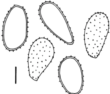
Fig. 3 Milesina blechni. a Urediniospores from Blechnum punctula-tum (South Africa, Kleinmond). Scale bar =10μm. b Urediniospores from Rumohra adiantiformis, optical section (South Africa, Klein-mond). Scale bar = 10μm. c Like b, focus on spore surface. Scale bar =10μm
Fig. 2 Milesina australis. Urediniospores from Blechnum occidentale (Costa Rica). Scale bar=10μm
also listed a specimen on Polypodium lycopodioides L. (= Microgramma mauritiana (Willd.) Tardieu, Polypo-diaceae) collected by Ethel Doidge in Natal. The specimen was originally assigned to Milesina dieteliana (Syd.) P. Magn. by Doidge (1927) and kept under this name as a member of the South African rust mycobiota in Doidge (1950). According to Faull (1932) it is essentially identical to M. nervisequa on C. viridis (as P. hastata). He therefore thought it“wisest, to diagnose it as M. nervisequa” despite the different host genera. M. dieteliana is a doubtful report, therefore, and should be excluded from the South African rust mycobiota. Doidge (1950) listed Polystichum adianti-forme (Forst.) J. Sm. (= Rumohra adiantiformis) as an additional host with a question mark. This specimen may however represent M. blechni (see above). Unfortunately, it is not present in PREM (S. Velthuysen, personal commu-nication) and could not be examined.
Faull (1932) measured 20–31×14–18 μm (average about
24×16μm) for the urediniospores of M. nervisequa on C. viridis (as P. hastata); 19–33×13–19 μm (average about 26×16 μm) for those on M. mauritiana (as Polypodium lycopodioides). I measured (20)22–35×14–19 μm (mean 26.5×15.9μm) for the urediniospores of the rust on Pellaea pteroides. The specimens on C. viridis were very similar but had slightly smaller urediniospores with slightly thicker spore walls. As the differences are small and as Pellaea and Cheilanthes are closely related and belong to the same family, Pteridaceae, I regard these specimens as belonging to one species and assign them to M. nervisequa. Pellaea pteroides is a new host for this species to my knowledge.
The following description was compiled from the speci-mens on C. viridis.
Uredinia on abaxial side of pinnules, scattered or in small groups, often confined to the area between adjacent leaf veins, bullate and more or less circular, c. 0.2–0.5 mm diam., releasing spores from an apical pore-like or irregular aperture, spore mass white to ivory, peridium dome-shaped, composed of polyangular cells measuring 8.5–13(16)×7– 12 μm (surface view); urediniospores (Fig. 4) ellipsoid, pyriform, obovoid or subclavate, rarely clavate, sometimes slightly deformed, 18.5–31×12.5–17 μm (mean 24.1× 15.1μm, for all specimens), spore wall very thin, c. 0.5– 1μm thick, hyaline, evenly and rather delicately echinulate by spines c. 2–3 μm apart, towards the base of the spore often slightly more densely and coarsely echinulate, germ pores not seen. Teliospores not observed.
Beside M. nervisequa, three other rust fungi have been described on Cheilanthes: Milesia wilczekiana (Maire) Kuprevicz & Tanzschel on Cheilanthes pteridioides C. Chr. from the Moroccan Atlas Mountains has larger urediniospores measuring 19–39×15–19 μm. Maire (1929) described them as resembling those of Milesia magnusiana (Jaap) Faull whose urediniospores average
35 × 20μm (Faull1932). The second species, Uredinopsis glabra Faull, has smooth urediniospores. Hyalopsora cheilanthis Arth. has pigmented urediniospores which measure 20–31×14–23 μm (Hiratsuka 1936). The latter species has to be renamed as the name H. cheilanthis applies to the uredinial stage (Arthur 1907) and is therefore a nomen anamorphosis. The same is true for the synonym H. pellaeicola Arth. which was also first applied to the uredinial stage by Arthur (1906). Both names are therefore inapplicable to the telial stage and I propose the new name Hyalopsora neocheilanthis for the holomorph:
Hyalopsora neocheilanthis R. Berndt, nom. nov.
Replaced synonym Hyalopsora cheilanthis (Peck) Arth. ex Arth. 1934. Manual of the rusts in United States and Canada, p. 11 (for the uredinial and telial stage, but inapplicable to the holomorph as applied to the uredinial stage first). A type was not designated, but probably is on Cheilanthes pringlei Davenp. from Arizona, USA (= type of H. cheilanthis as cited in Arthur 1907)
= Caeoma cheilanthis Peck 1883. Bull. Torrey Bot. Cl. 20:62 (uredinial stage)
= Hyalopsora cheilanthis (Peck) Arth. 1907. N. Amer. Fl. 7:113 (nom. illegit.: teleomorph name illegitimately applied to the uredinial stage)
= Uredo pellaeae Dietel & Neger 1899. Bot. Jahrb. 27:15
= Uredo pasadenae P. Syd. & Syd. 1904. Ann. Mycol. 2:31
= Hyalopsora pasadenae P. Syd. & Syd. 1915. Monogr. Uredin. III, p. 501 (nom. illegit.: teleomorph name illegitimately applied to the uredinial stage) = Hyalopsora pellaeicola Arth. 1906. Bull. Torrey Bot. Cl. 33:30 (nom. illegit.: teleomorph name illegitimately applied to the uredinial stage)
= Hyalopsora pellaeicola Arth. ex Hirats. f. 1936. Monogr. Pucciniastreae, p. 175 (for the uredinial and telial stage, but inapplicable to the holomorph as applied to the uredinial stage first)
The only authentic description of the telial stage appears to be in Arthur (1934), unfortunately without citing the specimen on which telia were found.
Milesia cf. magellanica Faull1932
Contr. Arnold Arboretum 2:31
= Milesina magellanica Hirats. f. 1936. Monogr. Pucciniastreae, p. 145 (nom. illegit.: teleomorph name illegitimately applied to the uredinial stage)
= Uredo magellanica (Faull) Hirats. f. 1957. Trans. Mycol. Soc. Japan 5:2, non Uredo magellanica Speg. 1899
Material investigated
M. magellanica On Asplenium magellanicum Klf. (Asple-niaceae): Chile, Región de los Lagos, Corral, Dec 1905, leg. R. Thaxter (PUR, type!).
M. cf. magellanica On A. rutifolium (Berg.) Kunze: Western Cape Province, Harkerville Forest Reserve E of Knysna,
along“Kranshoek Walk”, 26 Oct 2004, leg. E. Uhlmann & R. Berndt (PREM 59727). Knysna Forest N of Knysna, Goudveld Forest, near Rheenendal, along “Woodcutter Trail”, 25 Oct 2005, leg. A. Ritschel, E. Uhlmann & R. Berndt (PREM 59726). Eastern Cape Province, Amatola Mountains, Hogsback, 11 Dec 2006, leg. A.R. Wood (no. 697). On A. aethiopicum (Burm. f.) Becherer: Western Cape Province, Groenvlei lake E of Lake Pleasant, in milkwood scrub on the southern shore of Groenvlei, 26 Oct 2005, leg. R. Berndt (PREM 59725). Eastern Cape Province, Amatola Mountains, Hogsback, 11 Dec 2006, leg. A.R. Wood (no. 696).
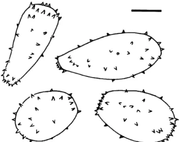
Fig. 4 Milesia nervisequa. a Urediniospores from Cheilanthes viridis, optical section (South Africa, Homtini Pass). Scale bar =10μm. b Like a, focus on spore surface. Scale bar =10μm. c Urediniospores from Pellaea pteroides (South Africa, Bain’s Kloof). Scale bar =10μm. d Like c, focus on spore surface. Scale bar =10μm
The following description comprises characters observed in the South African specimens.
Uredinia abaxial on fronds on blurred, yellowish brown or bleached patches of pinnules, scattered or in loose groops, small, 0.2–0.5 mm diam., straw–coloured to ochraceous, flatly pulvinate to bullate and covered by the epidermis, opening irregularly to liberate the white, powdery spore mass often deposited around the opening of the uredinia and on their surface, with dome–shaped peridium composed of subangular, ± isodiametric or flattened, slightly thick–walled cells, 9–14.5×8–13 μm, often with differently shaped, short, inflated or cylindrical, thin-walled, occasionally moderately thick-walled paraphysis-like hyphae bordering the base of the peridium; urediniospores (Figs. 5, 6 and 7) obovoid, ellipsoid to broadly ellipsoid, pyriform or subclavate, 22–29 (31.5)×14–18.5 μm (mean 25.7×16.7 μm) in Harkerville Forest specimen, 20–29×(13)15–19.5 μm (mean 24.3× 15.9μm) in Knysna Forest specimen, 22.5–34×14–19 μm (mean 27.5×16.1 μm) in Groenvlei specimen, spore wall hyaline, scarcely 1μm thick, rather evenly and moderately densely covered with delicate spines (spines c. 2–3 μm apart, 0.5–1 μm long), spines slightly coarser towards the hilum, germ pores not seen. Telia unknown.
A comparison of the South African specimens with other rust species known from the genus Asplenium showed that the spore characters tallied well with Milesia magellanica described from Chile (Fig.5, Table1). The spores were also very similar, however, to those of M. nervisequa (Fig.4, Table1).
In uredinia of the specimens on A. rutifolium, paraphysis-like hyphae were found which basally surrounded the uredinial peridium (Fig. 7b). It could not be decided with certainty whether such hyphae can develop in the hymenium as well. The specimens on A. aethiopicum lacked paraphysis-like hyphae around the uredinia and revealed additional differences. The urediniospores were slightly longer than in the specimens on A. rutifolium, their spore wall thinner, though sometimes thickened slightly at the hilum and the spore apex, and the peridial cells had more delicate walls. While the latter differences are minor, the presence of paraphysis-like hyphae may distinguish the rust on A. rutifolium as a distinct species. More specimens have to be studied, however, to prove the constancy of this character. As the type of M. magellanica is very scanty I did not section uredinia and therefore do not know whether paraphysis-like hyphae occur in them. Uredinia of the investigated specimens of M. nervisequa did not reveal paraphysis-like cells.
Paraphyses do not normally occur in the uredinia of Hyalopsora, Milesina or Uredinopsis (Moss1926). Magnus (1895,1901) described and illustrated uredinial paraphyses in Hyalopsora aspidiotus (Magn.) Magn. but his drawing (1895: Plate 23, Fig. 6) indicates that he depicted an old uredinium. In such uredinia thin-walled, paraphysis-like sterile hyphae may develop, perhaps after liberation of the majority of the urediniospores. The paraphysis-like hyphae observed in the present specimens looked dissimilar.
The question whether the South African rusts on Asplenium belong to M. magellanica or M. nervisequa or include an undescribed species cannot be settled at the moment. Because of the general similarity and the Asplenium hosts, I assign them to M. magellanica
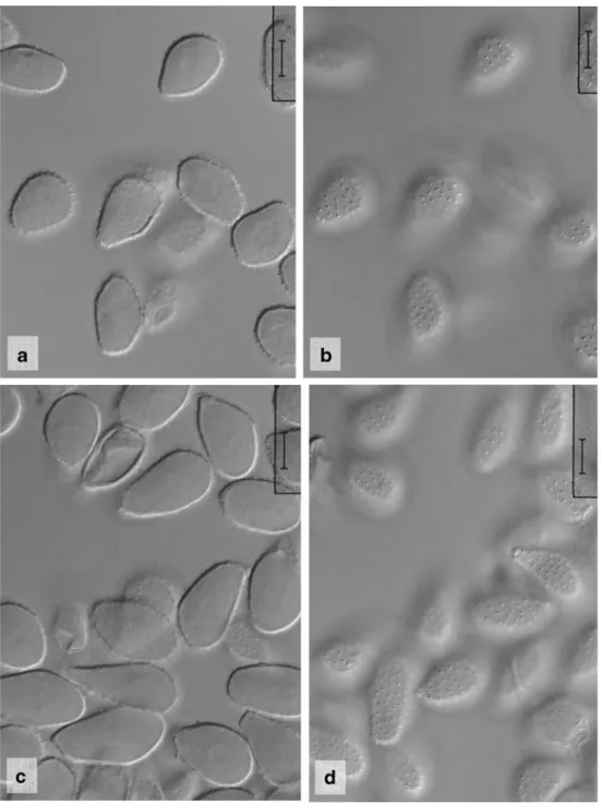
Fig. 5 Milesia magellanica. Urediniospores from type specimen on Asplenium magellanicum, optical section and view of spore surface (Chile). Scale bar =10μm
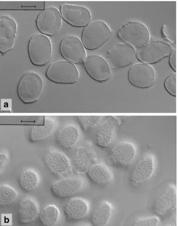
Fig. 6 Milesia cf. magellanica. Urediniospores from Asplenium rutifolium, optical section and view of spore surface (South Africa, Knysna). Scale bar =10μm
tentatively and not to M. nervisequa. To my knowledge, M. magellanica is only known from the type specimen. It would be a new member of the African rust mycobiota on two new host species, Asplenium aethiopicum and A. rutifolium. The rust will most probably prove to belong to the teleomorph genus Milesina. The name Milesina magellanica proposed by Hiratsuka (1936) is illegitimate, however, and a new name will have to be created when the telial stage is discovered.
Milesia silvae-knysnae R. Berndt, sp. nov. Etymology Named after the Knysna Forest, the southernmost large tract of afromontane forest. Uredinia abaxialia in pinnulis, sparsa vel laxe aggregata, minuta, 0.1–0.3 mm diam., straminea vel ochracea, vetera obscuriore brunnea, paulum bullata et epidermide tecta, peridio unistrato, ex cellulis plus minusve rotundatis, tenue
Fig. 7 Milesia cf. magellanica. a Urediniospores from Asple-nium rutifolium (South Africa, Knysna). Scale bar =10μm. b Uredinium from A. rutifolium with a part of the uredinial peridium (top of micrograph) and paraphysis-like hyphae (bottom). Scale bar =10μm. c Urediniospores from A. aethiopicum, optical section (South Africa, Groenvlei). Scale bar =10μm. d Like c, focus on spore surface. Scale bar =10μm
tunicatis composito; urediniosporae ellipsoideae, late ellip-soideae ad obovoideae, rariter clavatae, 26–36×(16)18– 22.5 μm (medium 40 sporae 29.8×20.2 μm), pariete hyalino, 0.5–1 μm crasso, subdense, inaequaliter delicate-que verrucoso, verrucis irregularibus, inter se (0.5)1–2 (3)μm distantibus, poris germinationis non visis.
In frondibus Polystichi pungentis (Kaulf.) C. Presl (Aspidiaceae).
Uredinia abaxial on the pinnules, scattered over the entire surface or in loose groups, tiny, 0.1–0.3 mm diam., straw-coloured to ochraceous, darker brown when old, slightly bullate and covered by the epidermis, opening irregularly to liberate the white spore mass, peridiate with a one-layered peridium made up by more or less round, thin-walled cells; urediniospores (Figs. 8 and 9) ellipsoid, broadly ellipsoid to obovoid, rarely clavate, 26–36×(16) 18–22.5 μm (mean of 40 spores 29.8×20.2 μm), spore wall hyaline, 0.5–1 μm thick, rather densely and finely verrucose, warts irregularly arranged and of different shape and seize, spaced at (0.5)1–2(3) μm, giving the spore profile a delicately wavy outline, germ pores not seen.
On fronds of Polystichum pungens (Kaulf.) C. Presl (Aspidiaceae).
Holotype (PREM 59737): Republic of South Africa, Western Cape Province, scenic road between Knysna and George, foot path through forest at “Big Tree” near Woodville N of Wilderness, 27 Oct 2004, leg. R. Berndt & E. Uhlmann. Isotype in Z+ZT.
Paratype (PREM 59737): Republic of South Africa, Western Cape Province, Knysna Forest N of Knysna, Goudveld Forest, near Rheenendal, along “Woodcutter Trail”, on P. pungens, 25 Oct 2005, leg. R. Berndt.
The present species will most probably belong to the genus Milesina because of the morphology and lack of
pigment of the urediniospores, the presence of a uredinial peridium and the fern host. It differs from the uredinial stages of other rust fungi on Polystichum spp. and from Milesina neoexigua R. Berndt, M. neovogesiaca R. Berndt,
Fig. 8 Milesia silvae-knysnae. Urediniospores from paratype, optical section and view of spore surface (South Africa, Knysna). Scale bar = 10μm
Table 1 Comparison of urediniospore measurements of Milesia magellanica, M. cf. magellanica and M. nervisequa
Specimen Urediniospore measurements Mean value
M. magellanica, type (own measurements and, below, from Faull1932) 20–28(29.5)×13–16 μm 23.4×14.7μm
18–30×14–20 μm 22×16μm
M. cf. magellanica on Asplenium rutifolium, Harkerville Forest 22–29(31.5)×14–18.5 μm, paraphysis-like hyphae present
25.7×16.7μm M. cf. magellanica on A. rutifolium, Knysna Forest 20–29×(13)15–19.5 μm, paraphysis-like
hyphae present
24.3×15.9μm M. cf. magellanica on A. rutifolium, Amatola Mts. 20–28×12–16 μm,paraphysis-like
hyphae present
23.4×14.0μm
M. cf. magellanica on A. aethiopicum, Groenvlei 22.5–34×14–19 μm 27.5×16.1μm
M. cf. magellanica on A. aethiopicum, Amatola Mts. 19.5–28×12.5–16 μm 23.7×14.5μm
M. nervisequa on Cheilanthes viridis (after Faull1932) 20–31×14–18 μm 24×16μm
M. nervisequa on Pellaea pteroides (20)22–35×14–19 μm 26.5×15.9μm
M. nervisequa on Microgramma mauritiana (after Faull1932) 19–33×13–19 μm 26×16μm
M. nervisequa on C. viridis, Phantom Pass 18.5–27×13–16 μm 22.1×14.9μm
M. whitei (Faull) Hirats. f. and M. winelandi Hirats. f., by the verruculose urediniospores.
During the study of M. silvae-knysnae it was noticed that the names Milesina exigua Faull and Milesina vogesiaca P. Syd. & Syd. are taxonomically incorrect and need to be replaced by new names:
Milesina neoexigua R. Berndt, nom. nov.
Replaced synonym Milesia exigua (Faull) Faull “comb. nov.” 1932. Contr. Arnold Arboretum 2:100 (nom. ana-morphosis misapplied to the uredinial and telial stage). The specimen on which the description of the telia was based has not been specified.
= Milesina exigua Faull 1931. J. Arnold Arboretum 12:218–219 (nom. illegit.: teliomorph name illegiti-mately applied to uredinial stage)
The teleomorph name Milesina exigua Faull is attached to the uredinial stage and thus inapplicable to the telial stage. Therefore, I propose the new name Milesina neo-exigua for the holomorph.
Milesina neovogesiaca R. Berndt, nom. nov.
Replaced synonym Milesia vogesiaca (P. Syd. & Syd.) Faull1932. Contr. Arnold Arboretum 2:103 (nom. anamor-phosis applied to the uredinial and telial stage). The specimen on which the description of the telia was based has not been specified.
= Milesina vogesiaca P. Syd. & Syd. 1910. Annal. Mycol. 8:491 (nom. illegit. et nudum: teliomorph name illegitimately applied to uredinial stage and putative immature teliospores)
= Uredo vogesiaca (P. Syd. & Syd.) Sacc. & Trotter in Sacc. 1912. Syll. Fung. 21:812 (new combination proposed assuming that no genuine teliospores were observed by the Sydows)
= Milesina vogesiaca P. Syd. & Syd. ex Hirats. f. 1936. Monograph of Pucciniastreae, p. 96 (description of teliospores but without Latin diagnosis, thus a nomen nudum not validly published and a later homonym of M. vogesiaca P. Syd. & Syd. preoccupied for the uredinial stage)
When proposing Milesina vogesiaca, Sydow and Sydow (1910) stated that a few immature teliospores were present which could not be described properly. Saccardo and Trotter (1912) consequently recombined M. vogesiaca to Uredo vogesiaca. Sydow and Sydow (1915) noted under Milesina vogesiaca (translated from German):“We found a few hyaline cells within the epidermal cells, but it remains an open question whether these were really teliospores.” It appears that the teleomorph name attached to the type merely anticipated that the fungus belonged to the genus Milesina and is therefore not valid. Even assuming that the Sydows found genuine teliospores, the name Milesina vogesiaca remains a nomen nudum which has never been validated by a Latin description of the teliospores. I consider the teliomorph name to be attached to the uredinial stage and thus not available for the holomorph. Therefore, the new name Milesina neovogesiaca is proposed.
Uredinopsis pteridis Dietel & Holw. in Diet. 1895 Ber. deutsch. bot. Ges. 13:331
Material investigated
On Pteridium aquilinum (L.) Kuhn (Dennstaedtiaceae): Western Cape Province, Barrydale, road to Heidelberg, at Tradouw Pass, 24 Oct 2004, leg. E. Uhlmann & R. Berndt (PREM 59733). Cape Town, Klaasen Rd. near Kirsten-bosch Botanical Garden, 2 Nov 2004, leg. E. Uhlmann &
Fig. 9 Milesia silvae-knysnae. a Urediniospores from paratype, optical section (South Africa, Knysna). Scale bar =10μm. b Like a, focus on spore surface. Scale bar =10μm
R. Berndt (PREM 59735). Swellendam, Marloth Nature Reserve, at trail to Duiwelsbos waterfall in afromontane forest, 20 Oct 2005, leg. A. Ritschel, E. Uhlmann & R. Berndt (PREM 59734). Hermanus, Fernkloof Nature Reserve, NW Lemoenkop, at path from the “Shelter” to the waterfall, 2 Nov 2005, leg. A. Ritschel, E. Uhlmann & R. Berndt (PREM 59732). Kleinmond, patch of afromon-tane forest in the Oudebos River valley, 4 Nov 2005, leg. E. Uhlmann & R. Berndt (PREM 59731). On Stenochlaena tenuifolia (Desv.) T. Moore (Blechnaceae): Kwa-Zulu-Natal, Umzinto, Raphia Palm Monument near Umlalazi Nature Reserve, 23 Mar 2006, leg. A.R. Wood (no. 657). The following description is based on the South African specimens on P. aquilinum.
Uredinia restricted to abaxial side of pinnules, on olive to brownish blurred leaf areas, sori scattered or close together but hardly confluent, bullate and more or less elliptic, c. 0.3-1.0 ×0.3–0.5 mm, rupturing laterally or at base of sori releasing the white to ivory spores as tongue-shaped or - rarely - tendril-like masses; urediniospores (Fig.10) ellipsoid, clavate or subfusiform, rounded at apex or subapiculate depending on the view onto the band of warts at the apex, 27–52×12–18 μm (mean 39.5× 14.9 μm), spore wall hyaline, scarcely 1 μm thick, with two, more or less opposite longitudinal bands of rather coarse interconnected warts or rods that reach from the hilum to the spore apex, bands up to 2μm high, at the apex about 3(−6) μm, between the bands spore wall with scattered small warts, which are round, elongated to striiform and sometimes aggregate to form a third, little pronounced band in addition to the ordinary longitudinal bands, germ pores not always visible, bizonate, located in upper and lower third of spores. Teliospores not found in South African specimens.
U. pteridis is distributed over wide areas of the world together with its host bracken fern (cf. Faull 1938a and Hiratsuka1958for distribution data). The species is very variable with regard to the size of the urediniospores, and this variability does not seem to be related to geographic distribution or host varieties (Faull1938a). The uredinio-spores are generally described as smooth with the exception of two longitudinal bands of cobs, but Ziller (1959) stated that the spores may be“sparsely verrucose-echinulate”. In the South African specimens, the spore wall was never smooth between the longitudinal bands and sometimes the warts even clustered and formed an additional though less pronounced band. The spores were very similar to those of Uredo verruculosa described from Venezuela (Berndt 1998). As long as a comprehensive taxonomic study of U. pteridis is not available I prefer to regard it as one highly variable species from which U. verruculosa may not be specifically different. The South
African specimens are therefore assigned to U. pteridis rather than U. verruculosa.
Recently, A.R. Wood collected a rust on Stenochlaena tenuifolia (Blechnaceae) in Natal. The sori were old and spores compressed or deformed so that they could not be measured reliably. However, they showed the typical surface ornament of U. pteridis with longitudinal bands and some interspersed warts and are therefore assigned to the latter tentatively. Stenochlaena would be a new host genus, Blechnaceae a new host family for U. pteridis. Milesia histiopteridis (Cunn.) Faull 1932
Contr. Arnold Arboretum 2:73
M. histiopteridis is known from New Zealand (McKenzie
1998) and New Guinea (Farr et al., no date). BPI 148015 listed in Farr et al. (no date) is supposed to stem from South Africa but is not accompanied by any additional collection data. The specimen is not listed in Crous et al. (2000
onwards). I consider the presence of M. histiopteridis in South Africa to be dubious, therefore, and do not present a description of the species. It is included into the key, however, as the host, Histiopteris incisa (Thunb.) J. Sm., occurs in southern Africa.
Fig. 10 Uredinopsis pteridis. a Urediniospores from Pteridium aquilinum (South Africa, Cape Town). Scale bar = 10 μm. b Urediniospores from P. aquilinum (South Africa, Swellendam). Scale bar =10μm
Discussion
The species of the rust genera Hyalopsora, Milesina and Uredinopsis are distributed throughout the temperate forests of the Northern Hemisphere with eastern Asia and eastern North America as centres of diversity (Faull1932,1938a; Hiratsuka1958). In all species whose life cycle is known, the spermogonia and aecia develop on members of the conifer genus Abies which is restricted to the Northern Hemisphere (Liu 1971). It is reasonable, therefore, to hypothesize that these rusts evolved on the Northern Hemisphere in temperate forests where Abies and ferns occur together. Unexpectedly, a few members of these rust genera thrive on ferns on the Southern Hemisphere, in Africa, South America or New Zealand, far beyond the
natural area of Abies. In South America, they occur for example in Ecuador (Milesia andina Faull, Milesina australis, Uredo semidiscifera R. Berndt), Colombia (Milesia columbiensis (Diet.) Arthur) and even in southern Chile (Milesia magellanica, Milesina australis, Hyalopsora neocheilanthis [reported as Uredo pellaeae]). In New Zealand, there are Uredo lindsaeae Henn., Milesia histiop-teridis and Hyalopsora polypodii (Pers.) Magnus which has its southernmost outpost on the North Island (Faull
1932; Hiratsuka 1958; McKenzie 1998; New Zealand Landcare Research 2005). Milesia nervisequa and Uredi-nopsis pteridis have been recorded previously in South Africa and another two or probably three new fern rusts are added in this paper to the South African rust mycobiota.
Urediniospores with two longitudinal bands of interconnected warts or rods reaching
from the hilum to the spore apex; spore wall smooth in-between the bands or sparsely
verrucose or/and with an additional longitudinal band of loosely crowded warts. On
Pteridium, possibly Stenochlaena: ... Uredinopsis pteridis
Urediniospores echinulate or verruculose, not with longitudinal bands of warts
Urediniospores verruculose
Urediniospores 22-34(36) x (15)17-22.5
µ
m (mean 28.0 x 19.2 µm). On
Polystichum: ... Milesia silvae-knysnae
Urediniospores smaller, 18-28 x 14-18 (mean c. 24 x 16). On Histiopteris;
not known from southern Africa with certainty: ... Milesia histiopteridis
Urediniospores echinulate
Urediniospores 26-41(44) x 17-24.5 µm, echinulate by moderately coarse
spines. On Blechnum or Rumohra: ... Milesina blechni
Urediniospores smaller, finely echinulate, not on Blechnum or Rumohra
On Cheilanthes, Pellaea, possibly Microgramma: ... Milesia nervisequa
On Asplenium: ... Milesia cf. magellanica
The obvious questions are how these rusts maintain their population in the absence of the Abies hosts and how they reached their locations on the Southern Hemisphere.
The propagation of the named species beyond the area of Abies can be explained by their ability to perpetuate by the conidial urediniospores which are able to carry rust infection from fern to fern (Faull1947). Biologically this means that these rusts cannot reproduce by ordinary sexuality but propagate vegetatively. Observations on fern rusts on the Northern Hemisphere suggest that asexual propagation may prevail locally even within the distribution range of Abies (McGinnis1971).
A probable explanation how fern-Abies-rusts came to outlying Southern Hemisphere locations is that their progenitors migrated from their original Northern Hemi-sphere area to the Southern HemiHemi-sphere using appropriate fern hosts in suitable habitats as “step stones”. In Africa, suitable habitats may be found in mountainous regions with cool, moist climate and fern-rich afromontane or alpine vegetation. A track of step stones has already been suggested for the immigration of northern temperate seed plants to eastern and southern Africa (Hedberg 1965; Linder 1990; cf. White 1978 for additional references). It is strange, however, that few reports of fern rusts have been made from eastern and central Africa. To my knowledge, only Uredinopsis pteridis has been reported from Kenya (Nattrass1961) and Zaire (sub“U. pteridis var. congensis” Henn., Hiratsuka1958). It is very likely that this scarcity of reports does not reflect absence or rarity of the rusts but rather the difficulty with which these inconspicuous fungi are discovered in the field as well as insufficient collecting in the region. A comparable track of migration can be hypothesized for the South American pucciniastraceous fern-rusts which may have proceeded from North America along the Cordillera south to southern Chile.
Other examples of rust species occurring on the Northern Hemisphere and in southern Africa are Pucciniastrum agrimoniae (DC.) Tranz., Coleosporium clematidis Barcl., C. ipomoeae (Schwein.) Burrill, and Melampsora hyper-icorum (DC.) Winter (Doidge 1927, 1950; Jackson 1931; Sydow and Sydow1915). P. agrimoniae alternates between Agrimonia spp. (Rosaceae) and Abies on the Northern Hemisphere and is known from southern outposts in Brazil and South Africa. Melampsora hypericorum (DC.) Winter is probably autoecious and known from a variety of Hypericum spp. (Hypericaceae) while Coleosporium clem-atidis Barcl. and C. ipomoeae (Schwein.) Burrill alternate between Pinus (Pinaceae) and species of Clematis (Ranun-culaceae) or Ipomoea (Convolvulaceae).
The putative South American-South African disjunction of M. magellanica would be difficult to explain and the
question whether the specimens collected in South Africa really belong to this species is pivotal. The easiest way out would be that the South African specimens represent a distinct species. However, no clear characters have been found so far that would allow one to distinguish this species morphologically. The southern African Milesia nervisequa is very similar to M. magellanica as has been mentioned above. It is known from Cheilanthes and Pellaea (Pterida-ceae) and has also been reported from Microgramma (Polypodiaceae). Should the rusts collected on Asplenium (Aspleniaceae) belong to M. nervisequa, too, this would be an unparalleled case of a Milesia infecting hosts of three different fern families. It is very unusual for species of Milesina, Uredinopsis and their Milesia anamorphs to parasitize fern hosts belonging to different families. Infection experiments carried out by various investigators showed that these rusts exhibit a high specificity to particular host species or at least host genera (Faull 1934, 1938b; Hunter 1936; Kamei 1940). Therefore, I tend to assume that the rust on Asplenium collected in South Africa is M. magellanica. Molecular taxonomic studies should help to solve the question whether the rusts on Asplenium from South Africa and South America are identical as soon as South American specimens become available for sequencing.
A contrary conclusion was drawn in the similar case of Milesina blechni on Rumohra and Blechnum. The rust on Rumohra was assigned to M. blechni despite the fact that the fern belongs to Dryopteridaceae and not Blechnaceae. In this case, however, the infected ferns were found in direct vicinity and the respective rusts were morphologically indistinguishable so that it is not reasonable to separate a new species just on grounds of different host families.
Air-borne transport of urediniospores by westerly winds might serve as an explanation for a possible disjunct occurrence of M. magellanica. Long distance transport of rust spores by wind currents is known to occur (Nagarajan and Singh 1990; Brown and Hovmøller 2002). It is questionable, however, whether urediniospores of Milesina are adapted for transport between continents. Their uredi-niospores tend to be sticky and to remain exposed on the infected fronds. As in most species of the genus Milesina, the urediniospores of Milesia magellanica are thin-walled and not visibly pigmented. Carotenoid pigments which are widespread in rust fungi (Zwetko and Pfeifhofer1991) have protective properties against UV-radiation (Lysenko and Demina1981). The lack of such protective properties may not be detrimental for sori or spores protected by host tissue (Rotem and Aust 1991) or spores exposed within a moist and shady forest habitat but could render the spores unsuitable for inter-continental transport.
Acknowledgements I thank the Northern Cape Department of Nature and Environmental Conservation and the Western Cape Nature Conservation Board in South Africa for issuing collecting and export permits, Alan R. Wood (ARC-PPRI, Stellenbosch) for rust-infected fern specimens from South Africa and helpful comments on this paper, and the Arthur Herbarium (PUR) for the loan of the type of Milesia magellanica. Ottmar Holdenrieder (ETH Zurich) is thanked for the permission to use microscopy facilities in his lab. I am grateful to the German Ministry of Education and Research (BMBF) for funding the field stays in Africa.
References
Arthur JC (1906) New species of Uredineae– IV. Bull Torrey Bot Club 33:27–34
Arthur JC (1907) North American Flora. Vol. 7, part 2. Order Uredinales. The New York Botanical Garden, New York, USA Arthur JC (1934) Manual of the rusts in United States and Canada.
Purdue Research Foundation, Lafayette, Indiana, USA
Berndt R (1998) New species of neotropical rust fungi. Mycologia 90:518–526
Brown JKM, Hovmøller MS (2002) Aerial dispersal of pathogens on the global and continental scales and its impact on plant disease. Science 297:537–541
Crous PW, Phillips AJL, Baxter, AP (2000 onwards) Phytopathogenic fungi from South Africa (online version, designed and hosted by Systematic Botany & Mycology Laboratory, United States Dept. Agriculture), http://nt.ars-grin.gov/fungaldatabases/southafrica/ Index.cfm)
Doidge EM (1927) A preliminary study of the South African rust fungi. Bothalia 2:1–228
Doidge EM (1950) The South African fungi and lichens to the end of 1945. Bothalia 5:1–1094
Farr FD, Rossman AY, Palm ME, McCray EB (without date) Fungal databases, Systematic Botany and Mycology Laboratory, ARS, USDA. Retrieved 18 Apr 2006 from http://nt.ars-grin.gov/ fungaldatabases/
Faull JH (1932) The genus Milesia. Contrib Arnold Arbor 2:1–138 Faull JH (1934) The biology of milesian rusts. J Arnold Arbor 15:50–85 Faull JH (1938a) Taxonomy and geographical distribution of the
genus Uredinopsis. Contrib Arnold Arbor 11:1–120
Faull JH (1938b) The biology of the rusts of the genus Uredinopsis. J Arnold Arbor 19:402–436
Faull JH (1947) Tropical fern hosts of rust fungi. J Arnold Arbor 28:309–319
Gjærum HB, Sunding P (1986) Flora of Macaronesia. Checklist of rust fungi (Uredinales). Sommerfeltia 4:1–42
Hedberg O (1965) Afroalpine flora elements. Webbia 19:519–529 Hiratsuka N (1936) A monograph of the Pucciniastreae. Mem Tottori
Agri Coll 4:1–374
Hiratsuka N (1958) Revision of taxonomy of the Pucciniastreae. Contributions from Laboratories of Phytopathology and Mycology,
Faculty of Agriculture, Tokyo University of Education, no. 31: 1–167
Hunter LM (1936) The life histories of Milesia scolopendrii, M. polypodii, M. vogesiaca and M. kriegeriana. J Arnold Arbor 17:26–37 Jackson HS (1931) The rusts of South America based on the Holway
collections– III. Mycologia 23:96–116
Kamei S (1940) Studies on the cultural experiments of the fern rusts of Abies in Japan. J Fac Agri Hokkaido Univ 57:1–189
Linder HP (1990) On the relationship between the vegetation and floras of the afromontane and the Cape regions of Africa. Mitt Inst Allg Bot Hamburg 23b:777–790
Liu T-S (1971) A monograph of the genus Abies. Dept. of Forestry, College of Agriculture, Natl. Taiwan University, Taipei, Taiwan R.O.C.
Lysenko SV, Demina NS (1981) Mechanisms of microbial protection from the effects of ultraviolet light. Biol Bull Acad Sci USSR 8:486–493
Magnus P (1895) Die Teleutosporen der Uredo aspidiotus Peck. Ber Dtsch Bot Ges 13:285–288
Magnus P (1901) Weitere Mittheilung über die auf Farnkräutern auftretenden Uredineen. Ber Dtsch Bot Ges 19:578–584 Maire R (1929) Champignons nord-africains nouveaux ou peux
connus. Bull Soc Hist Nat Afr Nord 20:279–282
McGinnis MR (1971) Selected aspects of the biology of Hyalopsora polypodii on Cystopteris fragilis. Mycologia 63:277–282 McKenzie EHC (1998) Rust fungi of New Zealand - An introduction,
and list of recorded species. NZ J Bot 36:233–271
Moss EH (1926) The uredo stage of Pucciniastreae. Ann Bot 40: 813–849
Nagarajan S, Singh DV (1990) Long-distance dispersion of rust pathogens. Annu Rev Phytopathol 28:139–153
Nattrass RM (1961) Host lists of Kenya fungi and bacteria. Mycol Pap 8:1–46
New Zealand Landcare Research (2005) NZFUNGI - New Zealand Fungi (and Bacteria) database,http://nzfungi.landcareresearch.co. nz/html/mycology.asp
Rotem J, Aust HJ (1991) The effect of ultraviolet and solar radiation and temperature on survival of fungal propagules. J Phytopathol 133:76–84
Saccardo PA, Trotter A (1912) Sylloge Fungorum, vol. 21. Padua, Italy Sydow P, Sydow H (1910) Mycotheca germanica Fasc. XVIII-XIX
(No. 851–950). Ann Mycol 8: 489–493
Sydow P, Sydow H (1915) Monographia Uredinearum Vol. III. Pucciniaceae (excl. Puccinia et Uromyces), Melampsoraceae, Zaghouaniaceae, Coleosporiaceae. Gebr Bornträger, Leipzig, Germany
White F (1978). The afromontane region. In: Werger MJA (ed) Biogeography and ecology of southern Africa, Vol. I. W. Junk, The Hague, The Netherlands, pp 463–513
Ziller WG (1959) Studies of western tree rusts. IV. Uredinopsis hashiokai and U. pteridis causing perennial needle rust of fir. Can J Bot 37:93–107
Zwetko P, Pfeifhofer HW (1991) Carotinuntersuchungen an Rostpilz-sporen. Bedeutung für die Physiologie und Taxonomie. Nova Hedwigia 52:251–266