DOI 10.1007/s00221-009-1743-3
R E S E A R C H A R T I C L E
Control of roll and pitch motion during multi-directional balance
perturbations
Ursula Margareta Küng · C. G. C. Horlings · F. Honegger · J. E. J. Duysens · J. H. J. Allum
Received: 30 April 2008 / Accepted: 10 February 2009 / Published online: 5 March 2009 © Springer-Verlag 2009
Abstract Does the central nervous system (CNS) inde-pendently control roll and pitch movements of the human body during balance corrections? To help provide an answer to this question, we perturbed the balance of 16 young healthy subjects using multi-directional rotations of the support surface. All rotations had pitch and roll compo-nents, for which either the roll (DR) or the pitch (DP) com-ponent were delayed by 150 ms or not at all (ND). The outcome measures were the biomechanical responses of the body and surface EMG activity of several muscles. Across all perturbation directions, DR caused equally delayed shifts (150 ms) in peak lateral centre of mass (COM) veloc-ity. Across directions, DP did not cause equally delayed shifts in anterior–posterior COM velocity. After 300 ms however, the vector direction of COM velocity was similar to the ND directions. Trunk, arm and knee joint rotations followed this roll compared to pitch pattern, but were di Ver-ent from ND rotation synergies after 300 ms, suggesting an intersegmental compensation for the delay eVects. Balance correcting responses of muscles demonstrated both roll and
pitch directed components regardless of axial alignment. We categorised muscles into three groups: pitch oriented, roll oriented and mixed based on their responses to DR and DP. Lower leg muscles were pitch oriented, trunk muscles were roll oriented, and knee and arm muscles were mixed. The results of this study suggest that roll, but not pitch components, of balance correcting movement strategies and muscle synergies are separately programmed by the CNS. Reliance on diVerentially activated arm and knee muscles to correct roll perturbations reveals a dependence of the pitch response on that of roll, possibly due to biomechani-cal constraints, and accounts for the failure of DP to be transmitted equally in time across all limbs segments. Thus it appears the CNS preferentially programs the roll response of the body and then adjusts the pitch response accordingly.
Keywords Balance corrections · Postural control · Muscle responses · CNS motor programs
Introduction
If balance corrections are diVerently organised in the roll (medio-lateral) and pitch (anterior–posterior) directions, exploring these diVerences may provide insights into the mechanisms underlying falls. A major inXuence on balance corrections is the biomechanical response of the body, which is diVerent in the roll and pitch planes. For a pure pitch perturbation, the trunk moves in pitch only. In con-trast, across a range of perturbation directions from pure roll to roll combined with pitch, both pitch and roll motions of the trunk occur (Carpenter et al. 1999; Grüneberg et al. 2005). Thus, if body motion is diVerent depending on the roll and pitch content of the stimulus, then it might be U. M. Küng · C. G. C. Horlings · F. Honegger · J. H. J. Allum (&)
Department of ORL, University ORL Clinic,
University Hospital, Petersgraben 4, 4031 Basel, Switzerland e-mail: [email protected]
U. M. Küng
e-mail: [email protected]
C. G. C. Horlings
Department of Neurology, Radboud University Medical Centre, Nijmegen, The Netherlands
J. E. J. Duysens
Department of Rehabilitation,
expected that the CNS takes this into account when execut-ing balance corrections, possibly by relyexecut-ing more on those muscles which act eYciently in the roll and pitch planes to correct the pitch motion induced by a roll perturbation.
There are two opposing viewpoints on the directional control of balance corrections. One viewpoint asserts that no diVerences exist between the roll and pitch commands issued by the CNS, rather a common movement strategy and muscle synergy is used regardless of perturbation direction (Henry et al. 1998a, 1998b; Park et al. 2004; Jones et al. 2008). According to this viewpoint, diVerences in movement responses or joint torques with perturbation direction can be explained by a simple directional re-weight-ing of the muscle responses along the body, accordre-weight-ing to the alignment of lines of muscle action with perturbation directions. It was suggested that this re-weighting would take into account the inherent diVerences in skeletal geome-try that lead to diVerent initial responses of the body to the perturbation in the pitch and roll directions. In contrast, the very fact that the timing of trunk velocity is very diVerent in the roll and pitch planes following multi-directional pertur-bations to stance, led others to believe that there were too many factors to be taken into account for a single direction-ally re-weighted response synergy to work eVectively (Allum et al. 2003; Carpenter et al. 1999, 2001). Some of the factors inXuencing diVerences in roll and pitch balance correcting strategies are the diVerences in the arrival of roll and pitch stimulus-related sensory information used to generate these strategies (Allum et al. 2008), the directional sensitivity of muscle responses (Carpenter et al. 1999) and the need for diVerent knee Xexing strategies in the response to roll and pitch tilts (Allum et al. 2008; Oude-Nijhuis et al. 2007).
Thus another viewpoint that has been developed is that the CNS controls roll and pitch joint torques separately. This idea is not new. Winter et al. (1996) suggested sepa-rate control of roll and pitch torques during quiet stance and others argued that this is the case for balance corrections (Allum et al. 2008; Carpenter et al. 2001; Matjacic et al. 2001). Matjacic et al. (2001) argued that control in the medio-lateral and anterior–posterior (AP) directions is decoupled based on the observation that net joint torques in pitch only and the roll only directions were identical to those elicited for combined pitch and roll perturbations of the same magnitude. It could however be argued that this does not implicate diVerent control in the two planes and may provide support for the viewpoint that a common torque strategy is utilized regardless of perturbation direc-tion (Henry et al. 1998a, b). Recent studies in the cat, how-ever, also support the concept of separate roll and pitch muscle synergies. Ting et al examined muscles activity in response to several directions of support surface translation and came to the conclusion that, despite the complex
num-ber of muscle patterns involved, these could be resolved into four patterns—two for the lateral directions (left and right) and two for AP directions (backwards–forwards) (Ting and Macpherson 2004; Torres-Oviedo et al. 2006). With three diVerent synergies required (those for lateral perturbations would be similar for the left and right direc-tions, just opposite in polarity), two have been aligned in opposite directions in the pitch plane (equivalent to diVer-ences in the toe-up and toe-down synergies in humans (Allum et al. 2003, 2008), it can be expected that the resul-tant balance correcting joint torques would have diVerent patterns in the roll and pitch planes too. This would lead to diVerent movement strategies for pitch and roll as con-cluded on the basis of studies on humans (Carpenter et al. 2001; Grüneberg et al. 2005; Matjacic et al. 2001; Winter et al. 1996).
One way to explore the diVerences in CNS action for the roll and pitch planes is to delay either the pitch or roll com-ponent of the stimulus and compare the response to that with no delay. If the roll and pitch correcting commands are organised separately, a delay in one command should not aVect the other. Grüneberg et al. (2005) used only delayed roll (DR) tilt stimuli with respect to pitch, in order to focus on the diVerent CNS response organization for these two planes. One of the roll stimulus delay times chosen, that with 150 ms delay, was designed to shift the earlier roll trunk motion to the time when trunk pitch motion normally occurs if there is no delay (ND) between roll and pitch components of the stimulus. In this way, both roll and pitch commands were forced to act at the same time. This approach worked well in that Grüneberg et al. (2005) were able to show that shifting the roll stimulus merely shifted, but did not alter the roll dependent amplitude characteris-tics of trunk motion or trunk muscle responses. Their results supported the idea that pitch motion is mainly con-trolled by the ankle muscles and roll motion by the hip and trunk muscles (Carpenter et al. 2001; Matjacic et al. 2001; Winter et al. 1996), but left a number of important issues unexplored. Most importantly, they did not explore the eVect of delaying the pitch component of the tilts in diVer-ent directions. The lack of an interaction with pitch move-ments for DR stimuli might not be true for delayed pitch (DP) stimuli. Second, Grüneberg et al. (2005) did not explore the eVect of the delays on the primary controlled variable, centre of mass (COM) movement. Third, they did not explore knee and arm (shoulder joint) motion. At these joints, an interaction between roll and pitch corrections could be expected (Allum et al. 2008, Bakker et al. 2006) in addition to any at the trunk. A study of arm and knee joint motion as well as trunk angular motion would seem crucial as these variables show high correlations to COM motion when instability is present (Küng et al. 2009). To explore these issues experiments with both DR and DP components
to tilt stimuli are required with measurements of shoulder and knee joint motion and muscle activity.
Thus the aim of this study was to provide supporting evi-dence for separate neural control of roll and pitch body motion during balance corrections. For this purpose, we investigated the balance corrections following support sur-face tilts with DR and DP stimulus components. One hypothesis we explored was that the biomechanical reactions of the human body in the roll and pitch planes are decoupled from one another and for this reason, the CNS controls motion in these planes independently (Grüneberg et al. 2005). We assumed that this could be revealed using delays in the roll and pitch components of tilt stimuli. An alternative hypothesis would be that one command is simply re-weighted by the CNS dependent on direction of body motion (Henry et al. 1998a, b; Jones et al. 2008). Neither hypothesis Wt our results, because of the interactions between pitch and roll responses in trunk motion, as well as knee and arm responses, following tilt stimuli with roll components.
Materials and methods Subjects
A total of 16 young healthy subjects without neurologic or orthopaedic deWcits were selected [mean age 27 § 1 (SEM) years; height 175 § 2.1 (SEM) cm; and weight 69 § 1.8 (SEM) kg]. All subjects gave witnessed informed, written consent to participate in the experiments according to the Declaration of Helsinki. The Institutional Ethical Review Board of Basel University Hospital approved the study. Protocol
The subjects’ feet were lightly strapped across the insteps with backward foot movement blocked by heel guides Wxed to the upper surface of a movable platform capable of rotat-ing in the pitch and roll directions. The heel guides were adjusted to ensure that the ankle joint axes were aligned with the pitch axis of the platform. The foot straps pre-vented stepping reactions when stimuli causing stance per-turbations occurred. The roll axis had the same height as the pitch axis and passed between the feet. The stance width was standardized (14 cm) and two handrails of adjustable height were located 40 cm from the sides of the platform centre. Subjects were informed that they were allowed to grasp the handrails if they needed support. One assistant was presented to lend support in case of a fall, but no falls, or near falls (deWned as a need to grasp the hand-rail or receive assistance) occurred.
Stimuli consisted of rotations of the platform in eight diVerent directions with a constant velocity of 60°/s and a
constant amplitude of 7.5°. Pitch and roll rotations of the platform were combined to reach the following resulting tilt directions deWned in laboratory coordinates (see schema in Fig.3): forward right (23°, 68°), backward right (113°, 158°), backward left (203°, 248°), and forward left (293°, 338°). We chose those stimulus directions for two reasons. First, each direction would have a pitch or roll component that could be delayed. Pure roll or pure pitch directions would not have both components, and second, to have com-parable directions to those used by Grüneberg et al. For all stimulus directions, either the roll or the pitch component of the stimulus could be delayed by 150 ms (DP or DR) or both components could occur simultaneously with ND. Each perturbation was presented in a random order eight times to the subject. To minimize fatigue, participants were given a 3–4 min seated rest after the 36th, 73th, 108th and 144th trial. Each trial was preceded by a random 5–15 s interstimulus delay, which was initiated automatically. During this time period, visual feedback of the subjects’ own AP and medio-lateral ankle torque was presented to the subject on a cross with light emitting diodes. This visual feedback was used to standardize prestimulus subject posi-tion across trials. Subjects were required to maintain AP ankle torque within a range of §5 Nm and medio-lateral torque within §10 Nm of their preferred stance reference values. In response to each perturbation, subjects were instructed to recover their balance as quickly as possible. The visual feedback was switched oV at stimulus onset for 5 s.
Data collection
Recordings of biomechanical and EMG data commenced 100 ms prior to perturbation onset and were collected for 1 s. To record EMG activity, pairs of silver–silver chloride electrodes were placed approximately 3 cm apart along the muscle bellies of left tibialis anterior, left soleus, left peron-eus longus, left rectus femoris, left biceps femoris, left glu-teus medius, left medial deltoid (pars acromialis) and bilaterally on paraspinals at the L1–L2 level of the spine. EMG recordings were analogue band-pass Wltered between 60 and 600 Hz, full-wave rectiWed, and low-pass Wltered at 100 Hz prior to sampling at 1 kHz.
Full body kinematics were collected using a three-dimensional optical tracking system with 21 infrared emit-ting diodes (IREDs) (Optotrak, Northern Digital). The Optotrak cameras sampled the IRED signals at 64 Hz and were placed approximately 4 m in front of the subject. IREDs were placed bilaterally on the following anatomical landmarks: frontally at the lateral malleolus; center of the patella; frontally at the greater trochanter; anterior superior iliac spine; radial styloid process; elbow axis; acromion; chin; angulus sterni; and on a headband placed just above
the ears. Three IREDs were placed at the front corners and the left side of the platform to deWne the pitch and roll movements of the platform. Subjects wore tight Wtting shorts and vests to prevent marker movements.
Support surface reaction forces of both feet were mea-sured from strain gauges embedded within the rotating sup-port surface. The strain gauges were located under the corners of the plate supporting each foot. From forces recorded perpendicular to the support surface by the strain gauges under the left foot and the distances to the centre of ankle joint rotation, the AP and lateral ankle torques were calculated for the left foot. Because a diVerence in strain gauge measures was used for torque calculations, an in Xu-ence of the platform mass on the torque measures was neg-ligible. A similar system measured forces and torques applied by the right foot. The torques from the left and right foot were added together and displayed to the subject as described above.
Data analysis
Primary variables of interest were COM displacement and velocity, trunk angular velocity, shoulder and knee joint angular velocity proWles as well as muscle responses of the legs, arms and the trunk.
Following analogue to digital data conversion, biome-chanical and EMG signals were averaged oZine across each perturbation direction. Zero latency was deWned as the onset of platform rotation. Subject averages were pooled to produce population averages for a single direction. The Wrst trial was excluded from data analysis to reduce habituation eVects entering the data (Keshner et al. 1987).
Kinematic analysis
Marker position data from the Optotrak system were digi-tally Wltered at 16 Hz using a zero phase shift 4th order but-terworth Wlter. Total body COM displacement and velocity were calculated separately for the AP, lateral and vertical directions using a 12 body segment adaptation (Visser et al. 2008) of a 14 segment model (Winter et al. 2003). Two trunk segments (upper and lower trunk) were used instead of four. In addition, we calculated the following angular displacements: absolute upper trunk angle (roll and pitch), pelvis, and head angle, ankle and knee joint angles. Abso-lute rotation angles of the planes deWned by pelvis trunk, and head body segments and the platform surface were cal-culated using three or four markers to deWne a plane on these segments. The rotation of this plane was calculated yielding an estimate of the segment rotation. Knee and ankle joint angles were calculated using the angle between the body segments either side of the joint. Arm abduction and rotation were calculated as the angle between the upper
arm and upper trunk segments (for further details see Bakker et al. 2006; Visser et al. 2008). Stimulus induced changes were calculated with respect to a pretrigger time interval of 90 ms ending 10 ms prior stimulus onset. We concentrated our analysis of body segment motion to that of the upper trunk, the arm angles with respect to the trunk and knee Xexion as these motions had been shown to have the strongest correlation to COM linear velocity, following tilt of the support surface (Küng et al. 2009). Peak velocity amplitudes and times for these variables were measured in both population and individual average traces.
EMG analysis
Each EMG response was corrected for background activity by subtracting the average level of prestimulus activity measured over a 90 ms period ending 10 ms prior to pertur-bation onset. Then techniques similar to those previously employed (Grin et al. 2007; Grüneberg et al. 2005) to deter-mine the response areas of balance correcting responses were used for analysis. Basically response areas were deW-ned over intervals from the onset of balance correcting muscle activity until 150 ms later. We considered only the Wrst 150 ms because due to the delay interval of 150 ms of the DP or DR stimuli, earlier short and medium latency activity in the delayed stimulus responses would also have contributed to measured ongoing balance correcting activ-ity. The onset of the balance correcting responses was de W-ned from the population response for the muscle based on the direction showing the greatest peak activity. From the time of peak activity, the analysis algorithm looked back-wards in time to locate the moment when the activity was last below the threshold given by the sum of the mean (set to zero after correction for background activity) plus 2.5 times the standard deviations of the prestimulus activity. Starting at this onset, areas were calculated over an interval of 150 ms for each individual response. As seen in Fig.7, this interval contains the primary burst of balance correct-ing activity.
Torque analysis
Anterior–posterior torque was calculated over an interval from 140 to 290 ms post stimulus onset, when the greatest changes are observed (Carpenter et al. 1999). Torque changes were calculated for left and right feet separately and summed to yield for total AP ankle torque.
Statistics
Our primary analysis concentrated on between-conditions comparisons of ND, DP and DR responses using a repeated measures ANOVA model (condition £ direction). SigniWcant
main eVects (p < 0.05) were further explored using one-way ANOVA and post hoc t test comparisons with a Bonferroni correction to account for the eVect of comparing three conditions at once.
Results
An examination of COM velocity in Fig.1 suggests that the stimulus delays of the platform motion were replicated in all body links with an equal delay for roll, but not for pitch. As we will show, the shift in the roll responses could be observed in balance corrections of all recorded body seg-ments, but not in pitch responses. In this respect, it is possi-ble to describe the roll responses as decoupled from those of pitch. To highlight the directional diVerences of roll and pitch responses, in support of separate neural controls for these directions of motion, our description of the results has been divided into four sections. First, we present a global picture by considering COM linear and trunk angular motion with respect to the three delay conditions. Second, results for knee and arm joint motions are described in order to reveal whether diVerences in roll and pitch cou-pling of motion occurring at these joints match those at the trunk. We concentrated on trunk angular motion as well as knee and arm joint motion, because we had identiWed in previous studies that these motions had the greatest eVect on COM motion (Küng et al. 2009; Oude-Nijhuis et al. 2007). Third, we examined the roll and pitch components
of ankle torque across directions and stimulus delay condi-tions in order to determine if coupling between roll and pitch responses was present in ankle torque. Finally, we analysed muscle activity at various joints with the aim of correlating this activity to segment motion in the roll and pitch directions and thereby establish a neural correlate as evidence for separate controllers in biomechanical responses.
Biomechanical responses of the COM: comparisons with trunk, knee and arm motion
COM motion: timing and vector directions
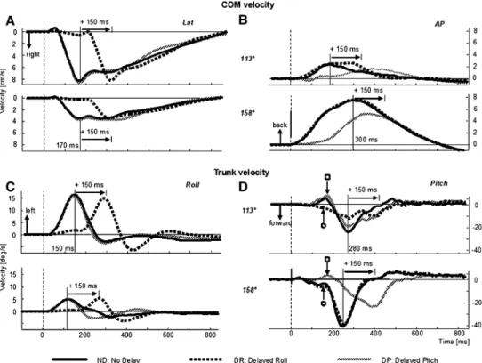
Figure1 shows examples of COM and trunk motion for two stimulus directions. One direction is 158° (lower graphs of Fig.1a–d), a pitch tilt almost purely backwards (toe-up) and the other direction is 113° (upper graphs of Fig.1a–d), a roll tilt almost purely right. Directions are illustrated by centre schema in Fig.3. In Fig.1, the lateral movement of the COM and the roll motion of the trunk were clearly shifted 150 ms with the DR stimulus, for both stimulus directions (Fig.1a, c). For the two directions shown (and for all other directions), the peak in lateral COM was at 170 ms for ND stimuli. This compared to a later peak in AP COM at 300 ms for directions 158° (Fig.1b) and 203° for ND stimuli. Thus the rationale for the 150 ms delay time, forcing roll and pitch trunk peak velocity to occur simultaneously was achieved. For other
Fig. 1 Average velocity plots
for COM velocity (a and b), and trunk velocity (c and d). Lateral and roll plots on the left, ante-rior–posterior and pitch plots on the right. Plots for two directions of platform tilt are shown 113° (right and slightly backwards) and 158° (toe-up and slightly right). Each plot for each of the three delay conditions is the average of eight responses from 15 subjects (120 responses). Stimulus onset is marked by a dotted vertical line. The times of peak velocity of each curve for the no delay (ND) condition is marked by a solid vertical line
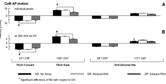
directions, the peak in AP COM was at diVerent times, compared to the time for near pitch (158°/203°) stimuli and was diVerently shifted with direction under the delay condi-tion. For example, as shown in Fig.1b, for the 113° direc-tion of tilt, the peak in AP COM for DR was shifted almost 150 ms (rather than having no shift as seen for 158° in Fig.1b) compared to ND stimuli. DiVerences in timing shifts across directions observed in Fig.1 for AP COM velocities are quantiWed in Fig.2. The time of the AP COM velocity was shifted for DP with respect to ND. For the more pitch directed tilts (23°/338°, 158°/203°), no shift occurred in AP COM velocity peak for DR stimuli as expected (Fig.2a). However, peaks in AP COM velocity were shifted for DR with respect to ND stimuli for the more laterally directed backward tilts (113°, 248°). These changes in AP COM motion with roll stimulus delay indi-cate an interaction between roll and pitch responses depen-dent on stimulus tilt direction.
Despite these changes in timing there were few changes in early vector directions of COM velocities. The polar plots of Fig.3a/b indicate the direction of the COM motion at the two time points, 170 and 300 ms, when these COM velocities have peaks in lateral and AP directions for the ND stimuli in stimulus directions 113° and 158°, respec-tively. If the COM motion is independently controlled in roll and pitch, then Wrst, the delay of the roll component of
the stimulus (DR) by 150 ms should cause the COM motion to be pitch oriented at 170 ms (seen in Fig.3a). Sec-ond, if the pitch component is delayed (DP) 150 ms, then motion should be laterally oriented at 170 ms (Fig.3a). However at 300 ms, when a shift in the vector orientation of COM might have been expected with delayed stimulus components, based on the earlier changes in COM velocity at 170 ms with stimulus delay, only slight diVerences in the vector orientation of COM velocity between DP, ND and DR stimuli were observed (Fig.3b). As this result indicates a compensation for earlier changes in COM velocity, we examined whether AP COM position at 800 ms was altered with stimuli delay. No change was found (P = 0.940). However for the DR stimuli, lateral COM position deviated downhill marginally less than for ND stimuli (P = 0.042). These results suggest that delaying roll and pitch compo-nents of the stimuli had no overall eVect on control of the COM velocity after 300 ms, despite the presence of interac-tion eVects between roll and pitch prior to 300 ms.
Trunk motion: timing and vector directions
Interactions between pitch and roll responses emerged before and after 300 ms for angular motion of the upper trunk, the knee and shoulder joints compared to the linear motion of the COM. At approximately 150 ms, when trunk roll velocity peaked for ND stimuli, DP stimuli revealed that roll component of the stimulus caused backward directed pitch motion of the trunk (marked open square in Fig.1d). This motion was smaller for the backwards DP perturbations, 158° and 203° with small roll components (Fig.1d). The pitch responses revealed with DR stimuli also caused trunk motion with a pitch component at 150 ms (marked open circle in Fig.1d), which was, however oppo-site in direction to that revealed by DP stimuli (see Fig.1d, see traces marked open circle and open square). Moreover, the pitch trunk velocity for roll directed DR stimuli (for example 113° DR traces seen in Fig.1d), appeared to have two peak values, one due to the pitch component of the stimulus at 280 ms (revealed by DR), the other 150 ms later due to pitch induced by the DR stimulus. This interaction in pitch responses was not seen for tilts in the two backward pitch directions (Fig.1d, 158°).
In contrast to COM, the vector directions of trunk velocities at 280 ms for DP and DR stimuli were not aligned with those of the ND stimuli, except for the two backward pitch directions (158° and 203°). For other tilt directions, vector directions of trunk motion were clearly diVerent for DR and DP stimulus at this time point (Fig.3d). The earlier timing of the peak in trunk pitch under ND conditions, the shift of the peak in comparison to ND for DR conditions and the lack of a 150 ms shift for DP stimuli were common characteristics of the more roll
Fig. 2 Times of peak COM AP velocity and trunk pitch velocity
across directions for the three delay conditions. The height of each col-umn represents the mean population value based on each subject’s mean response (average of eight responses) per direction and the ver-tical bars standard errors of the mean (SEM). Responses for directions with the same pitch stimulus component, but oppositely directed roll (e.g. 23° and 338°) have been pooled
oriented stimuli (68°/293°, 113°/248°, see Fig.2b). In comparison, the trunk pitch response for more pitch directed stimuli (23°/338°, 158°/203°) was clearly shifted for DP and not changed for DR conditions (Fig.2b). The diVerences in vector directions for COM at 300 ms and trunk at 280 ms suggests that roll components of the stim-uli induce pitch motion of the trunk, which is not mirrored in COM motion due to compensation at other body seg-ments, for example, the arms, so that by 300 ms for COM velocity (and 800 ms for COM position), no major diVer-ences can be observed in COM motion. The question also arises whether this trunk pitch motion is induced directly on the trunk by the tilt perturbations or is induced on the trunk by earlier movements at other body segments, for example, the knees. In the later case, this action would provide evidence of the CNS planning compensatory pitch responses with a roll command.
Amplitudes of COM and trunk angular velocities
Amplitudes of lateral COM velocity and trunk roll velocity were preserved across delay conditions. It made little diVer-ence if the amplitude was examined at the time of the peaks for each subject for each stimulus direction or if the ampli-tude at the times of the peak in the population average traces was taken (170 and 320 ms for lateral COM velocity and 150 and 300 ms for trunk roll velocity for DP and DR stimuli, respectively). There were no diVerences in the peak
amplitudes across stimulus direction [COM:
F(2,84) = 0.067; P > 0.5; trunk: F(2,87) = 0.021; P > 0.5].
In contrast, delay conditions caused small, but signiW-cant changes in the amplitudes of AP COM. A consistently reduced AP COM velocity with DP stimuli occurred for forwards (23°/338°) and backwards (158°/203°) directed stimuli (Fig.4).
In summary, these results indicate that when the pitch component of the tilt stimulus is delayed, then the timing of the peak velocities of AP COM and trunk pitch are not shifted an amount equal to the delay and amplitudes are not preserved. This eVect occurs preferentially for roll directed tilts, that is, those with a larger roll component than pitch. These eVects contrast with a lack of eVects of stimulus delay on lateral COM and trunk roll responses other than a shift of 150 ms for DR stimuli. This provides evidence that the roll component of balance correcting responses can be programmed by the CNS independently of the pitch response, but not vice versa. The question arises whether the lack of an eVect of the delays on COM motion after 300 ms, despite the clear eVect on trunk motion after 300 ms is due to a compensatory action of the CNS for trunk motion using knee and arm responses.
Knee angular velocities
In comparison to the trunk, Xexion of the knees has the sec-ond-most signiWcant inXuence on lateral and AP motion of
Fig. 3 Vector directions of
COM velocities at 170 (a) and 300 ms (b), and of trunk veloci-ties at 150 (c) and 280 ms (d) when these velocities peak (see vertical lines in Fig.1). For each delay condition, the vector direc-tion of velocity computed from anterior to posterior or pitch and lateral or roll velocities for the COM or trunk, respectively, is shown as a polar plot. The direc-tions of the spokes in the polar plot correspond to the directions of tilt indicated in the middle of the Wgure
the COM (Küng et al. 2009). The uphill knee Xexes and the downhill knee extends (up to approximately 2–3° to maxi-mum extension) for roll tilts of the support surface (Bakker et al. 2006; Allum et al. 2008). InsuYcient Xexion and extension leads to an unstable COM position and loss of balance (Küng et al. 2009). Appropriate correction for a roll and/or forward tilt is based on the diVerence in knee move-ments. Greater Xexion of the uphill knee and extension of the downhill knee provides a greater lateral shift of the COM uphill due to a greater diVerence between the knee movements. On the other hand, knee Xexion will also inXu-ence AP COM motion (Oude-Nijhuis et al. 2007). The greater the sum of the two knee Xexion movements (zero if one knee Xexes an equal amount to the extension of the other), the greater eVect on AP COM motion. Thus, we were interested in learning how delaying the roll and pitch components of the stimulus inXuences knee movements. For this purpose, we examined the diVerence and sum of knee Xexion movements.
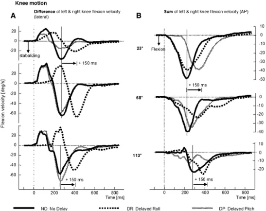
Knee Xexion movements occurred primarily for forward and/or roll tilts. Knee Xexion (both diVerential and summed) was small for backwards tilts (158° and 203°). There were two phases in the diVerence in knee Xexion– extension velocities with the Wrst relative knee extension having a peak at approximately 130 ms (preceding the peak in trunk roll velocity, compare Figs.5a and 1c). This was followed by relative uphill knee Xexion, which peaks at approximately 250 ms (see Table1), as the trunk roll veloc-ity uphill reduced to near zero (compare with trunk roll velocity traces in Fig.1c). Over perturbation directions, the roll velocity directed proWles of knee Xexion appeared to be decoupled from a dependence on the pitch eVect of knee Xexion, because proWle timing was equal for the ND and DP conditions and shifted 150 ms for the DR condition (see Fig.5a; Table1). No changes in the amplitude of diVeren-tial knee Xexion velocity occurred across delay conditions [F(2,81) = 0.224, P > 0.05].
The sum of the left and right knee velocities divided by two is shown for right and forward tilts of the platform in Fig.5b. If knee movements were used to predominantly con-trol the pitch rather than the roll displacement of body, then across directions all traces of the sum of left and right knee velocities should be similar to those for the 23° directed per-turbation. For this direction, the response to DR stimuli has the same proWle as ND stimuli and the response to DP stimuli is delayed 150 ms with respect to ND stimuli. For the pertur-bations with a greater roll than pitch component (see traces for 68° and 113° in Fig.5b), the proWle of the sum of the left and right knee velocity was diVerent. The response to DP was shifted earlier and the response to DR was shifted later (Fig.5b; Table1). These changes were consistent with changes in the amount of trunk pitch velocity present under ND and DR conditions for near roll perturbations.
Arm angular velocities
Arm movements also have a strong inXuence on COM movements (Küng et al. 2009). For lateral tilts, both arms moved laterally downhill, but the amount of abduction and adduction in each arm varies (Küng et al. 2009), hence we considered the diVerence in arm abduction velocities (see Fig.6). For roll and backward pitch tilts, both arms rotate forward with most rotation occurring for backwards tilts (Küng et al. 2009), hence we considered the sum of arm rotation velocities.
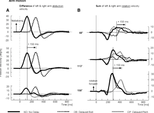
Arm abduction velocities, considered as the diVerence between the left and right arms were shifted exactly 150 ms for DR stimuli across all perturbation directions, with no shifts for DP stimuli (Fig.6 left). The timing of the peaks in arm abduction velocities was identical to those of trunk velocities, occurring at approximately 150 ms, and prior to the peak in the diVerence in knee Xexion velocities (com-pare trunk, arm and knee diVerence traces in Figs.1c, 5a, 6a; Table1).
Fig. 4 Mean amplitudes of
COM AP velocity measured from subjects’ individual mean response peaks (a). Measure-ments at 300 ms (for ND and DR stimuli) and 150 ms later for DP stimuli, times when population mean has a maximum value (b). The layout of the Wgure is identi-cal to that of Fig.2
For arm rotation velocities, a signiWcant diVerence equal to 150 ms in the time of peak velocity was observed in the backward pitch directions (158°/203°) between ND and DP stimuli, with no diVerence for ND and DR stimuli (Fig.6b).
For near roll stimuli (68°/293° and 113°/248°), the forward arm rotation did not follow this pattern, but instead matched the pattern seen for AP directed knee motion (compare responses to 68° and 113° tilts in Fig.6). The time of peak arm rotation (rotation as seen in the transverse plane) velocity shifted earlier in time as the perturbation direction was directed more forwards for DP stimuli with no signiWcant diVerence of the timing being observed for all predominantly roll directed stimuli (68°/293°, 113°/248°— see Table1). For DR stimuli, this peak shifted progres-sively later in time as perturbation direction was directed more forward with the diVerence between DR and ND changing from almost equal for backward stimuli (158° and 203°) to a delay of 150 ms for forward and roll stimuli (68° and 293°). In summary, as Table1 indicates the peak times of arm rotation responses were after those of knee “pitch-inducing” Xexion responses, possibly indicating that the arm responses were a compensation for the eVects of knee Xexion on AP COM velocity.
Ankle torques
Despite the shifts in knee velocity proWles (Fig.6), both roll and pitch ankle torques (summed for the left and right foot) were delayed consistent with these torques being decoupled from one another. Lateral torque magnitudes at the ankle were of the order of 1/20 of the AP torques. Summed AP torques from both ankles were shifted 150 ms by DP stimuli,
Fig. 5 Mean population traces
of the diVerence in right and left knee Xexion movements (a) leading to a lateral stabilisation of the body and sum of traces of the right and left knee Xexion (b) leading to trunk pitch. Respons-es for the three delay conditions and three directions of right tilt are shown. Knee responses for the direction 158° are small. The layout of the Wgure is identical to Fig.1 with peak responses marked by a solid vertical line
Table 1 Times of peak responses in knee and arm velocities
Knees: diVerences of left and right, mean (§SE)
23°/338° 68°/293° 113°/248°
ND 277.55 (§11.09) 270.51 (§12.41) 256.80 (§18.85) DR 322.00 (§19.28) 416.25 (§13.78) 394.68 (§16.30) DP 316.56 (§22.21) 257.51 (§17.44) 251.19 (§19.21) Knees: sum of left and right, mean (§SE)
23°/338° 68°/293° 113°/248°
ND 253.13 (§26.76) 231.08 (§12.27) 266.49 (§19.92) DR 256.62 (§16.66) 318.73 (§24.74) 361.11 (§17.14) DP 343.11 (§25.11) 278 68 (§19.11) 236.30 (§17.98) Arm: sum of the left and right, mean (§SE)
68°/293° 113°/248° 158°/203° ND 326.86 (§15.27) 295.43 (§10.91) 293.81 (§6.82) DR 452.01 (§14.48) 399.46 (§19.57) 323.07 (§24.90) DP 320.36 (§16.82) 342.03 (§14.07) 435.76 (§17.27) Arm: diVerence of the left and right, mean (§SE)
68°/293° 113°/248° 158°/203° ND 145.74 (§4.63) 138.78 (§2.43) 163.74 (§13.93) DR 293.19 (§1.86) 292.61 (§2.10) 339.05 (§29.71) DP 140.51 (§1.77) 143.72 (§4.25) 153.87 (§17.54)
but not at all by DR stimuli. Consider, for example, the AP ankle torque proWles for near roll stimuli (68°/293°, 113°/ 248°). The uphill and downhill ankle torques were changed by the DR and DP stimuli in one leg, however the torque responses of the contralateral legs had completely opposite polarities. Thus despite these changes at each leg, the eVect of the DR stimulus on combined ankle torques was minor even when ankle torque changed rapidly over the balance correcting interval we analysed (140–290 ms). Across all directions combined ankle torque was pitch oriented for ND stimuli and remained so for DR stimuli. The DP stimuli shifted ankle torque proWles 150 ms with unchanged ampli-tudes.
EMG responses
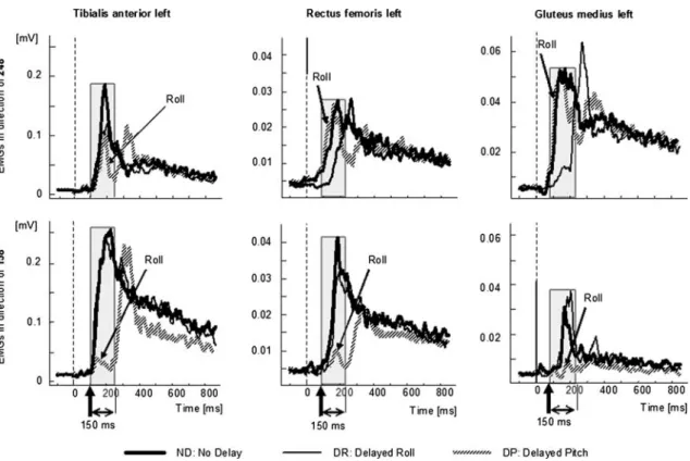
All muscles we examined had responses with varying sen-sitivities to the pitch and roll components of the stimulus. Figure7 provides three examples of these diVerences to delay stimulus for the tilt directions 158° (backwards) and 248° (sideways). Tibialis anterior had a pitch sensitivity as its maximal response (158° in Fig.7) was signiWcantly shifted 150 ms for DP stimuli, but not by DR stimuli. Glu-teus medius has a roll sensitivity as its maximum response (see traces for 248° in Fig.7) was shifted by DR, but not by DP stimuli. However, note that both these muscles were somewhat diVerently aVected by stimulus delay for direc-tions of tilt not eliciting the maximum response amplitude suggesting, for example, tibialis anterior had a roll sensitiv-ity for the 248° direction and gluteus medius had a pitch
sensitivity for the 158° direction, albeit weak. Responses of an intermediate muscle, rectus femoris with both pitch and roll sensitivity is illustrated by the middle traces of Fig.7.
To characterise the directional sensitivity of muscle responses, Fig.8 plots the response amplitudes measured as the area under the curve for the Wrst 150 ms after response onset for diVerent directions of tilt under the three delay conditions. Each spoke of these polar plots represents the amplitude for a direction of tilt. It is clear from Fig.8 that tibialis anterior shows little diVerence in response sensitiv-ity for ND and DR stimuli across directions reinforcing its classiWcation as a pitch directed muscle. Likewise, for glu-teus medius, there was little diVerence between ND and DP stimuli justifying its classiWcation as a roll directed muscle, as supported by the direction of its maximum responsive-ness for the ND condition (see arrows on polar plots). Nonetheless, tibialis anterior had a small roll, and gluteus medius had a small pitch sensitivity as indicated by the areas of the polar plots of responses to DP and DR stimuli, respectively, and the arrows indicating the direction of maximum sensitivity under these delay conditions. The polar plot of rectus femoris in Fig.8 provides an intermedi-ate picture. For this muscle, the area circumscribed by the polar plot is approximately the same size for DP and DR responses, both of which are less than the area of the plot for ND responses.
The area circumscribed in the polar plots, such as those of Fig.8 was used to categorize the muscle response types as shown in Fig.9. Thus, pitch sensitivity was deWned by the area circumscribed in the polar plot for DR stimuli
Fig. 6 Mean population traces
of the diVerence in right and left arm abduction movements (a), which precede a peak in trunk roll and traces of the sum of right and left forward rotation move-ments (b), which follow the sum of knee Xexion movements. Traces are shown for three delay conditions and three directions of right tilt. Arm responses for the direction 23° are small. The layout of the Wgure is similar to that of Figs.1 and 5
compared to the area for ND stimuli and that roll sensitivity by DP stimuli, compared to ND stimuli. If pitch sensitivity was signiWcantly greater than the roll sensitivity for a mus-cle, we termed this as a pitch sensitivity muscle and vice
versa, a roll sensitivity muscle. When the two sensitivities had less than a 15% diVerence in pitch and roll sensitivity, we termed the muscles as mixed sensitivity muscles. Figure9 shows how the various muscles we recorded were
Fig. 7 Mean population traces of EMG activity in tibialis anterior (left
set of traces), rectus femoris (middle set of traces) and gluteus medius (right set of traces). The upper row of traces is in response to a tilt in
the 248° direction, the lower set of traces for a 158° tilt. The onsets of the responses are marked by vertical arrows and the 150 ms averaging interval by a grey box
Fig. 8 Polar plots of muscle
activity over the 150 ms averag-ing intervals shown in Fig.9. The layout of the polar plots is identical to that shown in Fig.3. Each spoke represents the direc-tion of tilt and radial distance from the plot centre along the spoke the amplitude of the EMG response area according to the scales next to the polar plots. The amplitudes along the spokes are then joined and the direction of the centroid of the Wgure deW-nes the direction of maximum response sensitivity as indicated by the arrow. Note the diVerent directions of maximum sensitiv-ity for the three stimulus delay conditions for the three muscles
grouped. The ankle joint muscles, except for peroneus were classiWed as pitch muscles. The trunk muscles gluteus medius and paraspinals were classiWed as roll muscles. In contrast, knee (including peroneus) and arm muscles were classiWed as mixed muscles.
Discussion
The results of this work add further evidence to the concept that balance corrections in the roll and pitch directions are executed separately by neural command centres. If this command control is managed within the same neural centre or within closely connected, but diVerent neural centres for pitch and roll cannot be determined on the basis of this study. We have been able to extend the work of Grüneberg et al. (2005) for DR stimuli by demonstrating that a delay in the roll component of the stimulus is transmitted faithfully to occur in knee Xexion and arm abduction responses at the later time, regardless of the stimulus direction. In the sense that neither the stimulus direction nor the presence or not of a pitch component to the tilt stimulus inXuenced roll responses, this observation can be interpreted as a control by the CNS of roll decoupled from that of pitch. The Wnd-ings of Grüneberg et al. (2005), who used diVerent times of DR stimuli, plus earlier work on the biomechanical responses to combined roll and pitch tilts without delays (Allum et al. 2003, 2008; Carpenter et al. 1999), led us to develop the hypothesis that pitch motion would be
controlled independently of roll motion too, and that this pitch motion would occur mainly about the ankle and knee joints (Allum et al. 2003; Grüneberg et al. 2005). Accord-ing to this concept, there would be little interaction between the two forms of control. Our current results as well as recent work in vestibular loss subjects (Allum et al. 2008) indicates that pitch control is dependent on roll, with the dependence increasing as the size of the roll component of the stimulus increases. Thus it appears that CNS is not able to program pitch control independently from roll.
When the pitch component of the stimulus was delayed, trunk, knee and arm movements were not transposed faith-fully in time by the amount of the delay. Rather the amount of delay in the response proWle and its amplitude depended on the stimulus direction (see Fig.1, 2, 3, 5, 6). Changes in pitch movements prior to 120 ms in response to roll directed tilts as shown in right sets of traces in Figs.1, 5 and 6 are probably due to a purely biomechanical pitch response of the body to roll component of the stimulus. Carpenter et al. (1999) also noted an early pitch response of the trunk following pure roll tilts. An early eVect on roll responses in response to the pitch component of the stimu-lus was not observed in this study and not by Carpenter et al. (1999) for pure pitch tilts. The presence of these early biomechanical changes in pitch for the roll component of the stimulus implies the CNS must take these pitch changes into account when planning the response to the roll pertur-bation. Most of the later changes in pitch kinematics due to balance corrections could be traced to diVerences in knee
Fig. 9 ClassiWcation of muscle sensitivity based on response areas in the polar plots under the two delay conditions, DR and DP compared to the
Xexion movements between the left and right knees across stimulus directions. The Xexion of the uphill knee and exclusion of the downhill knee resisted lateral shift of COM downhill by holding the trunk tilted uphill. If not resisted suYciently, as in, for example, vestibular loss and spinal cerebellar ataxia (SCA) patients with insuYcient Xexion of the uphill knee and extension of the downhill knee an unstable lateral motion of the COM resulted (Allum et al. 2008; Küng et al. 2009). Because the diVerential knee action is insuYcient in these patients, a reversal of trunk motion downhill occurs. The marked instability in SCA subjects following roll tilts and the inability to program pitch responses to forward tilt adequately (Bakker et al. 2006) would suggest that centres responsible for executing the separate roll and pitch commands, postulated on the basis of the current results, may lie in the vestibulo cerebel-lum.
It is interesting to speculate whether our results could be explained as an inability of the CNS to generate roll correc-tions without the use of knee motion, which also induces motion in the pitch plane. It appears that the CNS has other choices in programming roll corrections. For example, when knees Xexion was blocked artiWcially, subjects suc-cessfully corrected for roll perturbations using greater than normal arm movements at expense of greater COM motion (Oude-Nijhuis et al. 2008). Thus diVerential knee Xexion may also help control the amount of COM pitch directed motion.
Our analysis of EMG responses under diVerent stimulus delay conditions indicates, as described before, that ankle muscles predominantly control pitch and trunk muscles roll motion of the body (Grüneberg et al. 2005). Our new Wnd-ings are that arm and knee muscle responses act on both roll and pitch motion of the body as these are equally sensitive to roll and pitch plane components of the stimulus. We assume this action is the primary manner in which the CNS maintains the controlled variable, presumably COM veloc-ity, at a minimum.
The key contribution of knee movements to controlling roll and pitch motion of the body contrasts with the low sensitivity of early passive knee movements to stimulus direction (Allum et al. 2008). This may have the advantage that the knee muscles help control body motion with eVer-ent signals enhancing later proprioceptive feedback in a feedforward manner without early proprioceptive knee responses contributing signiWcantly to the sensory signals initiating and modulating balance corrections. A similar function may be exercised by arm muscles in which stretch reXexes relating to tilt stimuli have not been observed (Allum et al. 2002).
It is interesting to speculate why balance control centres coordinate roll balance corrections, as if these were totally decoupled from those of pitch, but not vice versa. One
reason could be simply biomechanical in that roll move-ment of the trunk occurs earlier than that of pitch following a combined roll and pitch tilt of the support surface (Allum et al. 2002; Carpenter et al. 1999). Furthermore, complete proprioceptive and vestibular information on the stimulus roll characteristics appears to reach the CNS prior to the arrival of pitch directed information (Allum et al. 2008). Thus from timing considerations alone, the CNS may need to carry out the necessary programming and release of the response to roll tilt prior to that for the pitch tilt.
This report presented data on eight directions of tilt each with a roll and pitch component. For completeness, it would have been advisable to have the pure pitch and roll directions as well as 45° directions with equal components of pitch and roll for the three delay conditions. Data for the pure pitch and roll directions are available in prior publica-tions (Allum et al. 2008; Bakker et al. 2006). Also we have no reason to believe that responses from the directions 45, 135, 225 and 315 in our nomenclature could not be pre-dicted from the current results either side of these directions that is from the current 23, 63, 113, 158, 203, 248, 293 and 338 responses. An expansion of our protocol on the same subjects would probably have been too tiring for them.
These results add further evidence to the diVerences between the control strategies in centres generating balance correcting responses for bipedal and quadrupedal stance (Allum et al. 2008). Some authors speculated on the simi-larities based on pitch plane responses (Dunbar et al. 1986; Horak and Macpherson 1996). Roll responses are funda-mentally diVerent between bipedal and quadrupedal stance. Firstly, in humans, the motion of the upper trunk is in the opposite direction to that of the pelvis on roll tilt (Allum et al. 2002, 2008). This is a completely diVerent biome-chanical response from that of quadrupeds, where trunk and pelvis move in the same direction as the roll tilt of the sup-port surface (Macpherson et al. 2007). The movement of the trunk on the pelvis provides a completely diVerent bio-mechanical situation in biped stance. In quadrupeds, the uphill knee must Xex in order to shift the body laterally uphill, whereas in bipeds the uphill knee must Xex in order to hold the trunk in the uphill position compensating for the lateral shift of the pelvis downhill. The current research indicates that the functional pitch plane eVect of the knee action in bipeds is presumably not present in quadrupeds due to stance on four legs. We presume that the presence of a diVerent knee action plus simultaneous forelimb action in quadrupeds leads to a diVerent and possibly reduced pitch motion during roll balance control in quadrupeds. Nonethe-less, it would be interesting to explore changes in balance corrections for roll tilt when humans are asked to respond to roll tilt in a quadrupedal position in order to determine if the roll responses can be programmed separately from pitch as indicated here.
In the sagittal plane, increasing use of knee movements was seen, dependent on stimulus velocity (Runge et al. 1999). These results provide a conceptional focus diVerent from the notion that the degrees of freedom are reduced in balance control (Bernstein 1967; Horak and Nashner 1986). While this concept might hold for backward tilts, in the roll plane, the control of the degrees of freedom is quite com-plex as the knees are controlled independently as are the arms leading to a cross-coupling eVect of motion in the pitch plane. The essential question we have tried to address is whether the CNS programs the roll and pitch movement of body independently. The very fact that either the pitch or the roll component of the stimulus could be delayed 150 ms, yet at the completion of the balance correction the overall eVect on COM motion was identical to the eVect with no delay, would suggest that the CNS programs the roll and pitch motion independently, but that both responses interact in a linear manner biomechanically. Interestingly, when individual segments were examined, it was very apparent that the underlying segment motions in the roll and pitch planes were not independent of one another. SpeciWcally this is very apparent from knee muscle responses, which have almost equal pitch and roll plane sensitivities. The delay of knee Xexion movements with respect to trunk roll, and arm movements with respect to the knees, indicates that these movements are not a biome-chanical response to the tilt stimulus, rather a compensating balance correction acting to stabilise early trunk motion.
We have assumed here that biomechanical responses prior to 120 ms, interact linearly and that the only eVect of the delay was to shift the early biomechanical responses of 150 ms. For our 7.5° support surface tilts, this appears to be a valid approximation, as segment angle changes are maxi-mally 6° (Allum et al. 2003). Amplitudes of pitch responses were generally not altered. Roll amplitudes were unchanged with delay conditions. For larger amplitude tilts, this may not be a valid approximation. The amplitude of tilt for which the eVect of the delay leads to a fundamentally diVerent response needs to be investigated, possibly with modelling techniques. The lack of such an investigation limits the application of our Wndings to tilt amplitudes greater than those we investigated.
Although there was a clear interaction between the pitch and roll motion of the trunk, knees and arms induced by tilt of the support surface, delaying either the pitch or roll com-ponent did not inXuence the overall COM velocity response of the body after 300 ms. We had not expected that changes we observed with stimulus delays to pitch would lead to interactions at this level and be compensated in COM responses. Again this reinforces our conclusion that the CNS can program balance corrections in the pitch and roll planes independently of one another, even if interactions exist between the two planes at the level of the arms and
knees. Interestingly the form interactions took implied that roll control is programmed Wrst and the pitch control must take into account previously occurring eVects on pitch due to roll commands. In this sense, pitch control is not inde-pendent of roll. The question arises within this context as whether the roll Wrst action represents a preferred plane of action. Preferred planes of action for head–neck move-ments has been suggested as a technique to simplify sen-sory–motor transformations serving motor control and a way to minimize neural operations (Graf et al. 1995).
Acknowledgments This project was supported by a grant from the Swiss National Research Foundation (No. 32000-117950) to J.H.J. Allum.
ConXict of interest statement The authors declare that they have no
conXict of interest, Wnancial or otherwise, related to the submitted manuscript or the associated research.
References
Allum JHJ, Carpenter MG, Honegger F, Adkin AL, Bloem BR (2002) Age-dependent variations in the directional sensitivity of balance corrections and compensatory arm movements in man. J Physiol 542:643–663
Allum JHJ, Carpenter MG, Honegger F (2003) Directional aspects of balance corrections in man. Employing multidirectional perturba-tions to better understand dynamic postural control in normal and balance-deWcient populations. IEEE Eng Med Biol Mag 22:37–47 Allum JHJ, Oude Nijhuis LB, Carpenter MG (2008) DiVerences in coding provided by proprioceptive and vestibular sensory signals may contribute to lateral instability in vestibular loss subjects. Exp Brain Res 184:391–410
Bakker M, Allum JHJ, Vissser JE, Grüneberg C, van de Warrenburg BPC, Kremer BH, Bloem BR (2006) Postural responses to multi-directional stance perturbations in cerebellar ataxia. Exp Neurol 202:21–35
Bernstein N (1967) The co-ordination and regulation of movements. Pergamon, London
Carpenter MG, Allum JHJ, Honegger F (1999) Directional sensitivity of stretch reXexes and balance corrections for normal subjects in the roll and pitch planes. Exp Brain Res 129:93–113
Carpenter MG, Allum JHJ, Honegger F (2001) Vestibular inXuences on human postural control in combinations of pitch and roll planes reveal diVerences in spatiotemporal processing. Exp Brain Res 140:95–111
Dunbar DC, Horak FB, Macpherson JM, Rushmer DS (1986) Neural control of quadrupedal and bipedal stance: implications for the evolution of erect posture. Am J Phys Anthropol 69:93–105 Graf W, de Waele C, Vidal PP, Wang DH, Evinger C (1995) The
ori-entation of the cervical vertebral column in unrestrained awake animals. II. Movement strategies. Brain Behav Evol 45:209–231 Grin L, Frank J, Allum JHJ (2007) The eVect of voluntary arm
abduc-tion on balance recovery following multidirecabduc-tional stance pertur-bations. Exp Brain Res 178:62–78
Grüneberg C, Duysens J, Honegger F, Allum JHJ (2005) Spatio-tem-poral separation of roll and pitch balance-correcting commands in humans. J Neurophysiol 94:3143–3158
Henry SM, Fung J, Horak FB (1998a) EMG responses to maintain stance during multidirectional surface translations. J Neurophysiol 80:1939–1950
Henry SM, Fung J, Horak FB (1998b) Control of stance during lateral and anterior/posterior surface translations. IEEE Trans Rehabil Eng 6:32–42
Horak FB, Macpherson JM (1996) Postural orientation and equilib-rium. In: Rowell LB, Shepherd JT (eds) Handbook of physiology. Exercise: regulation and integration of multiple systems, sect. 12. Oxford University Press, New York, pp 255–292
Horak FB, Nashner LM (1986) Central programming of postural movements: adaptation to altered support-surface conWgurations. J Neurophysiol 55:1369–1381
Jones SL, Henry SM, Raasch CL, Hitt JR, Burn JY (2008) Responses to multi-directional surface translations involve redistribution of proximal versus distal strategies to maintain upright posture. Exp Brain Res 187:407–417
Keshner EA, Allum JH, Pfaltz CR (1987) Postural coactivation and adaptation in the sway stabilizing responses of normals and patients with bilateral vestibular deWcit. Exp Brain Res 69:77–92 Küng UM, Horlings CGC, Honegger F, Kremer HPH, Bloem BR, van de Warrenburg BPC, Allum JHJ (2009) Postural instability in cer-ebellar ataxia: correlactions of knee, arm and trunk movements to center of mass velocity. Neuroscience 159:390–404
Macpherson JM, Everaert DG, Stapley PJ, Ting LH (2007) Bilateral vestibular loss in cats leads to active destabilization of balance during pitch and roll rotations of the support surface. J Neuro-physiol 97:4357–4367
Matjacic Z, Voigt M, Popovic D, Sinkjaer T (2001) Functional postural responses after perturbations in multiple directions in a standing man: a principle of decoupled control. J Biomech 34:187–196
Oude-Nijhuis L, Bloem BR, Munneke M, Honegger F, Allum JHJ (2007) Incorporating voluntary knee Xexion into nonanticipatory balance corrections. J Neurophysiol 98:3047–3057
Oude-Nijhuis L, Hegeman J, Bakker M, Van Meel M, Bloem BR, Allum JHJ (2008) The inXuence of knee rigidity on balance corrections: a comparison with responses of cerebellar ataxia patients. Exp Brain Res 187:181–191
Park S, Horak FB, Kuo AD (2004) Postural feedback responses scale with biomechanical constraints in human standing. Exp Brain Res 154:417–427
Runge CF, Shupert CL, Horak FB, Zajac FE (1999) Ankle and hip pos-tural strategies deWned by joint torques. Gait Posture 10:161–170 Ting LH, Macpherson JM (2004) Ratio of shear to load ground-reac-tion force may underlie the direcground-reac-tional tuning of the automatic postural response to rotation and translation. J Neurophysiol 92:808–823
Torres-Oviedo G, Macpherson JM, Ting LH (2006) Muscle synergy organization is robust across a variety of postural perturbations. J Neurophysiol 96:1530–1546
Visser JE, Allum JHJ, Esselink RA, Speelman JD, Borm GF, Bloem BR (2008) Subthalamic nucleus stimulation and levodopa-resistant postural instability in Parkinson’s disease. J Neurol 255:205–210 Winter DA, Prince F, Frank JS, Powell C, Zabjek KF (1996) UniWed
theory regarding A/P and M/L balance in quiet stance. J Neuro-physiol 75:2334–2343
Winter DA, Patla AE, Ishac M, Gage WH (2003) Motor mechanisms of balance during quiet standing. J Electromyogr Kinesiol 13:49– 56