Tumours of the thymus: a cohort study of prognostic factors from
the European Society of Thoracic Surgeons database
Enrico Ruffini
a,*, Frank Detterbeck
b, Dirk Van Raemdonck
c, Gaetano Rocco
d, Pascal Thomas
e, Walter Weder
f,
Alessandro Brunelli
g, Andrea Evangelista
hand Federico Venuta
ion behalf of the European Association of
Thoracic Surgeons (ESTS) Thymic Working Group
a
Department of Surgery, Section of Thoracic Surgery, University of Torino, Torino, Italy
b Department of Surgery, Section of Thoracic Surgery, Yale University, New Haven, CT, USA c
Department of Thoracic Surgery, University Hospitals Leuven, Leuven, Belgium
d Department of Thoracic Surgery, National Cancer Institute, Pascale Foundation, Naples, Italy e
Department of Thoracic Surgery, Aix-Marseille University, Marseille, France
f Department of Thoracic Surgery, University Hospital, Zurich, Switzerland g Division of Thoracic Surgery, St. Jame’s University Hospital, Leeds, UK
h Unit of Clinical Epidemiology, Azienda Ospedaliera Città della Salute e della Scienza di Torino, Torino, Italy i
Department of Thoracic Surgery, University of Rome SAPIENZA; Policlinico Umberto I; Fondazione Eleonora Lorilard Spencer Cenci, Rome, Italy * Corresponding author. Division of Thoracic Surgery, University of Torino, Via Genova 3, 10126 Torino, Italy. Tel: +39-011-6705380; fax: +39-011-6705365;
e-mail: enrico.ruffi[email protected] (E. Ruffini).
Received 25 November 2013; accepted 11 December 2013
Abstract
OBJECTIVES: A retrospective database was developed by the European Society of Thoracic Surgeons, collecting patients submitted to surgery for thymic tumours to analyse clinico-pathological prognostic predictors.
METHODS: A total of 2151 incident cases from 35 institutions were collected from 1990 to 2010. Clinical-pathological characteristics were analysed, including age, gender, associated myasthenia gravis stage (Masaoka), World Health Organization histology, type of thymic tumour [thymoma, thymic carcinoma (TC), neuroendocrine thymic tumour (NETT)], type of resection (complete/incomplete), tumour size, adjuvant therapy and recurrence. Primary outcome was overall survival (OS); secondary outcomes were the proportion of incomplete resections, disease-free survival and the cumulative incidence of recurrence (CIR).
RESULTS: A total of 2030 patients were analysed for OS (1798 thymomas, 191 TCs and 41 NETTs). Ten-year OS was 0.73 (95% confidence interval 0.69–0.75). Complete resection (R0) was achieved in 88% of the patients. Ten-year CIR was 0.12 (0.10–0.15). Predictors of shorter OS were increased age (P < 0–001), stage [III vs I HR 2.66, 1.80–3.92; IV vs I hazard ratio (HR) 4.41, 2.67–7.26], TC (HR 2.39, 1.68–3.40) and NETT (HR 2.59, 1.35–4.99) vs thymomas and incomplete resection (HR 1.74, 1.18–2.57). Risk of recurrence increased with tumour size (P = 0.003), stage (III vs I HR 5.67, 2.80–11.45; IV vs I HR 13.08, 5.70–30.03) and NETT (HR 7.18, 3.48–14.82). Analysis using a propensity score indicates that the administration of adjuvant therapy was beneficial in increasing OS (HR 0.69, 0.49–0.97) in R0 resections.
CONCLUSIONS: Masaoka stages III–IV, incomplete resection and non-thymoma histology showed a significant impact in increasing recur-rence and in worsening survival. The administration of adjuvant therapy after complete resection is associated with improved survival. Keywords:Thymoma• Thymic carcinoma • Myasthenia gravis • Neuroendocrine thymic tumours • Staging • Surgery
INTRODUCTION
Thymic malignancies are uncommon tumours, with an estimated incidence of 2.5–3.2/106people. Thymomas, thymic carcinomas (TC) and neuroendocrine thymic tumours (NETT) are the three most important histological categories.
The rarity of thymic tumours has limited so far the possibility to set homogeneous management protocols as evidenced by a recent survey from the European Society of Thoracic Surgeons (ESTS) [1]. For this reason, the identification of prognostic predic-tors is of utmost importance to guide the clinician to the most
appropriate therapeutic treatment in these rare tumours. A recent review [2] of the available literature evaluating studies reporting multivariable analyses of prognostic factors confirmed the limitations in our current knowledge about prognostic factors in thymic tumours.
Collaborative retrospective databases offer the opportunity to collect a large series of patients in a relatively short time period, and for this reason, they are helpful in rare diseases such as thymic malignancies.
The ESTS retrospective database project was launched in 2011 among ESTS members to collect data of patients submitted to
© The Author 2014. Published by Oxford University Press on behalf of the European Association for Cardio-Thoracic Surgery. All rights reserved.
THORA
C
IC
Follow-up data collection was closed in December 2011. The aim of the present study was to investigate, using the largest database of thymic malignancies ever collected, several clinical-pathological prognostic predictors of incomplete resection, survival and recur-rence that have been previously tested with conflicting results in smaller observational studies.
MATERIALS AND METHODS
ESTS is a thoracic surgical organization open to European and non-European members around the world. An enquiry was sent to all ESTS members to ask for participation to the thymic data-base project and to send their data. Thirty-five institutions responded and joined the project: 27 from Europe, 3 from Asia and 5 from USA/Canada. Institutional Review Board approval was obtained at each institution.
The datafields included demographics, presence of myasthenia gravis (MG), histology [2004 World Health Organization (WHO) classification [3]], tumour size (continuous), stage according to Masaoka [4], completeness of resection, administration of induction or adjuvant treatment, type of surgical procedure, data for survival analysis, cause of death, recurrence, year of surgery and mean number of patients provided by the centres/year (≤4, 5–9, ≥10). Patients operated on or before 2004 were reclassified at each centre using the latest WHO histological classification [3]. In-formation about the site of recurrence was not sufficient to include this covariate in the predictor analysis.
Overall, data on 2244 patients were collected. Of these, 58 patients with not–otherwise-specified lesions, 19 with undeter-mined tumours and 16 with insufficient data were excluded, and the remaining 2151 patients form the basis of our report.
Study outcomes
Primary outcome was overall survival (OS) calculated from the date of surgery to the date of death from any cause. Secondary outcomes were the proportion of incomplete resections (micro-scopically or macro(micro-scopically), the disease-free survival (DFS) cal-culated from the date of surgery to the date of recurrence or death from any cause, and the cumulative incidence of recurrence (CIR) calculated from the date of surgery to the date of recurrence. In order to rule out a potential bias between patients with and without information about recurrence (missing recurrence date or recurrence status) out of the total of R0 resections, we compared OS curves of the two groups. The difference between the curves was not significant [HR 1.19 95% confidence interval (CI) 0.88– 1.60;P = 0.25]. We therefore assumed that no significant bias exists between the two populations.
Statistical analysis
OS and DFS were estimated by the Kaplan–Meier product-limit method. CIR was estimated considering death from any cause as a competing event. To account for the heterogeneity across centres, factors associated with OS and DFS were investigated using Cox proportional hazard models with shared frailty. CIR was analysed with proportional hazard frailty models for the subdistribution [5] including the same variables considered for the OS analysis. Factors associated to incomplete resection were investigated
centre as random effect.
The effect of adjuvant therapy on OS was investigated in patients after R0 resection and by subgroups. In order to adjust this non-randomized comparison and to reduce the loss of power in the subgroup analyses, a propensity score for the likelihood of receiving adjuvant therapy was calculated from eight covariates: age, gender, stage, tumour size, histology, MG, year of intervention and mean annual number of resections. Multivariable Cox proportional hazard models with shared frailty (for centre heterogeneity) were estimated including as predictor the adjuvant therapy along with the propensity score. Effect modifications by subgroups were evalu-ated by including in the models an interaction term between the covariate indicating the adjuvant therapy and the subgroup covari-ate of interest, adjusting for propensity score.
In all modelsfitted in this study, missing data were multiple imputed using the method of chained equations [6]. Combined estimates were obtained fromfive imputed datasets.
The statistical analysis was performed using STATA (version 11.1) (ice command for multiple imputation) and R (version 2.15.1) (cmprsk and crrSC packages for the competing event analyses).
RESULTS
The median number of patients submitted by each institution was 45 [interquartile range (IQR) 24–73, range 8–257]; 20 institutions (57%) reported <60 cases, 8 (23%) reported 60–99 cases and 7 (20%) reported at least 100 cases. The majority of institutions per-formed a follow-up schedule based on a 3- to 6-month computed tomography (CT) scan for thefirst 3 years, followed by annual CT scan lifelong. In most centres, more aggressive thymic tumours (TC and NETT) received a more strict imaging (CT) surveillance. The median follow-up time of surviving patients was 48 months (IQR 21–90). Fixed at 31 December 2011, the end of the follow-up data collection, the completeness of follow-up for the study was 73%.
Study
flow of the patient population and patient
characteristics
Figure1shows the studyflow of the 2151 patients in the database; 2030 had sufficient information for OS analysis and 1325 for DFS and CIR analysis.
Table1shows the patient characteristics and the number and percentages of missing information for the 2030 patients with suf-ficient information for OS analysis. Of these, there were 1798 thymomas, 191 TCs and 41 NETTs.
A complete resection was achieved in 1709 patients (88%). The resectability rates were 99, 92, 78 and 53% for stages I, II, III and IV, respectively. The rates were 97, 97, 91, 85, 77, 72 and 69% for types A, AB, B1, B2, B3, TC and NETT
Induction therapy (mostly chemotherapy, 71%) was adminis-tered in 239 patients, of whom 186 (80%) were at stage III/IV; ad-juvant therapy was administered in 853 patients and consisted of radiotherapy (n = 566), chemotherapy (n = 44) and combined chemo/radiotherapy (n = 243).
Frequency of study end points
Three hundred and twenty-four patients died during the follow-up. Ninety patients had recurrence out of 1325 R0 patients, with
complete information about the recurrence status. Two hundred and fourteen patients had either recurrence or died out of the 1325 patients evaluated for DFS and CIR analysis.
Predictors of incomplete resection
In 1709 patients with complete resection, recurrence occurred in 141 cases (8%); it increased with stage (stages I 3%, II 4%, III 22%, IV 40%) and histology (A 3%, AB 5%, B1 8%, B2 11%, B3 14%, TC 30%, NETT 37%).
Among the different examined clinico-pathological variables, the probability of an incomplete resection is higher in male patients (adjOR 1.61, 95% CI 1.18–2.20, P = 0.003); it increases with tumour size (adjOR 1.10 [ per 1 cm increase], 95% CI 1.01–1.21, P = 0.031), while it decreases in the presence of MG (adjOR 0.55, 95% CI 0.37–0.81, P = 0.002). With respect to A-AB-B1 thymoma, the probability of an incomplete resection is higher in B2-B3 thymoma (adjOR 5.91, 95% CI 4.02–8.70, P < 0.001), TC (adjOR 10.46, 95% CI 6.22–17.59, P < 0.001) and NETT (adjOR 7.77, 95% CI 3.45–17.51, P < 0.001).
Overall survival and disease-free survival analyses
Figure2reports the overall time-to-event curves for the different end points (OS, DFS and CIR). Five- and 10-year OS rates were 0.85 (95% CI 0.83–0.86) and 0.73 (95% CI 0.69–0.75), and DFS rates were 0.84 (95% CI 0.82–0.87) and 0.70 (95% CI 0.65–0.74). Thirty-day mortality was 1% (21 cases).Table2shows the analysis of OS predictors. The results of the DFS analysis paralleled those of OS analysis and were not reported. The risk of mortality increases with age [adjHR ( per 5 year increase) 1.19, 95% CI 1.14–1.24, P < 0.001] and with stage (III vs I adjHR 2.66, 95% CI 1.80–3.92, P < 0.001; IV vs I adjHR 4.41, 95% CI 2.67–7.26, P < 0.001). The mortality risk is also higher for TC (adjHR 2.39, 95% CI 1.68–3.40, P < 0.001), NETT (adjHR 2.59,
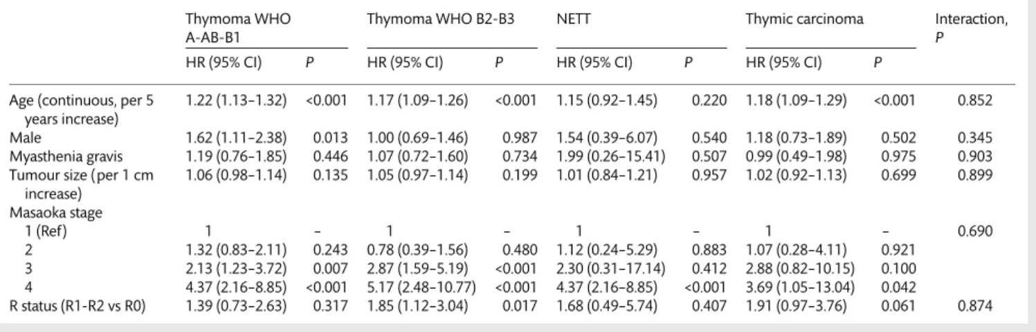
95% CI 1.35–4.99, P = 0.004) and after incomplete resection (adjHR 1.74, 95% CI 1.18–2.57, P = 0.007); a weak trend towards an increased risk was found in males (adjHR 1.25, 95% CI 0.99–1.58, P = 0.06). No evidence of a major effect modification by histology categories (thymoma, TC and NETT) was found for the evaluated prognostic factors (Table3).
Recurrence analysis
Cumulative incidence of recurrence was 0.05 (95% CI 0.04–0.07), 0.08 (95% CI 0.06–0.09) and 0.12 (95% CI 0.10–0.15) at 3, 5 and 10 years, respectively (Fig.2).
Significant predictors of higher risk of recurrence included (Table4) young age, non-MG status, increased tumour size, stages III and IV, NETT tumours and the most recent years of intervention (using as reference the earliest period, 1990–1995).
Adjuvant therapy
Radiation doses and chemotherapeutic regimens for adjuvant treatments varied among the centres. In general, a total dose of 40–60 Gy was employed for radiotherapy, while cisplatin-based regimens (mostly cisplatin/doxorubicin/cyclophosphamide-CAP, or cisplatin/doxorubicin/cyclophosphamide/vincristine-ADOC) were uniformly used. The analysis was undertaken on the subgroup of patients receiving adjuvant therapy after R0 resection. Complete information was available in 1662 patients out of 1709 R0 resec-tions (Fig.1). Adjuvant therapy was more frequently administered in younger patients (median age 54 vs 57) and in advanced stages (stage I 75/646, 12%; II 316/612, 52%; III 217/295, 74%; IV 56/91, 62%); the administration was also different according to histology (A-AB-B1 256/920, 28%; B2-B3 326/599, 54%; TC 78/116, 67%; NETT 16/27, 59%) (Table5). The subgroup analysis was undertaken using a Cox model with shared frailty adjusted for propensity score. Results are reported in Fig.3as forest plot. The overall effect
Figure 1:Studyflow diagram of the patient population for analysis of predictors. THORA
C
of adjuvant therapy on OS was significantly beneficial (HR 0.69, 95% CI 0.49–0.97). No strong evidence of an effect modification by specific subgroup was found. The overall effect of radiotherapy (alone or with chemotherapy) was nearly identical as in the whole adjuvant group (HR 0.66, 95% CI 0.46–0.94).
DISCUSSION
The results of our cohort study on patients submitted to surgical resection for thymic malignancies indicate that (i) independent negative predictors of OS were increased age, high Masaoka stages (III/IV), TC, NETT and incomplete resection; (ii) similar pre-dictors for DFS were observed; (iii) adjusted prepre-dictors of in-complete resection were male gender, increased tumour size, non-MG status, high-risk thymomas (WHO B2/B3), TC and NETT; (iv) the incidence of recurrence increases with the length of the follow-up. Independent predictors of recurrence were young age, non-MG status, increased tumour size, high Masaoka stages (III/ IV) and NETT and (v) exploratory analysis indicates that the admin-istration of adjuvant therapy after complete resection was effective in increasing OS without strong differences among subgroups.
Our analysis showed that several variables are independent pre-dictors of OS, incomplete resection and recurrence, confirming previous smaller observational studies.
Masaoka stage
The clinico-pathological staging system proposed by Masaoka has been repeatedly reported to impact survival and recurrence [7–9]; this is confirmed by our study. Previous reports included stages I and II in the same survival category, as the differences between them were not crucial. In the present study, stages I and II pre-sented similar adjusted hazard ratios, supporting evidence for a redefinition of their identities. In the absence of an officially recognized staging system, therefore, Masaoka stage remains the most reliable way to stage thymic tumours.
Complete resection
In our analysis, macro- and microscopic complete resection (R0) resulted an independent prognostic factor confirming previous reports [9,10]. This variable was clearly associated with stage and it was strongly prognostic of OS and DFS. An aggressive surgical Table 1: Patient characteristics (n = 2030)
Missing information Age, median (IQR) 56 (45;67) 3 (<1%) Males 1042 (51%) 1 (<1%) Myasthenia gravis 629 (35%) 243 (12%) T size, median (IQR) 6 (4;7) 527 (26%) Masaoka stage 1 672 (34%) 34 (2%) 2 699 (35%) 3 410 (21%) 4 215 (11%) Diagnosis
Thymoma WHO A-AB-B1 1018 (50%) 0 (0%) Thymoma WHO B2-B3 780 (38%) Thymic carcinoma 191 (9%) NETT 41 (2%) Complete resections (R0) 1709 (88%) 80 (4%) Intervention period 1990–1995 176 (9%) 0 (0%) 1996–2001 395 (19%) 2002–2007 858 (42%) 2008–2011 601 (30%) Mean number of patients treated yearly by centre
≤4 [19 (54%) centres] 532 (26%) 0 (0%) 5–9 [11 (31%) centres] 680 (33%) ≥10 [ 5 (14%) centres] 818 (40%) Induction therapy 239 (13%) 154 (8%) CT alone 170 (9%) RT alone 12 (1%) CT and RT 57 (3%) Adjuvant therapy 853 (44%) 76 (4%) CT alone 44 (2%) RT alone 566 (29%) CT and RT 243 (12%)
Data aren (%) unless otherwise indicated. Percentages are calculated on the basis of available data for each characteristics.
CT: chemotherapy; NETT: neuroendocrine thymic tumour; RT: radiotherapy.
approach withen bloc removal of the neighbouring organs was recommended to achieve complete resection [11], and this strat-egy is supported by ourfindings.
Recurrence
Recurrence has a distinct significance in thymic tumours. It may occur late in the course of the disease, particularly in early-stage
tumours. Patients with a recurrence may live for many years owing to the indolent behaviour of most thymomas, and the possibility of reresection and multimodality treatments may significantly prolong survival in these patients [12,13]. Our study indicates that the risk of recurrence increases with time. Thisfinding corroborates some evidence in smaller studies that lifelong surveillance is warranted after thymic malignancy resection. Recurrence was correlated with advanced stage, increased tumour size and NETT histology. Recurrence was also a robust negative prognostic factor for OS. Table 2: Analysis of overall survival predictors. Cox proportional hazard model with shared frailty. (n = 2030)
Univariable analysis Multivariable analysisa
HR (95% CI) P HR (95% CI) P Age (continuous, per 5 years increase) 1.16 (1.11–1.21) <0.001 1.19 (1.14–1.24) <0.001 Male 1.29 (1.03–1.62) 0.025 1.25 (0.99–1.58) 0.060 Myasthenia gravis 0.68 (0.51–0.89) 0.005 1.11 (0.84–1.48) 0.451 T size (continuous, per 1 cm increase) 1.07 (1.03–1.11) 0.001 1.04 (1–1.09) 0.073 Masaoka stage 1 (Ref) 1 – 1 – 2 1.38 (0.96–2.00) 0.084 1.09 (0.75–1.60) 0.655 3 3.58 (2.51–5.13) <0.001 2.66 (1.80–3.92) <0.001 4 7.43 (5.16–10.70) <0.001 4.41 (2.67–7.26) <0.001 Diagnosis
Thymoma WHO A-AB-B1 (Ref) 1 – 1 –
Thymoma WHO B2-B3 1.40 (1.07–1.82) 0.013 1.09 (0.82–1.46) 0.553 NETT 4.76 (2.55–8.87) <0.001 2.59 (1.35–4.99) 0.004 Thymic carcinoma 4.49 (3.27–6.15) <0.001 2.9 (1.68–3.40) <0.001 R status (R+ vs R0) 3.87 (2.82–5.30) <0.001 1.74 (1.18–2.57) 0.007 Year of intervention 1990–1995 (Ref) 1 – 1 – 1996–2001 1.25 (0.83–1.86) 0.282 1.09 (0.73–1.62) 0.690 2002–2007 1.44 (0.93–2.23) 0.099 1.05 (0.68–1.64) 0.816 2008–2011 1.27 (0.71–2.27) 0.427 1.07 (0.60–1.94) 0.812 Mean number patients (yearly)
≤4 (Ref) 1 – 1 –
5–9 1.09 (0.55–2.17) 0.797 1.75 (0.89–3.44) 0.102 ≥10 0.64 (0.28–1.50) 0.309 0.92 (0.41–2.06) 0.838 NETT: neuroendocrine thymic tumour.
aAll effects were unadjusted for T size, whereas T size effect was unadjusted for Masaoka stage.
Table 3: Analysis of overall survival predictors by histology categories of thymic malignancies (thymoma, thymic carcinoma and NETT). Cox proportional hazard model with shared frailty
Thymoma WHO A-AB-B1
Thymoma WHO B2-B3 NETT Thymic carcinoma Interaction, P HR (95% CI) P HR (95% CI) P HR (95% CI) P HR (95% CI) P Age (continuous, per 5
years increase)
1.22 (1.13–1.32) <0.001 1.17 (1.09–1.26) <0.001 1.15 (0.92–1.45) 0.220 1.18 (1.09–1.29) <0.001 0.852 Male 1.62 (1.11–2.38) 0.013 1.00 (0.69–1.46) 0.987 1.54 (0.39–6.07) 0.540 1.18 (0.73–1.89) 0.502 0.345 Myasthenia gravis 1.19 (0.76–1.85) 0.446 1.07 (0.72–1.60) 0.734 1.99 (0.26–15.41) 0.507 0.99 (0.49–1.98) 0.975 0.903 Tumour size (per 1 cm
increase) 1.06 (0.98–1.14) 0.135 1.05 (0.97–1.14) 0.199 1.01 (0.84–1.21) 0.957 1.02 (0.92–1.13) 0.699 0.899 Masaoka stage 1 (Ref) 1 – 1 – 1 – 1 – 0.690 2 1.32 (0.83–2.11) 0.243 0.78 (0.39–1.56) 0.480 1.12 (0.24–5.29) 0.883 1.07 (0.28–4.11) 0.921 3 2.13 (1.23–3.72) 0.007 2.87 (1.59–5.19) <0.001 2.30 (0.31–17.14) 0.412 2.88 (0.82–10.15) 0.100 4 4.37 (2.16–8.85) <0.001 5.17 (2.48–10.77) <0.001 4.37 (2.16–8.85) <0.001 3.69 (1.05–13.04) 0.042 R status (R1-R2 vs R0) 1.39 (0.73–2.63) 0.317 1.85 (1.12–3.04) 0.017 1.68 (0.49–5.74) 0.407 1.91 (0.97–3.76) 0.061 0.874 NETT: neuroendocrine thymic tumour.
THORA
C
Histology
The current WHO classification of thymic malignancies [3] includes two categories: thymoma and TC; each one is further divided into subtypes: 5 for thymoma and 11 for TC, including NETT. TC and NETT have been reported to portend a worse prog-nosis than thymomas [14–16]. In our study, NETT and TC were strong predictors of reduced OS, DFS and of increased recurrence. The prognostic significance of the different thymoma subtypes has not been confirmed, and a few studies showed that it is pri-marily related to the worse outcome of B3 tumours. Further, the histological differentiation between B3 and TC is difficult [17], with a reported wide interobserver variability. In the present analysis, high-risk thymomas (B2/B3) did not significantly differ from low-risk thymomas (A/AB/B1) for OS, DFS and recurrence, al-though B2-B3 thymomas showed an increased risk of being asso-ciated with incomplete resection. However, we are aware that the lack of a central review of the pathology specimens in the present series should be taken into account in the interpretation of the obtained results.
Tumour size
The prognostic significance of tumour size has been investigated in thymic neoplasms, and smaller tumours were generally found to be associated with improved survival and decreased risk of recurrence [18]. However, the size thresholds that have been used in these studies were somehow arbitrary. In the present study, we consid-ered tumour size measured as the largest diameter on the surgical specimen as continuous variable. Our analysis indicates that increased tumour size was not a significant predictor of either OS or DFS, although it was found to increase the risk of recurrence and of incomplete resection. Thisfinding may be taken into consideration in the indication of the follow-up schedule of these patients.
Myasthenia gravis
The role of MG has been addressed in several studies [19]; some authors speculate that MG patients are different from non-MG patients [20]. Our results somehow differ from these series, indi-cating that the presence of MG has no impact on OS and DFS. Non-MG patients, however, are at significantly higher risk of re-currence and of receiving incomplete resections. Although adjusted for other covariates, thisfinding might be explained by the more strict surveillance of MG patients leading to earlier thymoma detection.
Induction therapy
Administration of induction therapy has been suggested to improve resectability and survival and to decrease recurrence. Retrospective studies [21, 22] showed an encouraging pooled 5-year OS of 78% with a complete resection rate of 72% in locally advanced stages III–IVa thymic tumours, considerably higher than historical series using upfront surgery. Based on these studies, therefore, induction therapy is customarily performed in patients with tumours deemed unresectable at initial surgical evaluation. In the present study, an exploratory analysis of our data indicated a strong biased selection of patients receiving induction treatment that we could not control with the available information, thus preventing the possibility to obtain an unbiased estimate of its efficacy.
Table 4: Cumulative incidence of recurrence. Proportional hazard frailty models for the subdistribution on R0 patients (N = 1325)
HR (95% CI)a P Age (continuous, per 5 years increase) 0.91 (0.84–1.00) 0.039 Male 0.81 (0.55–1.19) 0.286 Myasthenia gravis 0.57 (0.33–0.98) 0.042 T size (continuous, per 1 cm increase) 1.16 (1.09–1.24) 0.003 Masaoka stage 1 (Ref) 1 – 2 1.46 (0.72–2.95) 0.288 3 5.67 (2.80–11.45) <0.001 4 13.08 (5.70–30.03) <0.001 Diagnosis
Thymoma WHO A-AB-B1 (Ref) 1 – Thymoma WHO B2-B3 1.22 (0.67–2.20) 0.515 NETT 7.18 (3.48–14.82) <0.001 Thymic carcinoma 1.50 (0.64–3.49) 0.349 Year of intervention 1990–1995 (Ref) 1 – 1996–2001 9.43 (2.82–31.51) <0.001 2002–2007 10.92 (3.32–35.92) <0.001 2008–2011 8.18 (1.49–44.77) 0.015 Mean number patients (yearly) treated yearly by centre
≤4 (Ref) 1 –
5–9 1.01 (0.41–2.46) 0.984 ≥10 1.08 (0.56–2.09) 0.820 NETT: neuroendocrine thymic tumour.
a
All effects were unadjusted for T size, whereas T size effect was unadjusted for Masaoka stage.
Table 5: Patients characteristics according to the adminis-tration of adjuvant therapy (R0 patients;n = 1662)
na Adjuvant therapy
administration
P* No
(n = 986) Yes(n = 676)
Age, median (IQR) 1659 57 (46;68) 54 (44;64) <0.001 Males 1661 464 (47%) 368 (54%) 0.003 Myasthenia gravis 1491 322 (36%) 234 (39%) 0.229 T size, median (IQR) 1270 6 (4;7) 5 (4;6) 0.015 Masaoka stage 1 1644 571 (58%) 75 (11%) <0.001 2 296 (30%) 316 (48%) 3 78 (8%) 217 (33%) 4 35 (4%) 56 (8%) Diagnosis Thymoma WHO A-AB-B1 1662 664 (67%) 256 (38%) <0.001 Thymoma WHO B2–B3 273 (28%) 326 (48%) Thymic carcinoma 38 (4%) 78 (12%) NETT 11 (1%) 16 (2%)
NETT: neuroendocrine thymic tumour.
aAnalysis performed on the R0 patients with appropriate information
on the administration of adjuvant therapy and on the characteristics of interest.
*P-values based on Mann–Whitney U-test and χ2
test for continuous variables and categorical variables, respectively.
Adjuvant therapy
Adjuvant treatment, mostly in the form of radiotherapy or com-bined chemoradiotherapy, is currently administered in up to 60% of the patients with invasive thymic tumours [1]. This attitude, however, is based on several historical series, and its real impact on survival and recurrence is still debated with no consistent evi-dence having emerged so far. Kondoet al. [23] found that prophy-lactic postoperative radiotherapy (PORT) neither prevented recurrence nor increased survival after complete resection of stages II–IV thymic tumours. In a large study based on the surveil-lance epidemiology and end results database, Forqueret al. [24] found on all-stage thymic tumours (thymoma and thymic carcin-omas) that PORT had no or even detrimental effect on local disease (Masaoka I), and a beneficial overall effect on OS in regional disease (stages II–III). Importantly, however, no survival differences were observed after ‘extirpative’ (i.e. R0) surgery. Finally, in a meta-analysis of retrospective studies from 1981 to 2008, Korstet al. [25] found no survival advantage for the use of PORT after complete resection of stages II–III thymomas. The bulk of evidence so far indicates that there is no convincing evidence of a consistent survival advantage of the use of PORT after com-plete resection of all-stage thymomas. In our study, the analysis of the prognostic effect of adjuvant therapy after complete resection was performed using a propensity score approach, minimizing the
bias resulting from unbalanced confounding factors and adjusting the comparison for a large number of covariates in the subgroup analyses. Our results indicate that adjuvant therapy provides an overall beneficial effect on OS, without a strong evidence of an effect modification by specific subgroup. Our finding may there-fore support the need for further prospective trials about the role of adjuvant treatment in thymic malignancies
The present study has several strengths and limitations. The main strength is that it is based on the largest database of patients with thymic tumours ever collected. The participating institutions used a homogeneous staging system and histological classi fica-tion. Several limitations result from the collection of data from centres of different volume activity, expertise and geographic areas and from the lack of a central review of pathology reports. Also, the follow-up was not complete for all patients, and the person-time at risk available for the OS analysis accounted for the 73% of the expected follow-up. Finally, in 15% of R0 recurrent patients, we had no information about the recurrence date, result-ing in a loss of statistical power of the analysis, and a consequent theoretical underestimate of the CIR.
In conclusion, despite the previously reported limits, this study on thymic tumours is of interest for its confirmatory results about the prognostic role of several factors, for the evidence of the efficacy of the current standard of care and because it provides suggestions for further investigational studies.
Figure 3:Forest plot of subgroup analysis for adjuvant vs no adjuvant therapy comparison, adjusted for propensity score (OS). Cox model proportional hazard models with shared frailty. Only R0 patients with available information on adjuvant therapy (n = 1662).
THORA
C
ACKNOWLEDGEMENTS
We thank Gianni Ciccone, from the Department of Epidemiology and Statistics, University of Torino, Italy, for his invaluable help in the statistical analysis of the data. We acknowledge the International Thymic Malignancy Interest Group (ITMIG) for the joint effort in the development of a common platform for the col-lection of the retrospective data.
The European Society of Thoracic Surgeons (ESTS) Thymic Working Group: Khaled AlKattan1, Alex Arame2, Majed Refai3, Caterina Casadio4, Paolo Carbognani5, Robert Cerfolio6, Giovanni Donati7, Christophoros N Foroulis8,
Cengiz Gebitekin9, David Gomez de Antonio10, Kemp H Kernstine11, Shaf Keshavjee12, Bernhard Moser13, Cosimo Lequaglie14, Moishe Liberman15, Eric
Lim16, Andrew G Nicholson16, Loic Lang-Lazdunski17, Maurizio Mancuso18, Nasser Altorki19, Mario Nosotti20, Nuria M Novoa21, Geoffrey Brioude22,
Alberto Oliaro23, Pier Luigi Filosso23, Salvatore Saita24, Marco Scarci25, Jan Schützner26, Alberto Terzi27, Alper Toker28, Hans Van Veer29, Marco Anile30,
Erino Rendina31, Luca Voltolini32, Wojciech Zurek33
1King Faisal Specialist Hospital, Alfaisal University, Riyadh, Saudi Arabia 2
Hopital Europeen Georges-Pompidou and Hopital Laennec, Paris, France
3Ospedali Riuniti, Ancona, Italy 4
University of Eastern Piedmont, Novara, Italy
5University of Parma, Parma, Italy 6
University of Alabama at Birmingham, Birmingham, AL, USA
7General Regional Hospital, Aosta, Italy 8
Aristotle University of Thessaloniki, A.H.E.P.A. University Hospital, Thessaloniki, Greece
9
Uludag University School of Medicine, Bursa, Turkey
10Hospital Universitario Puerta de Hierro Majadahonda, Madrid, Spain 11
University of Texas, Southwestern Medical Center and School of Medicine, TX, USA
12
Toronto General Hospital, University of Toronto, Toronto, Canada
13Medical University of Vienna, Vienna, Austria 14
IRCCS-CROB Centro Riferimento Oncologico della Basilicata, Rionero in Vulture, Italy
15Centre Hospitalier de l’Université de Montréal, University of Montreal,
Montreal, Canada
16Royal Brompton and Harefield NHS Foundation Trust and National Heart
and Lung Division, Imperial College, London, UK
17Guy’s Hospital, London, UK
18SS Antonio e Biagio e Cesare Arrigo Hospital, Alessandria, Italy 19
New York Presbyterian Hospital-Weill Cornell Medical Center, NY, USA
20IRCCS Fondazione Cà Granda Ospedale Maggiore Policlinico, Milano, Italy 21
University Hospital of Salamanca, IBSAL, Salamanca, Spain
22Hôpital Nord-Aix-Marseille University, Marseille, France 23
Department of Surgery, University of Torino, Torino, Italy
24Vittorio Emanuele Hospital, Catania, Italy 25
Papworth Hospital NHS Foundation Trust, Papworth Everard, Cambridge, UK
263rd Department of Surgery, First Faculty of Medicine, Charles University in
Prague and University Hospital Motol, Prague, Czech Republic
27Thoracic Surgery, Sacred Heart Hospital Negrar, Verona, Italy 28
Istanbul University, Istanbul Medical School, Istanbul, Turkey
29University Hospitals Leuven, Leuven, Belgium 30
University of Rome SAPIENZA; Policlinico Umberto I; Fondazione Eleonora Lorilard Spencer Cenci; Rome, Italy
31
University of Rome SAPIENZA, Ospedale S. Andrea, Fondazione Eleonora Lorilard Spencer Cenci; Rome, Italy
32
University Hospital of Siena, Siena, Italy
33Medical University of Gdansk, Gdansk, Poland
Conflict of interest: none declared.
REFERENCES
[1] Ruffini E, Van Raemdonck D, Detterbeck F, Rocco G, Thomas P, Venuta F, European Society of Thoracic Surgeons Thymic Questionnaire Working
among members of the European Society of Thoracic Surgeons. J Thorac Oncol 2011;6:614–23.
[2] Detterbeck F, Youssef S, Ruffini E, Okumura M. A review of prognostic factors in thymic malignancies. J Thorac Oncol 2011;6(Suppl. 3): S1698–1704.
[3] Muller-Hermelink HK, Engel P, Harris N. Tumours of the thymus. In: Travis W, Brambilla E, Muller-Hermelink H (eds). Tumours of the Lung, Thymus, Heart. Pathology and Genetics. Lyon: IARC Press, 2004, 145–98.
[4] Masaoka A, Monden Y, Nakahara K, Tanioka T. Follow-up study of thym-omas with special reference to their clinical stages. Cancer 1981;48: 2485–92.
[5] Katsahian S, Resche-Rigon M, Chevret S, Porcher R. Analysing multicentre competing risk data with mixed proportional hazards model for the sub-distribution. Stat Med 2006;225:4267–78.
[6] Royston P. Multiple imputation of missing values: update of ice. Stata J 2005;5:527–36.
[7] Rea F, Marulli G, Girardi R, Bortolotti L, Favaretto A, Galligioni Aet al. Long-term survival and prognostic factors in thymic epithelial tumours. Eur J Cardiothorac Surg 2004;26:412–8.
[8] Ruffini E, Filosso PL, Mossetti C, Bruna MC, Novero D, Lista P et al. Thymoma: inter-relationships among World Health Organization hist-ology, Masaoka staging and myasthenia gravis and their independent prognostic significance: a single-centre experience. Eur J Cardiothorac Surg 2011;40:146–53.
[9] Margaritora S, Cesario A, Cusumano G, Meacci E, D’Angelillo R, Bonassi S et al. Thirty-five-year follow-up analysis of clinical and pathologic out-comes of thymoma surgery. Ann Thorac Surg 2010;89:245–52.
[10] Regnard JF, Magdeleinat P, Dromer C, Dulmet E, de Montpreville V, Levi JF et al. Prognostic factors and long-term results after thymoma resection: a series of 307 patients. J Thorac Cardiovasc Surg 1996;112:376–84. [11] Venuta F, Rendina EA, Klepetko W, Rocco G. Surgical management of
stage III thymic tumors. Thorac Surg Clin 2011;21:85–91.
[12] Ruffini E, Mancuso M, Oliaro A, Casadio C, Cavallo A, Cianci R et al. Recurrence of thymoma: analysis of clinicopathologic features, treatment, and outcome. J Thorac Cardiovasc Surg 1997;113:55–63.
[13] Ruffini E, Filosso PL, Oliaro A. The role of surgery in recurrent thymic tumors. Thorac Surg Clin 2009;19:121–31.
[14] Cardillo G, Carleo F, Giunti R, Lopergolo MG, Salvadori L, De Massimi AR et al. Predictors of survival in patients with locally advanced thymoma and thymic carcinoma (Masaoka stages III and IVa). Eur J Cardiothorac Surg 2010;37:819–23.
[15] Ruffini E, Oliaro A, Novero D, Campisi P, Filosso PL. Neuroendocrine tumors of the thymus. Thorac Surg Clin 2011;21:13–23.
[16] Suster S, Rosai J. Thymic carcinoma. A clinicopathologic study of 60 cases. Cancer 1991;67:1025–32.
[17] Venuta F, Anile M, Diso D, Vitolo D, Rendina EA, De Giacomo Tet al. Thymoma and thymic carcinoma. Eur J Cardiothorac Surg 2010;37:13–25. [18] Wright CD, Wain JC, Wong DR, Donahue DM, Gaissert HA, Grillo HCet al.
Predictors of recurrence in thymic tumors: importance of invasion, World Health Organization histology, and size. J Thorac Cardiovasc Surg 2005; 130:1413–21.
[19] Kondo K, Monden Y. Thymoma and Myasthenia Gravis: a clinical study of 1,089 patients from Japan. Ann Thorac Surg 2005;79:219–24.
[20] Lucchi M, Ricciardi R, Melfi F, Duranti L, Basolo F, Palmiero G et al. Association of thymoma and Myasthenia Gravis: oncological and neuro-logical results of the surgical treatment. Eur J Cardiothorac Surg 2009;35: 812–6.
[21] Venuta F, Rendina EA, Coloni GF. Multimodality treatment of thymic tumors. Thorac Surg Clin 2009;19:71–8.
[22] Lucchi M, Melfi F, Dini P, Basolo F, Viti A, Givigliano F et al. Neoadjuvant chemotherapy for stage III and IVA thymomas: a single-institution experi-ence with a long follow-up. J Thorac Oncol 2006;1:308–13.
[23] Kondo K, Monden Y. Therapy for thymic epithelial tumors: a clinical study of 1320 patients from Japan. Ann Thorac Surg 2003;76:878–85.
[24] Forquer JA, Rong N, Fakiris AJ, Loehrer PJ, Johnstone PAS. Postoperative radiotherapy after surgical resection of thymoma: differing roles in localized and regional disease. Int J Radiat Oncol Biol Phys 2010;76:440–5. [25] Korst RJ, Kansler AL, Christos PJ, Mandal S. Adjuvant radiotherapy for
thymic epithelial tumors: a systematic review and meta-analysis. Ann Thorac Surg 2009;87:1641–7.