2053
CONCISE COMMUNICATIONS
Improved Detection of Enterotoxigenic Escherichia coli among Patients
with Travelers’ Diarrhea, by Use of the Polymerase Chain Reaction Technique
Juan-Pablo Caeiro,1M. Teresa Estrada-Garcia,2Zhi-Dong Jiang,2John J. Mathewson,2 Javier A. Adachi,2Robert Steffen,4 and Herbert L. DuPont1–3
1Department of Medicine, Baylor College of Medicine,2University
of Texas Houston, School of Public Health and Medical School, and3St. Luke’s Episcopal Hospital, Houston;4University of Zurich,
Zurich, Switzerland
This study sought to determine whether a specific polymerase chain reaction (PCR) for enterotoxigenic Escherichia coli (ETEC) toxins after chaotropic extraction of DNA from stool would increase the detection of ETEC over that of conventional oligonucleotide probe hy-bridization of 5 E. coli colonies per stool sample (a standard method). By DNA hyhy-bridization, 29 (21%) of 140 patients were positive for ETEC, and 59 (42%) of 140 were positive for ETEC when PCR was used. Sensitivity of the PCR assay was confirmed through spiked stool ex-periments to be∼100–1000 ETEC colonies per sample. Specificity of the assay was determined by showing an absence of ETEC by the PCR technique in a subgroup of 48 subjects and by confirming the presence of ETEC DNA of positive samples by dot blot procedure. PCR technique detected significantly more ETEC infections in these subjects than did the hybrid-ization method (P!.0001).
In 1971, enterotoxigenic Escherichia coli (ETEC) were shown to cause diarrhea in healthy volunteers [1]. ETEC cause diar-rhea through the action of heat-labile (LT) and heat-stable (ST) enterotoxins. ETEC strains may express only LT or ST or may express both LT and ST [2]. Diagnosis of ETEC infection relies on biologic or immunologic detection of the ST or LT in fecal E. coli isolates or on identification of the genes encoding for the toxins. It is common for laboratories studying ETEC to test for the presence of toxin genes by oligonucleotide probe hybridization of 5 E. coli–like colonies from the stool sample of each patient [3, 4].
Detection of ETEC by a multiplex polymerase chain reaction (PCR) assay in stool specimens directly processed with a chao-tropic solution and a DNA glass matrix has been reported to have greater sensitivity than other methods used to extract nu-cleic acid from stools [5]. The binding of DNA to glass particles in the presence of chaotropic agents is well documented [6]. The chaotropic agent guanidine thiocyanate (GuSCN) is a pow-erful agent for purifying and detecting both DNA and RNA, apparently because of its ability to lyse cells combined with its ability to inactivate nucleases.
This study compared ETEC toxin detection from specimens
Received 1 March 1999; revised 8 July 1999; electronically published 12 November 1999.
Presented in part: 36th annual meeting of the Infectious Diseases Society of America, Denver, 12–15 November 1997 (abstract 266).
Reprints or correspondence: Dr. Herbert L. DuPont, 6720 Bertner Ave., MC 1-164, Houston, TX 77030 (hdupont@sleh.com).
The Journal of Infectious Diseases 1999; 180:2053–5
q 1999 by the Infectious Diseases Society of America. All rights reserved.
0022-1899/1999/18006-0042$02.00
of patients with travelers’ diarrhea by 2 methods: oligonucleo-tide probes for LT and ST hybridized with 5 E. coli–like colonies per stool sample (our standard assay) and a multiplex PCR of DNA extracted from stool specimens by use of the chaotropic DNA glass matrix method, which simultaneously detects the genes encoding for LT and ST.
Methods
Clinical Specimens
We studied 140 stool samples from patients with diarrhea who had traveled to Guadalajara, Mexico (70 subjects), and Montego Bay, Jamaica (70 subjects), during the summer of 1997. Stool sam-ples from 48 healthy Americans living in Houston were used as negative controls. Patient stool specimens were subjected to mi-crobiologic analysis in our field laboratories in Guadalajara and Montego Bay. Five individual E. coli–like colonies and an aliquot of stools from each subject were immediately stored at2207C until processed.
DNA Hybridization Assay
As previously described, 5 individual E. coli–like colonies from each stool sample were grown in the field laboratory and fixed to Whatman 541 filters (Whatman, Clifton, NJ). Then they were hy-bridized with ST and LT oligonucleotides that were labeled by using T4 polynucleotide kinase and [32
P] ATP [7].
Preparation of Stool Specimens for PCR and DNA Purification Stool lysis. DNA was extracted from feces by a modification of a procedure that uses a chaotropic glass matrix method to obtain
2054 Caeiro et al. JID 1999;180 (December)
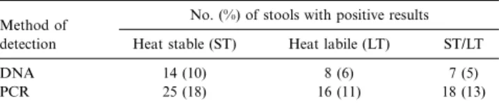
Table 1. Distribution of ETEC toxin–encoding genes as detected by PCR versus DNA hybridization in 140 stool samples from patients with travelers’ diarrhea in Mexico and Jamaica.
Method of detection
No. (%) of stools with positive results Heat stable (ST) Heat labile (LT) ST/LT
DNA 14 (10) 8 (6) 7 (5)
PCR 25 (18) 16 (11) 18 (13)
NOTE. ETEC, enterotoxigenic Escherichia coli; PCR, polymerase chain reaction.
DNA for ETEC detection by PCR [5]. In brief, 100 mg of a thawed stool sample was suspended in 1.5 mL of normal saline (0.85%) in microcentrifuge tubes. To remove debris from the stool, tubes were centrifuged at 183 g for 1 min. The supernatants were transferred to new tubes and centrifuged at 16,000 g for 5 min. The super-natants were then discarded; the pellets were washed in 1 mL of PBS and centrifuged as above. The supernatants were discarded once again, and the pellets were resuspended in 0.6 mL of chao-tropic solution (2.65 M guanidine thiocyanate, 5 mM dithiothreitol, 0.5% Tween 20, 0.15 M sodium acetate, 25 mM sodium citrate, 0.36% ammonium bromide, and 0.28 M sodium chloride, final pH 7.0) and were incubated at 657C for 20 min.
DNA purification. After incubation with chaotropic solution, 50 mL of resuspended glass matrix (GlasPac/GS; National Scientific Supply, San Rafael, CA) was added and incubated for 15 min at room temperature with continuous mixing. The suspension was centrifuged at 16,000 g for 1 min, and the supernatant was dis-carded. The matrix was resuspended in 1 mL of wash buffer (GlasPac/GS) and centrifuged at 16,000 g for 1 min. This wash step was repeated twice. The matrix pellet was dried at room tem-perature for 5 min, and the bound DNA was eluted by incubation with 100 mL of elution solution (10 mM Tris-HCl [pH 8.0], 0.1 mM EDTA) at 507C for 10 min with periodic mixing. The sus-pension was centrifuged at 16,000 g for 2 min, and the eluted DNA (supernatant) was carefully transferred to a new tube for PCR amplification.
PCR amplification. Oligonucleotide primers for LT and ST were selected on the basis of previously published sequences [5]. The final amplification mix contained 90 mL of PCR mix (10 mM Tris-HCl [pH 8.3]; 50 mM KCl; 2 mM MgCl2; 100 mg/mL gelatin; 5% glycerol; 1 mM [each] dATP, dCTP, dGTP and dTTP; and 2.5 U of AmpliTaq polymerase [Perkin-Elmer, Norwalk, CT]), 25 pmol of each of the 4 primers, and 10 mL of stool DNA solution. The reaction mixtures were heated to 507C for 2 min and 957C for 5 min and were subjected to 40 cycles (957C for 45 s and 507C for 45 s) and finally to an extension at 727C for 10 min in a DNA thermal cycler (Perkin-Elmer).
Detection of amplified products. We analyzed 20 mL of the amplified PCR products by 3% agarose electrophoresis gels in TBE buffer (89 mM Tris-HCL [pH 8.3], 89 mM boric acid, and 2.5 mM EDTA). In addition to the 1-kb DNA molecular weight marker (Gibco BRL, Gaithersburg, MD), 2 DNA controls were run si-multaneously with the samples. A positive control, an amplified PCR product from a stool sample spiked with H10407 ETEC strain, and a negative control, a PCR product from the stool of a healthy noninfected person spiked with E. coli strain JCP88, were included in the assay.
Dot blot analysis was done for a subset of 50 samples (25 from Mexico, 25 from Jamaica). Five microliters of PCR products was dot blotted onto nitrocellulose membranes, denatured with NaOH/ NaCl, and cross-linked with UV light. The membranes were hy-bridized with [32
P] ATP-labeled oligonucleotide probes for ST and LT, which were selected from published sequences, and visualized by autoradiography [5].
Sensitivity test. To assess the sensitivity of the PCR method, we performed experiments in which 100-mg samples of stools were spiked with varying concentrations (101
–107
cfu/g) of ETEC strain H10407, following MacFarland’s opacity standard.
Statistical Analysis
McNemar’s exact x2
test was used for correlated proportions of the 2 methods. The a level was set at 0.05.
Results
DNA hybridization. Of the 140 stool samples analyzed, 29 (21%) were positive for toxin genes by this method (table 1).
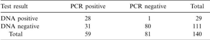
PCR products. The 140 PCR products from the same pa-tients were analyzed as described above using a 3% agarose gel and visualized with ethidium bromide. Positive samples yielded bands at∼450 bp for LT and at ∼190 bp for ST, as reported elsewhere [5]. By the multiplex PCR, 59 (42%) of 140 stool samples were positive. Of the 29 hybridization-positive isolates, 28 were positive by PCR (table 2). Fifty randomly selected PCR products were analyzed by dot blot oligonucleotide probes spe-cific for ST and LT. Samples positive by PCR were also positive using these specific DNA probes. The PCR method detected significantly (P!.0001) more ETEC in stool samples than the DNA hybridization method. When the 2 methods were com-pared regarding detection of toxin-encoding genes for LT and ST, the PCR method detected more LT and ST genes than the hybridization method (table 1).
Sensitivity test. PCR methodology detected ETEC toxin DNA (ST and LT) when ETEC concentrations in the stools were 100–1000 organisms/g stool.
Control samples. Stool samples from 48 healthy American volunteers without diarrhea were negative for the presence of ETEC when analyzed by the hybridization and PCR methodology.
Discussion
These results show that this PCR method is significantly more sensitive than a standard DNA hybridization technique in de-tecting ETEC in stools. Several factors make the multiplex PCR chaotropic DNA glass matrix assay a more sensitive, rapid, and less laborious technique for ETEC detection than the hy-bridization assay. The PCR method eliminates the need for a culture step, which depends on the presence of viable organisms in the stool. Microorganisms may be difficult to grow when their number is low and probably impossible if samples have been repeatedly frozen/thawed or kept frozen for months or
JID 1999;180 (December) Detection of Enterotoxigenic E. coli 2055
Table 2. Comparative results of polymerase chain reaction (PCR) versus DNA hybridization in 140 stool samples from patients with travelers’ diarrhea in Mexico and Jamaica.
Test result PCR positive PCR negative Total
DNA positive 28 1 29
DNA negative 31 80 111
Total 59 81 140
years. In the present study, stool samples that were frozen at 2207C for 6 months were analyzed by PCR after thawing with good results, and 28 (97%) of 29 fresh stool samples positive by DNA hybridization were detected by PCR. DNA extraction from stools using the chaotropic glass matrix method permits the isolation of most of the DNA present in a sample, thereby diminishing the problem of intrinsic PCR inhibitors.
Detection of shigellae, ETEC, and Campylobacter species by multiplex PCR using this DNA extraction procedure has been reported [8]. The simultaneous identification of several path-ogens from the same sample by use of this technology could be of great advantage in studies of acute diarrhea. Furthermore, the chaotropic DNA glass matrix method has a greater sen-sitivity than other techniques, such as conventional total nucleic acid extraction and ethanol precipitation [5]. The method used in the present study detected ETEC toxin DNA when ETEC concentrations were 100–1000 organisms/g stool. This is similar to the level of ETEC organisms (800) identified by the PCR-chaotropic method in another study [8].
The exquisite sensitivity of PCR can result in false-positive reactions due to contamination or carryover. Therefore, in this study, we physically separated the main PCR steps: DNA stool extraction, preparation of reaction mixture, and manipulation of PCR products [9]. Thus, the detection of increased ETEC toxin is most likely due to increased sensitivity and not to false-positive results. This is supported by the fact that the 40 ETEC-negative controls remained ETEC-negative when analyzed by the same method. The fact that 28 of 29 fecal samples identified as ETEC-positive by DNA hybridization were positive by PCR assay also suggests that the PCR assay is sensitive in detecting ETEC from fecal samples. When randomly selected PCR prod-ucts of 50 samples were analyzed by hybridization with oli-gonucleotide probes, the samples identified as positive by PCR were also positive by the former method, offering further ev-idence that the PCR was specific.
One problem related to the diagnosis of an infection based only on the detection of DNA is the high sensitivity of the PCR assay. It is also possible that viable or nonviable ETEC were present and detectable in stool from environmental (food, wa-ter) contamination of the gastrointestinal tract of the patients in the absence of symptomatic ETEC infection.
The finding of ETEC infection in persons who, without PCR study, would have an undetected enteropathogen may explain, in part, our results showing a benefit of antimicrobials in short-ening illness in cases of nonspecific illness [10]. In this study, which employed a PCR procedure, we found a higher per-centage of ETEC infection in a group of patients with travelers’ diarrhea than we found by a less sensitive DNA hybridization method. Thus, PCR may help to establish the true importance of ETEC in various populations (travelers, local populations in endemic areas, and foodborne outbreaks).
References
1. DuPont HL, Formal SB, Hornick RB, et al. Pathogenesis of Escherichia coli diarrhea. N Engl J Med 1971; 285:1–9.
2. Nataro JP, Kaper JB. Diarrheagenic Escherichia coli. Clin Microbiol Rev
1998; 11:142–201.
3. Hyams KC, Bourgeois AL, Merrell BR, et al. Diarrheal disease during operation desert shield. N Engl J Med 1991; 325:1423–8.
4. Bourgeois AL, Gardiner CH, Thornton SA, et al. Etiology of acute diarrhea among United States military personnel deployed to South America and West Africa. Am J Trop Med Hyg 1993; 48:243–8.
5. Stacy-Phipps S, Mecca JJ, Weiss JB. Multiplex PCR assay and simple prep-aration method for stool specimens detect enterotoxigenic Escherichia coli DNA during course of infection. J Clin Microbiol 1995; 33:1054–9. 6. Boom R, Sol CJ, Salimans MM, Jansen CL, Wertheim-van Dillen PM, van
der Noordaa J. Rapid and simple method for purification of nucleic acids. J Clin Microbiol 1990; 28:495–503.
7. Murray BE, Mathewson JJ, DuPont HL, Hill WE. Utility of oligodeoxy-ribonucleotide probes for detecting enterotoxigenic Escherichia coli. J In-fect Dis 1987; 155:809–11.
8. Oyofo BA, Mohran ZS, El-Etr SH, Wasfy MO, Peruski L. Detection of enterotoxigenic Escherichia coli, Shigella and Campylobacter spp. by mul-tiplex PCR assay. J Diarrhoeal Dis Res 1996; 14:207–10.
9. Kwok S, Higuchi R. Avoiding false positives with PCR. Nature 1989; 339: 327–8.
10. Ericsson CD, Johnson PC, DuPont HL, Morgan DR, Bitsura JM, de la Cabada FJ. Ciprofloxacin or trimethoprim-sulfamethoxazole as initial therapy for travelers’ diarrhea A placebo-controlled, randomized trial. Ann Intern Med 1987; 106:216–20.