Coronary vasomotor tone during static and
dynamic exercise
O. M. HESS, A. BORTONE, K. E I D , J. E. G A G E , H. NONOGI, J. GRIMM AND H. P. KRAYENBUEHL
Department of Internal Medicine, Medical Policlinic, Cardiology, University Hospital, Zurich, Switzerland
KEY WORDS: Coronary vasomotion, quantitative coronary arteriography, isometric and dynamic exercise, coronary sinus flow, normal and stenotic coronary arteries, coronary artery disease, endothelium-derived relaxing factor.
Coronary vasomotion is an important determinant of myocardial perfusion in patients with angina pectoris, and it influences not only normal but also stenotic coronary arteries. The ability of a stenotic coronary artery to change its size is dependent on the presence of a normal musculo-elastic wall segment within the stenosis (i.e., eccentric stenosis). Coronary vasoconstriction of normal and stenotic coronary arteries has been reported by Brown and coworkers (Circulation 1984; 70: 18-24) during isometric exercise.
The effect of dynamic exericse on coronary vasomotion was evaluated in one group of 13 patients with ischaemia-like symptoms and normal coronary arteries (group 1) and in a second group of 12 patients with coronary artery disease with exercise-induced angina pectoris (group 2). Luminal area of a normal and a stenotic vessel segment was determined by biplane quantitative coronary arteriography at rest, during supine bicycle exercise and 5min after administration of 16 mg sublingual nitroglycerin. Coronary sinus blood flow was measured in group 1 at rest and after 0-5mgkg~' intravenous dipyridamole using coronary sinus thermodilution. Coronary flow reserve was calculated from coronary sinus flow after dipyridamole divided by coronary sinus flow at rest.
In group 1, coronary vasodilation of the large (i.e., proximal) and the small (i.e., distal) coronary arteries was observed during exercise in seven patients (subgroup A). However, in the remaining six patients (subgroup B) coronary vasoconstriction of the small arteries (—24%, P < 0-001) was found during exercise, whereas the large vessels showed coronary vasodilation (+26%, P <0-001). Coronary flow reserve was significantly (P <0-05) larger in subgroup A (mean 2-5) than in subgroup B (mean 1 -2) with exercise-induced vasoconstriction of the small epicardial arteries.
In group 2 vasodilation of the normal (+23%, P < 0-001) and vasoconstriction of the stenotic coronary arteries (—29, P<0-001) was found during supine bicycle exercise. Administration of sublingual nitroglycerin at the end of the exercise test was accompanied by coronary vasodilation of both normal (+40%, P <0001 vs rest) and stenotic (+12%, NS vs rest) vessel segments.
It is concluded that isometric exercise is associated with reflex coronary vasoconstriction of the normal and stenotic vessel segments due to enhanced sympathetic stimulation. Dynamic exercise in patients with ischaemia-like symptoms and normal coronary arteries is accompanied by an abnormal dilatory response of the small coronary arteries in a subgroup of patients with reduced coronary flow reserve. Dynamic exercise in patients with coronary artery disease is, however, associated with coronary vasodilation of the normal and coronary vasoconstriction of the stenotic vessel segments. The nature of this exercise-induced vasoconstriction of stenotic coronary arteries is not clear, but might be related to endothelial dysfunction with an insufficient production of the endothelium-derived relaxing factor during exercise.
Address for correspondence: Otto M. Hess, M. D., This study was supported by the Swiss National Science Medical Policlinic, Cardiology, University Hospital, Foundation.
Raemistrasse 100, CH-8091 Zurich, Switzerland.
Introduction
Coronary vasomotion plays an important role in the regulation of coronary perfusion at rest and during exercise11"31. Not only normal but also stenotic coronary arteries show coronary vaso-motion at the site of the coronary stenosis since approximately 70% of all coronary stenoses have a normal musculo-elastic wall segment within the stenosis'4"51. Previous studies have reported that during isometric exercise not only normal but also stenotic coronary arteries show coronary vasoconstriction'6', probably due to enhanced sympathetic stimulation. Pretreatment with intracoronary nitroglycerin prevented vaso-constriction of both normal and stenotic coronary arteries, probably due to the direct vasodilating effect of nitroglycerin on the smooth vasculature of the coronary arteries. The purpose of the present study was (1) to evaluate the effect of dynamic exercise on coronary vasomotion in patients with ischaemia-like symptoms and normal coronary arteries, and (2) to study the effect of dynamic exercise on coronary vasomotion in patients with coronary artery disease with exercise-induced angina pectoris.
Patients and methods
25 patients were included in the present study. 13 patients with normal coronary arteries constituted group 1 and 12 patients with coronary artery disease group 2. Mean age was 48 years in group 1 and 53 years in group 2. A history of angina pectoris and/or ST-segment depression in the upright bicycle exercise test was present in all patients of both groups. Coronary arteriography revealed normal coro-nary arteries in all group 1 patients, and one-vessel disease in one patient, two-vessel disease in two and three-vessel disease in nine patients of group 2.
QUANTITATIVE CORONARY ARTERIOGRAPHY Biplane coronary arteriography was per-formed after an interval of at least 10 min after the last diagnostic coronary angiogram. Baseline coronary arteriography was carried out after the patient's feet had been attached to a bicycle ergometer (Siemens-Elema AG, model 380B). Repeat biplane coronary arteriography, with concurrent aortic and pulmonary artery pressure recordings, was carried out at the end of each
exercise level. Exercise was begun at 50-75 W and increased every 2 min in increments of 25-50 W and was terminated because of anginal pain, fatigue or ST-segment depression of more than 0-2 mV. Immediately after the exercise test 1 -6 mg sublingual nitroglycerin was administered and, 5 min later, biplane coronary arteriography was repeated.
Quantitative evaluation of biplane coronary angiograms was carried out in a blinded fashion. Tracings were made manually from the simul-taneous right and left anterior oblique projec-tion during diastasis or end-diastole. Each vessel segment was analysed four to six times and the results were averaged to reduce sampling error. A section of the catheter of known dimensions was traced as a scaling factor. The tracings were digitized and analysed on a PDP 11/34 computer'31. The luminal area of the proximal and distal left anterior descending and the proximal and distal left circumflex coronary artery were analysed in group 1 and the luminal area of a normal and a stenotic vessel segment, either of the left anterior descending or left circumflex coronary artery, was evaluated in group 2. Interobserver variability (n = 22) was 9-3% of the mean vessel area for monoplane and 7-9% for biplane measurements'31.
CORONARY SINUS BLOOD FLOW
Flow was measured by the coronary sinus thermodilution technique'71. Ten patients of group 1 were restudied 7-20 days (mean 10 days) after coronary arteriography on an ambulatory basis. A coronary sinus thermodilu-tion catheter was introduced from the right femoral vein into the coronary sinus and 50 ml saline was infused over 1 min to determine coronary sinus outflow according to the tech-nique of Ganz and coworkers'7'. The first measurement was carried out at rest and the second after intravenous infusion of 0-5mgkg"' dipyridamole over 15 min. Coronary flow ratio (i.e., coronary flow reserve) was calculated as the coronary sinus flow after dipyridamole divided by the coronary sinus flow at rest. The coronary resistance ratio was obtained by dividing the coronary resistance at rest by the coronary resistance after dipyridamole.
PATIENT SUBGROUPS
Patients with normal coronary arteries and ischaemia-like symptoms were divided into two
subgroups according to the behaviour of the small coronary arteries during exercise: Patients with vasodilation during exercise were allocated to subgroup A (n = 7) and patients with vasoconstriction of the small epicardial arteries during exercise to subgroup B (n = 6).
STATISTICS
Comparisons of haemodynamic and an-giographic data in response to a first and a second exercise level and to sublingual nitrogly-cerin were carried out by two-way analysis of variance for repeated measurements. Com-parisons between two groups or subgroups were performed by the unpaired Student /-test. In Figs 1-3 mean values ±1 standard error are reported.
Results
CORONARY LUMINAL AREA AT REST AND DURING EXERCISE
1. Patients with ischaemia-like symptoms and normal coronary arteries (Fig. 1).
Coronary vasodilation of the proximal and distal coronary arteries was found in all seven patients of subgroup A. The proximal vessels of the left anterior descending and left circumflex coronary artery increased by 2 1 % (/>< 0-001) and the distal vessels by 45% (P< 0001) during exercise. After sublingual administration of riitroglycerin, there was a further increase in luminal area of the proximal (+46%, P <0-001) and distal (+99%, P<0-001) coronary vessels. In contrast to subgroup A, there was coronary vasoconstriction of the distal coronary vessels (-24%, P < 0-001) during exercise in subgroup B. Nevertheless, the proximal coronary arteries showed, as in subgroup A, coronary vasodilation (+26%, P < 0-001) during exercise. After sublingual administration of nitroglycerin there was an increase in luminal area of both distal (+44%, P < 0 0 0 1 ) and proximal (+47%, P < 0-001) coronary arteries in subgroup B. Coronary flow reserve was significantly ( P < 005) reduced in subgroup B (1-2±0-3; n = 5) compared with subgroup A (2-5 ± 1 0 ; n = 5). Parallel to coronary flow reserve, coronary resistance ratio was significantly (/><0-05)
250 2 0 0 k 150 100 5 0 LADLCX P mean - 1 sem • P O 0 5 vs rest • P<0 01 vs rest • PO-OOI vs rest
rest 2minex max ex NTGsl
250
2 0 0
150
-100
rest 2minex maxex NTGS.L Figure 1 Response of normal coronary arteries to dynamic exercise in patients with ischaemia-like symptoms and normal coronary arteries (group 1). Proximal and distal vessel segments of the left anterior descending (LAD) and left circumflex (LCX) coronary artery were analysed at rest, after 2 min exercise (ex) and during maximal ex as well as Smin after 1-6mg sublingual nitroglycerin (NTG s.l.). Patients (n = 7) who showed vasodilation of both proximal and distal coronary arteries were designated subgroup A (a) and patients (n = 6) who showed vasoconstriction of distal but vasodilation of proximal coronary vessels were designated subgroup B (b). Luminal area is given in percent of control area. Abbreviations as in Fig. 1.
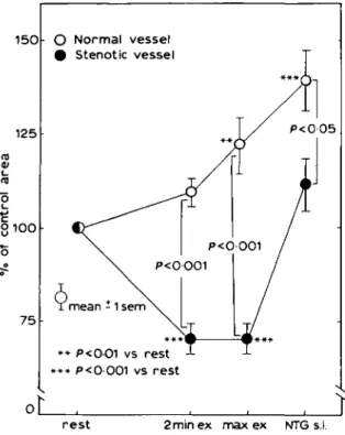
reduced in subgroup B (1-2 ±0-3; n = S) compared with subgroup A (2-7 ± 1-2; n = 5). 2. Patients with coronary artery disease and exercise-induced angina pectoris (Fig. 2). Coronary vasodilation of the normal coro-nary arteries was observed in patients with coronary artery disease (+23%, .P<0-001) during exercise. After sublingual administration of nitroglycerin, the luminal area of the normal vessel increased (+40%, P<0-001). Stenotic coronary vessels showed coronary vasoconstric-tion ( - 2 9 % , /)< 0 0 0 1 ) during exercise (Fig. 2) which was reversible after sublingual ad-ministration of nitroglycerin at the end of the exercise test (+12%, NS vs rest, P < 0-001 vs exercise). 1 5 0• O Normal vessel Stenotic vessel 125 01 flj O 8100 7 5 P<005 mean -1 sem • • P<OO1 vs rest * * * p < 0 0 0 1 vs rest
oL
rest 2minex max ex NTG s.l.
Figure 2 Responses of normal and stenotic coronary arteries to dynamic exercise in patients with coronary artery disease and exercise-induced angina pectoris. Normal vessels (open symbols) dilate during exercise to 123% of resting values, and further dilate to 140% after sublingual nitroglycerin. Stenotic vessels (closed symbols) show vasoconstriction during exercise, the stenosis narrowing to 71% of control, and vasodilation to 112% of control after sublingual nitroglycerin. Luminal area is given as % control area.
INFLUENCE OF STENOSIS SEVERITY ON CORONARY VASOMOTION
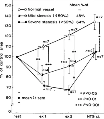
Patients with coronary artery disease were analysed separately for the influence of stenosis severity on coronary vasomotion. Three groups of coronary vessels were evaluated (Fig. 3): normal vessels (n = 7), stenotic vessels (n = 4) with mild stenosis (=£50% diameter stenosis) and stenotic vessels (n = 7) with severe stenosis (>50% diameter stenosis). Normal coronary arteries showed progressively coronary vasodila-tion during exercise and vasodilavasodila-tion was maximal after sublingual administration of nitroglycerin. Stenotic vessels showed, however, exercise-induced vasoconstriction which was more pronounced in severely stenotic than in mildly stenotic coronary arteries (Fig. 3). At the second exercise level, mild coronary stenoses showed only minor coronary vasoconstriction compared with the resting state ( - 1 0 % , NS) but severe coronary stenoses revealed significant coronary vasoconstriction during dynamic exer-cise (-32%, /»< 0-001).
Discussion
Epicardial coronary arteries show completely different reactions to isometric or dynamic exercise in patients with coronary artery disease'136'. This reaction to exercise is depend-ent on several factors and can be modified by the administration of different vasodilating substances. The purpose of the present study is to report on the effect of dynamic exercise on coronary vasomotion in patients with ischaemia-like symptoms and normal coronary arteries, as well as in patients with coronary artery disease with classic exercise-induced angina pectoris. EFFECT OF ISOMETRIC EXERCISE ON CORONARY VASOMOTION
Brown and coworkers1*1 studied the effect of isometric exercise on coronary luminal area in 11 patients with coronary artery disease. Normal and stenotic vessel segments showed coronary vasoconstriction (normal segment —14%, steno-tic segment —35%) during handgrip exercise. After pretreatment with intracoronary nitrogly-cerin, coronary vasoconstriction was abolished and there was coronary vasodilation of both normal (+29%) and stenotic (+32%) vessel segments during handgrip exercise. The authors concluded that enhanced o--adrenergic
stimula-150 140 130 120 CD 2 100 +•* c Q
s
9 0 o ?• 80 7 0 6 0 0 Meanest o—o Normal vessel —9—9 Mild stenosis (S50°/o) 45%> Tn_7
• — • Severe stenosis (>50°/o) 64%> >&-\
n - 1 ^ ^
1 1
n-l^>>^ • ^ 1 * ** nzAii***
X 'X- •X'I
^ 7
. ^ / J i -n-1.1/
* /V
T Jn=7 *p<o-O5 • mean i1 sem * * P<001 ^ *•** P<0001 • . > • rest e x 1 ex 2 NTG s.l.Figure 3 Influence of stenosis severity on coronary vasomotion of normal vessels (open symbols), mild stenoses (half open symbols) and severe stenoses (closed symbols) during exercise (exl: first, and ex2: second level of exercise) and 5 min after administration of 1-6 mg sublingual nitroglycerin (NTG s.l.) Normal vessels dilate progressively during exercise and after sublingual nitroglycerin, whereas stenotic vessel segments show exercise-induced vasoconstr-iction. Severe coronary stenoses constrict more than mild stenoses but both dilate only minimally after sublingual nitroglycerin. These data suggest that exercise-induced stenosis narrowing is dependent on the severity of coronary stenosis or, in other words, is dependent on the extent of the atherosclerotic process. Luminal area is given as % control area; Mean %st: Mean diameter stenosis (%); n: number of patients.
tion with increased levels of circulating epi-nephrine and norepiepi-nephrine during isometric exercise was probably responsible for coronary vasoconstriction of epicardial coronary arteries in patients with coronary artery disease.
EFFECT OF DYNAMIC EXERCISE ON CORONARY VASOMOTION
Normal epicardial arteries show coronary vasodilation during dynamic exercise. Several factors are responsible for coronary vasodila-tion, such as a higher perfusion pressure, an increase in coronary blood flow with release of
the endothelium-derived relaxing factor, an augmentation of circulating metabolites, as well as changes in neurohumoral regulation of the coronary arteries.
13 patients with normal coronary arteries and ischaemia-like symptoms were evaluated in the present study. Half were found to have abnormal coronary vasomotion of the small epicardial arteries during dynamic exercise (subgroup B). Not only was an abnormal dilatory response of the small coronary arteries observed during exercise, but also a reduced dilator capacity of the resistance vessels
( = arterioles) was found during dipyridamole infusion. The abnormal dilatory response is likely to be due to an abnormal neurohumoral regulation of coronary vasomotor tone, as was suggested by Cannon and coworkers18'91 and Greenberg and coworkers1101. Administration of sublingual nitroglycerin was associated with coronary vasodilation of the small epicardial coronary arteries, suggesting that these vessels potentially can react normally.
Dynamic exercise in patients with coronary artery disease was accompanied by coronary vasodilation of normal vessel segments but by coronary vasoconstriction of stenotic segments. This opposite effect of dynamic exercise on the coronary vasomotion of normal and stenotic coronary arteries must be explained by the following mechanisms: normal coronary arteries show vasodilation during dynamic exercise to meet the increased oxygen demands of the myocardium (see above). Stenotic coronary arteries behave somewhat paradoxically during dynamic exercise by further increasing the severity of stenosis. This phenomenon is related either to a passive collapse of the free vessel wall within the stenotic segment when coronary blood flow velocity is increased during exercise (Venturi mechanism)1'1 or to an insufficient production of endothelium-derived relaxing factor due to the atherosclerotic alterations of the vessel wall'31. Another possible mechanism involves increased platelet aggregation with turbulent flow during exercise and with release of thromboxane A2 and serotonin, both of which induce coronary vasoconstriction. Thus, the present results suggest that exercise-induced narrowing of coronary stenoses is due to active vasoconstriction and is an important mechanism in the production of myocardial ischaemia
during dynamic exercise in patients with coronary artery disease.
References
[1] Brown BG, Bolson EL, Dodge HT. Dynamic mechanisms in human coronary stenosis. Circulation 1984: 70: 917-22.
[2] Mates RE, Gupta RL, Bell AC, Klocke FJ. Fluid dynamics of coronary artery stenosis. Circ Res 1978; 42: 152-62.
[3] Gage JE, Hess OM, Murakami T, Ritter M, Grimm J, Krayenbuehl HP. Vasoconstriction of stenotic coronary arteries during dynamic exercise in patients with classic angina pectoris: reversibility by nitrogly-cerin. Circulation 1986; 73: 865-76.
[4] Freudenberg H, Lichtlen PR. The normal wall segment in coronary stenosis—a postmortal study. Z Kardiol 1981; 70: 863-9.
[5] Saner HE, Gobel FL, Salomonowitz E, Erlien DA, Edwards JE. The disease-free wall in coronary atherosclerosis: its relation to degree of obstruction. J Am Coll Cardiol 1985; 6: 1096-9.
[6] Brown BG, Lee AB, Bolson EL, Dodge HT. Reflex constriction of significant coronary stenosis as a mechanism contributing to ischemic left ventricular dysfunction during isometric exercise. Circulation 1984; 70: 18-24.
[7] Ganz W, Tamura K, Marcus HS, Donoso R, Yoshida S, Swan HJC. Measurement of coronary sinus blood flow by continuous thermodilution in man. Circulation 1971; 44: 181-95.
[8] Cannon RO, Watson RM, Rosing DR, Epstein SE. Angina caused by reduced vasodilator reserve of the small coronary arteries. J Am Coll Cardiol 1983; 1: 1359-73.
[9] Cannon RO, Schenke WH, Leon MB, Rosing DR, Urqhart J, Epstein SE. Limited coronary flow reserve after dipyridamole in patients with ergonovine-induced coronary vasoconstriction. Cir-culation 1987; 75: 163-74.
[10] Greenberg MA, Grose RM, Neubiirger N, Sil-verman R, Strain JE, Cohen MV. Impaired coronary vasodilator responsiveness as a cause of lactate production during pacing-induced ischemia in patients with angina pectoris and normal coronary arteries. J Am Coll Cardiol 1987; 9: 743-51.