PAEDIATRICS
Electrocardiographic and haemodynamic alterations caused
by three different test solutions of local anaesthetics to
detect accidental intravascular injection in children
J. Y. Mauch
1*, N. Spielmann
1, S. Hartnack
2and M. Weiss
11Department of Anaesthesia, University Children’s Hospital, Steinwiesstrasse 75, 8032 Zurich, Switzerland 2
Section of Veterinary Epidemiology, Vetsuisse Faculty, University of Zurich, Switzerland * Corresponding author. E-mail: jacqueline.mauch@kispi.uzh.ch
Editor’s key points
† The signs of local anaesthetic (LA) toxicity may be masked by general anaesthesia, and specific monitoring is recommended. † This study in children
found that a test dose of LA must contain
epinephrine for
intravascular injection to be detected.
† The effect depended on patient age.
† Monitoring of heart rate, arterial pressure, and ECG for T-wave changes should be mandatory during test dose injection.
Background.The aim of this study was to investigate ECG and haemodynamic alterations provoked by a test dose of bupivacaine, epinephrine, and their combination.
Methods.Paediatric patients undergoing general anaesthesia were randomized into three groups. After anaesthesia induction and tracheal intubation, 0.2 ml kg21(max. 3 ml) of the corresponding test solution was i.v. injected: bupivacaine 0.125% (Group B), bupivacaine 0.125% plus epinephrine 1:200 000 (Group BE), or epinephrine 1:200 000 (Group E). ECG was printed and analysed post hoc. Non-invasive arterial pressure (AP) was measured at 1 and 2 min after test dose injection. Increases in T-wave of≥25%, in heart rate (HR) of ≥10 beats min21, and in systolic AP of ≥15 mm Hg above baseline value were considered a positive result.
Results. A total of 105 children aged 0.2 –16 (median 6.8) yr were enrolled. Test dose injection provoked T-wave elevation in 0%, 85%, and 89% of patients in Groups B, BE, and E, respectively. A positive increase in HR was found in 0%, 68%, and 76%. A positive increase in AP at 1 min was found in 0%, 88%, and 94% and at 2 min in 0%, 42%, and 59%. A decrease in HR of ≥10 beats min21 was observed in 6%, 76%, and 69%. Alterations in T-wave and HR were significantly influenced by age.
Conclusions.ECG and haemodynamic alterations after i.v. injection of a local anaesthetic test dose were significantly influenced by epinephrine. T-wave elevation, increase in AP, and changes in HR are highly reliable variables, particularly when age is taken into account.
Keywords: anaesthesia, local; anaesthesia, regional; bupivacaine; drug toxicity; electrocardiography; epinephrine; heart rate
Accepted for publication: 7 October 2011
When combined general and regional anaesthesia is used for intra- and postoperative pain relief in children, neurological signs of local anaesthetic (LA) toxicity in the case of inadvertent intravascular administration of LA are blunted by general an-aesthesia, muscle paralysis, or both.1 Therefore, to timely detect an inadvertent intravascular administration of LA, other than neurological signs are needed to avoid sudden haemodynamic collapse by LA intoxication.2 3 Animal research in small pigs has shown that the systemic application of a standardized small test dose of bupivacaine with epineph-rine or epinephepineph-rine alone but not bupivacaine alone results in an increase in T-wave amplitude in the ECG and in heart rate (HR) increase.4The aim of this study is to elucidate whether these ECG findings and haemodynamic effects found in small pigs provoked by a test dose of epinephrine, bupivacaine, and their combination are reproducible from infancy to adolescence.
Methods
The study was approved by the local ethics committee (KEK-ZH-No. 2009-0025) and registered on ClinicalTrials.gov (NCT 01091766). Paediatric patients aged from 1 month to 16 yr, ASA classification I, undergoing an elective invasive procedure requiring general anaesthesia and tracheal intub-ation were recruited for this prospective randomized clinical trial. The patients were grouped according to age: infants (Group 1, n¼15), 1 –6 yr (Group 2, n¼30), 6 –12 yr (Group 3, n¼30), and 12 –16 yr (Group 4, n¼30). Less infants were included in Group 1 compared with other groups in order to guarantee a continuous distribution of age. Written parental and patient (Group 4) consent was obtained.
Premedication and route of induction (i.v. or inhalation) of general anaesthesia depended on the age and preference
of the patient. Tracheal intubation was performed in all patients after neuromuscular block using atracurium (Tracrium; GlaxoSmithKline, Mu¨nchenbuchsee, Switzerland). Anaesthesia was maintained by sevoflurane (Sevorane; Abbott AG, Baar, Switzerland) 2.5 vol% in oxygen/nitrous oxide (ratio 1:2). No atropine was given to any patient before test dose injection.
Monitoring consisted of pulse oximetry, ECG, non-invasive arterial pressure (AP) measurement, and end-tidal gas ana-lysis (sevoflurane, O2, N2O, and CO2). For application of the three different test solutions, the four age-dependent groups were each randomized into three groups. Group B received plain bupivacaine 0.125% (Bupivacaine 0.125%; Sin-tetica S.A., Mendrisio, Switzerland), Group BE received bupiva-caine 0.125%+epinephrine 1:200 000, and Group E received only epinephrine 1:200 000 (Adrenalin; Sintetica S.A.). Under stable cardiopulmonary and anaesthesia conditions, 0.2 ml kg21(max. 3 ml) of the corresponding test solution was i.v. administered followed by an injection of 10 ml normal saline. The amount of 0.2 ml kg21(max. 3 ml) test solution corresponded to the routinely used test dose of bupivacaine 0.125%+epinephrine 1:200 000 in the author’s anaesthesia department before injection of the total amount of LA for regional anaesthesia blocks. The two anaesthetists injecting the study medication were not blinded to the study drug for safety reasons in the case of an adverse event and were not involved in anaesthetic care of the study patient. ECG traces I and II were monitored and continuously printed for post hoc analysis by a single, blinded anaesthetist. Initial, maximal, and minimal HR and non-invasively measured AP were noted before and at 1 and 2 min after test dose injection.
The primary outcome was T-wave alterations. The second-ary outcomes were changes in HR and AP. According to current literature,2 5 6increases of ≥25% in T-wave ampli-tude, ≥10 beats min21in HR, and ≥15 mm Hg in systolic AP above baseline were considered each a positive response.
In addition, a decrease in HR of≥10 beats min21was taken as a positive finding.
The computer package NCSS 2007 (Version 07.1.20, Kays-ville, UT, USA) was used for statistical analysis. A P-value of ≤0.05 was considered significant. Linear and generalized linear models were performed with the software R (Version 2.11.1) using the packages lmtest7and nlme.8Model selec-tion was based on Akaike’s informaselec-tion criterion and visual checking of the residuals. The results of the models are given in estimated mean effects and standard errors (SEs).
P-values were derived from likelihood ratio tests.
Results
Overall 105 children were included in the study (Table 1). All patients aged .6 months received premedication with midazo-lam (0.5 mg kg21, max 15 mg, reduced dose in agreement with the anaesthetist in charge of the patient; applied orally or rectally as decided by the child/parents) except one 8-yr-old boy in Group BE. Inhalation induction of anaesthesia with sevoflurane was chosen in all except 17 patients (13 of them aged 12–16 yr), who had an i.v. induction with propofol and alfentanil (10 mg kg21) or fentanyl (2 mg kg21). In one boy (aged 14), randomized into study group BE, a test dose was not injected because of the therapy used to treat hypotension after induction of anaesthesia and data from this patient were excluded from further data analysis. During the study period, no critical or adverse events were observed or had to be treated. At baseline, using analysis of variance, no significant dif-ferences between the three treatment groups were detected for age (P¼0.990), systolic (P¼0.491) and diastolic (P¼0.453) AP, and HR (P¼0.902).
The incidence of positive T-wave elevation to test dose (all ages) was 0% in Group B, 85% in Group BE, and 89% in Group E (Table 2) (P,0.0001, Fisher’s exact test). A positive T-elevation to intravascular epinephrine administration (IVEA) occurred in median 20 (range 14–34) s. The median baseline
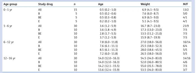
Table 1Patient characteristics, subdivided according to age group and study drug
Age group Study drug n Age Weight M/F
0– 1 yr All 15 0.5 (0.2 –1.0) 6.9 (4.1 –9.5) 13/2 B 5 0.5 (0.2 –0.6) 7.6 (6.0 –8.7) 5/0 BE 5 0.5 (0.3 –0.8) 6.9 (6.5 –9.0) 4/1 E 5 0.2 (0.2 –1.0) 5.1 (4.1 –9.5) 4/1 1– 6 yr All 30 3.6 (1.2 –5.9) 16.7 (8.7 –23.0) 21/9 B 10 3.6 (1.8 –4.9) 17.3 (13.0 – 23.0) 7/3 BE 10 2.8 (1.7 –5.5) 13.5 (11.2 – 21.0) 7/3 E 10 3.7 (1.2 –5.9) 15.9 (8.7 –19.9) 7/3 6– 12 yr All 30 7.8 (6.0 –11.8) 27.0 (18.0 – 56.0) 16/14 B 10 7.6 (6.1 –11.1) 25.5 (18.0 – 52.3) 6/4 BE 10 8.5 (6.1 –11.3) 28.0 (18.6 – 45.5) 5/5 E 10 7.2 (6.0 –11.8) 26.5 (18.0 – 56.0) 5/5 12 –16 yr All 30 14.0 (12.0 – 16.3) 54.5 (34.0 – 83.0) 14/16 B 10 14.0 (12.0 – 16.3) 52.0 (36.0 – 80.5) 4/6 BE 10 14.2 (12.1 – 15.5) 55.0 (35.5 – 78.0) 7/3 E 10 13.6 (12.4 – 15.9) 53.5 (34.0 – 83.0) 3/7
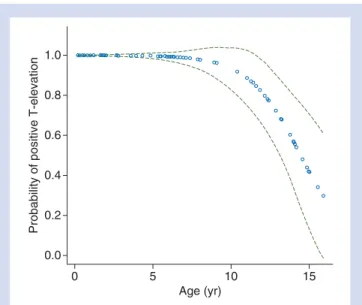
T-wave in ECG lead I was 0.15 (range 0.02–0.45) and 0.25 (0.05 –0.70) mV in ECG lead II. Relative increases in T-amplitude in lead II were larger than in lead I (Table3). In Groups BE and E, age was significantly associated with the probability of a positive T-wave elevation (P,0.0001), with older children having a reduced probability to show a positive T-elevation compared with the younger ones (Fig.1). Patients of Groups BE and E did not differ (P¼0.625). Gender was not significant (P¼0.621). Typical ECG traces of an infant and an adolescent are shown in Figure2.
A positive HR increase to test dose (all ages) were 0% in Group B, 68% in Group BE, and 76% in Group E (Table 2). Groups BE and E had a significantly higher maximum increase in HR during the observation period with 13.2 (SE1.7) and 16.1
(SE1.7) beats min21, respectively (both P,0.0001), compared
with Group B. Three highly influential data points affecting the age effect were removed. The maximum increase in HR was significantly influenced by age with a decrease of 0.3 (SE 0.14) beats min21 per year (P¼0.026), but not by sex
(P¼0.391) nor by i.v. induction (P¼0.114).
A positive decrease in HR (.10 beats min21) after test dose (all ages) was found in 6% in Group B, 76% in Group BE, and 69% in Group E (Table2). Groups BE and E had a significantly greater decrease in HR during the observation period with 14.1 (SE2.1) and 12 (SE2.1) beats min21, respectively (both
P,0.0001), compared with Group B (Table3). The maximum decrease in HR was significantly influenced by age with a de-crease of 0.5 (SE0.17) beats min21per year (P¼0.003), but not
by gender (P¼0.157) nor by i.v. induction (P¼0.137).
Intermittent loss of P-wave was observed in Groups BE and E (Table 3). In 10 patients, loss of P-wave occurred during HR increase at 26.5 s (range 23 –30) after IVEA. In seven patients, P-wave disappeared at 62.5 (44 –85) s after IVEA and during the decrease in HR.
A positive systolic AP increase to the test dose after 1 min (all ages) was seen in 0% in Group B, 88% in Group BE, and
94% in Group E (Table2). Groups BE and E had a significant increase of 35.8 (SE6.26) and 50.97 (SE6.26) mm Hg in
systol-ic AP compared with Group B (both P,0.0001) after 1 min (Table3). The interaction between the group effect and i.v. induction was found to be significant (P¼0.007). The effect of the interaction was to decrease the mean difference for a patient in Group E without i.v. induction from 50.1 to 29.4 mm Hg. Neither age (P¼0.303) nor gender (P¼0.115) was significantly associated with the difference in systolic AP after 1 min. After 2 min, the increases in Groups BE and E were less prominent with significant increases of 19.03 (SE2.03) and 18.51 (SE2.01) mm Hg compared with Group B
(both P,0.0001). The interaction between the group effect and i.v. induction was no longer significant (P¼0.583). Neither age (P¼0.503) nor gender (P¼0.253) had a signifi-cant influence.
Diastolic AP changes: after 1 min, Groups BE and E had a significant increase in diastolic AP with 26.3 (SE3.4) and 22.15
(SE 3.6) mm Hg compared with Group B (both P,0.0001).
Boys had a significantly lower diastolic AP with 5.1 (SE1.8)
mm Hg less than girls (P¼0.001). The interaction between the group effect and age was significant (P¼0.017). The effect of the interaction was to decrease the mean difference for a patient in Group BE by 1.1 mm Hg (P¼0.005) per year. Without three highly influential data points, the effect of the i.v. induction was not significant (P¼0.235). After 2 min, the increases in Groups BE and E were less prominent with significant increases of 10.05 (SE 1.8) and 7.08 (SE1.8)
mm Hg compared with Group B. The effect of age was found to be significant (P,0.001) with a decrease of 0.59 (SE0.15) mm Hg per year. Neither gender (P¼0.149) nor i.v.
induction (P¼0.272) was found to have a significant effect on diastolic AP after 2 min.
To summarize, the inclusion of T-wave and HR allows de-tection of an epinephrine-containing test dose with a reliabil-ity of 100% (Table4).
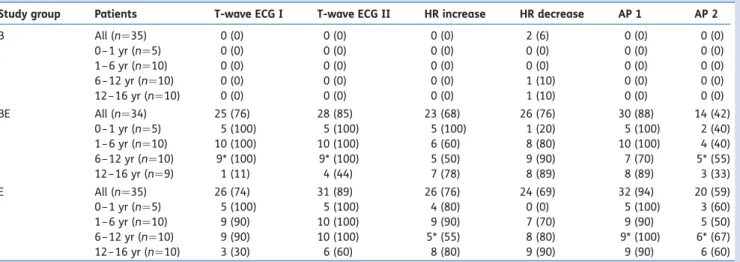
Table 2Number (%) of positive reactions such as T-wave elevation of≥25% in the ECG, increase or decrease in HR of ≥10 beats min21, and
increase in systolic AP of≥15 mm Hg at 1 (AP 1) and 2 (AP 2) min after test dose injection. *Because of a technical problem, results of one patient are missing
Study group Patients T-wave ECG I T-wave ECG II HR increase HR decrease AP 1 AP 2 B All (n¼35) 0 (0) 0 (0) 0 (0) 2 (6) 0 (0) 0 (0) 0– 1 yr (n¼5) 0 (0) 0 (0) 0 (0) 0 (0) 0 (0) 0 (0) 1– 6 yr (n¼10) 0 (0) 0 (0) 0 (0) 0 (0) 0 (0) 0 (0) 6– 12 yr (n¼10) 0 (0) 0 (0) 0 (0) 1 (10) 0 (0) 0 (0) 12 –16 yr (n¼10) 0 (0) 0 (0) 0 (0) 1 (10) 0 (0) 0 (0) BE All (n¼34) 25 (76) 28 (85) 23 (68) 26 (76) 30 (88) 14 (42) 0– 1 yr (n¼5) 5 (100) 5 (100) 5 (100) 1 (20) 5 (100) 2 (40) 1– 6 yr (n¼10) 10 (100) 10 (100) 6 (60) 8 (80) 10 (100) 4 (40) 6– 12 yr (n¼10) 9* (100) 9* (100) 5 (50) 9 (90) 7 (70) 5* (55) 12 –16 yr (n¼9) 1 (11) 4 (44) 7 (78) 8 (89) 8 (89) 3 (33) E All (n¼35) 26 (74) 31 (89) 26 (76) 24 (69) 32 (94) 20 (59) 0– 1 yr (n¼5) 5 (100) 5 (100) 4 (80) 0 (0) 5 (100) 3 (60) 1– 6 yr (n¼10) 9 (90) 10 (100) 9 (90) 7 (70) 9 (90) 5 (50) 6– 12 yr (n¼10) 9 (90) 10 (100) 5* (55) 8 (80) 9* (100) 6* (67) 12 –16 yr (n¼10) 3 (30) 6 (60) 8 (80) 9 (90) 9 (90) 6 (60)
Discussion
The main findings in this study were that after i.v. injection of an LA test dose, ECG and haemodynamic alterations were significantly influenced by epinephrine. I.V. injection of a test dose of plain bupivacaine had no effect on ECG, AP, or HR. After IVEA, manifestation of elevated T-wave and changes in HR were age-dependent, whereas an increase in systolic pressure was age-independent. A combination of all three of these cardiovascular variables allowed reliable detection of IVEA in all patients investigated.
Observation of needle and catheters for passive backflow of blood or the active aspiration of blood from needles or catheters in regional anaesthesia techniques have a failure rate of 57% to detect intravascular needle/catheter position.9 Although ultrasound-guided regional anaesthesia promises visual control of LA injection, bupivacaine-induced toxicity has been described.3 Similarly, the use of a less toxic LA may reduce the risk of systemic intoxication, but there are reports of LA toxicity, despite using ropivacaine or levobupi-vacaine.10 Thus, the ECG and haemodynamic alterations play an important role in the early detection of inadvertent intravascular LA administration.
Recent animal studies demonstrate that T-wave elevation associated with LA test dose is caused by epinephrine and
Table 3Quantitative changes in T-wave (ECG traces I and II), HR, and systolic AP at 1 and 2 min after test dose injection and number of patients with intermittent loss of P-wave. Results are median (range)
Study drug
Patients T-wave ECG I (elevation in % above baseline) T-wave ECG II (elevation in % above baseline) HR increase (beats min21above baseline) HR decrease (beats min21 below baseline) AP 1 (changes in mm Hg) AP 2 (changes in mm Hg) Loss of P-wave (number of pat.) B All (n¼35) 0 (0%) 0– 1 yr (n¼5) 0 (0 –17) 0 (0– 17) 0 (0– 0) 25 (26 to 24) 21 (25 to 3) 21 (25 to 1) 0 (0%) 1– 6 yr (n¼10) 0 (0 –0) 0 (0– 0) 0 (0– 2) 24 (26 to 22) 0 (26 to 6) 0 (28 to 6) 0 (0%) 6– 12 yr (n¼10) 0 (0 –0) 0 (0– 0) 0 (0– 5) 24 (210 to 0) 2.5 (25 to 8) 0 (28 to 7) 0 (0%) 12 –16 yr (n¼10) 0 (0 –0) 0 (0– 0) 0 (0– 2) 25 (210 to 21) 23 (223 to 5) 21.5 (219 to 7) 0 (0%) BE All (n¼34) 6 (18%) 0– 1 yr (n¼5) 166 (83 – 167) 200 (100 – 225) 27 (13 –35) 24 (219 to 0) 29 (21 –61) 14 (6 –45) 1 (20%) 1– 6 yr (n¼10) 100 (25 – 250) 175 (67 –275) 12.5 (4– 26) 219.5 (245 to 25) 30.5 (20 –44) 14 (7 –26) 1 (10%) 6– 12 yr (n¼10) 150 (502400) 145 (25 –700) 9.5 (1218) 222 (236 to 23) 26 (11 –44) 15 (5 –31) 2 (20%) 12 –16 yr (n¼9) 0 (0 –300) 0 (0– 300) 15 (5237) 217 (229 to 29) 25 (10 –58) 9 (5 –42) 2 (22%) E All (n¼35) 11 (31%) 0– 1 yr (n¼5) 133 (100 –366) 140 (133 – 200) 27 (9– 44) 0 (216 to 0) 27 (25 –49) 16 (12 –28) 0 (0%) 1– 6 yr (n¼10) 116.5 (0 –300) 200 (50 –400) 19.5 (8– 35) 216.5 (223 to 25) 26.5 (11 –38) 15 (7 –25) 2 (20%) 6– 12 yr (n¼10) 100 (0 –200) 145 (29 –250) 10 (0– 39) 221.5 (243 to 0) 29 (18 –66) 18 (8 –37) 5 (50%) 12 –16 yr (n¼10) 0 (0 –200) 39.5 (0– 350) 15.5 (7– 28) 222 (232 to 29) 34.5 (14 –62) 17 (7 –30) 4 (40%) 1.0 0.8 0.6 0.4 0.2 0.0
Probability of positive T-elevation
0 5 10
Age (yr)
15
Fig 1 Probability of occurrence of T-elevation (ECG lead II) after i.v. injection of an epinephrine-containing test dose (BE, E) related to patient’s age. Green dotted lines indicate 95% confi-dence interval.
that a test dose of plain bupivacaine did not alter the ECG.4 In contrast, T-elevations provoked by plain bupivacaine occur after intravascular injection of a large, potentially toxic dose.11 The current study in children confirms the results from animal investigations.4
In the past, studies investigating the sensitivity of an LA test dose in children have reported different and not always convincing sensitivities of T-elevation, AP, and HR to detect intravascular injection.2Owing to possible fatal conse-quences of a systemic LA intoxication, the use of an epinephrine-containing test dose has been recommended anyhow,2 12 13but due to unequal results and weak evidence, it is often not adhered to in clinical practice (several personal communications). Confounding factors of the test dose effects observed in previous studies might be related to the
anaesthetic agent used, the administration of atropine, pre-medication, patent’s age and sex, the ECG lead observed, the LA investigated, or the amount of epinephrine applied. Therefore, the results have to be interpreted within the spe-cific setting. Polaner and colleagues14 investigated positive intravascular test dose criteria during propofol– remifentanil anaesthesia and obtained quite different results compared with the current study conducted during sevoflurane anaes-thesia. Thus, our results are only valid under sevoflurane-based anaesthesia.
In several investigations, the ECG lead II was the preferred lead to assess T-wave elevations.6 15 16 There is only one study investigating ECG lead and usefulness of the T-wave criterion in anaesthetized children aged 6 –49 months.17 The authors conclude that the ECG lead does not affect
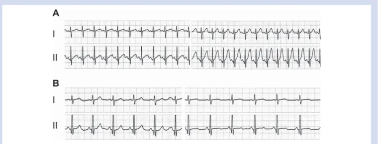
Fig 2ECG traces lead I and II before (left) and after (right) injection of a test dose bupivacaine+epinephrine. (A) 6-month-old boy. (B)
13.2-yr-old boy.
Table 4 Number (%) of positive reactions upon test dose injection if multiple variables were combined. *In one patient, max. HR and AP at 1 min (AP 1) failed to be recorded
Study drug Patients T-wave or HR increase T-wave or HR increase or HR decrease T-wave or AP 1 HR increase or AP 1 HR increase or HR decrease or AP 1 B All (n¼35) 0 (0) 2 (6) 0 (0) 0 (0) 2 (6) 0 – 1 yr (n¼5) 0 (0) 0 (0) 0 (0) 0 (0) 0 (0) 1 – 6 yr (n¼10) 0 (0) 0 (0) 0 (0) 0 (0) 0 (0) 6 – 12 yr (n¼10) 0 (0) 1 (10) 0 (0) 0 (0) 1 (10) 12 –16 yr (n¼10) 0 (0) 1 (10) 0 (0) 0 (0) 1 (10) BE All (n¼34) 33 (97) 34 (100) 33 (97) 31 (91) 33 (97) 0 – 1 yr (n¼5) 5 (100) 5 (100) 5 (100) 5 (100) 5 (100) 1 – 6 yr (n¼10) 10 (100) 10 (100) 10 (100) 10 (100) 10 (100) 6 – 12 yr (n¼10) 10 (100) 10 (100) 10 (100) 8 (80) 9 (90) 12 –16 yr (n¼9) 8 (89) 9 (100) 8 (89) 8 (89) 9 (100) E All (n¼35) 35 (100) 35 (100) 35 (100) 33 (97) 35 (100) 0 – 1 yr (n¼5) 5 (100) 5 (100) 5 (100) 5 (100) 5 (100) 1 – 6 yr (n¼10) 10 (100) 10 (100) 10 (100) 10 (100) 10 (100) 6 – 12 yr (n¼10) 10 (100) 10 (100) 10 (100) 9* (100) 10 (100) 12 –16 yr (n¼10) 10 (100) 10 (100) 10 (100) 9 (90) 10 (100)
recognition of T-elevation. In the current study, we found a slight superiority of lead II over lead I in adolescents but not in younger children. Tanaka and Nishikawa6found a sig-nificant negative correlation between the patient’s age and increase in T-wave. In the current study, the appearance of a positive T-elevation was age-dependent too with a high reliability up to about 12 yr of age. In adults, a decrease of ≥25% of T-wave amplitude is the typical reaction upon injec-tion of an epinephrine-containing test dose.18 According to the literature,6 15maximal change in T-wave amplitude occurred within 1 min of test dose administration. As reported, T-elevation most often was the first sign, followed by HR increase and later on by changes in AP.2 Current results are comparable with other reports,9which found a transient increase in T-wave of 33 –250% (mean 157%) lasting over 15–30 s.
False-negative results for HR increase to detect intravas-cular injection of an epinephrine-containing test dose were reported to be 29% of children during halothane anaesthe-sia.19Other authors using sevoflurane for general anaesthe-sia found a sensitivity of 71%16and 100%,6respectively. The current investigations showed a remarkable age-dependency of HR upon test dose injection. As observed in the current study, secondary HR decrease and loss of P-wave are reported in the literature.12
Reported mean maximal increases in systolic AP of 225 and 36 mm Hg17are consistent with our results 1 min after test dose injection. The values obtained after 2 min were remarkably lower, most likely due to the short half-life of epinephrine. Therefore, it is important to perform AP meas-urement at 1 min intervals during injection of test dose and remaining LA volume.
Most studies investigating the effects of an i.v. injected epinephrine-containing test dose in children use epinephrine in a dose of 0.5 mg kg21.5 6 14 16 17 19 Recom-mendations for clinical use2 are based on these observa-tions. In daily practice, remarkably higher test doses were applied.9 20 As proposed by Gerheuser and Roth,12 in the author’s institution, a test dose of 0.2 ml kg21LA with epi-nephrine 5 mg ml21 has been used routinely for many years, providing excellent safety with no systemic LA intoxi-cation and no adverse events observed upon test dose injection.
The clinical implications of this study are that LA test dose must contain epinephrine to detect intravascular injection by ECG and by changes in HR and AP. ECG monitoring is manda-tory and lead II might be preferable to lead I. Not solely an increase in HR but also a decrease can be associated by intra-vascular test dose injection and has to be taken as a positive result. Most of the test dose effects are age-dependent in dif-ferent extents. Therefore to achieve best safety profile, mul-tiple variables have to be observed.
In conclusion, this study demonstrates that ECG and haemodynamic alterations are significantly influenced by epinephrine-containing test doses. Since effects on T-elevation, HR, and BP are age-dependent, all three para-meters should be assessed to detect intravascular injection
of an epinephrine-containing test dose with a reliability of 100%.
Declaration of interests
None declared.Funding
Departmental resources.References
1 Badgwell JM, Heavner JE, Kytta J. Bupivacaine toxicity in young pigs is age-dependent and is affected by volatile anesthetics. An-esthesiology 1990; 73: 297 –303
2 Tobias JD. Caudal epidural block: a review of test dosing and rec-ognition of systemic injection in children. Anesth Analg 2001; 93: 1156– 61
3 Lin EP, Aronson LA. Successful resuscitation of bupivacaine-induced cardiotoxicity in a neonate. Paediatr Anaesth 2010; 20: 955– 7
4 Mauch J, Kutter AP, Madjdpour C, et al. Electrocardiographic alterations during intravascular application of three different test doses of bupivacaine and epinephrine: experimental study in neonatal pigs. Br J Anaesth 2010; 104: 94– 7
5 Tanaka M, Nishikawa T. Simulation of an epidural test dose with intravenous epinephrine in sevoflurane-anesthetized children. Anesth Analg 1998; 86: 952– 7
6 Tanaka M, Nishikawa T. Evaluating T-wave amplitude as guide for detecting intravascular injection of a test dose in anesthetized children. Anesth Analg 1999; 88: 754 –8
7 Zeileis A, Hothorn T. Diagnostic checking in regression relation-ships. R News 2002; 2: 7– 10. Available from http://CRAN.R-project.org/doc/Rnews/
8 Pinheiro J, Bates D, DebRoy S, Sarkar D; The R Core Team. nlme: Linear and Nonlinear Mixed Effects Models. R package version 3.1-96, 2009. Available from http://cran.r-project.org/web/ packages/nlme/index.html
9 Fisher QA, Shaffner DH, Yaster M. Detection of intravascular injec-tion of regional anaesthetics in children. Can J Anaesth 1997; 44: 592– 8
10 Available from http://www.lipidrescue.org (accessed 22 August 2011)
11 Mauch J, Kutter APN, Madjdpour C, et al. Electrocardiographic changes during continuous intravenous application of bupiva-caine in neonatal pigs. Br J Anaesth 2010; 105: 437 –41 12 Gerheuser F, Roth A. Epidural anesthesia. Anaesthesist 2007; 56:
499– 523
13 Neal JM, Bernards CM, Buttwerworth JF, et al. ASRA practice ad-visory on local anesthetic systemic toxicity. Reg Anesth Pain Med 2010; 35: 152–61
14 Polaner DM, Zuk J, Luong K, Pan Z. Positive intravascular test dose criteria in children during total intravenous anesthesia with pro-pofol and remifentanil are different than during inhaled anesthe-sia. Anesth Analg 2010; 110: 41– 5
15 Tanaka M, Nishikawa T. The efficacy of a simulated intravascular test dose in sevoflurane-anesthetized children: a dose–response study. Anesth Analg 1999; 89: 632 –7
16 Kozek-Langenecker SA, Marhofer P, Jonas K, Macik T, Urak G, Semsroth M. Cardiovascular criteria for epidural test dosing in sevoflurane- and halothane-anesthetized children. Anesth Analg 2000; 90: 579– 83
17 Ogasawara K, Tanaka M, Nishikawa T. Choice of electro-cardiography lead does not affect the usefulness of the T-wave criterion for detecting intravascular injection of an epi-nephrine test dose in anesthetized children. Anesth Analg 2003; 97: 372 – 6
18 Tanaka M, Nishikawa T. A comparative study of hemodynamic and T-wave criteria for detecting intravascular injection of the
test dose (epinephrine) in sevoflurane-anesthetized adults. Anesth Analg 1999; 89: 32 –6
19 Desparment J, Mateo J, Ecoffey C, Mazoit X. Efficacy of an epidural test dose in children anesthetized with halothane. Anesthesi-ology 1990; 72: 249– 51
20 Sparks JW, Seefelder C. Neonatal T-wave elevation from a posi-tive epidural test dose. Paediatr Anaesth 2005; 15: 706– 7