Pleural tumors as sole primary
manifestation of acute
promyelocytic leukemia
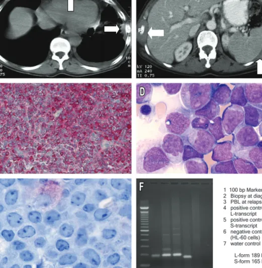
A 66-year-old woman was admitted with progressive weakness, a weight loss of 22 pounds in the last 4 months, and diffuse thoracic pain. Laboratory analysis revealed anemia of 108 g/l, normal thrombocytes (242 ·109/l) and normal leukocytes (4.3 ·109/l). CT scan demonstrated four pleural tumors with up to 8 cm in diameter without evidence of disease elsewhere (Figure 1A, B). Bone scintigraphy indicated hot spots over the ribs on both sides. Biopsy of the pleural tumors showed cells staining positive for myeloperoxidase, CD34, CD43, CD45 and lysozyme, indicating myelogenous sarcoma (Figure 1C). Chromosomal translocations such as t(9;22) (BCR-ABL), t(8;21) (AML1-ETO), inv(16) (CBFb-MYH11), as well as mutations of the FLT3 gene were excluded by PCR in cells from the pleural tumors. Bone marrow biopsy was normal suggesting the diagnosis of an exclusively extramedullary manifestation of acute myeloid leukemia (AML) in the pleura. The patient was treated with two cycles of chemotherapy, including
daunorubicin and cytarabine in the first cycle, and cytarabine alone in the second cycle. At the end of therapy, CT scan showed residual pleural tumors of up to 2 cm.
Five months later the patient reported costal pains again. CT scan demonstrated pleural tumors progressive in size and number. The patient was admitted because of nasal bleeding due to severe thrombocytopenia (6 ·109/l). In addition, 64% of peripheral leukocytes (8.2 ·109/l) were myeloid blasts. The bone marrow was highly infiltrated with myeloid blasts having an identical immunophenotype as the cells from the pleural tumors at diagnosis (Figure 1D, E). A relapse of AML was diagnosed and a therapy with hydroxyurea was initiated. The patient died 7 days later of pulmonary infection.
Surprisingly, the cytogenetic analysis at relapse (finished post mortem) indicated the presence of t(15;17) specific for the acute promyelocytic leukemia (APL) subtype of AML [1]. Retrospective analysis revealed that PML/RARa transcripts were detectable in pleural tumors already at diagnosis (Figure 1F). However, bone marrow and peripheral blood at diagnosis were negative by PCR for PML/RARa transcripts.
letters to the editor
Annals of OncologyAlmost all cases of APL are defined at the molecular level by the presence of a balanced reciprocal translocation t(15;17) resulting in the PML-RARa fusion gene [1]. APL is the most curable subtype of AML [2, 3]. With current therapy, including all trans retinoic acid (ATRA) and anthracycline-based chemotherapy, 70%–80% of patients are free of disease at 5 years [2, 3].
Extramedullary disease in APL may occur at relapse, but it is a very rare clinical event at diagnosis, with half of the reported cases involving the skin [4, 5]. Other sites involve the central nervous system, gingival, lymph nodes and spleen. To our knowledge this is the first report of pleural tumors as sole extramedullary manifestation of APL at diagnosis. Our case indicates that determination of t(15;17) should be included in the panel of markers assessed at diagnosis of all AML patients, including patients with exclusively extramedullary AML. This also allows timely diagnosis of APL at unusual clinical presentation and thus possibly live-saving therapy with ATRA.
B. U. Mueller1, A. U. Buerger1, M. Solenthaler2, E. M. Garamvoelgyi3, E. Oppliger-Leibundgut4, M. F. Fey5& T. Pabst5*
Departments of1Internal Medicine and2Hematology,3Institute of Pathology;4Laboratory for Molecular Diagnostics and5Institute of
Medical Oncology, University Hospital, 3010 Bern, Switzerland (*E-mail: thomas.pabst@insel.ch)
references
1. Rowley JD, Golomb HM, Dogherty C. 15/17 translocation: a consistent chromosomal change in acute promyelocytic leukemia. Lancet 1977; 1: 549–550.
2. Chou WC, Dang CV. Acute promyelocytic leukemia: recent advances in therapy and molecular basis of response to arsenic therapies. Curr Opin Hematol 2005; 12: 1–6.
3. Mistry AR, Pedersen EW, Solomon E, Grimwade D. The molecular pathogenesis of acute promyelocytic leukaemia. Implications for the clinical management of the disease. Blood 2003; 17: 71–97.
4. Menendez A, Gonzalez A, Cabrera H et al. Clinical spectrum of extramedullary acute promyelocytic leukemia. Eur J Haematol 2000; 64: 201–203.
5. Wiernik PH, De Bellis R, Muxi P, Dutcher JP. Extramedullary acute promyelocytic leukemia. Cancer 1996; 78: 2510–2514.
doi:10.1093/annonc/mdj062
Published online 15 November 2005
Figure 1. (A, B) CT scan at diagnosis showing large pleural tumors. (C) Pleural biopsy demonstrating blasts staining positive for myeloperoxidase, indicating granulocytic sarcoma. (D) Bone marrow aspirate and (E) bone marrow biopsy at relapse, indicating diffuse infiltration with myeloid blasts. (F) RT-PCR analysis detecting PML-RARa transcripts in the pleural tumors at diagnosis (lane 2) as well as in leukemic blasts at relapse (lane 3).
Annals of Oncology