HAL Id: hal-01834263
https://hal.umontpellier.fr/hal-01834263
Submitted on 27 Jan 2021
HAL is a multi-disciplinary open access
archive for the deposit and dissemination of
sci-entific research documents, whether they are
pub-lished or not. The documents may come from
teaching and research institutions in France or
abroad, or from public or private research centers.
L’archive ouverte pluridisciplinaire HAL, est
destinée au dépôt et à la diffusion de documents
scientifiques de niveau recherche, publiés ou non,
émanant des établissements d’enseignement et de
recherche français ou étrangers, des laboratoires
publics ou privés.
Severe Osteoarthritis of the Knee: A Phase I
Dose-Escalation Trial
Yves-Marie Pers, Lars Rackwitz, Rosanna Ferreira, Oliver Pullig, Christophe
Delfour, Frank Barry, Luc Sensebe, Louis Casteilla, Sandrine Fleury, Philippe
Bourin, et al.
To cite this version:
Yves-Marie Pers, Lars Rackwitz, Rosanna Ferreira, Oliver Pullig, Christophe Delfour, et al..
Adi-pose Mesenchymal Stromal Cell-Based Therapy for Severe Osteoarthritis of the Knee: A Phase
I Dose-Escalation Trial.
Stem Cells Translational Medicine, Wiley, 2016, 5 (7), pp.847 - 856.
�10.5966/sctm.2015-0245�. �hal-01834263�
Human Clinical Article
Adipose Mesenchymal Stromal Cell-Based
Therapy for Severe Osteoarthritis of the Knee:
A Phase I Dose-Escalation Trial
YVES-MARIEPERS,a,bLARSRACKWITZ,cROSANNAFERREIRA,aOLIVERPULLIG,d
CHRISTOPHEDELFOUR,eFRANKBARRY,fLUCSENSEBE,gLOUISCASTEILLA,h,iSANDRINEFLEURY,g,h,i PHILIPPEBOURIN,g,jDANI`ELENO¨EL,bFRANÇOISCANOVAS,kCATHERINECYTEVAL,lGINALISIGNOLI,m JOACHIMSCHRAUTH,nDANIELHADDAD,nSOPHIEDOMERGUE,oULRICHNOETH,c
CHRISTIANJORGENSEN,a,bON BEHALF OF THEADIPOA CONSORTIUM
Key Words. Osteoarthritisx Adipose mesenchymal stromal cells x
Intra-articular injectionx Therapeutic potential x Regenerative medicine x Phase I clinical trial
ABSTRACT
Osteoarthritis (OA) is the most widespread musculoskeletal disorder in adults. It leads to cartilage damage associated with subchondral bone changes and synovial inflamma-tion, causing pain and disability. The present study aimed at evaluating the safety of a dose-escalation protocol of intra-articular injected adipose-derived stromal cells (ASCs) in patients with knee OA, as well as clinical efficacy as secondary endpoint. A bicentric, uncontrolled, open phase I clinical trial was conducted in France and Germany with regulatory agency approval for ASC expansion procedure in both countries. From April 2012 to December 2013, 18 consecutive patients with symptomatic and severe knee OA were treated with a single intra-articular injection of autologous ASCs. The study design consisted of three consecutive cohorts (six patients each) with dose esca-lation: low dose (23 106cells), medium dose (103 106), and high dose (503 106). The primary outcome parameter was safety evaluated by recording adverse events through-out the trial, and secondary parameters were pain and function subscales of the West-ern Ontario and McMaster Universities Arthritis Index. After 6 months of follow-up, the procedure was found to be safe, and no serious adverse events were reported. Four pa-tients experienced transient knee joint pain and swelling after local injection. Interest-ingly, patients treated with low-dose ASCs experienced significant improvements in pain levels and function compared with baseline. Our data suggest that the intra-articular injection of ASCs is a safe therapeutic alternative to treat severe knee OA pa-tients. A placebo-controlled double-blind phase IIb study is being initiated to assess clin-ical and structural efficacy. STEMCELLSTRANSLATIONALMEDICINE2016;5:847–856
SIGNIFICANCE
Although this phase I study included a limited number of patients without a placebo arm, it showed that local injection of autologous adipose-derived stem cells was safe and well tolerated in patients with knee osteoarthritis. This study also provides en-couraging preliminary evidence of efficacy. Larger and controlled long-term studies are now mandatory to confirm whether this new strategy of cell therapy can improve pain and induce structural benefit in osteoarthritis.
INTRODUCTION
Osteoarthritis (OA) is a multifactorial, slowly progressive degenerative disorder of the joints leading to irreversible damage of the cartilage, sclerosis of subchondral bone, and synovial inflammation [1]. As a consequence
of increasing longevity and obesity, the cost of OA to the health care system rapidly grows. Current treatment strategies have no impact on the progressive degeneration of joint tissues. In this context, the use of mesenchymal stromal stem cells (MSCs) is an attractive therapeutic option thanks to
a
Clinical Immunology and
Osteoarticular Diseases Therapeutic Unit, Lapeyronie University Hospital, Montpellier, France;bINSERM, U1183, Saint-Eloi Hospital, Montpellier, France;
c
Department of Orthopaedic Surgery, K¨onig-Ludwig-Haus, University of W¨urzburg, W¨urzburg, Germany;
d
Fraunhofer Institute for Interfacial Engineering and Biotechnology IGB, Translational Center“Regenerative Therapies for Oncology and
Musculoskeletal Diseases,” W¨urzburg, Germany;eDepartment for Cell and Tissue Pathobiology of Tumor, Hospital Saint Eloi, Montpellier, France;
f
Regenerative Medicine Institute, Galway University, Galway, Ireland;
g
Etablissement Français du Sang, Toulouse, France;hINSERM U1031 STROMAlab, Toulouse, France;iCNRS, Universit ´e Toulouse III, UPS UMR5273 F-31 432 STROMAlab, Toulouse, France;
jUnivercell Biosolutions, Toulouse,
France;kDepartment of Orthopaedic Surgery, Lapeyronie University Hospital, Montpellier, France;lDepartment of Radiology, Lapeyronie Hospital, Montpellier, France;mLaboratory of Immunorheumatology and Tissue Regeneration, Istituto Ortopedico Rizzoli, Bologna, Italy;nMRB Research Center Magnetic Resonance Bavaria, W¨urzburg, Germany;oMaxillofacial, Plastic Reconstructive, and Aesthetic Surgery Department, Gui de Chauliac Hospital, Montpellier, France
Correspondence: Christian Jorgensen, M.D., Ph.D., Clinical Immunology and Osteoarticular Diseases Therapeutic Unit, CHRU Lapeyronie, 371, avenue du doyen Gaston Giraud, 34295 Montpellier, France; INSERM U1183, IFR3, Universit ´e Montpellier I, Hopital Saint Eloi Batiment INM, 80 rue Augustin Fliche BP, 74103-34091 Montpellier cedex 5, France. Telephone: (33) 4 67 33 77 98; E-Mail: christian.jorgensen@ inserm.fr
Received September 12, 2015; accepted for publication January 13, 2016; published Online First on May 23, 2016. ©AlphaMed Press
1066-5099/2016/$20.00/0 http://dx.doi.org/ 10.5966/sctm.2015-0245
S
TEMC
ELLST
RANSLATIONALM
EDICINE2016;5:847
–856 www.StemCellsTM.com
©AlphaMed Press 2016their chondrogenic and anti-inflammatory properties [2]. Adipose tissue-derived MSCs (ASCs) share similar properties with bone marrow-derived MSCs but are easier to collect for clinical applica-tion, with higher isolation yields. Indeed, intra-articular (IA) injec-tion of ASCs prevented OA onset in a collagenase-induced murine knee OA model and reduced synovitis, osteophyte formation, and cartilage degeneration [3]. Furthermore, intra-articular injection of 2 or 6 million autologous ASCs improved the cartilage degrada-tion score and significantly reduced knee synovitis in a biomechan-ical induced OA rabbit model [4].
Using an established Good Manufacturing Practice (GMP) procedure based on ASCs expanded for 2 weeks in the presence of platelet lysate [5], we conducted a proof-of-concept phase I clinical trial to assess the safety and efficacy of intra-articular injection of autologous ASCs in patients with active and severe knee OA.
PATIENTS ANDMETHODS Study Design
A phase I, prospective, bicentric, single-arm, open-label, dose-escalating clinical trial of a single injection of autologous ASCs in patients with severe primary knee OA was conducted from March 2012 to April 2014 in two hospitals: CHRU Montpellier (France) and the Department of Orthopedic Surgery at the Uni-versity of W¨urzburg (Germany). No placebo group was scheduled because of ethical issues (including late-stage knee OA patients associated with liposuction procedure without active therapy benefit). The study protocol was approved by the local ethics committees of both institutions (Comit ´e de Protection des Per-sonnes of Montpellier [UF8606-120203] and Ethik-Kommission bei der Medizinischen of W¨urzburg) and by the national compe-tent authorities (TC301; EudraCT no. 2011-000183-10).
Patient Selection and Enrollment
A total of 48 outpatients with knee OA were screened (Fig. 1). Eighteen consecutive patients with primary femorotibial knee OA diagnosed according to the clinical and radiological criteria of the American College of Rheumatology were enrolled in this study after written informed consent was obtained [6].
Inclusion Criteria
Patients 50–75 years of age with symptomatic primary knee OA and radiographic changes of grade 3 to 4 according to the Kellgren-Lawrence scale in the targeted knee were included [7]. To obtain histologic analysis for safety issues, the medical board required end-stage knee OA patients with an indication of knee prosthesis in the year after inclusion. Symptomatic primary knee OA was defined by daily knee pain for at least 12 months before study inclusion.
Exclusion Criteria
Patients were excluded if they had secondary arthritis (related to rheumatoid arthritis, spondyloarthritis, previous articular frac-tures, postinfectious arthritis, and crystal arthropathies), autoim-mune disorders, or previous malignancies in the past 5 years. Previous administration of oral/intra-articular corticosteroids and injection of hyaluronic acid derivatives within 6 months be-fore screening examination were also exclusion criteria.
Treatment Allocation
Eligible patients were consecutively allocated to the treatment groups, three arms with different doses (23 106, 103 106, and 503 106cells) (Fig. 1). The starting dose of 23 106cells has been defined based on the No Observed Adverse Effect Level obtained after IA administration determined in preclinical studies Figure 1. Flow chart of the clinical trial. Abbreviation: ASC, adipose-derived stromal cell.
performed in goat and rabbit models of OA, adjusted by allome-tric factors (weight and size of the knee joint compared with hu-man) [4] (data not shown).
First, the patients underwent outpatient liposuction under lo-cal anesthesia, and autologous ASCs were produced and prepared at a single GMP facility (Etablissement Français du Sang Midi-Pyr ´enn ´ees, France), as summarized in the supplemental online data. Fourteen days after isolation, ASCs were recovered and un-derwent defined quality control before shipping (supplemental online data). A single IA dose of ASCs was injected into the knee joint (volume, 5 ml) under ultrasound control.
Cell Preparation and Expansion of ASCs
The procedure has been described [5]. The stromal vascular fraction (SVF) was obtained by means of collagenase digestion. Al-iquots of 10 g of adipose tissue were mixed with 34 ml of collagenase solution (NB6; Coger, Paris, France, http://www. cogerbio.com) and incubated at 37°C for 45 minutes. Enzymatic di-gestion was stopped by the addition of complete culture medium (CCM) containing minimum essential medium (MacoPharma, Tourcoing, France, http://www.macopharma.com), human plate-let growth factor-enriched plasma, 10 mg/ml ciprofloxacin, and 1 U/ml heparin. After homogenization, the digested suspen-sion was passed through sterile 100-mm filters. The cells were centrifuged at room temperature for 10 minutes at 600g. The supernatant was discarded, and the SVF was resuspended in 20 ml of CCM. An aliquot of the SVF was removed for quality con-trol: cell count, viability, phenotyping (CD34, CD45, and CD14), and sterility.
Cells from the SVF were then seeded in a 1,270-cm2CellStack culture chamber (MacoPharma) at a density of 43 103cells per cm2in CCM, by use of a seeding kit (MacoPharma), at 37°C in an
atmosphere saturated with moisture and 5% CO2. After an initial
24-hour incubation, the nonadherent cells were removed. The adherent cells were washed once with Dulbecco’s phosphate-buffered saline (PBS), and CCM medium was added for 7 days. The medium was completely replaced at days 4 and 6 of culture with the use of medium exchange kits (MacoPharma). At day 8 (primary culture, P0), the cells were harvested with the use of a detachment kit (MacoPharma) according to the following protocol. After aspi-ration of the medium and washing with Dulbecco’s PBS, 50 ml of irradiated trypsin solution was added for 5 minutes at room temperature. After inhibition of trypsin activity by the addition of CCM, the cells were collected in a transfer bag (MacoPharma). An aliquot of the cell suspension was aseptically removed for cell count, viability, phenotyping (CD34, CD45, and CD14), measures of hTERT messenger RNA contents by quantitative reverse-transcription polymerase chain reaction, and assessment of microbial testing.
The cells were seeded in 1,270-cm2CellStack culture cham-bers at a density of 23 103cells per cm2and incubated for 6 days. The CCM was completely replaced at days 11 and 13. At day 11, an aliquot of culture medium was aseptically removed for my-coplasma and endotoxin testing. At day 14, the cells were har-vested according to the procedure described above. The cell suspension was placed in a transfer bag (MacoPharma) and washed with Dulbecco’s PBS. The ASCs were then resus-pended in a solution containing 3.6% human albumin (pro-vided by Laboratoire Français du Fractionnement et des Biotechnologies, Courtaboeuf, France) and a polyionic solu-tion containing glucose. An aliquot of the ASC suspension was aseptically removed for cell count, and its quality was evaluated as described above.
Flow cytometry analyses were performed as follows. Briefly, ASCs (23 105cells) were stained with saturating amounts of Table 1. Patient demographic and baseline characteristics of each group (low, medium, and high dose, n = 6 each)
Characteristic Low dose Medium dose High dose
Age, yr 63.26 4.1 65.56 8.1 65.26 2.3 Women 3 (50) 3 (50) 4 (66.7) BMI, kg/m2 28.86 1.5 26.966 3.1 27.16 2.4 Kellgren-Lawrence system Grade III 2 (33) 1 (17) 0 Grade IV 4 (66) 5 (83) 6 (100) WOMAC (0–100 scale) Pain subscale 62.56 15.5 36.66 14.6 34.06 25.6 Stiffness subscale 58.56 27.9 54.56 17.9 45.36 31.5 Function subscale 63.66 16.7 44.46 17.9 37.36 26.5 Total index 60.76 18.6 47.26 14.7 38.86 27.3 Global knee pain VAS (0–100 mm) 776 15.7 63.76 20.5 43.76 25.4 PGA (0–100 mm) 306 21 326 17.9 46.76 20.7 KOOS index (0–100 mm) 346 15 426 9 45.26 13.6 SAS index (0–40 mm) 296 6 266 5 19.56 7.6 SF-36 Physical scale 30.96 8.2 29.96 6.2 35.76 10.6 Mental scale 55.96 8.3 51.96 10.2 53.66 7.8 Data are presented as n (%) or mean6 SD.
Abbreviations: BMI, body mass index; WOMAC, Western Ontario and McMaster Universities Osteoarthritis Index; VAS, visual analog scale; PGA, patient global assessment; KOOS, Knee Injury and Osteoarthritis Outcome; SAS, short arthritis assessment scale; SF-36, short-form 36 (quality of life). Pers, Rackwitz, Ferreira et al. 849
monoclonal antibodies conjugated with fluorescein isothiocyanate (FITC) or phycoerythrin (PE) and their respective isotype controls for 30 minutes in the dark at 4°C in PBS/0.5% human albumin and 0.1% sodium azide. After washing, the labeled cells were analyzed by flow cytometry (EPICS XL-MCL flow cytometer; Beckman-Coulter, Nyon, Switzerland, http://www.beckmancoulter.com). FITC anti-CD14, FITC anti-CD45, PE anti-CD34, PE anti-CD73, PE anti-CD90, PE anti-CD105, and immunoglobulin G1 PE and FITC were from BD Phar-mingen (Le Pont de Claix, France, http://www.bdbiosciences.com).
Release Criteria of ASCs
Release criteria were defined as negative for microbial testing on SVF, intermediate product (P0), and final product (P1); negative for mycoplasma testing on adipose tissue and cul-ture medium at day 11; endotoxin testing negative on culcul-ture medium at day 11; and absence of hTERT detection by quan-titative reverse-transcription polymerase chain reaction on intermediate product (P0). Finally, on active substance, cellu-lar viability had to be.90%. The percentage of positive cells Table 2. Summary of adverse events during the clinical trial
Variable
0–3 Months 3–6 Months
Low dose Medium dose High dose Low dose Medium dose High dose
AEs 10 4 12 8 2 1
Patients with AEs 6 (100%) 4 (67%) 5 (83%) 6 (100%) 2 (33%) 3 (50%)
Patients with serious AEs 0 1 0 0 0 0
Patients with serious infectious events 0 0 0 0 0 0 Biological changesa CRP.1–3 ULN 2 — — 1 — — ALT.1–3 ULN — 1 — — — — CPK.1–3 ULN — — — 1 — 1 Mild neutropenia, 900–1,499 cells per mm3 1 — — 1 — — Infections Nasal congestion 1 1 — — — —
Rhinitis and pharyngitis — — 3 — — —
Influenza syndrome 1 — — — —
Urinary tract infection 1 — — — — —
Dental infection 1 — — — — — Musculoskeletal disorders Joint effusion/swelling, treated kneeb 1 — 3 1 — — Sciatic pain — — — 1 — —
Low back pain — — 2 — — —
Trauma to the treated knee — — 1 — 2 —
Skin erythema around the
treated knee — — — 1 — — Shoulder pain 2 — — — — — Hip pain 1 — — — — — Neurological disorders Headache — — 1 — — — Gastrointestinal disorders Diarrhea — — 1 — — — Eye disorders Cataract — — — 2 — — Conjunctivitis — — 1 — — — Cardiovascular disorders Right coronary artery stenosisc
— 1 — — — —
a
Participants who had normal values at baseline.
bFive AEs related to ASCs. c
One serious AE not related to ASCs.
Abbreviations:—, no data; AE, adverse event; ALT, alanine aminotransferase; CPK, creatinine phosphokinase; CRP, C-reactive protein; ULN, upper limit of normal.
for hematopoietic markers (CD45 and CD14) had to be lower than 2%, and for mesenchymal markers, higher than 90% for CD90 and CD73 and higher than 80% for CD105. The per-centage of positive cells for CD34 had to be less than 10%.
Karyotype analyses were performed, on final product, for 15 productions. Because of the time required for performing the karyotype analysis, results were obtained after release. Karyotype analyses revealed no clonal abnormalities. Results Table 3. Effect of autologous ASC injection on OA clinical outcomes.
Outcome D, 1 week p value D, 3 months p value D, 6 months p value Low dose (23 106cells
injected) WOMAC pain 236.0 6 10.2 ,.001 241.7 6 10.2 ,.01 230.7 6 10.7 ,.05 WOMAC stiffness 241.2 6 10.6 ,.01 246.8 6 10.6 ,.001 235.3 6 11.1 ,.05 WOMAC function 244.0 6 12.2 ,.01 237.4 6 9.9 ,.01 235.7 6 10.5 ,.01 WOMAC total 238.6 6 8.6 ,.001 241.2 6 8.5 ,.001 233.1 6 8.9 ,.001 VAS pain 251.5 6 12.7 ,.01 254.4 6 12.7 ,.01 241.2 6 13.3 ,.05 KOOS index 34.96 8.7 ,.01 38.06 8.7 ,.001 31.86 9.1 ,.01 SAS index 215.2 6 4.8 ,.05 216.3 6 4.8 ,.01 211.3 6 5.0 .09 OARSI/OMERACT responders, % ND 83.3 80.0 SF-36 Physical scale 6.66 4.4 .34 12.86 4.4 ,.05 8.26 4.6 .33 Mental scale 252.9 6 3.0 .75 20.9 6 3.7 .99 24.0 6 3.8 .60 Medium dose (103 106cells
injected) WOMAC pain 26.3 6 9.5 .85 29.7 6 9.9 .65 212.4 6 9.9 .47 WOMAC stiffness 227.7 6 10.9 .052 216.2 6 11.4 .37 230.1 6 11.4 ,.05 WOMAC function 212.7 6 10.7 .51 29.9 6 11.2 .71 220.9 6 11.2 .19 WOMAC total 219.7 6 9.1 .11 212.7 6 9.6 .43 222.9 6 9.1 .054 VAS pain 220.8 6 11.9 .22 222.2 6 11.9 .18 227.0 6 11.9 .09 KOOS index 5.96 6.5 .69 4.96 6.5 .79 17.26 6.5 ,.05 SAS index 24.8 6 3.6 .41 24.8 6 3.6 .41 211.7 6 3.6 ,.05 OARSI/OMERACT responders, % ND 60.0 60.0 SF-36 Physical scale 20.62 6 4.0 .99 2.16 4.0 .92 5.46 4.0 .42 Mental scale 4.76 5.8 .76 0.16 5.8 .99 3.26 5.8 .91 High dose (503 106cells
injected) WOMAC pain 3.46 14.9 .99 223.7 6 14.9 .29 220.3 6 14.9 .41 WOMAC stiffness 20.7 6 7.1 .99 230.8 6 17.1 .21 225.8 6 17.1 .32 WOMAC function 7.96 14.9 .91 226.0 6 14.9 .23 221.8 6 14.9 .35 WOMAC total 24.1 6 15.3 .99 226.8 6 16.0 .26 222.6 6 16.0 .38 VAS pain 210.3 6 16.4 .86 221.3 6 16.4 .44 219.7 6 17.1 .54 KOOS index 4.86 12.5 .96 18.56 12.5 .34 20.06 13.1 .32 SAS index 21.5 6 6.3 .99 27.3 6 12.2 .52 29.3 6 6.6 .38 OARSI/OMERACT responders, % ND 60.0 60.0 SF-36 Physical scale 22.1 6 6.8 .98 0.66 6.8 .99 1.96 6.8 .98 Mental scale 1.46 6.6 .99 1.36 6.6 .99 0.56 6.6 .99 Data are mean6 SD unless noted otherwise. All indices and scores are on a natural 0–100 mm or normalized 0–100 scale, except the SAS (0–40 mm). Baseline values are reported in Table 1.D represents the mean change from baseline (at 1 week, 3 months, and 6 months postinjection) in the OA patients for clinical outcome parameters.
Abbreviations: ASC, adipose-derived stromal cell; KOOS, Knee Injury and Osteoarthritis Outcome Score; ND, not determined; OA, osteoarthritis; OARSI, Osteoarthritis Research Society International; OMERACT, Outcome Measures in Rheumatology; SAS, Short Arthritis Assessment Scale; SF-36, short-form 36 (quality of life); VAS, visual analog scale; WOMAC, Western Ontario and McMaster University Osteoarthritis Index.
Pers, Rackwitz, Ferreira et al. 851
for release criteria obtained for the three cohorts are presented in the supplemental online Appendix.
Outcome Measures
Primary Endpoint
Incidence, relatedness, and severity of treatment-emergent sus-pected unexsus-pected serious adverse reactions, serious adverse events, and adverse events (AEs) were documented at each visit throughout the study. Laboratory tests (hematology, blood chem-istry, and urinalysis), vital signs, and physical examinations of the patients were assessed systematically. A 12-week safety period was implemented between subject 1 and subject 2 of the first co-hort receiving the low dose, and the safety medical board autho-rized continuation with patients 2 to 6. A further 4-week safety period was scheduled between the other two cohorts.
Secondary Endpoints
Secondary efficacy endpoints were assessed by measuring the Western Ontario and McMaster Universities Arthritis Index (WOMAC), pain visual analog scale (VAS), the Patient Global As-sessment (PGA), the Short Arthritis AsAs-sessment Scale (SAS), and the Knee Injury and Osteoarthritis Outcome Score (KOOS index) [8]. A 0- to 100-mm VAS was used to assess WOMAC pain (5 ques-tions), physical function (17 quesques-tions), and stiffness (2 questions) subscales. Osteoarthritis Research Society International (OARSI)/ Outcome Measures in Rheumatology response was defined as 20% improvement compared with baseline VAS and WOMAC
[9]. Quality of life was measured by the short-form 36 (SF-36) questionnaire [10].
Secondary imaging endpoints included delayed gadolinium-enhanced magnetic resonance imaging of cartilage (dGEMRIC) and T1rhoMRI for selected German patients at 3–4 months after
ASC injection [11]. MRIs were evaluated by a radiologist blinded to the administered dose. dGEMRIC and T1rhomaps were motion
corrected and zerofilled and then derived using anatomical land-marks and an automated fit algorithm [12].
Histology
Upon request of the ethics committee, a total knee arthroplasty (TKA) was originally scheduled 3 months after ASC injection for all patients to obtain histologic analysis. However, if a patient re-fused TKA, knee arthroscopy with biopsy could be performed. No standardized protocol was planned for biopsies. Cartilage and synovial samples were fixed for 24 hours in 10% neutral for-mol and embedded in paraffin. Sections of 5-mm thickness were stained with hematoxylin and eosin, Alcian blue, or Toluidine blue. Immunohistochemistry was performed on a Benchmark Ul-tra Ventana automat with the following antibodies: protein S100 (1:3,200, polyclonal; Dako, Carpinteria, CA, http://www.dako. com), CD34 (1:100, QBEND/10; Dako), and Ki67 (1:100, mono-clonal mouse, clone Mib-1; Dako). The OARSI cartilage OA histo-pathology grading system was performed by an experienced anatomopathologist who was blinded to the treatments [13].
Statistical Analysis
All values are expressed as mean6 SD. The significance of differ-ences was assessed by Wilcoxon test or one-way analysis of var-iance and corresponding nonparametric tests. A value of p, .05 was considered statistically significant. All analyses were per-formed using GraphPad Prism software version 6.0 (GraphPad Software, La Jolla, CA, http://www.graphpad.com).
RESULTS
Characteristics of Patients
All three cohorts had similar baseline characteristics for age, sex, and body mass index, and 83% of patients were grade IV on the Kellgren-Lawrence scale (Table 1). Eleven patients were included in France and seven in Germany. Baseline levels for pain and func-tion (WOMAC, KOOS, SAS scores) were different between the co-horts (Table 1). Disease activity at baseline was higher in the group of patients injected with the low dose of ASCs, with higher VAS and WOMAC values. All patients completed the 6-month follow-up. Only one patient with persistent joint swelling and knee pain underwent TKA surgery at 6 months.
Safety and Tolerance Profile of IA Injection of Autologous ASCs
No AE associated with liposuction and IA injection was observed in this study (Table 2). No serious infectious AEs related to ASC injection occurred during follow-up (Table 2). Laboratory tests, vital signs, and electrocardiograms indicated no local or systemic safety concerns. One severe adverse event, unstable angina pectoris without in-creased cardiac markers, was reported in 1 patient 3 months after ASC injection. The patient’s risk factors included hypertension and hyperlipidemia. Five minor AEs reported by four patients were Figure 2. WOMAC pain and function improvement during the study.
Abbreviation: WOMAC, Western Ontario and McMaster Universities Arthritis Index.
potentially related to the procedure: slight knee pain/joint ef-fusion occurred during the first week after ASC injection that resolved with nonsteroidal anti-inflammatory drugs in three patients and spontaneously (without medication) in one patient (Table 2).
Otherwise, a small increase in creatinine phosphokinase was observed in two patients and in alanine aminotransferase in one patient. There was also a mild decrease of neutrophil count in one patient who presented with a low baseline count (1,500/mm3) and high variability of neutrophil count, independent of IA injec-tion, during follow-up.
Efficacy Profile of Autologous ASC Injection on OA Clinical Outcomes
Mean changes from baseline to 1 week, 3 months, and 6 months in clinical outcomes are summarized in Table 3. Improvement for all clinical outcome parameters (pain, function, and mobility) regard-less of the injected dose was observed (Fig. 2). However, statistical significance was detected only for patients treated with the low dose. Finally, all patients except one refused to have the previ-ously scheduled TKA.
MRI Evaluation
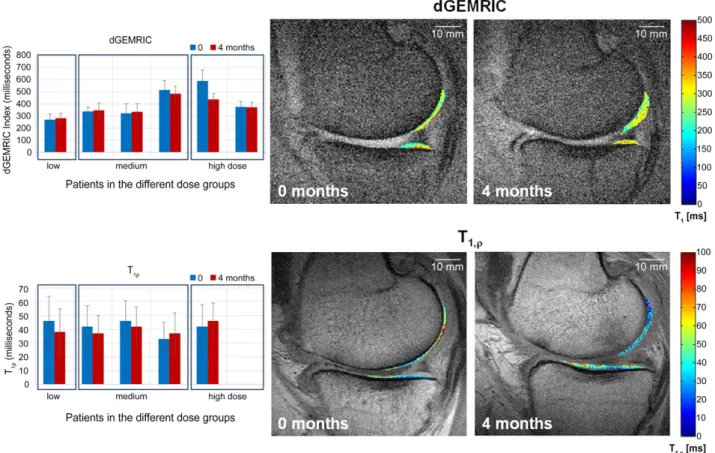
Among the 7 patients included in Germany, quantitative dGEM-RIC (6 patients) and T1rho(5 patients) maps were acquired and
analyzed before and 4 months after therapy (Fig. 3). In these parameter maps, the dGEMRIC index increased in three se-lected patients with time, whereas the T1rhovalues decreased
at the same time. For the other three patients, the opposite effect was observed. Thus, the positive changes were only lim-ited and suggested a possible cartilage improvement in three of six patients. In conclusion, within this small number of pa-tients, we did not observe any correlation between MRI and clinical changes.
Histologic Analysis
Histologic analysis of cartilage and synovium at 3 months was available for 11 of 18 patients after arthroscopy. All samples showed signs of severe OA (OARSI histologic grading.3). Oste-oarthritic chondrocytes stained positive for PS100 and negative for CD34 or Ki67 (Fig. 4). Significant synovial inflammation was ab-sent in two cases, whereas weak or moderate inflammation and synovial hyperplasia with diffuse interstitial lymphocytic infiltrate were observed in five and four cases, respectively. In one pa-tient (case 2) who received a low dose of ASCs, we observed a sheet of cells that could be interpreted as a stem cell graft on cartilage surface (Fig. 4). These cells showed rare Ki67 nuclear staining and weak PS100 staining and were CD34 negative. Fi-nally, none of the synovial or cartilage samples showed any tumor proliferation.
Figure 3. dGEMRIC and T1rhomagnetic resonance imaging (MRI) of selected patients. The graphs on the left show the dGEMRIC (n = 6) and T1rho
(n = 5) values before and 4 months after cell therapy. Increasing dGEmRIC and decreasing T1rhovalues are each known to correspond to increasing
glycosaminoglycan/proteoglycan content and thus improved cartilage condition. On the right, the corresponding dGEMRIC and T1rhomaps are
shown as a color-coded overlay on an anatomical MRI for a patient receiving a low cell dose. The observed values in the cartilage change in the time course can be easily seen and correspond to an increase in cartilage condition. Abbreviation: dGEMRIC, delayed gadolinium-enhanced magnetic resonance imaging of cartilage.
Pers, Rackwitz, Ferreira et al. 853
DISCUSSION
This pilot trial reached its predetermined primary outcome pa-rameters, i.e., safety of IA injection of ASCs in patients with knee OA. Our results are similar to those reported from other studies, in critical limb ischemia or fistulae in inflammatory bowel disease, where ASCs have been injected locally without reported side ef-fects [5, 14]. Additionally, we report clinical improvement with a reduction in pain levels and WOMAC score in all three groups, even though statistically significant results were obtained only in the low-dose group. Actually, the large variability in the range of the initial clinical parameters as well as the limited sample size may explain why statistical significance was not reached at 6 months. However, when compared with historical control stud-ies, our approach seems very encouraging. For example, in a thor-oughly double-blind study on hyaluronic acid treatment into the knee, the WOMAC pain score decreased by 22.96 1.4 mm be-tween baseline and 6 months [15]. In the present study, WOMAC pain score decreased by 30.76 10.7 mm in the group receiving low-dose ASCs. Furthermore, the average difference from base-line to 6 months on the WOMAC subscale scores (pain, function, and stiffness) is higher than the recommended minimal percep-tible clinical improvement of 10 mm [16]. Additionally, a study
comparing hyaluronic acid with saline solution reported 54.6% OARSI responders in the saline group after 13 weeks [17]. This score is lower than the OARSI response obtained with the three different ASC doses at the same time point in the present study, with 83.3% in low-dose, 60% in medium-dose, and 60% in high-dose groups. In a recent controlled study with steroid as compar-ator, the magnitude of the placebo effect led to a decrease in WOMAC pain score of 20 mm at 6 months versus baseline [18]. They recorded 52.1% OARSI responders at 6 months, which is lower than obtained in our groups. These studies suggest that ASC therapy might be more efficient than a possible placebo effect.
Our results are also consistent with those obtained in a recent study in patients with a larger heterogeneity in age and less severe forms of OA [11]. In a recent similar study from Jo et al., the high-est efficiency was found at the highhigh-est dose (1003 106cells) in patients who presented the highest levels of pain at baseline (VAS and WOMAC) [19]. In our study, the group of patients injected with 23 106cells exhibited the best response to ASC treatment, whereas they had higher baseline pain and WOMAC scores com-pared with those receiving higher doses. One possible reason for this inverse dose effect of ASC therapy might be the higher level of inflammation in the lowest dose group, as reflected by the highest Figure 4. Histologic findings. (A): Vascular congestion and weak lymphocytic infiltrate of the synovial (case 8) (magnification,350). (B): Osteoarthritic cartilage OARSI grade.3 (case 4) (325). (C): Toluidine blue staining (case 2) (magnification, 3100). (D): Stem cell stroma shows an Alcian blue depleted matrix compared with the strong staining of osteoarthritic cartilage (case 2) (magnification,3100). (E): Weak PS100 stain-ing of possible stem cells on the cartilage surface and strong PS100 stainstain-ing of chondrocytes (case 2) (magnification,3100). Abbreviations: OARSI, Osteoarthritis Research Society International.
level of pain at baseline. The inflammatory milieu might have primed the injected ASCs to exert their immunomodulatory func-tions more efficiently than in the groups where the inflammation was lower. We therefore cannot rule out that the treatment re-sponse was partly dependent on the initial disease activity. Orozco et al. published another interesting study on the treat-ment of knee OA with autologous MSCs derived from bone mar-row [20]. They injected 40 3 106 cells into the knee joint. Improvement of cartilage morphology and quality was observed in almost all patients using MRI T2mapping, suggesting a possible
structural benefit of stem cell therapy.
The potential mode of action of ASCs for the treatment of OA includes at least three different biological effects. The first is di-rect differentiation of ASCs into chondrocytes, whereas the others are related to a possible paracrine effect of secreted bioactive molecules, including anti-inflammatory and chondro-protective mediators. However, the capacity of MSCs to differen-tiate into chondrocytes is probably not critical in the observed therapeutic effect. Preliminary studies in rabbits and goats have shown that cartilage regeneration did not occur at the expense of chondrogenic differentiation of the injected cells but may be strongly related to a secondary stimulation of endogenous pro-genitor cells through paracrine effects [21]. MSCs contributed to the repair of damaged articular cartilage through homing, engraftment, production of cartilage matrix, and reduction of lo-cal inflammation [22–25]. Stromal cells have been shown to pos-sess immunomodulatory and antifibrotic properties, to protect cells from oxidative stress and apoptosis, and to stimulate prolif-eration and chondrogenic differentiation in coculture through se-cretion of growth factors [23]. In preclinical models of OA or experimental models of inflammatory diseases such as arthritis and experimental encephalitis, the benefit of ASC injection was related to secretion of anti-inflammatory factors including hepa-tocyte growth factor, human leukocyte antigen G5, or interleukin-1 receptor antagonist [26]. The immunomodulatory properties of adipose-derived MSCs are even stronger than those from other tissue sources [27]. Whether the in vitro capabilities of MSCs from different tissue sources reflect the in vivo situation has still to be elucidated. Nevertheless, there is an obvious variation among do-nors that could be related to differences in isolation, expansion, and freezing/thawing procedures. Altogether, these data suggest that MSCs can reduce synovitis and favor an appropriate environ-ment for tissue regeneration through expression of active growth factors or recruitment of endogenous progenitors.
CONCLUSION
Although this phase I study included a limited number of patients without a placebo arm, we were able to show that this innovative treatment was safe and well tolerated in patients with knee OA. We also provided encouraging preliminary evidence of efficacy. Larger and controlled long-term studies are now mandatory to confirm whether this new strategy of cell therapy can improve pain and induce structural benefit. Moreover, it is likely that sim-ilar therapeutic procedures based on autologous ASCs can be
extended in the future to other joints, such as the hip joint, or in-dications such as intervertebral disc degeneration.
ACKNOWLEDGMENTS
We thank all the patients for their participation in the study and Dr. Mazen Hamoui and Prof. Andrea Facchini for their coopera-tion and support during the study. M.R. and U.N. are currently af-filiated with the Department of Orthopaedic and Trauma Surgery, Evangelisches Waldkrankenhaus Spandau, Berlin, Germany. Trial registration number: NCT01585857. The research leading to these results has received funding from the European Union Sev-enth Framework Programme FP7/2007-2013 under grant agree-ment 241719. Work in the laboratory INSERM U844 was also supported by the Inserm Institute, the University of Montpellier, and the Agence Nationale pour la Recherche for support of the national infrastructure:“ECELLFRANCE: Development of a na-tional adult mesenchymal stem cell based therapy platform.”
AUTHORCONTRIBUTIONS
Y.-M.P.: provision of study patients, collection and assembly of data, data analysis and interpretation, manuscript writing, final approval of manuscript; L.R.: provision of study patients, collec-tion and assembly of data, manuscript writing, final approval of manuscript; R.F.: administrative support, provision of study pa-tients, collection and assembly of data, data analysis and interpre-tation, final approval of manuscript; O.P.: administrative support, collection and assembly of data, data analysis and interpretation, manuscript writing, final approval of manuscript; C.D., F.C., and C.C.: provision of study material, final approval of manuscript; F.B.: conception and design, administrative support, manuscript writing, final approval of manuscript; L.S., S.F., and P.B.: concep-tion and design, administrative support, provision of study mate-rial, final approval of manuscript; L.C. and G.L.: conception and design, administrative support, final approval of manuscript; D.N.: conception and design, administrative support, data analy-sis and interpretation, manuscript writing, final approval of man-uscript; J.S. and D.H.: provision of study material, collection and assembly of data, data analysis and interpretation, manuscript writing, final approval of manuscript; S.D.: collection of data, manuscript writing, final approval of manuscript; U.N.: concep-tion and design, administrative support, provision of study patients, collection and assembly of data, data analysis and inter-pretation, manuscript writing, final approval of manuscript; C.J.: conception and design, financial support, provision of study pa-tients, data analysis and interpretation, manuscript writing, final approval of manuscript.
DISCLOSURE OFPOTENTIALCONFLICTS OFINTEREST
S.F. has uncompensated research funding from European Union Seventh Framework Programme FP7/2007-2013, grant agree-ment 241719. D.H. is employed by MRB Research Center. The other authors indicated no potential conflicts of interest.
REFERENCES
1 Findlay DM. If good things come from above, do bad things come from below? Arthri-tis Res Ther 2010;12:119.
2 Jorgensen C, Djouad F, Bouffi C et al. Multipo-tent mesenchymal stromal cells inarticular diseases. Best Pract Res Clin Rheumatol 2008;22:269–284.
3 ter Huurne M, Schelbergen R, Blattes R et al. Antiinflammatory and chondroprotective
effects of intraarticular injection of adipose-derived stem cells in experimental osteoarthri-tis. Arthritis Rheum 2012;64:3604–3613.
4 Desando G, Cavallo C, Sartoni F et al. Intra-articular delivery of adipose derived stromal cells Pers, Rackwitz, Ferreira et al. 855
attenuates osteoarthritis progression in an ex-perimental rabbit model. Arthritis Res Ther 2013;15:R22.
5 Bura A, Planat-Benard V, Bourin P et al. Phase I trial: The use of autologous cultured adipose-derived stroma/stem cells to treat pa-tients with non-revascularizable critical limb is-chemia. Cytotherapy 2014;16:245–257.
6 Altman R, Asch E, Bloch D et al. Develop-ment of criteria for the classification and reporting of osteoarthritis. Classification of os-teoarthritis of the knee. Arthritis Rheum 1986; 29:1039–1049.
7 Kellgren JH, Lawrence JS. Radiological as-sessment of osteo-arthrosis. Ann Rheum Dis 1957;16:494–502.
8 Bellamy N, Buchanan WW, Goldsmith CH et al. Validation study of WOMAC: A health status instrument for measuring clinically important patient relevant outcomes to anti-rheumatic drug therapy in patients with osteo-arthritis of the hip or knee. J Rheumatol 1988; 15:1833–1840.
9 Pham T, van der Heijde D, Altman RD et al. OMERACT-OARSI initiative: Osteoarthritis Re-search Society International set of responder criteria for osteoarthritis clinical trials revisited. Osteoarthritis Cartilage 2004;12:389–399.
10 Ware JE Jr., Sherbourne CD. The MOS 36-item short-form health survey (SF-36). I. Conceptual framework and item selection. Med Care 1992;30:473–483.
11 Koh YG, Choi YJ, Kwon OR et al. Second-look arthroscopic evaluation of cartilage lesions after mesenchymal stem cell implantation in os-teoarthritic knees. Am J Sports Med 2014;42: 1628–1637.
12 Eckstein F, Burstein D, Link TM. Quantita-tive MRI of cartilage and bone: DegeneraQuantita-tive
changes in osteoarthritis. NMR Biomed 2006; 19:822–854.
13 Pritzker KP, Gay S, Jimenez SA et al. Oste-oarthritis cartilage histopathology: Grading and staging. Osteoarthritis Cartilage 2006;14:13–29. 14 Lee WY, Park KJ, Cho YB et al. Autologous adipose tissue-derived stem cells treatment demonstrated favorable and sustainable thera-peutic effect for Crohn’s fistula. STEM CELLS
2013;31:2575–2581.
15 Berenbaum F, Grifka J, Cazzaniga S et al. A randomised, double-blind, controlled trial com-paring two intra-articular hyaluronic acid prep-arations differing by their molecular weight in symptomatic knee osteoarthritis. Ann Rheum Dis 2012;71:1454–1460.
16 Ehrich EW, Davies GM, Watson DJ et al. Minimal perceptible clinical improvement with the Western Ontario and McMaster Universities osteoarthritis index question-naire and global assessments in patients with osteoarthritis. J Rheumatol 2000;27:2635– 2641.
17 Strand V, Baraf HS, Lavin PT et al. A mul-ticenter, randomized controlled trial comparing a single intra-articular injection of Gel-200, a new cross-linked formulation of hyaluronic acid, to phosphate buffered saline for treat-ment of osteoarthritis of the knee. Osteoarthri-tis Cartilage 2012;20:350–356.
18 Leighton R, Akermark C, Therrien R et al; DUROLANE Study Group. NASHA hyaluronic acid vs. methylprednisolone for knee osteoarthri-tis: A prospective, multi-centre, randomized, non-inferiority trial. Osteoarthritis Cartilage 2014;22:17–25.
19 Jo CH, Lee YG, Shin WH et al. Intra-articular injection of mesenchymal stem cells for the treatment of osteoarthritis of the knee:
A proof-of-concept clinical trial. STEM CELLS
2014;32:1254–1266.
20 Orozco L, Munar A, Soler R et al. Treat-ment of knee osteoarthritis with autologous mesenchymal stem cells: Two-year follow-up results. Transplantation 2014;97:e66–e68.
21 Murphy JM, Fink DJ, Hunziker EB et al. Stem cell therapy in a caprine model of osteoar-thritis. Arthritis Rheum 2003;48:3464–3474.
22 Hoogduijn MJ, Crop MJ, Peeters AM et al. Human heart, spleen, and perirenal fat-derived mesenchymal stem cells have immunomodulatory capacities. Stem Cells Dev 2007;16:597–604.
23 Puissant B, Barreau C, Bourin P et al. Im-munomodulatory effect of human adipose tissue-derived adult stem cells: Comparison with bone marrow mesenchymal stem cells. Br J Haematol 2005;129:118–129.
24 Wolbank S, Peterbauer A, Fahrner M et al. Dose-dependent immunomodulatory effect of human stem cells from amniotic mem-brane: A comparison with human mesenchymal stem cells from adipose tissue. Tissue Eng 2007; 13:1173–1183.
25 Ya~nez R, Lamana ML, Garc´ıa-Castro J et al. Adipose tissue-derived mesenchymal stem cells have in vivo immunosuppressive properties applicable for the control of the graft-versus-host disease. STEMCELLS2006;24: 2582–2591.
26 Maumus M, Jorgensen C, No¨el D. Mesen-chymal stem cells in regenerative medicine ap-plied to rheumatic diseases: Role of secretome and exosomes. Biochimie 2013;95:2229–2234. 27 Mattar P, Bieback K. Comparing the im-munomodulatory properties of bone marrow, adipose tissue, and birth-associated tissue mesenchymal stromal cells. Front Immunol 2015;6:560.