HAL Id: hal-01789033
https://hal.archives-ouvertes.fr/hal-01789033
Submitted on 17 May 2018HAL is a multi-disciplinary open access archive for the deposit and dissemination of sci-entific research documents, whether they are pub-lished or not. The documents may come from teaching and research institutions in France or abroad, or from public or private research centers.
L’archive ouverte pluridisciplinaire HAL, est destinée au dépôt et à la diffusion de documents scientifiques de niveau recherche, publiés ou non, émanant des établissements d’enseignement et de recherche français ou étrangers, des laboratoires publics ou privés.
Allosteric nanobodies uncovered a role of hippocampal
mGlu2 receptor homodimers in contextual fear
consolidation.
Pauline Scholler, Damien Nevoltris, Dimitri de Bundel, Simon Bossi, David
Moreno-Delgado, Xavier Rovira, Driss El Moustaine, Michaël Mathieu, Emilie
Blanc, Heather Mclean, et al.
To cite this version:
Pauline Scholler, Damien Nevoltris, Dimitri de Bundel, Simon Bossi, David Moreno-Delgado, et al.. Allosteric nanobodies uncovered a role of hippocampal mGlu2 receptor homodimers in contextual fear consolidation.. Nature Communications, Nature Publishing Group, 2017. �hal-01789033�
Allosteric nanobodies uncovered a role of hippocampal mGlu2 receptors in context 1 fear consolidation 2 3 4 5 6
Pauline Scholler*,1,2, Damien Nevoltris*,2,3, Dimitri de Bundel1, Simon Bossi4, David
Moreno-7
Delgado1, Xavier Rovira1, Driss El Moustaine1, Michaël Mathieu1, Emilie Blanc1, Heather
8
McLean4, Elodie Dupuis2, Gérard Mathis2, Eric Trinquet2, Hervé Daniel4, Emmanuel Valjent1,
9
Daniel Baty3, Patrick Chames3,#, Philippe Rondard1#, Jean-Philippe Pin1#
10 11 12 13
1 Institut de Génomique Fonctionnelle, CNRS UMR5203, INSERM U1191, Univ. Montpellier,
14
Montpellier, France 15
2 Cisbio Bioassays, Codolet, France
16
3 Aix Marseille Univ, CNRS, INSERM, Institut Paoli-Calmettes, CRCM, Marseille, F-13009,
17
France 18
4 CNRS UMR9197, Université Paris-Sud, Institut des Neurosciences Paris-Saclay, France.
19 20 21
* Equal contribution 22
# To whom correspondence should be addressed.
23 24 25 26
Abstract 27
28
Antibodies have enormous therapeutic and biotechnology potential. G protein-coupled 29
receptors (GPCRs), one of the main targets in drug development, are of major interest in 30
antibody development programs. Metabotropic glutamate receptors are dimeric GPCRs that 31
can control synaptic activity in a multitude of ways. Here we identify llama nanobodies that 32
specifically recognize mGlu2 receptors, among the eight subtypes of mGluR subunits. Two of 33
these nanobodies act as positive allosteric modulators (PAMs) on homodimeric mGlu2. One 34
of them potentiates agonist actions on activated mGlu2, but has no effect on mGlu2-4 35
heterodimers. This PAM enhances the inhibitory action of the orthosteric mGlu2/mGlu3 36
agonist, DCG-IV, at mossy fiber terminals in the CA3 region of hippocampal slices. It also 37
impairs contextual fear memory demonstrating for the first time the functional role of mGlu2 38
homodimers in living animals. These data also highlight the potential of developing 39
antibodies with allosteric actions on GPCRs to better define their roles in vivo. 40
41 42
43
There is growing interest in developing either activating or inactivating antibodies with 44
therapeutic potential1,2, but also as innovative tools to decipher the functional roles of cell
45
surface proteins3. G protein-coupled receptors (GPCRs), that are the main targets for small
46
therapeutic molecules, are now considered as promising targets for therapeutic antibodies4-7.
47
Single chain antibodies from camelids, such as llamas, are particularly well suited for such 48
purposes, being more prone to target specific conformations of their targets6,8,9. Such tools
49
have already proven their potential for pharmacological actions6,10, structural studies8,11, and
50
use as biosensors3.
51
In the central nervous system, glutamate, the main excitatory neurotransmitter, exerts its fast 52
actions via ionotropic receptors, but also modulates synaptic activity via GPCRs, so called 53
metabotropic glutamate receptors (mGluRs)12-14. Eight genes encoding mGluRs are found in
54
mammalian genomes, and are classified into three groups. While group-I receptors (mGlu1 55
and mGlu5) are mainly post-synaptic receptors that contribute to glutamatergic synaptic 56
responses, group-II (mGlu2 and 3) and -III (mGlu4, 6, 7 and 8) are mainly pre-synaptically 57
located, and inhibit transmitter release at various types of synapses12. As such, mGluRs are
58
considered to be interesting targets for the treatment of various brain diseases including 59
psychiatric or neurodegenerative diseases12,13.
60
Among the various mGluR subtypes, mGlu2, but also mGlu3 and 5, open new possibilities 61
for novel antipsychotic drugs13,15. However studies on the roles of mGlu2 are made difficult
62
by the limited number of specific tools. Indeed, there are no specific mGlu2 antibodies to 63
determine their precise localization in the brain16. Moreover, because of the high
64
conservation of the orthosteric glutamate binding site located in the Venus flytrap 65
extracellular domain (VFT) of these receptors17, only very few selective agonists have been
66
reported18,19. Efforts were concentrated on the development of positive allosteric modulators
67
(PAMs) interacting with the less conserved 7 transmembrane domains (7TM)17. Although
68
subtype selective PAMs have been identified, a number of limitations for their development 69
have been observed20,21. Although knock out lines are available12,13, one cannot exclude
70
compensation during development. Eventually, mGluRs, and especially mGlu2 have been 71
reported to associate with other mGlu subunits to form heterodimers22-24, and evidence for
72
mGlu2-4 heterodimers in cortico-striatal terminals have recently been provided23. These
73
observations strengthen the need for more specific tools to better characterize the functional 74
roles of homo or heterodimeric mGluRs containing the mGlu2 subunit. 75
In the present study, we aimed at identifying nanobodies25,26 that recognize specific
76
conformations of the mGlu2 receptor. This led us to identify two nanobodies that specifically 77
bind to the active form of the mGlu2. Accordingly, these nanobodies act as PAMs, enhancing 78
the agonist action at mGlu2 receptors in transfected cells and in brain slices. When injected 79
in the hippocampus, these nanobodies also enhance the effect of a group-II mGluR agonist 80
in the fear-conditioning test, demonstrating their possible use to decipher the physiological 81
role of mGlu2 receptors in the brain. These data nicely illustrate novel possibilities to develop 82
mGlu allosteric modulators for numerous therapeutic actions, and exemplify the use of 83
nanobodies to allosterically modulate GPCRs. 84
85 86
Results: 87
Identification of mGlu2 selective nanobodies 88
To identify nanobodies recognizing mGlu2 receptors, HEK-293 cells transiently expressing 89
both rat and human mGlu2 were injected in llamas, and VHH encoding sequences were 90
amplified to generate a phage display library27. By screening the later using a purified mGlu2
91
receptor reconstituted into nanodiscs, several positive clones were isolated and three of 92
them, DN1, DN10 and DN13 were retained for analysis. FRET based binding data (Fig. 1a)
93
revealed that all three nanobodies bind to mGlu2 in the presence of ambient glutamate 94
produced by the cells, and not to any other mGluR (Fig. 1b).
95
Because glutamate concentration in the blood is sufficient to fully activate mGlu2 receptors, 96
we expected that some of our identified nanobodies could preferentially bind to the active 97
form of the receptor (Fig 1c). Indeed, whereas DN1 displays the same affinity for the active
98
and inactive forms of mGlu2 (Fig. 1d and Supplementary Table 1), DN10 and DN13
99
specifically bind to the active form stabilized by the orthosteric agonist, LY379268 (Fig. 1e,f
100
and Supplementary Table 1). No binding was detected on the inactive form of the receptor in 101
the presence of the antagonist LY341495 (Fig. 1e,f). Note that in the absence of any added
102
ligand, and under conditions leading to very low extracellular glutamate concentrations in the 103
assay medium through the co-transfection of the receptor with the high affinity glutamate 104
transporter EAAC1, no binding of DN10 and DN13 to mGlu2 could be detected 105
(Supplementary Fig. 1a). This is in contrast to the conditions used for the screening and first 106
characterization of the nanobodies where binding could be detected under basal condition, 107
likely due to the presence of enough glutamate produced by the cells in the assay (Fig. 1b).
108
According to these data, the specificity of both DN10 and DN13 was further examined on the 109
eight mGlu subtypes in the presence of a saturating concentration of either agonists or 110
antagonists (Supplementary Fig. 1b and 1c, respectively). It demonstrated that these two
111
nanobodies only bind on the active form of mGlu2 receptor. 112
DN10 and DN13 are positive allosteric modulators of mGlu2 113
A possible effect of both DN10 and DN13 was first examined using a mGlu2 biosensor28.
114
This sensor makes use of the large movement between the VFTs that occurs upon receptor 115
activation, leading to an increase in distance between the N-terminal SNAP tags carried by 116
each subunit28. Such movement lead to a large decrease in lanthanide-based resonance
117
energy transfer (LRET) measured in a time resolved manner (TR-FRET) (Fig. 2a). In the
118
presence of an EC20 concentration of LY379268, both DN10 and DN13 were found to
119
activate the biosensor in the high nanomolar range (Fig. 2b). A similar agonist effect of both
120
DN10 and DN13 was observed using a functional assay based on the activation of 121
phospholipase C by this Gi/o-coupled receptor using a chimeric Gqi protein (Supplementary
122
Fig. 2). When the extracellular glutamate concentration was maintained as low as possible 123
using EAAC1, both DN10 and DN13 clearly increased mGlu2 agonist LY379268 potency 124
(Fig. 2c,d), revealing their positive allosteric effect. However, whereas DN13 retains minimal 125
agonist activity under such conditions (Fig. 2d), DN10 was still able to activate mGlu2 (Fig.
126
2c). Such observations were confirmed using the inositol-phosphate accumulation assay
127
(Fig. 2e,f). These data demonstrate that both DN10 and DN13 act as PAMs on mGlu2, but 128
that DN10 has, in addition, an intrinsic agonist activity (known as ago-PAM29).
129 130
DN13 binds at an epitope specific of the active conformation 131
The binding ability of the nanobodies on various mGlu2 constructs lacking the 7TM domains, 132
the cysteine-rich domain, or the entire extracellular domain, revealed that all three 133
nanobodies bind to the VFT (Supplementary Fig. 3). Moreover, competition studies revealed
134
that DN10 and DN13 share part of their binding epitope, while DN1 binds at a different site 135
(Supplementary Fig. 4). To identify the epitope recognized by DN13, we made use of the 136
inability of these nanobodies to bind to the homologous mGlu3 receptor, and their lack of 137
affinity for the inactive form of the receptor. The major conformational change in the VFT 138
dimer occurring upon receptor activation is the relative reorientation of the VFTs leading to a 139
close apposition of the second lobes, which are distant in the inactive form (Fig. 1c)28.
140
Accordingly, a specific crevice at the interface of the second lobes is formed in the active 141
form of the dimer only, and a few residues in that area were found to be different between 142
mGlu2 and mGlu3 receptors (Fig. 3a-c and Supplementary Table 2). These include Leu226,
143
Arg445 and Ile450 in protomer A (Fig. 3c) (Gln, Thr and Met in mGlu3, respectively), and
144
Ser246, Ala248, Ala249, Glu251 and Gly252 in protomer B (Ile, Lys, Ser, Asp and Ser in 145
mGlu3, respectively). Docking experiments conducted with a 3D model of DN13 and a 146
mGlu2 VFT dimer model suggest that DN13 interacts at that site (Fig. 3a-c). This is further
147
demonstrated by our observation that DN13 does not interact with a mGlu2 mutant in which 148
these residues were replaced by their mGlu3 equivalent (Fig. 3d and Supplementary Table
149
3) despite their normal expression and coupling properties (Supplementary Fig 5). In
150
contrast, DN13 binds with a nanomolar affinity on the mGlu3 mutant bearing these residues 151
from mGlu2 (Fig. 3e and Supplementary Table 3).
152
There is increasing evidence suggesting that mGlu2 receptors can exist not only as 153
homodimers, but also as heterodimers, associated with any group-III mGluRs22,23. Because
154
the epitope recognized by DN13 involves both subunits in the mGlu dimer, we predicted that 155
it would be specific for the mGlu2 homodimer relative to the heterodimers made of mGlu2 156
and a group-III mGlu subunits. Indeed, DN13 did not bind to the mGlu2-4 heterodimer 157
(Supplementary Fig. 6a), nor did it potentiate agonist action on this heterodimer as revealed 158
using either a mGlu2-4 specific biosensor, and a functional assay using subunit combination 159
that ensure targeting to the cell surface of the heterodimer only30 (Supplementary Fig. 6b,c).
160
DN13 potentiates the pre-synaptic effect of mGlu2 161
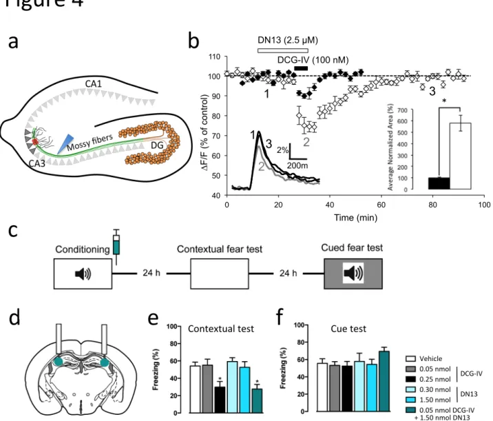
The mGlu2 gene is well expressed in hippocampal dentate gyrus granule neurons, where the 162
receptors are targeted to mossy fiber terminals that contact pyramidal neurons in the CA3 163
area (Fig. 4a). These terminals may also contain mGlu3, mGlu4 and mGlu7 that are also
164
expressed by granule neurons31,32. In acute hippocampal slices, we examined the effect of
165
nanobodies on mossy fiber terminal mGlu2 activation by quantifying presynaptic calcium 166
transients evoked by electrical stimulation of mossy fibers, using photometric measurements 167
of the fluorescent Ca2+ sensitive dye, magnesium green-AM33. We found that saturating
168
concentrations of DN13 (2.5 µM), did not affect the amplitude of Ca2+ transients in mossy
169
fiber terminals when applied alone but clearly enhanced the inhibitory effect of low 170
concentrations (100 nM) of the group-II mGluR agonist DCG-IV (24 ± 1.4 % and 8.3 ± 0.9 %
171
inhibition with and without DN13, respectively, p < 0.001, n = 9) (Fig. 4b). In the presence of
172
DN13, the effect of a low concentration of DCG-IV nearly reached the maximal effect 173
observed with saturating concentrations of the drug (30.5 ± 1.9 %, n = 6). In addition, and as 174
expected for a PAM effect, the off rate of the inhibitory action of a saturating concentration of 175
DCG-IV (5 µM) is prolonged in the presence of DN13 (Supplementary Fig. 7).
176
The hippocampal mossy fiber pathway projecting from the dentate gyrus to the CA3 is 177
critically involved in memory processing. Infusion of the group-II mGluR agonist DCG-IV into 178
the CA3 area was previously shown to block contextual fear memory consolidation in mice34.
179
Consistent with this observation we found that DCG-IV infused into the CA3 area (Fig. 4d
180
and supplementary Fig. 8) specifically disrupted contextual fear memory consolidation (Fig.
181
4e) without affecting cued fear memory consolidation (Fig. 4f) when immediately following
182
conditioning (Fig. 4c). DN13 did not affect fear memory consolidation when applied alone,
183
but potentiated the effect of low concentrations of DCG-IV, demonstrating the involvement of 184
mGlu2 (Fig. 4e).
185 186
Discussion 187
188
In the present study, we describe three nanobodies that specifically recognize mGlu2 189
in the nanomolar range, being then the first mGlu2 selective antibodies. Most interestingly, 190
while one of these does not discriminate between the different conformations of the receptor, 191
the two others, DN10 and DN13 exclusively bind to the active form, interacting at a site 192
exclusively found in the active form of the dimer. In the study we have further characterized 193
the DN13 nanobody that behaves as a PAM with no intrinsic agonist activity. Indeed, DN13 194
potentiated the action of mGlu2 agonists not only in heterologous expression systems, but 195
also in brain slices and in vivo. Because the binding epitope of DN13 involves residues from 196
both subunits, it is indeed inactive on mGlu2-4 heterodimers, indicating that the effects 197
observed both in brain slices and in vivo at the level of the hippocampal CA3 area likely 198
involves mGlu2 homodimers. 199
Despite the identification of both mGlu2 and mGlu3 in the early 90's, and the interest 200
they have generated for the development of anxiolytic and antipsychotic drugs, studies of 201
these two receptors have been hampered by the lack of specific pharmacological tools. Until 202
now, available antibodies have not been able to discriminate between mGlu2 and mGlu316,
203
and only few selective ligands have been developed18,19. However, the availability of mGlu2
204
and mGlu3 knockout mice (or mouse lines) coupled with more sophisticated pharmacology 205
has strengthened the interest in targeting mGlu2 specifically for antipsychotic effects35.
206
Today, the most selective and promising ligands are mGlu2 PAMs that bind to a hydrophobic 207
cavity in the 7TM36. Accordingly, such molecules show high hydrophobicity enabling them to
208
pass through the blood brain barrier. However, this also limits their effective concentration in 209
the cerebrospinal fluid, and increases the chance for off target activity20. Despite the
210
therapeutic potential of PAMs, so far only orthosteric mGlu2-3 agonists have reached phase 211
3 clinical trails for anxiety and schizophrenia35 but have had very limited success. Our
212
nanobodies are the first mGlu2 selective PAMs that do not target the 7TM domain, but rather 213
the VFT domain. These molecules reveal new possibilities to develop selective mGlu2 PAMs 214
that are designed to target this new site, without the limiting hydrophobic properties. 215
Although mGluRs were thought to exist exclusively as homodimers, recent data has 216
revealed that both group-I, and group-II/III mGluRs can associate to form multiple types of 217
heterodimers22. Among these, the heterodimeric mGlu2-4 receptor is likely present in
striato-218
cortical terminals, as illustrated by co-immunoprecipitation experiments, and the selective 219
action of one mGlu4 PAM23. This is in agreement with our observation that DN13 interacts
220
with both subunits in the mGlu2 VFT dimer and was found to be inactive on the mGlu2-4 221
heterodimer. This finding will lead to multiple possibilities to develop selective nanobodies for 222
mGlu homo and heterodimers comprised of specific mGlu subunits. 223
Although group-II mGluR agonists are well known for their anxiolytic and 224
antipsychotic properties, recent data also suggest that they act to consolidate context fear 225
memory34. This may result from a reduction in pre-synaptic glutamate release following
226
activation of group-II mGluRs located at the mossy fiber terminals in the CA3 area of the 227
hippocampus. These terminals originate from the granule neurons of the dentate gyrus that 228
also express mGlu316 and as such, that the inhibitory action of the group-II agonist DCG-IV
229
reported at these synapses may involve either mGlu2 or mGlu3. Our data using DN13, an 230
mGlu2 selective PAM, suggest that only mGlu2 receptors are involved in the DCG-IV effect. 231
Eventually, the DN13-mediated potentiation of the DCG-IV effect confirmed that a selective 232
activation of mGlu2 can prevent the consolidation of context fear memory. These data also 233
argue against the involvement of an mGlu2 receptor heterodimer containing mGlu4, and 234
likely the other group-III mGlu7 subunit both expressed in hippocampal granule 235
neurons16,31,32.
236
Taken together, our data are the first to report the development of PAM nanobodies 237
acting at a GPCR. Antibodies show increasing potential in therapeutics, although mainly by 238
targeting proteins other than GPCRs1. Since GPCRs still represent important targets for
239
therapeutic interventions, these membrane receptors have only recently been highlighted as 240
possible targets for antibody-based biologics4,5. So far, such possibilities have been validated
241
through the identification of antibodies inhibiting chemokine receptors4,6. Here we extend the
242
use of this approach revealing the feasibility to develop nanobodies with very selective PAM 243
activity at mGlu2 GPCRs, thus offering a way to better identify their actions in vivo, as well as 244
localizing activated receptors within the brain. Although an access to the brain would be 245
needed for targeting these central receptors for therapeutic intervention, conditions have 246
been reported to facilitate brain penetration of nanobodies37. Moreover, mGlu receptors are
247
not only expressed in the CNS, but also at the periphery where they have a role in the 248
regulation of cardiovascular38 and immune systems39, as well as in cancer40. Taken together,
249
mGluR targeting nanobodies offer interesting possibilities for therapeutic intervention. 250
251 252
Acknowledgements 253
We thank L Prézeau, D Maurel and C Vol from ARPEGE (Pharmacology
Screening-254
Interactome)facility at the Institut de Génomique Fonctionnelle (Montpellier, France) for their 255
help in various microplate assays. Funding was provided by the Centre National de la 256
Recherche Scientifique (CNRS), the Institut National de la Santé et de la Recherche 257
Médicale (INSERM), the University of Montpellier, Cisbio Bioassays, the Fondation 258
Recherche Médicale (FRM DEQ20130326522) and the Fondation Bettencourt Schueller to 259
JPP; the Fond Unique Interministériel of the french government (FUI, Cell2Lead project) to 260
GM, JPP and DB; the Agence Nationale de la Recherche (ANR-15-CE18-0020-01) to PR. 261
262
Author Contributions: 263
JPP, PR, PC, DB, EV, HD, HML, ET, GM, ED designed the research. 264
DN generated the nanobody phage display library and performed the screening and primary 265
characterization of the nanobodies, DEM set up the conditions and prepared the purified 266
mGlu2 receptor in nanodiscs, PS, DMD, MM, EB performed the in vitro characterization of 267
the nanobodies, XR performed the in silico studies, SB performed the experiments on 268
hippocampal slices, DDB performed the in vivo experiments. 269
JPP, PR and PS wrote the paper, with inputs from HML, PC and EV. 270
271
Author Information 272
The authors declare competing financial interests. Correspondence and requests for 273
materials should be addressed to JPP (jean-philippe.pin@igf.cnrs.fr), PR
274 (philippe.rondard@igf.cnrs.fr) or PC (patrick.chames@inserm.fr) 275 276 277 278
Figure legends: 279
280
Figure 1: Nanobodies DN1, DN10 and DN13 specifically interact with mGlu2 receptors. a) 281
Cartoon illustrating the principle of the TR-FRET binding assay. The receptor fused to a 282
SNAP-tag (black circled labeled "S") is labeled with Lumi4-Tb (light blue ball) while the 283
nanobody (purple) bearing a c-Myc epitope at its C-terminus is labeled with 200 nM of anti-c-284
Myc antibody (green) coupled to d2 fluorophores (orange). Binding of the nanobody to the 285
receptor is then measured by a TR-FRET signal. b) Specific TR-FRET binding data obtained 286
with the indicated mGlu receptor and either DN1, DN10, DN13 or a control irrelevant 287
nanobody in cells under basal condition with ambient glutamate. Data are mean ± SD of 288
triplicates from a typical experiment representative of three experiments. c) Cartoon 289
illustrating the main active (left), and inactive (right) conformation of an mGlu2 dimer, 290
stabilized by an agonist (glutamate or LY379268) or the antagonist LY341495, respectively. 291
d,e,f) Saturation binding curves obtained with DN1 (d), DN10 (e) and DN13 (f) on mGlu2 292
receptors under control conditions with low extracellular glutamate thanks to the co-293
expression of the high affinity glutamate transporter EAAC1 (buffer), in the presence of the 294
agonist LY379268 (1 µM), or the antagonist LY341495 (10 µM). Data are mean ± sem of 295
three individual experiments each performed in triplicates. 296
297 298
Figure 2: DN10 and DN13 are positive allosteric modulators of mGlu2. a) Cartoon illustrating 299
the principle of the mGlu2 sensor assay, where the VFT movement associated with receptor 300
activation results in a decrease in TR-FRET signal measured between Lumi4-Tb and SNAP-301
Green labeled SNAP tags. b) DN10 and DN13 concentration dependent decrease the sensor 302
signal in the presence of EC20 concentration of agonist LY379268 (0.1 nM), indicative of
303
receptor activation. DN10 (c) and DN13 (d) dose-dependently potentiate the effect of 304
LY379268 on the mGlu2 sensor. DN10 (e) and DN13 (f) dose-dependently potentiate the 305
effect of LY379268 on the production of inositol phosphate in mGlu2-transfected cells. Note 306
that the lower the IP-One Gq HTRF® ratio, the higher the amount of inositol monophosphate 307
produced (IP1). Data are means ± sem of three individual experiments each performed in 308
triplicates. 309
310
Figure 3: DN13 interacts at the lobe 2 crevice on the activated mGlu2 VFT dimer. a-b) View 311
of the proposed docking of DN13 (orange) on the mGlu2 extracellular domain dimer (a, 312
lateral view, b, top view). c) Detailed view of the proposed docking of DN13 illustrating 313
proposed residues involved in selectivity, shown are Leu226 and Arg445 in protomer A, and 314
Ser246, Ala248 (yellow), Ala249, and Glu251 from protomer B. d) Saturation binding curves 315
of DN13 on mGlu2 WT, mGlu2 bearing mGlu3 specific residues from protomer A (mut A), 316
mGlu2 bearing mGlu3 residues from protomer B (mut B), mGlu2 A248K mutant. e) 317
Saturation binding curves of DN13 on mGlu3 WT, mGlu3 bearing the mGlu2 residues on 318
protomer A (mut A), and mGlu3 bearing all identified residues of mGlu2 (mut AB). Data are 319
mean ± SD of triplicates from a typical experiment representative of three experiments. 320
321
Figure 4: mGlu2 receptors in the CA3 area of the hippocampus control contextual fear 322
consolidation. a) A schematic view of the hippocampus illustrating the granule neurons of the 323
dentate gyrus (DG) projecting to the pyramidal neurons in the CA3 area via the mossy fibers. 324
b) DN13 potentiates the inhibitory effect of a mGlu2-mGlu3 agonist (DCG-IV, 100 nM) on
325
presynaptic evoked calcium transients in the CA3 area (red box in panel a) in response to 326
electrical stimulation of the mossy fibers (blue arrow head in panel a). Data are normalized 327
amplitudes of peak fluorescence transients (∆F/F) evoked by five stimulations of mossy 328
fibers (delivered at 100 Hz). Insert on the right shows the average normalized area 329
corresponding to the depressant effect of DCG-IV alone (black bar, 100 ± 5.4%, n=10) and in 330
the presence of DN13 (white bar, 578.5 ± 69%, n=9), p<0.001. Insert on the left displays 331
superimposed fluorescence changes in one of these experiments recorded at the indicated 332
times. Each trace is an average of 10 consecutive trials. Since the variance was different 333
between DCG-IV and DCG-IV + DN13 groups, the Welch test was applied for statistical 334
analysis. c) Schematic of the experimental protocol used for the contextual fear consolidation
335
examination in mice, and drug infusion site (d). (e) Contextual fear memory expression 336
ANOVA: F5,35 = 6.025, P = 0.0004. (f) Cued fear memory. ANOVA: F5,35 = 0.3053, P =
337 0.9066. * P < 0.05 vs PBS and ** P < 0.01 vs PBS. 338 339 340 341 342
Methods 343
344
Reagents, cell lines, antibodies and plasmids 345
HEK293 cells were cultivated in DMEM (Thermo Fischer Scientific, Courtaboeuf, France) 346
complemented with 10% (v/v) fetal bovine serum. All drugs (DCG-IV, LY341495, LY379268, 347
LY487379) were from Tocris Bioscience (Bristol, UK). All HTRF® reagents, labeled 348
monoclonal antibodies Anti-c-myc-d2 and Anti-6His-d2, labeled ligands (SNAP-Lumi4-Tb, 349
SNAP-Red and CLIP-Red) and SNAP-tag mGluR plasmids, were a kind gift from Cisbio 350
Bioassays (Codolet, France). The pRK5 plasmids encoding wild-type rat mGluR subunits, 351
with a HA-tag and with SNAP or CLIP inserted just after the signal peptide, were previously 352
described (Scholler et al Nat Chem Biol 2017). Point mutations were introduced in the SNAP-353
tag mGlu2 or mGlu3 plasmids according to the QuikChange mutagenesis protocol (Agilent 354
Technologies, Santa Clara, CA, USA). 355
356
Llama immunization and library construction 357
Two llamas (Lama glama) were immunized subcutaneously 4 times with 5x10⁷ HEK293T
358
cells transfected with rat mGluR2 and human mGluR2. VHH library constructions were 359
performed in E. coli TG1 strain as previously described27,41. Library diversities were above
360
109 transformants.
361 362
Selection of nanobodies by phage display 363
20 μL of the bacteria library was grown in 50 mL of 2YTAG (2YT ⁄ ampicillin 100 μg/mL ⁄ 2% 364
glucose) at 37°C with shaking (250 rpm) to an OD600 between 0.5 and 0.7. Bacteria were
365
infected by KM13 helper phage using a multiplicity of infection of 20 during 30 min at 37°C 366
without shaking. The culture was centrifuged for 15 min at 3000 g, and bacterial pellet was 367
re-suspended in 250 mL of 2YTA / kanamycine (50 μg/mL) for an overnight phage-368
nanobodies production at 30°C with shaking. The overnight culture was split in 10 vials and 369
centrifuged for 20 min at 3000 g. Five mL of 80% PEG8000, 2.5 mM NaCl were added to the 370
supernatant in a new clean vial and incubated for 1 h on ice to induce phage particle 371
precipitation. The solution was centrifuged for 20 min at 3000 g at 4°C and the phage-372
containing pellet was re-suspended in 1 mL PBS. Another centrifugation step (2 min, 14000 373
g) was performed to eliminate bacterial contaminant, and 200 µL of PEG8000 NaCl was 374
added to supernatants in a new vial. After 30 min on ice and a last centrifugation (5 min, 375
14000 g), phage-containing pellets were re-suspended in 1 mL PBS to obtain ready to used 376
phage-nanobodies for selections. 377
To obtain mGluR2 specific clones, a first round of selection (S1) was performed on Maxisorp 378
plates (Nunc, Maxisorp®) coated 24 h at 4°C with purified human mGlu2 receptor 379
reconstituted in nanodiscs42. Before selection on purified mGluR2, phage-nanobodies library
380
was depleted by incubation with empty nanodiscs (without receptor) to eliminate anti-381
nanodisc antibodies and to reduce non-specific binding. Remaining phages and purified 382
mGluR2 coated wells were saturated with 2% milk/PBS during 1 h at 4°C, and then phages 383
and antigen were incubated together during 2 h at 4°C for selection with shaking. Wells were 384
then washed 10 times with PBS, and bound phages were finally eluted with 1 mg/ml trypsine 385
solution (Sigma-Aldrich, Saint-Quentin Fallavier, France) during 30 min at room temperature 386
with shaking. Phages were rescued and reamplified by infection of TG1 and phage 387
production as above, yielding S1 polyclonal phage population. 388
To avoid non-specific selection against proteins that composed nanodics and to select 389
antibodies against mGlu2 receptor in a cellular context, a second round of selection (S2) was 390
performed on HEK293T cells transfected with rat mGluR2 (2x10⁷ cells). S1 polyclonal phage 391
population and cells were saturated in 2% milk/PBS during 1 h at 4°C, and incubated 392
together in the same conditions as described previously. After five PBS washes, bound 393
phages were eluted using trypsin solution (1 mg/ml) during 30 min at room temperature. 394
Phages were again rescued in TG1 and infected bacteria corresponding to S2 were plated. 395
Individual TG1 colonies from S2 were picked and grown in two different 96-deep-well plates 396
in 400 µL of 2YTAG. After overnight growth, half of the culture was frozen at -80°C in 20% 397
glycerol for backup, and the rest of culture was used for soluble nanobodies production 398
induced by isopropyl-β-26-D-thiogalactopyranoside (IPTG). Nanobody concentrations in 399
supernatant were estimated at 100-500 nM using the DoubleTag check kit (Cisbio 400
Bioassays) according to manufacturer’s recommendations. 401
402
Production and purification of nanobodies 403
For large-scale nanobody production, positive phagemids from screening step were 404
transformed in E. coli BL21DE3 strain. A single colony was grown into 10 ml of LB 405
supplemented with 100 µg/mL ampicillin, 1% (wt/vol) glucose and 1 mM MgCl2 overnight at
406
37°C with shaking. Then 1 L of LB supplemented with 100 µg/mL ampicillin, 0.1% (wt/vol) 407
glucose and 1 mM MgCl2 was inoculated with 10 ml of the preculture and incubated until an
408
OD600 of 0.7. The nanobody expression was then induced with 1 mM IPTG (final
409
concentration) and bacteria were grown overnight at 28°C with shaking. Bacteria were then 410
collected by centrifugation for 10 min at 5,000 g, re-suspended in 15 mL of ice-cold TES 411
buffer (0.2 M Tris, 0.5 mM EDTA, 0.5 M sucrose, pH 8), and incubated for at least 1 h at 4°C 412
on a shaking platform. 30 mL of TES/4 buffer (TES buffer diluted 4 times in water) were then 413
added to the solution and further incubated for at least 45 min at 4°C on a shaking platform. 414
The periplasmic extract was recovered by collecting the supernatant after centrifugation of 415
the suspension for 30 min at 10,000 g at 4°C. The His-tagged nanobodies were then purified 416
from the periplasmic extract by using Ni-NTA purification (Qiagen, Hilden, Germany) 417
according to the manufacturer’s instructions. 418
419
Nanobody labeling 420
Nanobobies were dialysed overnight at 4°C and incubated (250 µg of nanobodies at 2 421
mg/ml) with the fluorophore-NHS (d2-NHS (Cisbio Bioassays, Codolet, France) into 422
carbonate buffer pH 9, and Lumi4-Tb-NHS (Cisbio Bioassays, Codolet, France) in phosphate 423
buffer 50 mM at pH 8, or acceptor and donor labeling, respectively) at a molar ratio of 6, for 424
45 min at room temperature. Nanobodies were then purified by gel filtration column (NAP-5) 425
in phosphate buffer 100 mM pH 7. The final molar ratio (corresponding to the number of 426
fluorophore per nanobodies) was calculated as the fluorophore concentration/conjugated 427
nanobody concentration, and the conditions set up for a ratio between 2 and 3. The 428
concentration of fluorophores in the labeled fraction was calculated as the OD/ɛ for each 429
fluorophore (OD at 340 nm and ɛ=26,000 M-1.cm-1 for Lumi4-Tb, and OD at 650 nmand
430
ɛ=225,000 M-1.cm-1 for d2), while that of nanobodies was determined by the OD
280. The
431
conjugated concentration calculated as OD280-(ODfluo/Rzmax)/ɛ nanobody, with Rz
432
max=ODfluo/OD280. Purified labeled fractions were supplemented with 0.1% BSA and kept at
-433
20°C. 434
435
Time resolved fluorescence resonance energy transfer (TR-FRET) assay 436
Binding and competition experiments assays were performed using HEK-293 cells 437
transfected with rat SNAP-tagged mGluR by electroporation as previously described22. 24 h
after transfection, cells were labeled with 300 nM SNAP-Lumi4-Tb in DMEM-GlutaMAX 439
(Thermo Fischer Scientific) for 1 h at 37°C, and then washed three times with Krebs buffer. 440
Depending on the experiments, either 100,000 cells per well were seeded in white 96-well 441
plates (Greiner, Kremsmünster, Austria). For epitope mapping, cells were transfected with 442
Lipofectamine 2000 (Thermo Fischer Scientific) according to the manufacturer’s instructions. 443
20,000 cells/well were used for a white 384SV-well plates (Greiner). The His6- and
c-Myc-444
tagged nanobodies were incubated with the drugs and transfected cells and revealed by 200 445
nM of Anti-6His-d2 or Anti-c-myc-d2. When using d2 labeled nanobody, the anti-tag antibody 446
was replaced by 5 µL of Krebs buffer. After 2h incubation at 4°C, d2 acceptor TR-FRET 447
signal (665 nm) and Tb donor signal (620 nm) were measured using a 50 µs delay and a 450 448
µs integration upon excitation at 337 nm on a PHERAstar FS (BMG LabTech). TR-FRET 449
ratio (665 nm / 620 nm x 104, Cisbio Bioassays patent US5,527,684) was calculated for
450
preventing interference due to medium variability and chemical compound or to normalize 451
experiments when using cells expressing different receptors levels. 452
For the mGlu2 TR-FRET biosensor, the SNAP-tagged mGlu2 homodimer was labeled with 453
SNAP-Lumi4-Tb and SNAP-Red substrates as previously reported28,30.
454
Measurement of inositol phosphate accumulation in HEK293 cells transiently expressing the 455
mGlu receptors and a chimeric Gqi9 protein (enabling the coupling of mGlu2 to the Gq
456
pathway) after a 24 h transfection with Lipofectamine 2000 was determined using the IP-One 457
Gq kit (Cisbio Bioassays) according to manufacturer’s recommendations as previously 458
described43.
459 460
In silico analysis of DN13 binding site 461
The homology models of DN13 nanobody and the extracellular domain of mGlu2 were
462
generated with Modeller 9.1244 based on the crystal structure of β
2-adrenoceptor bound
463
nanobody (PDB 3P0G) and the mGlu3 amino terminal domain as a template (PDB Code
464
2E4U) 45, respectively, using the loop optimization method. The sequences of template and
465
modeled proteins were aligned with ClustalW246. From 100 models generated, the top ten
466
classified by DOPE score47 were visually inspected and the best scored structure with
467
suitable loops was chosen. The closed-closed mGlu2 dimeric state was constructed by
468
superimposition with the crystal structure of the active state of the extracellular domain of 469
mGlu1 (PDB code 1ISR)48. A comparison with the very recently published structure of mGlu2
470
in active state (PDB code 4XAS)49 demonstrates a close similarity with a Cα RMSD of 1.36
471
for the dimer and 0.86 for the monomer. The maximum structural divergence is found in the 472
loops whereas the parts analyzed in the mutational study are very accurately located in the 473
model. 474
A docking based approach was used to find the binding site of DN13 nanobody in mGlu2 475
according to a previously described methodology50. Briefly, ZDOCK 3.0 program 51 was used
476
to perform an exhaustive rigid-body search in the six-dimensional rotational and translational 477
space. The three rotational angles were sampled with 6° spacing, and the three translational 478
degrees of freedom were sampled with a 1.2 Å spacing. For each set of rotational angles, 479
only the best translationally sampled prediction was retained resulting in 54,000 predictions. 480
The 2,000 first ranked predictions were clustered with MMTSB Tool Set52 using K-means and
481
a radius of 2.5 Å. The ten most populated clustered were visually inspected to avoid 482
structural violations and symmetric results. Discovery Studio 4.0 (BIOVIA – A Dassault 483
Systèmes brand – 5005 Wateridge Vista Drive, San Diego, CA 92121 USA) was used for 484
protein structure visualization and PDB file editing purposes. Images were generated with 485
UCSF Chimera software53. The multiple sequence alignment visualization and analysis was 486
performed with Jalview2 software54.
487 488
Slice preparation and calcium transient recordings 489
Experiments were performed using hippocampal slices prepared from twenty six 21-25 day-490
old male Sprague-Dawley rats. No experiments were excluded from the analysis. In 491
accordance with guidelines from the Centre National de la Recherche scientifique (CNRS, 492
France), animals were killed by decapitation after anesthesia with 2-bromo-2-chloro-1, 1, 1-493
trifluoroethan, and the brain was removed rapidly and put in an ice-cold cutting solution (75 494
mM sucrose, 25 mM glucose, 25 mM NaHCO3, 2.5 mM KCl, 87 mM NaCl, 1.25 mM KH2PO4,
495
7mM MgCl2, 0.5 mM CaCl2). Parasagittal hippocampal slices, 350 µm thick, were prepared
496
using a Vibroslicer (Motorised Advance Vibroslice MA752, Campden Instruments) according 497
to 55. Slices were then placed in oxygenated (saturated with 95% O
2 and 5% CO2) artificial
498
CSF (138.6 mM NaCl, 3 mM KCl, 1.15 mM KH2PO4, 1.15 mM MgSO4, 24 mM NaHCO3, 2
499
mM CaCl2, 10 mM glucose) and left to recover at room temperature for at least 1 h. Slices
500
were then transferred to the recording chamber, where they were maintained at 29–30°C and 501
perfused with oxygenated artificial CSF as above. 502
Presynaptic calcium transients were recorded by photometry according to Regehr and 503
collaborators33,56. A solution of 100 µM of the membrane-permeant calcium dye Magnesium
504
Green-AM was delivered during 40 min at the level of the stratum lucidum in CA3, where 505
mossy fibers contact proximal dendrites from pyramidal cells. After loading of the mossy 506
fiber, slices were left for at least 30 min to allow diffusion of the fluorochrome in the fibers. A 507
stimulation electrode filled with artificial CSF was placed between the fluorochrome loading 508
site and the measurement window. To measure the effect of mGlu2 ligands on evoked 509
presynaptique calcium influx, a train of five 100 Hz stimulations is delivered every 30 s to the 510
mossy fiber to induce presynaptic calcium transients, in the presence or absence of the 511
indicated drugs or nanobodies. During acquisition, a GABAA receptor antagonist (bicuculline
512
methiodide) was added to the artificial CSF to block any GABAergic component. 513
Measurements of intracellular calcium variations were performed on an epifluorescence 514
microscope (Zeiss axioskop 2), with a mercury lamp (Mercury short Arc HBO, 103 W) for 515
excitation (485 nm excitation filter), and a 530 nm emission filter. The measurement window 516
was localized at some distance from the loading site, allowing the selective recording of 517
loaded mossy fiber with a high signal to noise ratio. The basal fluorescence (F) and the 518
amplitude of the fluorescence peak after mossy fiber stimulation (∆F), and the ∆F/F ratio 519
were measured in real time. Each recoding was then analyzed individually using Microsoft 520
Excel before pooling them all together. Statistical significance was assessed by either an 521
unpaired Student’s t test or a Welch’s t. The similarity of variance between each group of 522
results was tested using Ficher’s test with α = 0.02. (n) indicates the number of cells included
523
in the statistics. 524
525 526
Contextual fear memory 527
Cannula implantation. Mice were bilaterally implanted with an infusion cannulae (26 gauge, 528
2.5 mm, Plastics One, Roanoke, VA, USA) aimed at the dorsal hippocampal CA3 using flat 529
skull coordinates: AP:-1.6 mm, ML ±2.5 mm, DV:-1.5 mm. The cannulae were fixed to the 530
skull using anchor screws and acrylic dental cement (AgnTho’s, Lidingö, Sweden). Following 531
surgery, mice were placed on a heating mat and a dummy cannula was inserted into each 532
guide cannula to seal off the opening. Mice were allowed to recover from surgery for a 533
minimum of one week during which time they were handled and habituated to the drug 534
infusion procedure on a daily basis. 535
Fear conditioning. Pavlovian fear conditioning was performed in a conditioning box (20 cm 536
width × 20 cm length × 20 cm height) placed within a sound proof chamber (Panlab, 537
Barcelona, Spain). Different contexts were used: (A) white walls, metal grid on black floor, 538
washed with 1% acetic acid, or (B) black walls, white rubber floor, washed with 70% ethanol). 539
Mice were conditioned in context A. After 2 min habituation, mice received three pairings 540
(60–120 s variable pairing interval) of a conditioned stimulus (CS: 4 kHz, 80 dB, 30 s tone) 541
with an unconditioned stimulus (US: 2 s, 0.6 mA scrambled footshock) using a freezing 542
system (Panlab). After 24 hours, contextual fear was tested by placing the mice in context A 543
for 5 min and after another 24 hours, cued fear was tested by first placing the mice in context 544
B for 2 min and after which the CS was presented twice (120 s intertrial interval). Freezing 545
was measured using a load cell coupler (Panlab) and was defined as the lack of activity 546
above a body weight-corrected threshold for a duration of 1 s or more as analyzed using 547
Freezing software (Panlab). 548
Drug infusions. Drug infusions were made using an injection cannula (33 gauge, 3.5 mm, 549
Plastics One). Immediately following fear conditioning, mice were gently scruffed and an 550
injection cannula was inserted into each guide cannula. The injection cannulae were 551
designed to protrude 1.0 mm from the tip of the guide cannula and effectively penetrated into 552
the hippocampal CA3. Drugs or vehicle were infused at a flow rate of 0.10 μl per min and in a 553
total volume of 0.25 μl per infusion site. Following infusion, the injection cannula was left in 554
place for 1 min to allow drugs to diffuse from the cannula tip. Dummy cannulae were then 555
inserted into each of the guide cannula and mice were returned to the homecage. At the end 556
of each experiment, correct implantation of the guide cannulae was histologically verified on 557
40 μm slices obtained from brains fixed in 4% paraformaldehyde. 558
559
References 560
1 Chames, P., Van Regenmortel, M., Weiss, E., & Baty, D. Br J Pharmacol 157 (2),
561
220-233 (2009). 562
2 Sevigny, J. et al. Nature 537 (7618), 50-56 (2016).
563
3 Irannejad, R. et al. Nature 495 (7442), 534-538 (2013).
564
4 Hutchings, C.J., Koglin, M., & Marshall, F.H. MAbs 2 (6), 594-606 (2010).
565
5 Webb, D.R., Handel, T.M., Kretz-Rommel, A., & Stevens, R.C. Biochem Pharmacol
566
85 (2), 147-152 (2013). 567
6 Mujic-Delic, A., de Wit, R.H., Verkaar, F., & Smit, M.J. Trends Pharmacol Sci 35 (5),
568
247-255 (2014). 569
7 Jo, M. & Jung, S.T. Exp Mol Med 48, e207 (2016).
570
8 Steyaert, J. & Kobilka, B.K. Curr Opin Struct Biol 21 (4), 567-572 (2011).
571
9 Staus, D.P. et al. Nature 535 (7612), 448-452 (2016).
572
10 Jahnichen, S. et al. Proc Natl Acad Sci U S A 107 (47), 20565-20570 (2010).
573
11 Rasmussen, S.G. et al. Nature 477 (7366), 549-555 (2011).
574
12 Nicoletti, F. et al. Neuropharmacology 60 (7-8), 1017-1041 (2011).
575
13 Niswender, C.M. & Conn, P.J. Annu Rev Pharmacol Toxicol 50, 295-322 (2010).
576
14 Pin, J.-P. & Bettler, B. Nature 540 (7631), 60-68 (2016).
577
15 Moghaddam, B. & Adams, B.W. Science 281 (5381), 1349-1352 (1998).
578
16 Ferraguti, F. & Shigemoto, R. Cell Tissue Res 326 (2), 483-504 (2006).
17 Rondard, P., Goudet, C., Kniazeff, J., Pin, J.-P., & Prezeau, L. Neuropharmacology 580
60, 82-92 (2011). 581
18 Johnson, M.P. et al. Psychopharmacology (Berl) 179 (1), 271-283 (2005).
582
19 Nielsen, C.K. et al. Med. Chem. Commun. 2, 1120-1124 (2011).
583
20 Flor, P.J. & Acher, F.C. Biochem Pharmacol 84 (4), 414-424 (2012).
584
21 Gregory, K.J. & Conn, P.J. Mol Pharmacol 88 (1), 188-202 (2015).
585
22 Doumazane, E. et al. FASEB J 25 (1), 66-77 (2011).
586
23 Yin, S. et al. J Neurosci 34 (1), 79-94 (2014).
587
24 Pandya, N.J. et al. Proteomics (2016).
588
25 Hamers-Casterman, C. et al. Nature 363 (6428), 446-448 (1993).
589
26 Lauwereys, M. et al. EMBO J 17 (13), 3512-3520 (1998).
590
27 Behar, G. et al. FEBS J 276 (14), 3881-3893 (2009).
591
28 Doumazane, E. et al. Proc Natl Acad Sci (USA) 110 (15), 5754-5755 (2013).
592
29 Christopoulos, A. et al. Pharmacol Rev 66 (4), 918-947 (2014).
593
30 Scholler, P. et al. Nat Chem Biol, in press (2017).
594
31 Shigemoto, R. et al. J Neurosci 17 (19), 7503-7522 (1997).
595
32 Wright, R.A. et al. Neuropharmacology 66, 89-98 (2013).
596
33 Regehr, W.G. & Atluri, P.P. Biophys J 68 (5), 2156-2170 (1995).
597
34 Daumas, S., Ceccom, J., Halley, H., Frances, B., & Lassalle, J.M. Learn Mem 16 (8),
598
504-507 (2009). 599
35 Patil, S.T. et al. Nat Med 13 (9), 1102-1107 (2007).
600
36 Schaffhauser, H. et al. Mol Pharmacol 64 (4), 798-810 (2003).
601
37 Li, T. et al. FASEB J 26 (10), 3969-3979 (2012).
602
38 Moore-Morris, T. et al. Mol Pharmacol 75 (5), 1108-1116 (2009).
603
39 Fallarino, F. et al. Nat Med 16 (8), 897-902 (2010).
604
40 Nicoletti, F. et al. Trends Pharmacol Sci 28 (5), 206-213 (2007).
605
41 Alvarez-Rueda, N. et al. Mol Immunol 44 (7), 1680-1690 (2007).
606
42 El Moustaine, D. et al. Proc Natl Acad Sci U S A 109 (40), 16342-16347 (2012).
607
43 Trinquet, E. et al. Anal Biochem 358 (1), 126-135 (2006).
608
44 Sali, A. & Blundell, T.L. J Mol Biol 234 (3), 779-815 (1993).
609
45 Muto, T., Tsuchiya, D., Morikawa, K., & Jingami, H. Proc Natl Acad Sci U S A 104
610
(10), 3759-3764 (2007). 611
46 Larkin, M.A. et al. Bioinformatics 23 (21), 2947-2948 (2007).
612
47 Shen, M.Y. & Sali, A. Protein Sci 15 (11), 2507-2524 (2006).
613
48 Tsuchiya, D., Kunishima, N., Kamiya, N., Jingami, H., & Morikawa, K. Proc Natl Acad
614
Sci U S A 99 (5), 2660-2665 (2002). 615
49 Monn, J.A. et al. Journal of Medicinal Chemistry 58 (4), 1776-1794 (2015).
616
50 Casciari, D., Seeber, M., & Fanelli, F. BMC Bioinformatics 7, 340 (2006).
617
51 Pierce, B.G., Hourai, Y., & Weng, Z. PLoS One 6 (9), e24657 (2011).
618
52 Feig, M., Karanicolas, J., & Brooks, C.L., 3rd. J Mol Graph Model 22 (5), 377-395
619
(2004). 620
53 Pettersen, E.F. et al. J Comput Chem 25 (13), 1605-1612 (2004).
621
54 Waterhouse, A.M., Procter, J.B., Martin, D.M., Clamp, M., & Barton, G.J.
622
Bioinformatics 25 (9), 1189-1191 (2009). 623
55 Bischofberger, J., Engel, D., Li, L., Geiger, J.R., & Jonas, P. Nat Protoc 1 (4),
2075-624
2081 (2006). 625
56 Regehr, W.G. & Tank, D.W. J Neurosci Methods 37 (2), 111-119 (1991).
626 627
Figure 1
c
d
f
a
e
DN10
ACTIVE INACTIVE Bi nd in g n orma lize d H T R F ra tio AgonistS
FRET
Nanobody Anti-c-myc [DN1] (nM)S
b
mGlu1 mGlu2 mGlu3 mGlu4 mGlu5 mGlu6 mGlu7 mGlu8 4 0 1
DN13
DN1
Antagonist [DN10] (nM) [DN13] (nM) Buffer Agonist Antagonist Bi nd in g n orma lize d H T R F ra tio Bi nd in g n orma lize d H T R F ra tio Bi nd in g - H T R F ra tio (x 10 4) DN1 DN10 DN13 irrelevantWith ambient glutamate
3
2
Lobe 1 Lobe 2
Figure 1: Nanobodies DN1, DN10 and DN13 specifically interact with mGlu2 receptors. a) Cartoon illustrating the principle of the TR-FRET binding assay. The receptor fused to a SNAP-tag (black circled labeled "S") is labeled with Lumi4-Tb (light blue ball) while the nanobody (purple) bearing a c-Myc epitope at its C-terminus is labeled with 200 nM of anti-c-Myc antibody (green) coupled to d2 fluorophores (orange). Binding of the
nanobody to the receptor is then measured by a TR-FRET signal. b) Specific TR-FRET binding data obtained with the indicated mGlu receptor and either DN1, DN10, DN13 or a control irrelevant nanobody in cells under basal condition with ambient glutamate. Data are mean ± SD of triplicates from a typical experiment
representative of three experiments. c) Cartoon illustrating the main active (left), and inactive (right) conformation of an mGlu2 dimer, stabilized by an agonist (glutamate or LY379268) or the antagonist LY341495, respectively. d,e,f) Saturation binding curves obtained with DN1 (d), DN10 (e) and DN13 (f) on mGlu2 receptors under control conditions with low extracellular glutamate thanks to the co-expression of the high affinity glutamate transporter EAAC1 (buffer), in the presence of the agonist LY379268 (1 µM), or the antagonist LY341495 (10 µM). Data are mean ± sem of three individual experiments each performed in
Figure 2
HIGH FRET LOW FRET
ACTIVE INACTIVE Agonist nanobody