HAL Id: hal-02346195
https://hal-amu.archives-ouvertes.fr/hal-02346195
Submitted on 7 Jan 2020
HAL is a multi-disciplinary open access
archive for the deposit and dissemination of
sci-entific research documents, whether they are
pub-lished or not. The documents may come from
teaching and research institutions in France or
abroad, or from public or private research centers.
L’archive ouverte pluridisciplinaire HAL, est
destinée au dépôt et à la diffusion de documents
scientifiques de niveau recherche, publiés ou non,
émanant des établissements d’enseignement et de
recherche français ou étrangers, des laboratoires
publics ou privés.
Distributed under a Creative Commons Attribution| 4.0 International License
oxide causes triptan-induced medication overuse
headache
Caroline Bonnet, Jizhe Hao, Nancy Osorio, Anne Donnet, Virginie Penalba,
Jérôme Ruel, Patrick Delmas
To cite this version:
Caroline Bonnet, Jizhe Hao, Nancy Osorio, Anne Donnet, Virginie Penalba, et al.. Maladaptive
activation of Nav1.9 channels by nitric oxide causes triptan-induced medication overuse headache.
Nature Communications, Nature Publishing Group, 2019, 10 (1), �10.1038/s41467-019-12197-3�.
�hal-02346195�
Maladaptive activation of Nav1.9 channels by nitric
oxide causes triptan-induced medication
overuse headache
Caroline Bonnet
1
, Jizhe Hao
1
, Nancy Osorio
1
, Anne Donnet
2
, Virginie Penalba
1
, Jérôme Ruel
1
& Patrick Delmas
1
Medication-overuse headaches (MOH) occur with both over-the-counter and pain-relief
medicines, including paracetamol, opioids and combination analgesics. The mechanisms that
lead to MOH are still uncertain. Here, we show that abnormal activation of Nav1.9 channels
by Nitric Oxide (NO) is responsible for MOH induced by triptan migraine medicine. Deletion
of the
Scn11a gene in MOH mice abrogates NO-mediated symptoms, including cephalic and
extracephalic allodynia, photophobia and phonophobia. NO strongly activates Nav1.9 in dural
afferent neurons from MOH but not normal mice. Abnormal activation of Nav1.9 triggers
CGRP secretion, causing artery dilatation and degranulation of mast cells. In turn, released
mast cell mediators potentiates Nav1.9 in meningeal nociceptors, exacerbating in
flammation
and pain signal. Analysis of signaling networks indicates that PKA is downregulated in
tri-geminal neurons from MOH mice, relieving its inhibitory action on NO-Nav1.9 coupling. Thus,
anomalous activation of Nav1.9 channels by NO, as a result of chronic medication,
promotes MOH.
https://doi.org/10.1038/s41467-019-12197-3
OPEN
1Aix-Marseille-Université, CNRS, Laboratoire de Neurosciences Cognitives, UMR 7291, CS8011, Bd Pierre Dramard, 13344 Marseille, France.2Centre
d’évaluation et de traitement de la douleur, Hôpital de la Timone, Marseille, France. Correspondence and requests for materials should be addressed to
P.D. (email:patrick.delmas@univ-amu.fr)
123456789
C
hronic headache encompasses many different headache
diagnoses and include chronic migraines, chronic
tension-type headaches, medication-overuse headaches (MOH),
and other types of daily persistent headaches. Migraine, a
fre-quently incapacitating neurovascular disorder, affects hundreds of
millions of individuals worldwide
1. It is characterized by a severe,
debilitating and throbbing unilateral headache accompanied by a
host of neurological symptoms including hypersensitivity to
visual and auditory stimulation, nausea and vomiting, and a
variety of autonomic, cognitive and motor disturbances
2,3.
Current antimigraine drugs target trigeminovascular 5-HT1B/
1D, 5-HT1F, and CGRP receptors. These different antimigrainemedications induce adverse side effects, including MOH, which is
a worldwide health problem with a prevalence range of 1–2%
with a 3:1 female to male ratio. MOH is a severe form of headache
where the patients are prone to develop primary headaches with
unsuccessful treatments. Patients with migraine or tension-type
headaches have a higher potential for developing MOH than
other primary headaches. MOH does not develop in persons
without a history of headache when medication is being used for
other conditions, such as inflammatory diseases. In addition,
virtually all medication for headaches may lead to MOH,
including opioids, ergotamine, butalbital-containing medicine,
triptans or a combination thereof. Thus, MOH is generated in
headache-prone persons by the interaction between headache
medication and pre-existing headache disorder pathways.
The precise mechanisms that lead to MOH development are
largely unknown. However, multiple factors may be implicated,
including
genetic
predisposition,
sensitization
within
the
trigeminal (TG) system, abnormal cortical pain processing
and decreased antinociceptive activity of the supraspinal
struc-tures
4–7.
Multiple studies indicate that migraine medication induces
sensitization of peripheral and central pain pathways. For
instance, chronic use of opioids and triptans in animals has been
shown to increase the level of calcitonin gene-related peptide
(CGRP), which is involved in neurogenic inflammation and
headache pain
8,9. These animals develop a persistent
hypersen-sitivity or latent sensitization to provocative triggers, such as
environmental stress stimulus and the well-known human
migraine trigger nitric oxide (NO). This latent sensitization
per-sists long after discontinuation of drug administration and
pro-duce a state of generalized cutaneous allodynia that was detected
in periorbital regions and hind paw.
To probe molecular mechanisms that lead to MOH, we
developed a MOH mouse model based on the sustained
admin-istration of sumatriptan, a HT receptor agonist selective for
5-HT1D
and 5-HT1B
subtypes. We applied the MOH model to
transgenic mice for studying the role of the nociceptor-specific
voltage-gated Nav1.9 channel in headache. The function of this
channel in the TG pain pathway is still poorly understood
10–12.
Nav1.9 channel is known to generate a persistent, tetrodotoxin
(TTX)-resistant Na
+current that promotes sustained neuronal
activity in dorsal root ganglion (DRG) neurons
13–17. Nav1.9 is
known
to
contribute
to
both
inflammatory
18–22and
neuropathic
22,23somatic pain in animal models, and variants of
the Scn11a gene encoding Nav1.9 in humans lead to congenital
insensitivity to pain and to painful syndromes
24–27.
Using molecular, electrophysiological, and behavioral
approa-ches, we show that mice chronically treated with sumatriptan
display increased responsiveness of Nav1.9 to NO, leading to
headache/migraine-like symptoms including generalized
allody-nia, photophobia, and phonophobia. MOH mice show deficit in
PKA-mediated inhibition of NO–Nav1.9 coupling, causing
hyperactivity of meningeal nociceptors and inflammation in the
meninges. Thus, our data identify abnormal activation of Nav1.9
by NO as a central determinant of triptan-induced MOH and
pave the way for the development of mechanism-based treatment
strategies that can improve the management of primary
headaches.
Results
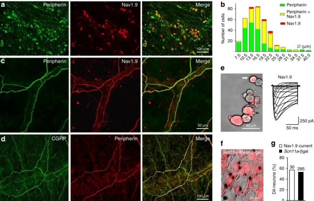
Nav1.9 is expressed in meningeal nociceptors. Immunostaining
of mouse TG cryosections indicated that Nav1.9 is expressed in
32% of TG neurons (Fig.
1
a). Nearly all immunopositive neurons
(96%, n
= 174) exhibited small-diameter or medium-diameter
cell bodies (<27 µm, average largest diameter
⌀ = 17.4 ± 0.3 µm)
(Fig.
1
b), while a minority of them (3.8%, n
= 7 out of 181)
displayed large cell bodies (⌀ ≥ 27 µm). Small peripherin-positive
fibers showed features of apposition with meningeal arteries in
whole mount of mouse dura mater (Supplementary Fig. 1A).
Dual-labeling showed that 89% (n
= 124/139) and 91% (n = 101)
of peripherin-positive meningeal
fibers were immunoreactive for
Nav1.9 and CGRP, respectively (Fig.
1
c, d), suggesting that
Nav1.9 and CGRP co-distribute in a large proportion of
menin-geal
fibers. Double-labeling for Nav1.9 and CGRP could not be
made due to the different
fixation conditions of primary
anti-bodies. However, the quasi-totality of
β-gal -positive TG neurons
exhibited CGRP staining in cryosections from Scn11a-GAL
reporter transgenic mice (n
= 63/64) (Supplementary Fig. 1B).
Few Nav1.9-positive meningeal
fibers were also found to be
immunoreactive for NF200, possibly corresponding to some
lightly myelinated sensory
fibers (Supplementary Fig. 1C, D). A
majority (57%) of retrogradely labeled (DiI) dural afferent
neu-rons was found to express Nav1.9 current using patch clamp
recording (Fig.
1
e, g). Consistently, 54% of DiI
+-dural afferent
neurons from Scn11a-GAL reporter transgenic mice exhibited
β-gal enzymatic activity (Fig.
1
f, g). Together, these data provide
evidence that about half of dural afferent neurons expresses
functional Nav1.9 channels.
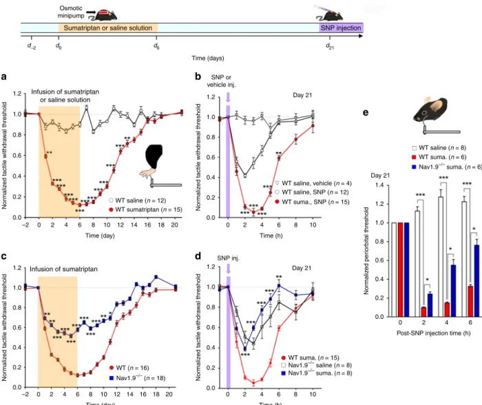
Scn11a gene inactivation abrogates NO-induced allodynia in
MOH mice. To probe the role of Nav1.9 channels in MOH, we
developed a model of MOH in Nav1.9
−/−mice and their
wild-type (WT) littermates using triptan medicines and assessed
quantitatively behavioral correlates of headache and migrainous
symptoms, including generalized allodynia, photophobia, and
phonophobia. Osmotic minipump infusion of sumatriptan in
WT mice for 6 days (0.6 mg/kg/day) produced a
time-dependent reduction in mechanical withdrawal thresholds of
the hind paw relative to saline-treated WT mice (Fig.
2
a).
Recovery of paw sensory thresholds to sumatriptan
pre-infusion values occurred within 18–20 days after minipump
implantation (Fig.
2
a). Latent sensitization to the migraine
trigger NO was tested at day 21, once sensory thresholds were
returned to pre-sumatriptan baseline levels (Fig.
2
b). Injection
of the NO donor sodium nitroprusside (SNP, 0.03 mg/kg) into
the loose skin over the neck evoked strong extracephalic tactile
allodynia in sumatriptan-treated WT mice compared with
saline-treated WT mice (Fig.
2
b). Heightened SNP-induced
allo-dynia in sumatriptan-treated WT mice was still observable up to
46 days after minipump implantation (Supplementary Fig. 2A).
Heightened mechanical allodynia induced by SNP injection at day
21 was absent in sumatriptan-treated Nav1.9
−/−mice (Fig.
2
d).
Moreover, saline-treated Nav1.9
−/−and WT mice displayed
similar SNP-induced (basal) allodynia (Fig.
2
b, d), indicating that
Nav1.9 contributes to the SNP-induced heightened allodynia in
sumatriptan-treated WT mice, but plays no apparent role in
SNP-induced basal allodynia in saline-treated animals.
Because females have increased risk of developing migraine
and MOH, we tested whether infusion of sumatriptan for 6 days
(0.6 mg/kg/day) produced mechanical hypersensitivity and latent
sensitization to NO in WT female mice as observed for the
opposite gender. Chronic sumatriptan produced a strong
reduction in mechanical withdrawal thresholds of the hind paw
relative to saline-treated WT female mice and to
sumatriptan-treated Nav1.9
−/−female mice (Supplementary Fig. 2B).
Injec-tion of SNP (0.03 mg/kg) at day 21 once sensory thresholds had
returned to pre-sumatriptan baseline values caused significantly
stronger mechanical allodynia in sumatriptan-treated WT female
mice compared to saline-treated WT female mice (Supplementary
Fig. 2C). SNP-induced heightened mechanical allodynia was
absent in sumatriptan-treated Nav1.9
−/−female mice
(Supple-mentary Fig. 2D), reaching similar amplitude to that caused by
SNP in saline-treated Nav1.9
−/−female mice (Supplementary
Fig. 2D). This series of experiments shows that Nav1.9, as
observed in male mice, had no role in SNP-induced allodynia in
saline-treated animals, but contributes to the heightened SNP
allodynia in sumatriptan-treated female mice.
SNP injection at day 21 was also found to reduce mean
periorbital von Frey thresholds (periorbital allodynia) in
sumatriptan-treated WT male mice compared with
saline-treated WT male mice (Fig.
2
e). Deletion of Nav1.9 significantly
attenuated the SNP-induced periorbital allodynia in
sumatriptan-treated animals (Fig.
2
e). Altogether, these data indicate that
Nav1.9 contributes to NO-induced cephalic and extracephalic
tactile allodynia in MOH mice.
Lack of NO-induced visual and auditory symptoms in Nav1.9
KO mice. Besides pain, disabling symptoms of MOH often
include photophobia and/or phonophobia
28. To evaluate
light-aversive behavior (photophobia) of mice after SNP injection, we
used the light–dark transition test
29. Two hours after SNP
injection, sumatriptan-treated WT mice spent significantly more
time in the dark chamber relative to saline-treated WT mice
(Fig.
3
a). Sumatriptan-treated Nav1.9
−/−mice challenged with
SNP showed no signs of photophobia as the animals spent no
more time in the dark box than saline-treated WT or Nav1.9
−/−mice treated likewise (Fig.
3
a).
We further quantified the sound sensitivity (phonophobia) of
mice following SNP injection by measuring the intensity of sound
required to induce the acoustic startle reflex (ASR). We exposed
mice to pseudo-random order of 100 ms-long white noise bursts
ranging from 60 to 120 dB SPL at 10 kHz. The ASR threshold
of saline-treated WT mice injected with SNP was 97 ± 2 dB SPL
(n
= 7), which was significantly different than that of
sumatriptan-treated WT mice, which showed a lower ASR threshold of 90 ± 1.5
dB SPL (n
= 6) (Fig.
3
b). Because one decibel equals 10 times the
common logarithm of the power ratio, a decrease in ~10 dB SPL in
ASR corresponds to a 10-fold increase in sound sensitivity. SNP
induced no changes in ASR threshold in sumatriptan-treated
Nav1.9
−/−mice compared to saline-treated Nav1.9
−/−mice
(Fig.
3
b). We then examined the prepulse inhibition (PPI) of the
ASR. This response provides an operational measure of
Peripherin
a
c
d
Nav1.9 Merge
Peripherin Nav1.9 Merge
CGRP Peripherin Merge 100 μm 50 μm 100 μm 60 80
b
e
f
Peripherin Peripherin + Nav1.9 Nav1.9 40 Number of cells Scn11a-driven β-gal 40 μm 25 μmg
Nav1.9 Nav1.9 current Scn11a-βgal 50 ms 80 30 285 60 40 20 0 250 pA Dil-neurons (%) 20 ∅ (μm) 7.5 10.513.516.519.522.5 25.528.531.534.537.540.5Fig. 1 Nav1.9 is expressed in meningeal nervefibers and dural afferent neurons. a Cryosections of a mouse TG were double-labeled for peripherin and
Nav1.9. Images are projections of seven consecutive optical sections spanning 9µm. Rightmost panel: merged image. b Histogram showing the distribution
of TG neurons immunopositive for peripherin (green), Nav1.9 (red), or both (yellow) as a function of cell body diameter (⌀). Bin size = 3 µm. c Mouse
whole-mount of dura mater was double-labeled for peripherin and Nav1.9. Images are projections of 16 consecutive optical sections spanning 22µm.
Rightmost panel: merged image.d Immunostaining for CGRP and peripherin in mouse dura mater. Images are projections of 21 consecutive optical sections
spanning 23µm. Rightmost panel: merged image. e Left panel: TG neurons cultured 2 days after DiI application through a cranial window in the parietal
bone. Right panel: recording of Nav1.9 current in the retrogradely labeled TG neuron indicated by an arrow. The current was evoked by 100 ms-voltage
steps from−80 to −5 mV from a Vh of −100 mV. CsF-containing patch pipette solution. f Image from a Scn11alacZmouse showingβ-galactosidase
expression (dark dots) in DiI+afferent neurons (red) from the TG.g Histogram showing the percentage of DiI+afferent neurons exhibiting Nav1.9 current
sensorimotor gating reflecting the sensitization of mice to weak
sounds
30. PPI was measured as the innate reduction of the startle
reflex induced by a weak pre-stimulus sound (Fig.
3
c). Following
SNP injection at day 21, the PPI value was 18.5 ± 2% in
saline-treated WT mice but reached 42.4 ± 2.5% in sumatriptan-saline-treated
WT mice (Fig.
3
c, d), indicating sensitization to sounds. By
contrast, the PPI value was not significantly different in
saline-treated versus sumatriptan-saline-treated Nav1.9
−/−mice (Fig.
3
c, d).
Together, these data indicate that Nav1.9 is essential to the
development of NO-induced symptoms of central sensitization
observed in MOH mice.
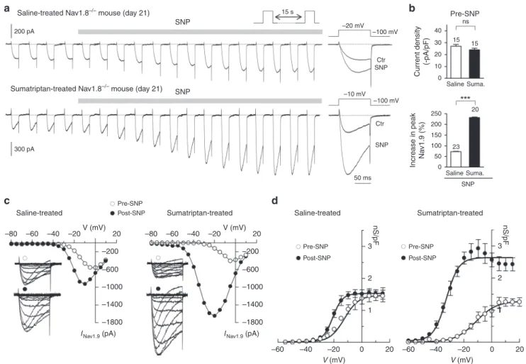
Triptan overuse promotes coupling of NO-cGMP to Nav1.9
channels. We investigated the molecular basis that promotes
increased sensitivity of MOH mice to NO. Given the difficulty to
unambiguously isolate Nav1.9 from Nav1.8 currents when using
CsCl-based pipette solution
11,13,14, retrogradely labeled dural
afferent neurons were studied from Nav1.8
−/−mice. We
pro-vided evidence that inactivation of the Scn10a gene encoding
Nav1.8 did not affect sumatriptan-induced latent sensitization
and hypersensitivity to SNP in MOH mice (Supplementary
Fig. 3).
Nav1.9 current recorded in dural afferent Nav1.8
−/−neurons
cultured at day 21 (i.e. 21 days after pump implantation) was
identifiable from its slow activation kinetics and incomplete
inactivation, producing
‘persistent’ TTX-resistant Na
+currents.
Neither the mean Nav1.9 peak current density (Fig.
4
b), nor the
level of Nav1.9 mRNA expression (at day 21) (Supplementary
a
c
d
b
e
d–2 d0 Osmotic minipumpSumatriptan or saline solution
d6 SNP or vehicle inj. Day 21 Day 21 WT suma. (n = 15) Day 21 d21 1.0 0.8 0.6 0.4 1.2 1.0 0.8 0.6 0.4 0.2 1.4 0.0 0.2 1.2 0.0 1.0 0.8 0.6 0.4 0.2 1.2 0.0 0 2 4 6 8 Infusion of sumatriptan or saline solution Infusion of sumatriptan 10 2 4 6 0
Post-SNP injection time (h)
2 4 6 8 0 10 12 WT saline (n = 12) WT saline (n = 8) WT suma. (n = 6) WT saline, vehicle (n = 4) WT saline, SNP (n = 12) WT suma., SNP (n = 15) Nav1.9–/– (n = 18) Nav1.9–/– suma. (n = 6) Nav1.9–/– saline (n = 8) Nav1.9–/– suma. (n = 8) WT (n = 16) WT sumatriptan (n = 15) Time (day) Time (days) Time (h) 14 16 18 –2 20 –2 0 2 4 6 8 10 12 0 2 4 6 8 10
Time (day) Time (h)
14 16 18 20 Nor maliz ed tactile withdr a w al threshold Nor maliz ed tactile withdr a w al threshold 1.0 0.8 0.6 0.4 0.2 1.0 0.8 0.6 0.4 0.2 1.2 0.0 1.2 0.0 SNP inj. Nor maliz ed tactile withdr a w al threshold Nor maliz ed per iorbital threshold Nor maliz ed tactile withdr a w al threshold SNP injection
Fig. 2 Deletion of Nav1.9 prevents NO-induced generalized allodynia in MOH mice. a Infusion of sumatriptan (0.6 mg/kg/day), but not saline solution (0.9%), decreases withdrawal thresholds to tactile stimuli applied to the hind paws of WT mice. Hind paw withdrawal threshold was tested using von Frey filaments (inset). **p < 0.01, ***p < 0.001 compared to saline with Mann–Whitney non-parametric test. Top inset: schematic of mouse treatment over time. b Changes in mechanical withdrawal thresholds of the hind paws induced by injection of SNP (0.03 mg/kg) in WT mice pre-treated (red symbols) or not
(open circles) with sumatriptan. Data illustrated depict SNP responses 21 days after minipump implantation. **p < 0.01, ***p < 0.001 compared to WT
saline, SNP with Mann–Whitney non-parametric test. c Hind paw withdrawal responses of sumatriptan-treated Nav1.9−/−mice compared with
sumatriptan-treated WT littermates. *p < 0.05, **p < 0.01, ***p < 0.001 compared to WT with Mann–Whitney non-parametric test. d Comparison of
SNP-induced changes in hind paw withdrawal thresholds in sumatriptan-treated WT mice (red symbols), saline-treated Nav1.9−/−mice (open squares) and
sumatriptan-treated Nav1.9−/−mice (blue squares). All tests were made at day 21. **p < 0.01, ***p < 0.001 compared to WT sumatriptan with two-way
ANOVA followed by Student–Newman–Keuls test. e Normalized periorbital withdrawal threshold plotted as a function of time after SNP injection in
saline-treated WT mice (open bars), sumatriptan-saline-treated WT mice (red bars) and sumatriptan-saline-treated Nav1.9−/−mice (blue bars). Data illustrated depict SNP
Fig. 4) was significantly different in TG neurons from
saline-treated and sumatriptan-saline-treated mice.
SNP (1 mM) increased Nav1.9 peak current (measured at peak
I–V) by 232 ± 11% (n = 20) in dural afferent neurons from
sumatriptan-treated Nav1.8
−/−mice, whereas it had little effect
(72.7 ± 4%, n
= 23) in control dural afferent neurons (Fig.
4
a, b).
Current–voltage relationships determined before and after SNP
application in sumatriptan-treated dural neurons showed a
22 mV negative shift in activation V1/2
value (from
−11.3 ± 1.8
to
−32.7 ± 1.02 mV). This shift was also associated with a ~2-fold
increase in Nav1.9 maximum conductance (from
−1.28 to −2.64
nS/pF) (Fig.
4
c, d). By contrast, SNP induced a ~−6 mV shift in
dural afferent neurons from saline-treated mice (from
−14.47 ±
1.3 to
−20.8 ± 0.8 mV), which was not associated with change in
Gmax
(Fig.
4
d).
Because the cyclic guanosine monophosphate (cGMP) signal
pathway plays an important role in NO signaling, we sought to
determine the involvement of the soluble guanylyl cyclase (sGC)
in the activation of Nav1.9. Application of methylene blue (100
µM), a sGC inhibitor, abolished the effect of SNP on Nav1.9 in
dural afferent neurons from sumatriptan-treated Nav1.8
−/−mice
(Supplementary Fig. 5). Consistently, the cell-permeable cGMP
analog 8-Br-cGMP (1 mM) strongly increased Nav1.9 current
and negatively shifted V1/2
activation by 19 mV (from
−13.5 to
−32.5 mV) in saline-treated Nav1.8
−/−mice (Supplementary
Fig. 6A–C). These data indicate that NO-cGMP pathway activates
Nav1.9 in sumatriptan-treated, but not in saline-treated dural
afferent neurons.
Relief of PKA inhibition causes NO coupling to Nav1.9 in
MOH mice. To probe the molecular changes in TG neurons at
day 21 from sumatriptan-treated mice, we made qPCR analysis of
cGMP-linked signaling molecules. Relative mRNA quantification
showed there was a three-fold decrease of the transcript of protein
kinase cAMP-activated catalytic subunit alpha (PKA-Cα) but no
changes (0.5 < RQ < 2) for the cyclic nucleotide
phosphodies-terases 3a, 3b and 5a (PDE3a, PDE3b, and PDE5a), the sGC, the
predominant receptor for NO, the Protein Kinase G type I
(PKG-I) and the Adenylyl Cyclase type III (AC-II(PKG-I) (Supplementary
Fig. 7A). Pre-treatment of 8-Br-cAMP, a membrane permeable
cAMP analog, inhibited cGMP-mediated activation of Nav1.9 in
dural afferent neurons from saline-treated mice, although it had
no effect per se on Nav1.9 (Supplementary Fig. 7B–D), indicating
that cAMP can inhibit cGMP coupling to Nav1.9 in TG neurons.
Consistently, we found that DiI
+-dural afferent neurons showed
strong staining for the catalytic PKA subunit phospho T197
antibody that recognizes the active form of the PKA protein
(Supplementary Fig. 8A, B). In addition, the mean intensity
(arbitrary unit) per pixel of PKA subunit phospho T197 staining
was significantly (p = 0.03, unpaired t-test) reduced from 131 ± 4
100 25 20 15 10 5
Auditory threshold (day 21) Light sensitivity (day 21)
a
c
d
Sound stimulus (60–120 dB SPL, 10 kHz) Strain gauge 7 10 ns 40 30 20 10 0 ns 6 6 7 5 ns 8 9 12 12 Saline Saline 10 % a.u. 10 % a.u. Saline 70 dB SPL 115 dB SPL 70 dB SPL 115 dB SPL Sumatriptan Saline Sumatriptan Saline Sumatriptan 10 6Acoustic startle reflex threshold after
SNP injection (dB SPL)
Pre-pulse inhibition (%)
Time in the dark after SNP injection (min)
95
90 85
80 WT
Sound sensitization after SNP injection (day 21) Sound sensitization after SNP injection (day 21)
Nav1.9–/– WT 100 ms PPI Sumatriptan Sumatriptan PPI PPI PPI Nav1.9–/– Nav1.9–/–mice WT mice WT 30 min test Light box Dark box
Light
Nav1.9–/–
b
Fig. 3 Deletion of Nav1.9 prevents NO-induced photophobia and phonophobia in MOH mice. a Light sensitivity of sumatriptan-treated and saline-treated
WT and Nav1.9−/−mice 2 h after injection of SNP (0.03 mg/kg) at day 21. Visible light: 380–740 nm, 2600 lx. Behavioral tests were made for a duration
of 30 min. ns not significant; **p < 0.01; Mann–Whitney test. b ASR threshold in sumatriptan-treated and saline-treated WT and Nav1.9−/−mice 2 h after
injection of SNP at day 21. ns not significant; **p < 0.01; Mann–Whitney test. c SNP-induced pre-pulse inhibition: the PPI was measured using the protocol
illustrated (top panels) in saline- (middle panel) and sumatriptan- (bottom panel) WT and Nav1.9−/−mice 2 h after injection of SNP. a.u. arbitrary unit.
d Comparison of the PPI in sumatriptan-treated or saline-treated WT and Nav1.9−/−mice 2 h after SNP injection. Tests were made at day 21. ns not
(n
= 44 DiI
+-neurons) in control mice injected with saline
solution to 118 ± 5 (n
= 50 DiI
+-neurons) in mice treated with
sumatriptan (data not shown). Changes in PKA expression in
TGs from sumatriptan-treated mice (n
= 8) were also evaluated
by quantifying band intensities on phospho T197 PKA blots and
comparing to blot band intensities in saline-treated mice (n
= 8)
as controls. Densitometry analysis of background-subtracted blots
from 20 µg of total lysate showed a 22% decrease in phospho
T197 PKA expression in sumatriptan-treated mice versus control
mice (Supplementary Fig. 8C). This decrease however did not
reach significant level due to sample variability (Supplementary
Fig. 8D).
Collectively, these data suggest that nitrergic activation of
Nav1.9 in sumatriptan-treated mice may result from a decrease in
PKA activity in dural afferent neurons and subsequent relief of
PKA-mediated inhibition of NO–Nav1.9 coupling.
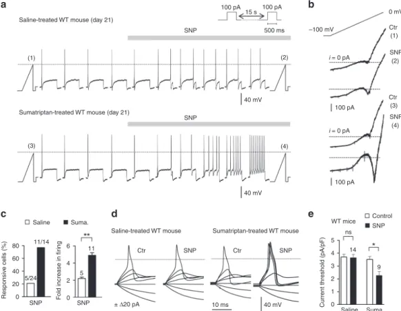
Nav1.9-mediated hyperexcitability causes central sensitization.
Firing activity of retrogradely labeled dural afferent neurons was
studied at day 21. SNP-mediated Nav1.9 activation converted
phasic discharges into multi-action potential (AP) responses in
78.5% of dural afferent neurons from sumatriptan-treated WT
mice (Fig.
5
a–c). In addition, SNP reduced by 35% the current
threshold for
firing in neurons from sumatriptan-treated WT
mice (Fig.
5
d, e). By contrast, SNP had no significant effects on
the
firing response and AP current threshold in dural afferent
neurons from saline-treated WT mice (Fig.
5
c, d). Moreover, SNP
caused no changes in excitation or AP current threshold in dural
afferent neurons from sumatriptan-treated Nav1.9
−/−mice
(Supplementary Fig. 9).
We tested whether Nav1.9-mediated hyperexcitability regulates
the secretion of CGRP, a key player in headache pathogenesis
(Fig.
6
a). SNP, at low concentrations, had no significant effects on
basal secreted CGRP levels in TG cultures from saline-treated
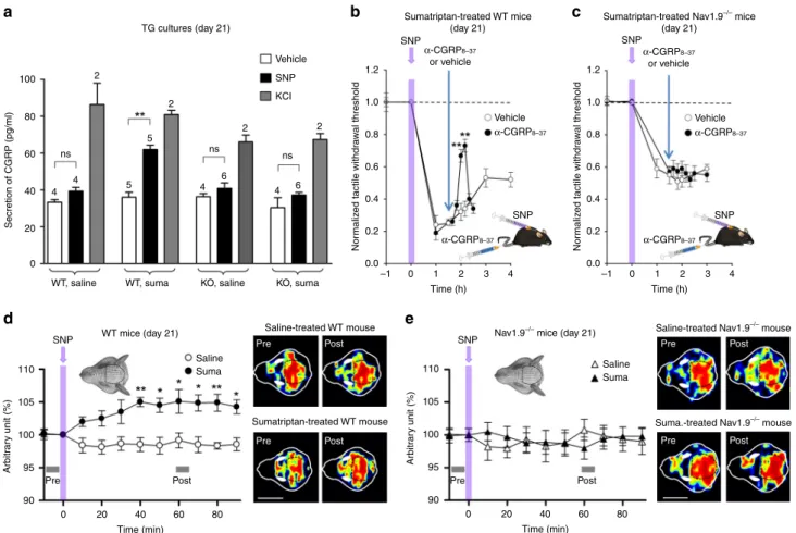
WT mice but enhanced CGRP secretion (+70%) in TG cultures
from sumatriptan-treated WT mice. Enhanced secretion of CGRP
by SNP was not observed in TG cultures from
sumatriptan-treated Nav1.9
−/−mice (Fig.
6
a).
The in vivo consequence of Nav1.9-dependent CGRP secretion
was tested on SNP-mediated extracephalic mechanical allodynia
at day 21 using the CGRP antagonist
α-CGRP8-37. Injection of
Saline-treated Nav1.8–/– mouse (day 21)
a
b
c
d
Saline-treated Saline-treated V (mV) V (mV) Sumatriptan-treated Sumatriptan-treated Pre-SNP Current density (-pA/pF) Increase in peak Na v1.9 (%) Pre-SNP Post-SNP Pre-SNP Post-SNP Pre-SNP Post-SNP –80 –60 –40 –20V (mV) 20 V (mV) –200 –600 –1000 –1400 –1800 –60 –40 –20 0 20 –60 –40 –20 0 20 –200 3 2 1 –600 –1000 –1400 –1800INav1.9 (pA) INav1.9 (pA)
–80 –60 –40 –20 20 nS/pF 3 2 1 nS/pF
Sumatriptan-treated Nav1.8–/– mouse (day 21)
SNP 15 s –20 mV –10 mV 50 ms –100 mV 40 ns 15 15 20 23 30 20 10 0 250 200 150 100 50 0 Saline Suma. Saline SNP Suma. –100 mV Ctr SNP Ctr SNP SNP 200 pA 300 pA
Fig. 4 Sumatriptan treatment promotes activation of Nav1.9 by NO. a Nav1.9 current exposed to 1 mM SNP in dural afferent neurons from saline-treated
(top panel) and sumatriptan-treated (bottom panel) Nav1.8−/−mice. CsCl-only-based patch pipette solution. Right-most traces: superimposed Nav1.9
currents before and after SNP application.b Nav1.9 current density (top panel) in dural afferent neurons from saline-treated and sumatriptan-treated
Nav1.8−/−mice. ns, not significant; unpaired t-test. Bottom panel: mean increase in Nav1.9 peak current induced by SNP (1 mM) in dural afferent neurons
from saline-treated and sumatriptan-treated Nav1.8−/−mice. ***p < 0.001; unpaired t-test. c Nav1.9 I–V determined in dural afferent neurons from
saline-treated and sumatriptan-saline-treated Nav1.8−/−mice before and after SNP exposure. Insets: superimposed Nav1.9 current traces evoked by voltage steps from
−80 to +10 mV from a Vh of −100 mV. Note that not all traces are shown for clarity sake. d Activation curves for Nav1.9 current determined in DiI+-dural
neurons before and after SNP application. Boltzmannfits gave V1/2values of−14.47 ± 1.3 and −20.8 ± 0.8 mV before and after SNP in saline-treated
Nav1.8−/−mice (n = 11) and of −11.3 ± 1.8 and −32.7 ± 1.02 mV before and after SNP in sumaptriptan-treated Nav1.8−/−mice (n = 9), respectively. All
α-CGRP8-37
(1 mg/kg), but not the vehicle, reduced SNP-induced
extracephalic allodynia in sumatriptan-treated WT mice,
indicat-ing that released CGRP contributes to central sensitization
(Fig.
6
b). Importantly, injection of
α-CGRP8-37
had no effects on
residual SNP-induced allodynia in sumatriptan-treated Nav1.9
−/−animals (Fig.
6
c), providing further evidence that Nav1.9
activation by SNP is a prerequisite for CGRP release from
meningeal nociceptors.
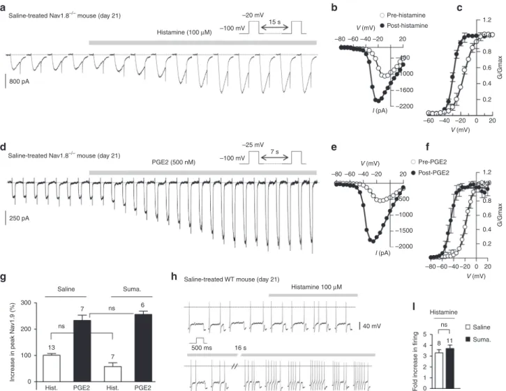
Nav1.9 mediates vasodilatation and mast cell degranulation.
Functional consequences of Nav1.9 activation on meningeal
microcirculation was examined at day 21 using a laser Doppler
blood perfusion scanner. SNP (0.03 mg/kg), injected through
the jugular vein, caused a gradual increase of meningeal blood
flow in sumatriptan-treated WT mice. No change was seen in
saline-treated WT mice and in sumatriptan-treated Nav1.9
−/−mice (Fig.
6
d, e). We further tested whether activation of
meningeal nociceptors by SNP causes degranulation of dural
mast cells (MCs) and pain amplification through a
Nav1.9-dependent mechanism. The MC stabilizing agent, sodium
cromoglycate (SCG, 10 mg/kg i.p.), injected 30 min prior to
SNP, significantly reduced both the intensity and duration of
the SNP-induced heightened allodynia in sumatriptan-treated
WT mice (Supplementary Fig. 10B) but had no significant
effects on SNP-induced basal allodynia in saline-treated WT
mice (Supplementary Fig. 10A) and on SNP-induced allodynia
in
sumatriptan-treated
Nav1.9
−/−mice
(Supplementary
Fig. 10C).
We
finally sought to determine whether MC degranulation
contributes to dural afferent terminal excitation through
receptor-driven modulation of Nav1.9. Patch clamp recordings
showed that the MC mediators histamine and PGE2 are
powerful activators of Nav1.9 current in dural afferent neurons
(Fig.
7
). Superfusion of histamine and PGE2 increased peak
Nav1.9 current by 100.5 ± 25% and 233 ± 40%, respectively, in
saline-treated Nav1.8 KO mice (Fig.
7
a–g). Both mediators
caused a substantial leftward shift in the activation curve of
Nav1.9 (Fig.
7
c, f) and strongly increased the
firing rate of dural
afferent neurons (Fig.
7
h, i). These effects were seen irrespective
of the mouse treatment (Fig.
7
g). By contrast, CGRP, SP, and
neurokinin A, which are potentially released by meningeal
terminals, had no detectable effects on Nav1.9 current in dural
afferent neurons from either saline-treated or
sumatriptan-treated mice (Supplementary Fig. 11A–D). Thus, some MC
mediators have the capacity to activate Nav1.9, resulting in a
feedforward loop that potentiates neurogenic inflammation and
nociceptive transmission. Collectively, these results indicate
that Nav1.9 acts as a hub in meningeal nociceptors and
contributes to maladaptive nociceptive signal, neurogenic
inflammation, meningeal vasodilatation, and mast cell
degra-nulation (Fig.
8
).
Saline-treated WT mouse (day 21)
Sumatriptan-treated WT mouse (day 21)
Sumatriptan-treated WT mouse
WT mice
Current threshold (pA/pF)
Control SNP ns 5 4 14 9 3 2 1 0 Saline-treated WT mouse 100 pA 100 pA 15 s SNP SNP 500 ms 40 mV 40 mV 40 mV 10 ms Saline 80 6 4 2 0 11/14 5/24 SNP SNP ± Δ20 pA Ctr SNP Ctr SNP 5 11 Responsiv e cells (%) F
old increase in fir
ing 60 40 20 0 Suma. Saline Suma. –100 mV i = 0 pA i = 0 pA 100 pA 100 pA 0 mV Ctr (1) (1) Ctr (3) (3) (2) (2) SNP (4) (4) SNP
a
b
c
d
e
Fig. 5 Nav1.9 activation by NO lowers excitability threshold and enhancesfiring in dural afferent neurons. a Effect of SNP (1 mM) on DiI+dural afferent
neurons from saline-treated (upper panel) and sumatriptan-treated (bottom panel) WT mice. KCl-based intracellular solution throughout.bI–V
relationships determined using a slow (50 mV/s) voltage ramp command in DiI+dural neurons illustrated ina before (1,3) and during (2,4) SNP
application. Note the activation of Nav1.9 (inwardlyflowing current) by SNP (4). c Percentage of DiI+neurons responding to SNP (left panel) and mean
change in theirfiring rate (right panel). Protocol as in a. **p < 0.01; Mann–Whitney test. d Generation of APs before and after SNP exposure in dural
afferent neurons from saline-treated or sumatriptan-treated WT mice. Steady bias currents were used to maintain the neurons at ~−65 mV. e Comparison
of normalized current threshold for AP before and during SNP application in DiI+dural neurons from saline-treated and sumatriptan-treated WT mice. ns
Discussion
How chronic exposure to abortive medication leads to MOH
remains unclear. Our study suggests that chronic use of triptans
induces MOH through abnormal activation of meningeal Nav1.9
by NO. Thus, triptan-overuse headache derives from Nav1.9
activation in the TG system, triggering pain facilitation through
central and peripheral sensitization and inflammation in the
meninges.
The complex pathophysiology behind MOH is still only partly
known. Mechanisms involved may differ from one class of
overused drug to another. Previous studies have shown that
chronic use of opioids and triptans increases CGRP levels which
is well known to be involved in neurogenic inflammation and
headache pain
8,31. Impaired diffuse noxious inhibitory controls
6and central sensitization are also seen in MOH patients
4,32. Many
of these phenomena are similar to mechanisms seen in
depen-dence processes
33,34.
Our approach to modeling MOH symptoms was the
quanti-fication of increased sensory sensitivity in response to NO, one of
the
most
common
reported
trigger
for
headache
and
migraine
8,9,35,36. NO donors reliably trigger headache in normal
subjects, but trigger migraine and severe pain in migraineurs
36,
and this condition is accompanied by an increase in blood levels
of CGRP, which is directly linked to the severity of headache
pain
37–39. In our study, MOH following NO infusion was
evaluated using multiple headache-like responses in each
indivi-dual animal, including cutaneous facial and extracephalic
allo-dynia as well as aversion to light and noise. Our data show that
chronic sumatriptan treatment of mice induces a state of dormant
sensitization
characterized
by sharp,
exacerbated
sensory
responses to NO
8,9,40.
Clinical and preclinical studies have consistently demonstrated
increased excitability of the TG system after medication overuse.
TG neuronal hyperexcitability may facilitate the process of
per-ipheral and central sensitization. We show that NO was capable
of producing strong activation of dural afferent neurons in MOH
mice and that sensory hypersensitivity and MOH-associated
symptoms were prevented by deleting Nav1.9 but not Nav1.8.
Nav1.9 activation sustains the hyperexcitability of meningeal
nociceptors and lowers the threshold response for afferent pain
signaling. Importantly, coupling of NO to Nav1.9 was weak under
normal conditions, consistent with the observation that deleting
Nav1.9 had no impact on SNP-induced (basal) cutaneous
allo-dynia in saline-treated mice. Thus, chronic sumatriptan treatment
promotes coupling of NO to Nav1.9 channels in dural afferent
neurons, thus lowering the threshold of the animal’s susceptibility
to respond to initiating factors of headache. Importantly, we
found that hypersensitivity to SNP was greater in MOH female
than in male mice, which parallels the sexual dimorphism
reported in MOH and migraine in humans.
TG cultures (day 21)
a
d
e
b
c
WT mice (day 21) Sumatriptan-treated WT mice (day 21)Sumatriptan-treated Nav1.9–/– mice (day 21) SNP SNP SNP α-CGRP8–37 or vehicle α-CGRP 8–37 or vehicle α-CGRP8–37 α-CGRP8–37 SNP α-CGRP8–37 SNP 100 ns 4 4 4 4 2 2 Vehicle Vehicle α-CGRP8–37 Vehicle SNP Saline Saline-treated WT mouse Pre Post Sumatriptan-treated WT mouse Pre Post
Saline-treated Nav1.9–/– mouse
Pre Post
Suma.-treated Nav1.9–/– mouse
Pre Post Suma KCI 6 6 2 2 5 5 ns ns Secretion of CGRP (pg/ml) Arbitr ar y unit (%) 80 60 40 20 0 90 Pre 0 20 40 60 80 Post 95 100 105 110
WT, saline WT, suma KO, saline KO, suma
1.0 0.8 0.6 0.4 0.2 1.2 0.0 –1 0 1 2 Time (h) Time (min)
Nav1.9–/– mice (day 21) SNP Saline Suma Arbitr ar y unit (%) 90 Pre 0 20 40 60 80 Post 95 100 105 110 Time (min) 3 4 –1 0 1 2 Time (h) 3 4 Nor maliz ed tactile withdr a w al threshold 1.0 0.8 0.6 0.4 0.2 1.2 0.0 Nor maliz ed tactile withdr a w al threshold
Fig. 6 Nav1.9 activation sustains CGRP release, which contributes to central sensitization and meningeal vasodilatation. a Effects of SNP (1 mM), HBSS
(vehicle), and KCl (40 mM) on CGRP secretion in TG cultures from WT or Nav1.9−/−mice treated or not with sumatriptan. Then number refers to the
number of triplicates. **p < 0.01; Mann–Whitney test. b, c Effect of intravenous injection of α-CGRP8–37(1 mg/kg) or its vehicle (NaCl 0.9%) on
SNP-induced mechanical paw allodynia in sumatriptan-treated WTb or Nav1.9−/−c mice.**p < 0.01 compared to α-CGRP8–37pre-injection with
Wilcoxon-matched paired test (n = 5 per group). Behavioral tests were carried out at day 21. d–e SNP-induced meningeal blood flow changes in WT d and Nav1.9−/−
e mice, treated or not with sumatriptan. *p < 0.05, **p < 0.01; Mann–Whitney test (n = 6 per group). Right panels: representative laser Doppler images
taken before and 60 min after SNP injection. The blue color represents low perfusion areas, green and yellow refer to higher perfusion and red shows the highest microperfusion. Scale: 8 mm
What then favors Nav1.9 activation by NO/cGMP in
sumatriptan-treated mice? PKA-Cα transcripts were found to be
down-regulated in TGs from sumatriptan-treated animals at day
21, suggesting that constitutive activity of PKA, in control
ani-mals, inhibits NO–Nav1.9 coupling. This is consistent with the
observation that cAMP pretreatment inhibited cGMP-mediated
activation of Nav1.9 in dural afferent neurons. How chronic
stimulation of Gi-coupled 5HT1B/D receptors, which are
notably expressed in TG neuron plasma membrane
41, leads to
long-term changes of PKA gene transcription remains to be
determined.
Genetic deletion of Nav1.9 prevented SNP-induced meningeal
vasodilatation, indicating that Nav1.9-dependent excitation of TG
neurons was a prerequisite for this effect. Thus, Nav1.9 activation
by SNP not only causes hyperexcitability of nociceptors but also
triggers the release of vasoactive neuropeptides in the meninges.
In vitro experiments further indicated that NO-induced release of
CGRP was prevented by deleting Nav1.9. These data indicate that
Nav1.9-dependent release of CGRP, and possibly other
neuro-peptides including SP, plays a pivotal role in the vasodilatation of
meningeal blood vessels. Our data however do not specify
whe-ther the vasodilatation contributes to MOH-related symptoms or
whether this is a side-phenomenon
42. Therefore, if vasodilatation
of meningeal arteries contributes to the propagation of the
cas-cade of symptoms, it is likely in conjunction with other factors.
CGRP is also poised to enhance headache pain by central
mechanisms. Consistently, our results show that SNP-induced
extracephalic allodynia was transiently alleviated by acute
administration
of
the
CGRP
antagonist
α-CGRP8–37
in
sumatriptan-treated animals. Because, extracephalic allodynia is a
manifestation of central sensitization
43, these data argue that
Nav1.9-dependent CGRP release may also occurs at postsynaptic
structures, such as the trigeminal nucleus caudalis, activity of
which may sensitize thalamic neurons. These results call for an
Saline-treated Nav1.8–/– mouse (day 21)Saline-treated Nav1.8–/– mouse (day 21)
Saline-treated WT mouse (day 21) Histamine (100 μM) Histamine 100 μM 40 mV Histamine Saline Suma. PGE2 (500 nM) 250 pA 800 pA
a
d
e
f
b
c
g
h
l
I (pA) –2200 –1600 –1000 –400 Saline 7 ns 6 300 200 Increase in peak Na v1.9 (%) 100 0Hist. PGE2 Hist. PGE2
5 ns 11 8 F o ld increase in fir ing 4 3 2 1 0 ns 13 7 Suma. 500 ms 16 s –80 –60 –40 –20 20 –500 –1000 –1500 –2000 –80 –60 –40 –20 0 V (mV) 20 –60 –40 –20 0 V (mV) 20 0.2 0.4 0.6 0.8 1.0 1.2 G/Gmax 0.2 0.4 0.6 0.8 1.0 1.2 G/Gmax I (pA) V (mV) –25 mV –100 mV 7 s –80 –60 V (mV) Pre-histamine Post-histamine Pre-PGE2 Post-PGE2 –40 –20 20 –20 mV –100 mV 15 s
Fig. 7 Mast cell mediators activate Nav1.9 in dural afferent neurons. a Nav1.9 current challenged with 100µM histamine in a DiI+dural afferent neuron
from a saline-treated Nav1.8−/−mouse. CsCl-only-based patch pipette solution.bI–V relationships from the cell depicted in a determined before and after
histamine exposure.c Averaged activation curve for Nav1.9 determined before and after histamine application in DiI+neurons (n = 13). Single Boltzmann
fits gave V1/2values of−15 ± 0.3 and −29.3 ± 0.15 mV before and after histamine application, respectively. d Nav1.9 current challenged with 500 nM PGE2
in a DiI+dural afferent neuron from a saline-treated Nav1.8−/−mouse. CsCl-only-based patch pipette solution.eI–V relationships from the cell depicted in
d determined before and after PGE2 exposure. f Averaged activation curve for Nav1.9 determined before and after PGE2 application in DiI+dural afferent
neurons (n = 7). Single Boltzmann fits gave V1/2values of−17.3 ± 0.6 and −45.6 ± 1.1 mV before and after PGE2 application, respectively. g Increase in
Nav1.9 current (at peakI/V as in b and e) induced by histamine in DiI+neurons from saline-treated or sumatriptan-treated Nav1.8−/−mice. ns not
significant, Mann–Whitney test. h Effects of histamine on a DiI+dural afferent neuron from a WT mouse treated with saline solution. Voltage responses
were evoked by depolarizing pulses (+50 pA) applied every 8 s. KCl-based intracellular solution. i Mean increase in firing of dural afferent neurons in
early use of anti-Nav1.9 drugs that target meningeal nociceptors,
before the development of central sensitization.
Our data demonstrate that Nav1.9-mediated release of
neuro-peptides from meningeal nociceptors could trigger degranulation
of dural MCs. CGRP and SP, which are often co-released, are
particularly important in this regard as they are known to
sti-mulate MC degranulation in different systems
44. Histamine, a
major amine released from MCs
45, and PGE2
another
pro-inflammatory MC mediators
46, are well-recognized
migraino-genic substances known to induce pain upon infusion into
human subjects diagnosed with migraine. Our data show that
both substances cause a prominent increase of Nav1.9 and
pro-mote neuronal hyperexcitability, resulting in a vicious,
self-reinforcing cycle of sterile inflammation and nociception (cf
Fig.
8
). Because MCs express a variety of 5-HT receptors, it is
possible that chronic treatment with sumatriptan reinforces the
possibility that MCs respond to peptide-induced degranulation.
However, the reported effects of 5-HT on MCs have generally
been found to be mediated by 5-HT1A and not by 5HT1B/D
receptors
47.
In conclusion, our study identifies NO-induced Nav1.9 channel
activation as a triggering mechanism for MOH-related
symp-toms. The way in which medication overuse transforms episodic
migraine into chronic daily headache is still unknown. The
pic-ture that emerges from our study is that abnormal sensitivity of
meningeal Nav1.9 channels to the migraine trigger NO may cause
the headache phase in MOH patients. Activation of Nav1.9 may
be a common denominator of overused drugs and migraine
triggers. Therefore, the use of Nav1.9 channel inhibitors, in
combination with sumatriptan or other headache medications,
may represent a new acute and preventive option for migraine
treatment.
Methods
Animals. This project was approved by the Institutional Review Board of the regional ethic committee. All animals were used in accordance with the European Community guiding in the care and use of animals (2010/63/UE). All efforts were
made to minimize the number of animals used and their suffering. Mice (10
–12-week-old adult males and females, C57Bl/6J background) used in this study were
Nav1.9−/−, Nav1.8−/−(gifts from N.J. Wood, see refs.19,23) and their WT
littermates. The Scn11a-GAL reporter mouse was previously described15. An IRES/
LacZpA cassette followed by a loxP/neo/loxP cassette was inserted into the end of exon 5 of the Scn11a gene.
Infusion of sumatriptan and injection of SNP. Alzet osmotic minipumps (model
1007D, Charles River, France) with a nominalflow rate of 0.5 μl/h for 6 days were
used for drug infusion. The minipumps were implanted subcutaneously under
anesthesia with isoflurane. The day of the pump implant was considered as day 0.
Drugs administered by infusion were sumatriptan (0.6 mg/kg/day, Sigma, St. Louis USA) and its vehicle (NaCl, 0.9%). SNP was injected subcutaneously into the loose skin over the neck at 0.03 mg/kg. Infusion of sumatriptan/saline and injection of SNP/saline were made blind, e.g. the investigator was not aware of the content of the minipump, nor of the nature of the solution (SNP or vehicle) injected on day 21. Animals were randomly assigned to treatment groups.
Tactile sensory testing. Hind paw mechanical threshold was assessed using von
Freyfilaments (Bioseb, France) as described20. For facial testing, mice were
sub-jected to a von Frey stimulus applied to the forehead surface, repeated three times (at minimum 30 s interval). The head withdrawal tactile sensory threshold was the lowest force to elicit withdrawal in 2 of 3 trials. Data points were normalized to the control sensory threshold values determined just before minipump implantation (i.e. at day 0) or before SNP injection (i.e. at day 21, H0).
Light aversive test. To evaluate the light-aversive behavior after SNP injection, we used a light–dark test. The light-aversion chamber consisted of two equally sized compartments (10 cm width, 13 cm length, and 13 cm height), one painted white and lacking a top, the other painted black and fully enclosed. A corridor (7.2 cm width, 7.2 cm length, and 13 cm height) connects the two compartments. Mice were acclimatized at least 1 h in their cages in the testing room and were allowed to explore the light-aversion chamber 10 min before testing. The light-aversive behavior was examined 2 h after SNP injection (0.03 mg/kg) and during 30 min.
The thermal-neutralfiber-optic source is located in the middle of white box and
produces a light intensity of 2600 lx inside the lit area of the white chamber. Mouse
transitions were interpreted to be a reflection of light aversion.
Acoustic startle threshold and prepulse inhibition tests. The Startle Reflex
System (Bioseb, France) was used to measure the acoustic sensitivity of mice. The hardware consisted of an isolation cabinet that minimizes the effects of extraneous noise and vibrations. The startle chamber is situated in the center of the isolation cabinet (250 (W) × 250 (D) × 250(H) mm) and a restrainer (90 × 30 mm) was used to restrain minimally the animal during testing. Mice were acclimatized in the restrainer for 5 min per day, one week before the testing session. Mice were also acclimatized in their cages in the testing room, 2 h before the testing session. Acoustic stimuli for behavioral tests were based on previously published studies defining criteria applicable to mice at around 2 months of age. The waveforms
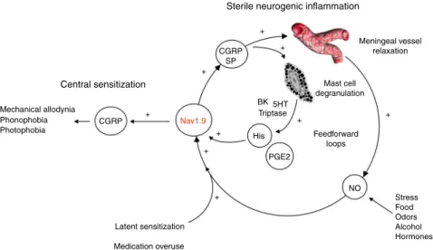
Central sensitization
Sterile neurogenic inflammation
Mechanical allodynia Phonophobia Photophobia CGRP Latent sensitization Medication overuse BK 5HT Mast cell degranulation Feedforward loops Stress Food Odors Alcohol Hormones Meningeal vessel relaxation Triptase His PGE2 NO CGRP SP Nav1.9 + + + + + + + + +
Fig. 8 Central role of Nav1.9 in MOH mechanisms. The following scenario summarizes the contribution of Nav1.9 to MOH. NO, which may be released from different sources, activates Nav1.9 channels in dural afferent neurons from chronically treated mice with triptans. Nav1.9 activation by NO increases the excitability of meningeal nociceptors, which sensitizes central structures leading to extracephalic allodynia, photophobia, and phonophobia. Nav1.9-dependent secretion of CGRP in the meninges, possibly in combination with other mediators, causes degranulation of resident mast cells. By releasing histamine and PGE2, MCs retro-excite meningeal nociceptors through Nav1.9 potentiation. Vasoactive peptides also contribute to vascular relaxation that may further facilitate endothelial (and possibly extravascular) NO production. The consequence is a vicious circle that leads to enhanced activation of meningeal nociceptors and maladaptive pain
generated by the mouse’s movement during the startle response were analyzed using the Packwin software (Panlab, Inc., Harvard apparatus).
Two hours after SNP injection, the mouse was acclimatized for 5 min in the presence of a background noise level of 60 dB SPL. The mouse was then exposed to random trials of white noise bursts ranging from 60 to 120 dB SPL at 10 kHz, frequency that falls within the most sensitive region of the mouse audiogram. Each sound intensity was presented three times. The ASR threshold was taken as the minimum intensity required to elicit a response in two out of third trials. The PPI is a reliable, robust quantitative phenotype that is useful for probing sensory functions. Testing consisted of a series of 115 dB SPL-test pulses immediately preceded or not by a prepulse of 70 dB SPL at 10 kHz. PPI was calculated as
follows: PPI (%)= (1–(startle response with prepulse)/(startle response without
prepulse)) × 100.
Cultures of TG neurons. Mice were anesthetized at day 21 with isoflurane and killed by decapitation. TGs were dissected out and freed from their connective tissue sheaths. TG neurons were incubated in enzyme solution containing 2 mg/ml of collagenase IA (Sigma) for 45 min at 37 °C. The tissue was washed several times and triturated in Hanks’ balanced salt solution. The resulting suspension was
fil-tered (70 µmfilters) and centrifuged (800×g for 5 min) and plated on poly-L-lysine/
laminin (0.05 and 0.01 mg/ml, respectively) coated Nunclon dishes. Culture medium was Dulbecco’s modified Eagle’s medium supplemented with 10%
heat-inactivated FCS, 100 U/ml penicillin–streptomycin, 2 mML-glutamine, 25 ng/ml
nerve growth factor (NGF), and 2 ng/ml glial-derived neurotrophic factor (GDNF). Patch clamp recordings. Patch pipettes had resistances of 2 MΩ. Voltage clamp
recordings of Na+currents used the following intracellular solutions:
(CsF-con-taining, in mM), 30 CsF, 100 CsCl, 10 HEPES, 10 EGTA, 8 NaCl, 1 MgCl2, 1 CaCl2,
4 MgATP, 0.4 Na2GTP (pH 7.35, 300 mOsm/l); (CsCl-based, in mM), 130 CsCl,
10 HEPES, 10 EGTA, 8 NaCl, 1 MgCl2, 1 CaCl2, 4 MgATP, 0.4 Na2GTP (pH 7.35,
300 mOsm/l). The extracellular solution contained (in mM): 60 NaCl, 110 sucrose,
3 KCl, 1 MgCl2, 10 HEPES, 2.5 CaCl2, 10 glucose, 10 TEA–Cl, 0.0005 TTX,
1 amiloride, 0.05 La3+(pH 7.4, 305 mOsm/l). For current clamp recording, the
intracellular solution (KCl-based) consisted of (mM): 115 KCl, 10 HEPES,
10 EGTA, 8 NaCl, 1 MgCl2, 1 CaCl2, 4 MgATP, 0.4 Na2GTP. The extracellular
solution consisted of (in mM) 131 NaCl, 3 KCl, 1 MgCl2, 10 HEPES, 2.5 CaCl2,
10 glucose (pH 7.4, 305 mOsm/l). All chemicals were from Sigma-Aldrich, except TTX (Alomone Labs).
PCLAMP 9.2 (Axon Instruments Inc.) and PRISM 4.0 (GraphPad) software suites
were used to perform linear and nonlinearfitting of data. Conductance–voltage curves
were calculated from the peak current according to the equation G= I/(V−Erev),
where V is the test pulse potential and Erevthe reversal potential calculated according
to the Nernst equation. The activation curve (G−V) was fitted using the Boltzmann
function: G/Gmax= 1/(1 + exp[(V1/2−V)/k]), where G/Gmaxis the normalized
conductance, V1/2is the potential of half-maximum channel activation, and k is the
steepness factor.
Tracer application onto the dura. Mice were anesthetized with isoflurane. Throughout surgery, the core temperature of the mouse was monitored and maintained by an homeothermic blanket system for rodents. Two small cranial windows were made in parietal bones and the retrograde nerve tracer DiI (DiI
tissue labeling paste, Invitrogen) was applied onto the dura. The boneflaps were
then replaced after the procedure with bone wax in order to prevent tracer spreading. Animals were euthanized for TG extraction 2 days after DiI application. Tissue preparation, immunostaining, and confocal imaging. For Nav1.9 immunostaining, the tissues were cryoprotected in PBS containing 4% sucrose during 30 min and then incubated for at least 1 h in PBS plus 20% sucrose at 4 °C. The TGs were frozen in OCT embedding matrix bathed in chilled isopentane. The TGs were then sagitally cryosectioned at 14–18 µm, transferred to SuperFrostPlus
slides and stored at−80 °C until processed. Whole mount dura maters were
transferred to SuperFrostPlus slides, frozen on dry ice and stored at−80 °C until
processed. Primary antibodies used and dilutions were as follows: anti-peripherin 1/400 (mouse monoclonal, Millipore, Temecula, CA); anti-NF200 1/400 (chicken polyclonal, Aves Labs, Tigard, OR); anti-CD31 (1/400, rat polyclonal, BD
Bios-ciences, Belgium); anti-Nav1.9 L23, (1/100, rabbit polyclonal)10; and anti-CGRP (1/
300, goat polyclonal, AbCam).
For CD31 immunostaining, the tissues werefixed for 30 min at room
temperature (RT) in Antigenfix (Microm Microtech, France) before being cryoprotected and frozen as described above. Slides with fresh frozen tissues or fixed with Antigenfix tissues were thawed at RT and then incubated for 90 min at RT in blocking solution containing 3% BSA and 0.1% Triton X-100. Primary antibodies were diluted in PBS containing 3% BSA and applied to tissues to be
incubated overnight at 4 °C in sealed humidified chambers.
For CGRP immunostaining, the tissues werefixed in 4% PFA in PBS for 3 h at
RT then cryoprotected in PBS containing 25% sucrose overnight at 4 °C. The
tissues were transferred to SuperFrostPlus slides, frozen and stored at−80 °C until
processed. Slides with PAF-fixed tissues were thawed at RT and then incubated for
1 h30 min at RT in blocking solution containing 5%fish gelatin and 0.2% Triton
X-100. CGRP antibodies were diluted in PBS containing 5%fish gelatin and applied
to tissues to be incubated overnight at RT in sealed humidified chambers.
After incubation with the primary antibodies,fixed and fresh frozen tissues
were identically proceeded. Tissues were washed three times for 5 min in PBS and incubated for 45 min at RT with secondary antibodies diluted in blocking buffer. Secondary antibodies were: Alexa Fluor 647-conjugated donkey anti-mouse (1/400, Life Technologies), TRITC-conjugated donkey anti-rabbit (1/400, Jackson ImmunoResearch, Suffolk, UK), TRITC-conjugated donkey anti-rat (1/100, Jackson ImmunoResearch), Alexa Fluor 488-conjugated donkey anti-goat (1/200, Life Technologies). After six 5 min-washes in PBS, sections were mounted in Mowiol (Sigma-Aldrich). Images were acquired using a LSM 780 laser-scanning confocal microscope (Zeiss), initially processed using ZEN software (Zeiss) and
later exported into Adobe Photoshop (Adobe Systems, San Jose, CA) forfinal
processing.
Staining for beta-galactosidase enzyme activity. Evaluation ofβ-gal enzymatic
activity was performed on transgenic mice that expressβ-gal at the Scn11a loci.
Once dissected out, the TGs were slightlyfixed 15 min in PBS containing 2% PFA,
0.2% glutaraldehyde, and 2 mM MgCl2. The tissues were washed with two changes
of PBS containing 2 mM MgCl2for 5 and 8 min. The staining is achieved with an
overnight incubation of TGs in PBS added with 4 mM Ferrocyanide, 4 mM
Fer-ricyanide, 2 mM MgCl2, 1% Tween-20, and 0.2 mg/ml X-gal at 37 °C. The TGs
werefinally post-fixed for 15 min in PBS containing 2% PFA, 0.2% glutaraldehyde,
and 2 mM MgCl2, and washed four times in PBS. The TGs were cryoprotected in
PBS containing 4% sucrose for 30 min and then incubated overnight in PBS plus 20% sucrose at 4 °C. The TGs were then frozen in OCT embedding matrix (Cellpath, Hemel Hempstead, UK) bathed in chilled isopentane. TGs were then
sagitally cryosectioned at 14–18 µm, transferred to SuperFrostPlus slides (Fisher
Scientific, Houston, TX), dried at RT and mounted in Mowiol (Sigma-Aldrich).
CGRP staining in TGs fromScn11a-GAL reporter mice. β-gal enzymatic activity
in TGs from Scn11a-GAL reporter transgenic mice was revealed as above, except
that glutaraldehyde was omitted from thefixative solution. Once achieved, TGs
were post-fixed for 2 h in PBS containing 2% PFA and 2 mM MgCl2, washed four
times in PBS and then cryoprotected in PBS containing 25% sucrose overnight. TGs were then frozen in OCT embedding matrix bathed in chilled isopentane and sagitally cryosectioned at 14–18 µm, transferred to SuperFrostPlus slides, dried at
RT and stored at−80 °C. Slides with TGs were thawed for 15 min at RT and then
incubated for 1 h 30 min at RT in blocking solution containing 3% BSA and 0.1% Tx-100. CGRP antibody (rabbit polyclonal #PC205L, Millipore) were diluted at 1/ 200 in blocking buffer and applied to tissues to be incubated overnight at 4 °C in
sealed humidified chambers. Tissues were washed three times for 10 min in PBS
and incubated 40 min at RT in PBS containing 3% BSA added with secondary antibodies Alexa Fluor 488-conjugated donkey anti-rabbit (1/200, Life Technolo-gies). After successive 5 min washes in PBS, sections were mounted in Mowiol
(Sigma-Aldrich). Images were acquired using a conventionalfluorescence
micro-scope (Zeiss Axio-observer) with constant acquisition settings.
Quantitation of anti-PKA immunostaining. The TG ganglia were carefully dis-sected out from NaCl-treated and sumatriptan-treated mice whose dura were
DiI-stained (see above). The TGs werefixed in 4% PFA in PBS for 2 h 30 min at 4 °C
then cryoprotected in PBS containing 25% sucrose overnight at 4 °C. The TGs were then frozen in OCT embedding matrix bathed in chilled isopentane and stored at −80 °C until processed. The day before PKA staining, the TGs were sagitally
cryosectioned at 14 µm, transferred to SuperFrostPlus slides and stored at−80 °C.
Slides with PAF TGs were thawed 15 min at RT and then incubated for 30 min at
RT in blocking solution containing 3%fish gelatin and 0.05% saponin. PKA
antibodies (rabbit polyclonal #ab75991, AbCam) were diluted at 1/200 in blocking buffer and applied to tissues to be incubated overnight at 4 °C in sealed humidified chambers. Tissues were washed three times for 10 min in PBS and incubated for 3 h
at RT in PBS containing 3%fish gelatin added with secondary antibodies Alexa
Fluor 488-conjugated donkey anti-rabbit (1/200, Life Technologies). After four 5 min washes in PBS, sections were stained with DAPI, washed once in PBS and mounted in Mowiol (Sigma-Aldrich).
In order to compare thefluorescence intensities of PKA immunostaining
between NaCl-treated and sumatriptan-treated DiI-positive TG neurons, images were acquired using a LSM 780 laser-scanning confocal microscope (Zeiss) using constant acquisition settings. ImageJ software was used to measure intensities of fluorescence on planar projections of confocal raw images spanning 6 µm. Elliptical regions of interest encompassing the soma of each DiI-positive neuron were defined in the DAPI image to exclude staining of the nucleus. The measurements were repeated in an adjacent area out of the tissue and the resulting background
intensities were subtracted from the somafluorescence signal. Results are expressed
as mean grey value intensity per pixel meaning the sum of grey values of all pixels in the ROI divided by the number of pixels.
PKA western blot analysis. TGs were collected at 21 days after sumatriptan or saline infusion (T0), scrapped in TE buffer (66 mM Tris pH 6.8, 2% SDS, 10% glycerol, 0.1 M DTT, and antiprotease 2×) and sonicated 3 × 5 s. The protein