HAL Id: inserm-01305109
https://www.hal.inserm.fr/inserm-01305109
Submitted on 20 Apr 2016
HAL is a multi-disciplinary open access archive for the deposit and dissemination of sci-entific research documents, whether they are pub-lished or not. The documents may come from teaching and research institutions in France or abroad, or from public or private research centers.
L’archive ouverte pluridisciplinaire HAL, est destinée au dépôt et à la diffusion de documents scientifiques de niveau recherche, publiés ou non, émanant des établissements d’enseignement et de recherche français ou étrangers, des laboratoires publics ou privés.
in a Family with Thrombophilia
Israel Fernández-Cadenas, Anna Penalba, Cristina Boada, Caty Carrerra Msc,
Santiago Rodriguez Bueno, Adoración Quiroga, Jasone Monasterio, Pilar
Delgado, Eduardo Anglés-Cano, Joan Montaner
To cite this version:
Israel Fernández-Cadenas, Anna Penalba, Cristina Boada, Caty Carrerra Msc, Santiago Rodriguez Bueno, et al.. Exome Sequencing and Clot Lysis Experiments Demonstrate the R458C Mutation of the Alpha Chain of Fibrinogen to be Associated with Impaired Fibrinolysis in a Family with Thrombophilia. Journal of Atherosclerosis and Thrombosis, Japan Atherosclerosis Society, 2016, �10.5551/jat.30676�. �inserm-01305109�
1 Journal of Atherosclerosis and Thrombosis Vol. 22, No. ●
Original Article
Exome Sequencing and Clot Lysis Experiments Demonstrate the
R458C Mutation of the Alpha Chain of Fibrinogen to be Associated
with Impaired Fibrinolysis in a Family with Thrombophilia
Israel Fernández-Cadenas1, 2, Anna Penalba2, Cristina Boada2, Caty Carrerra MsC2, Santiago Rodriguez Bueno3,
Adoración Quiroga4, Jasone Monasterio4, Pilar Delgado2, Eduardo Anglés-Cano5 and Joan Montaner2 1 Stroke pharmacogenomics and genetics laboratory, Fundació Docencia i Recerca MutuaTerrassa, Hospital Mutua de Terrassa,
Terrassa, Spain
2 Neurovascular Research Laboratory and Neurovascular Unit. Neurology and Medicine Departments-Universitat Autònoma de Barcelona. Vall d’Hebrón Hospital, Barcelona, Spain
3 Servicio de Hematología. Hospitals “Vall d’Hebron”, Barcelona, Spain
4 Vascular Biology and Haemostasis Research Unit, Vall d’Hebrón Hospital, Barcelona, Spain
5 Inserm UMRS 1140, Therapeutic Innovations in Haemostasis, Université Paris Descartes, Paris, France
Aim: We report the study of a familial rare disease with recurrent venous thromboembolic events that
remained undiagnosed for many years using standard coagulation and hemostasis techniques.
Methods: Exome sequencing was performed in three familial cases with venous thromboembolic
dis-ease and one familial control using NimbleGen exome array. Clot lysis experiments were performed to analyze the reasons of the altered fibrinolytic activity caused by the mutation found.
Results: We found a mutation that consists of a R458C substitution on the fibrinogen alpha chain
(FGA) gene confirmed in 13 new familial subjects that causes a rare subtype of dysfibrinogenemia characterized by venous thromboembolic events. The mutation was already reported to be associated with a fibrinogen variant called fibrinogen Bordeaux. Clot-lysis experiments showed a decreased and slower fibrinolytic activity in carriers of this mutation as compared to normal subjects, thus demon-strating an impaired fibrinolysis of fibrinogen Bordeaux.
Conclusions: The exome sequencing and clot-lysis experiments might be powerful tools to diagnose
idiopathic thrombophilias after an unsuccessful set of biochemical laboratory tests. Fibrinogen Bor-deaux is associated with impaired fibrinolysis in this family with idiopathic thrombophilia.
J Atheroscler Thromb, 2015; 22: 000-000.
Key words: Coagulation, Dysfibrinogenemia, Fibrinogen, Thrombosis, Genetics
Introduction
Exome sequencing is an efficient strategy to search for genetic variants underlying rare Mendelian disorders. Here we report a family with an autosomal dominant heritable disease characterized by venous thromboembolic events that has been studied since 9 Address for correspondence: Israel Fernández-Cadenas, Stroke pharmacogenomics and genetics laboratory, Fundació Docencia i Recerca MutuaTerrassa, Hospital Mutua de Terrassa, Sant Antoni 19, 08221, Terrassa, Spain
E-mail:
Received: April 1, 2015
Accepted for publication: August 27, 2015
years. After multiple coagulative and biochemical tests performed in a university hospital, only a high con-centration of tissue plasminogen activator (t-PA) was detected and the diagnosis remained elusive. Exome sequencing techniques in 2014 have finally permitted the identification of idiopathic thrombophilia, and proper clinical advice has been given to relatives affected by the disease.
Aim
Our aim was to diagnose a family with a mende-lian genetic disease using all the biochemical and genetic strategies available and to check the
fibrinoly-Journal of Atherosclerosis and Thrombosis
Accepted for publication: August 27, 2015
Published online: November 17, 2015
Tests for Coagulation System
Coagulation factors activity was determined by measuring activated partial tromboplastin time (APTT) in citrate plasma samples using the semi-automated coagulometer ST4 (Diagnostica Stago-Roche, Asnières, France) following manufacturer’s users guide. Factors diluent, APTT reagents, deficient serums, normal control and calibration plasma were from IZASA (Werfen Group, Barcelona, Spain). Activities are indicated as a percentage of normal con-trol plasma.
Thrombin clotting times of plasma were done in a mechanical test system containing 75μl of plasma and 75μl of thrombin. For reptilase times, 50μl of plasma and 100μl of enzyme were used. The reptilase clotting time was measured at 37℃ during one min-ute and the thrombin time at 37℃ during two min-utes.
Tests for Coagulation System Regulation
Thrombin-antithrombin complex: Citrate tubes were used to obtain plasma samples and commercial ELISA kit was used to determine TAT complex fol-lowing manufacturer user guide (Enzygnost TAT micro, Dade Behring Marburg GmbH, Marburg, German).
Protein C activity: Protein C activity was assayed by ProtClot Kit, following manufacturer’s users guide (IZASA, Spain).
Protein S activity: Protein S activity was assayed by ProS Kit, following manufacturer’s users guide (IZASA, Spain).
Antithrombin Ⅲ activity was assayed by a com-mercial kit, following manufacturer’s users guide (IZASA, Spain).
sis activity of fibrinogen Bordeaux found in that fam-ily.
Materials and Methods Study Population
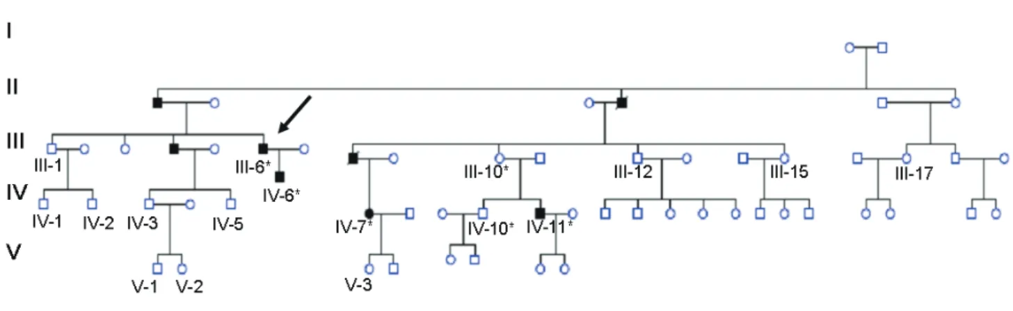
Several members of a family had episodes of venous thromboembolism (VTE), mainly deep vein thrombosis of the lower extremities or pulmonary embolism as seen in Fig. 1. The proband (Ⅲ-6) suf-fered acute dyspnea when he was 36. A ventilation-perfusion scan showed multiple bilateral ventilation-perfusion defects, and an ultrasonography revealed a bilateral deep vein thrombosis of the lower limbs. Placement of a Greenfield cava filter was required. He also reported to have abundant epistaxis as the only accompanying symptom. The most aggressive cases were those of a male (Ⅲ-8) and his daughter. He suffered right leg deep vein thrombosis and pulmonary embolism in 1991, received anticoagulation therapy for 6 months, and was then shifted to aspirin; 10 years later, he suf-fered a lethal massive pulmonary embolism. His daughter (Ⅳ-7) suffered a pulmonary embolism in the early postnatal period of her second child. Patients signed an informed consent, and this study was approved by our Hospital Ethics committee.
Biochemical Laboratory Tests
Routine biochemical and coagulation tests were performed according to standard procedures. Bio-chemical studies consisted the evaluation of thrombin time; reptilase time; levels of fibrinogen, PAI-1, alpha-2-antiplasmin, protein C, protein S, antithrombin, plasminogen, thrombin-antithrombin complex, t-PA, D-dimer, and beta-tromboglobulin; Fearnley test, coagulation factors activity, and t-PA zymography1-5).
Fig. 1. Pedigree of the family
In black, subjects with documented venous thrombotic events, including pulmonary venous thrombosis. *
indicates that the patient harbored the R458C mutation. The arrow indicates the proband.
3 Exome Sequencing for Thrombophilia
fication was performed by PCR. PCR was performed in a mixture containing 100-200 ng DNA, 5 μl (5 μM) forward primer 5′-ATGTAAGTCCAGGGACAAGG- 3′, 5 μl (5 μM) reverse primer 5′-GGTGAGAAGAA ACCTGGGAA-3′, 5 μl 10× PCR buffer, 4 μl dNTP buffer, 0.5 μl (5 U μl−1) Taq polymerase (TaKaRaBio
Inc.), and up to 50 μl water. The samples were sub-jected to denaturation at 95℃ for 5 min, followed by amplification consisting of 30 cycles of denaturation at 94℃ for 1 min, annealing at 55℃ for 30s, and extension at 72℃ for 1 min, with a final extension at 72℃ for 5 min, in a GeneAmp PCR system 2700 (Applied Biosystems, Foster City, CA, USA). Ampli-cons were purified with the QIAquick PCR purifica-tion kit and subjected to cycle sequencing on a Gene-Amp PCR System 2700 with BigDye-labeled termina-tors (Applied Biosystems) and analyzed in an ABI Prism_310 DNA sequencer.
Results
Biochemical laboratory tests detected normal lev-els or activity for fibrinogen, PAI-1, alpha-2-antiplas-min, protein C, protein S, antithrombin, plasmino-gen, thrombin-antithrombin complex, D-dimer, beta-tromboglobulin, and coagulation factors activity.
Thrombin time (21.5 s; normal range: 18-25 s) and reptilase time (14.2 s; normal range <22 s) were also normal for the proband and Ⅳ-6 patient (Fig. 1).
Fearnley test results were abnormally prolonged, indi-cating an impaired lysis; however, t-PA antigen levels measured by ELISA were high in cases, including the proband (23.5 ng/ml), compared with the normal range (0.15-13.4 ng/ml). The majority of t-PA appeared to be in complex with PAI-1 as detected by zymography (Supplemental Fig. 2). However, after an
extensive study, the cause of the disease remained undiagnosed.
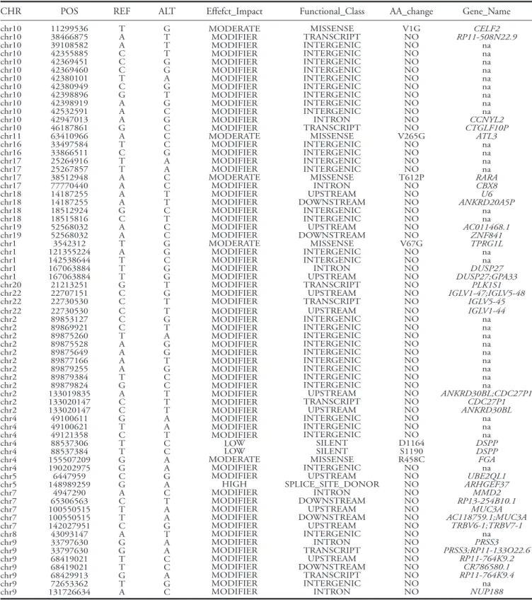
After quality control analysis of exome sequenc-ing results, 21.787 new mutations not previously described as polymorphisms in dbSNP or 1.000 Genomes project were identified. Sixty nine of these mutations were found in heterozygous in three cases and were not present in the control subject (Table 1)
following an autosomal dominant heritability. In 6 out of 69 mutations an amino acid change was identi-fied with a moderate or high impact on the protein function coded by the gene, based on Polyphen results (http://genetics.bwh.harvard.edu/pph2/) (Table 1).
The six mutations and genes were: R458C of FGA
gene, Splice-site of ARHGEF37 gene, V67G of TPRG1L, T612P of RARA, V265G of ATL3 and V1G
of CELF2. Tests for Fibrinolytic System
The presence and identity of t-PA and its inhibi-tors in circulating blood were analyzed using the euglobulin fraction of plasma by direct and reverse fibrin autography (zymography) following SDS-PAGE performed as described previously1) (expanded
meth-ods of tPA zymography are in the supplemental mate-rial).
Fearnley test: Tests were carried out in duplicate. In each case blood dilutions were made with phos-phate buffer pH 7.4 and clots were made with bovine or human thrombin at 10 units/ml. The tubes were kept in ice-cold water until firm clots appeared and then transferred to a water-bath at 37℃. When the clots were set the tubes were rolled between the palms of hands in order to loosen the clots. The tubes were left in a water-bath at 37℃ and the clots were observed for lysis at regular intervals.
The clot lysis assay was performed following pre-vious methods6). The slopes were calculated as follow.
We selected three points in the graph. Point 1 (start of clot formation), Point 2 (OD maximum) and Point 3 (end of clot lysis). Each of these points is indicated by two coordinates (OD and time in seconds). The slope of the clot formation is represented by the formula (OD2-OD1) / (t2-t1). The slope of the fibrinolysis is represented by the formula (OD3-OD2) / (t3-t2).
PAI-1, t-PA measurements: PAI-1 and t-PA were assayed by ELISA methods using kits from Biopool-Menarini, PAI-1 and t-PA antigens were from Bio-pool-Menarini as well.
Alpha-2-antiplasmin and Plasminogen activity: Alpha-2-antiplasmin and Plasminogen were assayed by commercial kits, following manufacturer’s users guide (IZASA, Spain).
Exome Sequencing
Exome sequencing was performed in three famil-ial cases (Ⅳ-6, Ⅳ-7, and Ⅳ-11) and one familial con-trol (Ⅲ-12) (Fig. 1). The results were validated in the
same samples by Sanger sequencing (Supplemental Fig. 1). In the second validation phase, 13 new
famil-ial cases and controls were analyzed.
Exome sequencing was performed in the Centro Nacional de Analisis Genómicos (CNAG, Barcelona, Spain). The Illumina TruSeq DNA Sample Prep kit and the NimbleGen SeqCap EZ Exome were used for library preparation and exome enrichment, respec-tively.
PCR Primers and Conditions
For the validation study the Exon 5 of the FGA gene, where the R458C mutation was located,
ampli-Journal of Atherosclerosis and Thrombosis
Accepted for publication: August 27, 2015
Published online: November 17, 2015
Table 1. Mutations obtained after exome sequencing analysis
The table shows the mutations that have not been described previously in dbSNP or 1000 genomes project and were present in the three cases and absent in the familial control.
CHR POS REF ALT Effefct_Impact Functional_Class AA_change Gene_Name
chr10 chr10 chr10 chr10 chr10 chr10 chr10 chr10 chr10 chr10 chr10 chr10 chr10 chr11 chr16 chr16 chr17 chr17 chr17 chr17 chr18 chr18 chr18 chr18 chr19 chr19 chr1 chr1 chr1 chr1 chr1 chr20 chr22 chr22 11299536 38466875 39108582 42355885 42369451 42369460 42380101 42380949 42398896 42398919 42532591 42947013 46187861 63410966 33497584 33866511 25264916 25267857 38512948 77770440 14187255 14187255 18512924 18515816 52568032 52568032 3542312 121355224 142538644 167063884 167063884 21213251 22707151 22730530 T A A C C C T C G A A A G A T C T T A A A A G C A A T A T T T G C C G T T T G G A G T G C G C C C G A A C C T T C T C C G G C G G T G T MODERATE MODIFIER MODIFIER MODIFIER MODIFIER MODIFIER MODIFIER MODIFIER MODIFIER MODIFIER MODIFIER MODIFIER MODIFIER MODERATE MODIFIER MODIFIER MODIFIER MODIFIER MODERATE MODIFIER MODIFIER MODIFIER MODIFIER MODIFIER MODIFIER MODIFIER MODERATE MODIFIER MODIFIER MODIFIER MODIFIER MODIFIER MODIFIER MODIFIER MISSENSE TRANSCRIPT INTERGENIC INTERGENIC INTERGENIC INTERGENIC INTERGENIC INTERGENIC INTERGENIC INTERGENIC INTERGENIC INTRON TRANSCRIPT MISSENSE INTERGENIC INTERGENIC INTERGENIC INTERGENIC MISSENSE INTRON UPSTREAM DOWNSTREAM INTERGENIC INTERGENIC UPSTREAM DOWNSTREAM MISSENSE INTERGENIC INTERGENIC INTRON UPSTREAM TRANSCRIPT UPSTREAM TRANSCRIPT V1G NO NO NO NO NO NO NO NO NO NO NO NO V265G NO NO NO NO T612P NO NO NO NO NO NO NO V67G NO NO NO NO NO NO NO CELF2 RP11-508N22.9 na na na na na na na na na CCNYL2 CTGLF10P ATL3 na na na na RARA CBX8 U6 ANKRD20A5P na na AC011468.1 ZNF841 TPRG1L na na DUSP27 DUSP27;GPA33 PLK1S1 IGLV1-47;IGLV5-48 IGLV5-45 chr22 chr2 chr2 chr2 chr2 chr2 chr2 chr2 chr2 chr2 chr2 chr2 chr2 chr4 chr4 chr4 chr4 chr4 chr4 chr4 chr5 chr5 chr7 chr7 chr7 chr7 chr7 chr8 chr9 chr9 chr9 chr9 chr9 chr9 chr9 22730530 89853127 89869921 89875260 89875528 89875649 89877166 89879255 89879384 89879824 133019835 133020147 133020147 49100611 49100621 49121358 88537306 88537384 155507209 190202975 6447959 148989259 4947290 65306563 100550515 100550515 142027951 43093147 33797630 33797630 68419021 68419021 68429913 72653362 131726634 C C C T A A A A T G A C C G T C T T G G C G A C T T C A G G T T G T A T G T A G G T G C C T T T A A T C C A A G A C T A A G T A A C C A G C MODIFIER MODIFIER MODIFIER MODIFIER MODIFIER MODIFIER MODIFIER MODIFIER MODIFIER MODIFIER MODIFIER MODIFIER MODIFIER MODIFIER MODIFIER MODIFIER LOW LOW MODERATE MODIFIER MODIFIER HIGH MODIFIER MODIFIER MODIFIER MODIFIER MODIFIER MODIFIER MODIFIER MODIFIER MODIFIER MODIFIER MODIFIER MODIFIER MODIFIER UPSTREAM INTERGENIC INTERGENIC INTERGENIC INTERGENIC INTERGENIC INTERGENIC INTERGENIC INTERGENIC INTERGENIC UPSTREAM TRANSCRIPT UPSTREAM INTERGENIC INTERGENIC INTERGENIC SILENT SILENT MISSENSE INTERGENIC UPSTREAM SPLICE_SITE_DONOR INTRON DOWNSTREAM UPSTREAM DOWNSTREAM UPSTREAM INTERGENIC INTRON TRANSCRIPT UPSTREAM DOWNSTREAM TRANSCRIPT INTERGENIC INTRON NO NO NO NO NO NO NO NO NO NO NO NO NO NO NO NO D1164 S1190 R458C NO NO NO NO NO NO NO NO NO NO NO NO NO NO NO NO IGLV1-44 na na na na na na na na na ANKRD30BL;CDC27P1 CDC27P1 ANKRD30BL na na na DSPP DSPP FGA na UBE2QL1 ARHGEF37 MMD2 RP13-254B10.1 MUC3A AC118759.1;MUC3A TRBV6-1;TRBV7-1 na PRSS3 PRSS3;RP11-133O22.6 RP11-764K9.2 CR786580.1 RP11-764K9.4 na NUP188
CHR: Chromosome number, Pos: Nucleotide position on the chromosome, Ref: Reference sequence at the position, Alt: Alternative sequence at this position, Effect_impact: Effects are categorized by ‘impact’: High, Moderate, Low and Modifier depending on results of Polyphen web tool. Functional_Class: the functional class of the variant, AA_change: the change in amino acid produced by a variant in a coding region, given in the following format using one letter amino acid codes: Amino acid in the reference-amino acid position in the protein-amino acid in the variant. Gene_Name: the name of the gene overlapping this position in HGNC gene symbol format, na: non applicable. The same variant position may appear in more than one row, since the same variant can affect different transcripts and could be located in different genes.
5 Exome Sequencing for Thrombophilia
result of controls, indicating an altered clot formation process.
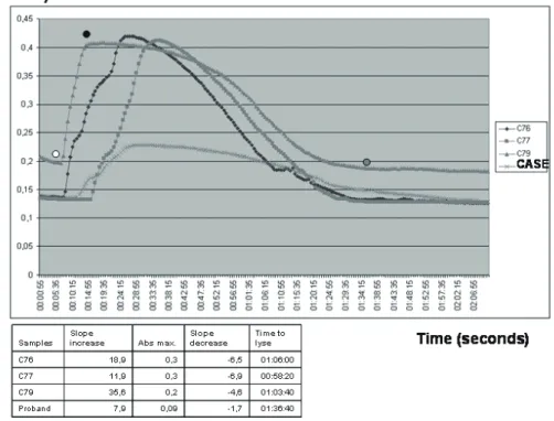
The internal fibrin lysis time process after t-PA administration was delayed in both abnormal fibrino-gens from proband and Ⅳ-6 patient vs. three healthy controls (95.8±0.28 min vs. 62.3±4.04 min, respec-tively).
The lysis rate was markedly reduced. The slope of the turbidity decrease diminished approximately 3-4 times in proband and Ⅳ-6 compared with con-trols (patients: 1.65±0.07 OD/sec vs. controls: 6± 1.22 OD/sec). The diminished slopes and delayed fibrin lysis are associated with clot lysis difficulty, probably due to the difficult binding of t-PA to fibrin.
Discussion
Familial dysfibrinogenemia is a coagulation dis-order with a bleeding tendency due to a functional One of these six mutations, R458C mutation in
FGA, was previously described7). FGA codifies for the
alpha chain subunit of fibrinogen, and mutations located in this gene have been associated with dysfi-brinogenemia, a disease with VTE episodes in some rare cases. Validation tests in three cases and control subjects and the analysis of thirteen new family indi-viduals confirmed the link between the R458C muta-tion and the disease (Supplemental Table 1). In
addi-tion, an asymptomatic subject (Ⅳ-10) (Fig. 1)
harbor-ing a R458C mutation was detected; the patient had no history of venous thromboembolic events at the time of evaluation.
The proband and Ⅳ-6 patient showed an impaired fibrin polymerization and an impaired fibri-nolysis based on clot lysis experiments (Fig. 2 and Supplemental Table 2). The slope of the turbidity
increase diminished approximately 2-3 times, and the final turbidity was 67% less than that of the mean
Fig. 2. Clot lysis experiment of the proband
C76, C77, and C79 were healthy controls. The graph shows the lower slope decrease of the case com-pared with three controls, indicating difficulty to lyse the clot. The slopes were calculated selecting three points in the graph. Point 1 (when the clot begins to form, white dot), Point 2 [point of maximum Absorbance (Abs.), black dot], and Point 3 (When just lyses, gray dot). Each of these points are indi-cated by two coordinates (OD and time in seconds). The slope of the clot formation is represented by the formula (Abs2-Abs1)/(t2-t1). The slope of the fibrinolysis is represented by the formula (Abs3-Abs2)/(t3-t2).
In addition, the time for fibrinolysis was higher in cases compared with controls. Time was measured in hours: minutes: seconds. Abs: Absorbance at 450 nm. Slope was measured in absorbance units/time in seconds.
Journal of Atherosclerosis and Thrombosis
Accepted for publication: August 27, 2015
Published online: November 17, 2015
study.
The R458C mutation causes a fibrinogen variant previously identified as fibrinogen Bordeaux7). The
base substitution found in fibrinogen Bordeaux implies the presence of an unpaired Cys residue. We observed in patients with the R458C mutation a delayed fibrin lysis process (the slope of the turbidity decrease was diminished in the clot-lysis experiments and the time that t-PA needs to do the complete fibri-nolysis was prolonged), suggesting an impaired tPA-induced fibrinolysis. This impaired fibrinolysis was also observed with Fearnley tests. This effect has been previously observed in dysfibrinogenemia patients caused by a less permeable fibrin that is more difficult to lyse compared with normal fibrin11). Hanss and
collegues hypothesized that the R458C mutation pro-duces an aberrant thrombus that blocked the correct binding of t-PA to fibrin, inhibiting fibrinolysis and causing thrombosis. We speculate that our clot lysis experiment suggests the same hypothesis of Hanss and colleagues. The region of the alpha chain of fibrinogen molecule from amino acid 392 to 610, where our mutation is located, participates in fibrinolysis regula-tion, providing sites for plasminogen and t-PA bind-ing, as it has been previously described9, 10).
Conse-quently, the R458C could modify plasminogen–tPA binding to fibrin delaying normal fibrin lysis, as we observed in our clot lysis experiment. We hypothesize that the increased t-PA levels in plasma (measured by ELISA and zymography) are due to an attempt to lyse the altered thrombus. This is the first time that has been described the biological mechanism of the impaired fibrinolysis in fibrinogen Bordeaux, however further studies are needed to confirm these hypothe-ses. In addition clot-lysis experiments could be a use-ful strategy to diagnose idiopathic thrombophilias. In our case the fibrinogen Bordeaux was associated with an impaired fibrinolysis detectable by clot-lysis experi-ments. However, experiments focused on the diagnos-tic usefulness of clot-lysis experiments in clinical prac-tise await further demonstration.
In summary, we could use exome sequencing to diagnose a family with dysfibrinogenemia that remained undiagnosed for 9 years. We suggest that exome sequencing could be a quick and economically affordable technique to diagnose monogenic idio-pathic thrombophilias when current clinical biochem-istry tests fail to find the cause. and importantly, we have observed an impaired fibrinolysis associated with fibrinogen Bordeaux, which could be the mechanism associated with the thrombotic accidents.
alteration of the circulating fibrinogen. In several dys-fibrinogenemias, an altered fibrinogen generates aber-rant thrombus that in some rare cases increases the risk of venous thrombosis.
Dysfibrinogenemia may be due to mutations in
FGA, FGB, or FGG that code for the alpha, beta, and
gamma chains of fibrinogen, respectively. More than 580 cases of abnormal fibrinogens have been reported8). In our study, the origin of idiopathic
thrombophilia was impossible to elucidate using the routine biochemical tests. Only the exome sequencing analysis was useful to find the causes of the familial thrombophilia disease. We studied three affected cous-ins and a control subject of the family without throm-boembolic events. We found a heterozygous R458C mutation in FGA, and we considered this as a patho-logical mutation on the following considerations. First, the mutation was previously described in a fam-ily with dysfibrinogenemia and venous thromboem-bolic disease7), and it is included in the Human
Genome Variation database. Second, the mutation was present in five cases and was absent in 11 healthy family controls. Third, the mutation changes a conser-vative arginine to cysteine, probably affecting the sec-ondary structure of the protein, as has been suggested previously9, 10).
The biochemical tests to determine the origin of venous thromboembolic events were performed according to the conventional protocol of our hospi-tal. Only the t-PA antigen and zymography assays detected higher t-PA and tPA-PAI-1 complexes in cases compared with controls. In addition, the Fearn-ley test detected an impaired fibrinolysis. Other clini-cal biochemistry results obtained, including fibrinogen levels and reptilase and thrombin times, were within the control range.
Thrombin and reptilase times are the gold stan-dard clinical tests to identify dysfibrinogenemias. Fibrinogen levels could be normal in dysfibrogenemias but thrombin and reptilase times are prolonged. How-ever, this is not the case in some rare cases of dysfi-brinogenemia. This problem was documented by Hanss and colleagues7) who reported a patient with
the R458C mutation and normal thrombin and repti-lase times, as we observed in our study.
This type of dysfibrinogenemias is very difficult to diagnose due to the normal results of fibrinogen levels and normal thrombin and reptilase times. In cases with idiopathic genetic thrombophilias, when all biochemical tests have been inconclusive and familial heritability has been observed, the use of exome sequencing could be an important clinical tool to per-form the diagnosis of the disease, as suggested by our
7 Exome Sequencing for Thrombophilia
fibrinolysis. Haematologica, 2012; 97: 1864-1872 3) Chakrabarti R, Fearnley G. R: The fibrinolytic potential
as a simple measure of spontaneous fibrinolysis. J Clin Pathol, 1962; 15: 228-230
4) Fearnley GR: Fibrinolysis. Ann R Coll Surg Engl, 1967; 41: 51-54
5) Clauss A: [Rapid physiological coagulation method in determination of fibrinogen]. Acta Haematol, 1957; 17: 237-246
6) Carter AM, Cymbalista CM, Spector TD, Grant PJ; EuroCLOT Investigators: Heritability of clot formation, morphology, and lysis: the EuroCLOT study. Arterioscler Thromb Vasc Biol, 2007; 27: 2783-2789
7) Hanss M, Vergnes C, Rugeri L, Ffrench P, DE Mazan-court P: A new electrophoretic variant of fibrinogen asso-ciated with venous thromboembolism, fibrinogen Bor-deaux Aalpha Arg439-->Cys. J Thromb Haemost, 2008; 6: 1422-1424
8) Hanss M, Biot F: A database for human fibrinogen vari-ants. Ann N Y Acad Sci, 2001; 936: 89-90
9) Tsurupa G, Medved L: Fibrinogen aC domains contain cryptic plasminogen and t-PA binding sites. Ann N Y Acad Sci, 2001; 936: 328-330
10) Medved L, Tsurupa G, Yakovlev S: Conformational changes upon conversion of fibrinogen into fibrin. The mechanisms of exposure of cryptic sites. Ann N Y Acad Sci, 2001; 936: 185-204
11) Marchi R, Carvajal Z, Meyer M, Soria J, Ruiz-Saez A, Arocha-Piñango CL, Weisel JW: Fibrinogen Guarenas, an abnormal fibrinogen with an Aalpha-chain truncation due to a nonsense mutation at Aalpha 467 Glu (GAA)-->stop (TAA). Thromb Res, 2006; 118: 637-650
Acknowledgments and Notice
We want to thank the family members, especially Florencio and María del Pilar.
The Laboratory of Stroke Genetics and Pharma-cogenomics is a part of the International Stroke Genetics Consortium (ISGC, www.strokegenetics. com) and coordinates the Spanish Stroke Genetics Consortium (Genestroke, www.genestroke.com). Ⅰ. F-C. is supported by the Miguel Servet programme (CP12/03298), Instituto de Salud Carlos Ⅲ. This study was funded by the Fundació Docència I Recerca MutuaTerrassa, Hospital Universitari Mutua de Ter-rassa grant (EXCLOP project).
Conflicts of Interest
The authors declare nothing to disclose.
References
1) Gaussem P, Grailhe P, Angles-Cano E: Sodium dodecyl sulfate-induced dissociation of complexes between human tissue plasminogen activator and its specific inhibitor. J Biol Chem, 1993; 268: 12150-12155
2) Lacroix R, Plawinski L, Robert S, Doeuvre L, Sabatier F, Martinez de Lizarrondo S, Mezzapesa A, Anfosso F, Leroyer AS, Poullin P, Jourde N, Njock MS, Boulanger CM, Anglés-Cano E, Dignat-George F: Leukocyte- and endothelial-derived microparticles: a circulating source for
Journal of Atherosclerosis and Thrombosis
Accepted for publication: August 27, 2015
Published online: November 17, 2015
2.5% Triton X-100. After washing-off excess Triton X-100 with distilled water, the gel was carefully over-laid on 1% agarose gel containing 1 mg/mL of bovine fibrinogen, 100 nM plasminogen, and 0.2 NIH U/ mL of bovine thrombin. For reverse fibrin zymogra-phy, the fibrin gel was supplemented with 0.05 IU/ mL of urokinase. Zymograms were allowed to develop at 37℃ for 24 h and photographed at regular intervals using dark-ground illumination. Active proteins in the samples were identified by reference to the migration of known markers (u-PA and t-PA). When required, the fibrin-agarose indicator gel was supplemented with antibodies (10 μg/mL) directed against specific plas-minogen activators.
Supplemental Material Expanded Methods
Fibrin zymography. The presence and identity of t-PA and its inhibitors in circulating blood were ana-lyzed using the euglobulin fraction of plasma by direct and reverse fibrin autography following SDS-PAGE performed as described previously (Supplemental ref-erence 1). Briefly, MPs were lysed in 100 mM Tris-HCl buffer, pH 8.1, containing 1% Triton X-100. MPs lysates (10 μL from 2.105 MPs) and reference
proteins (10 μL of t-PA 5 nM and uPA) were electro-phoresed in a 7.5% polyacrylamide gel under non-reducing conditions. SDS was then exchanged with
9 Exome Sequencing for Thrombophilia
a) b)
c) d)
Supplemental Fig. 1. Validation electropherograms of the three cases
(a, b and c) and one control (d) analyzed with exome sequencing
The electropherograms confirmed the C to T change, substituting an argi-nine for a cysteine at 458 position of FGA protein. The arrow indicates the substitution.
tPA uPA 127 127 III-6 III-6 IV-11 IV-11 IV-6 pre post pre post pre post pre
Supplemental Fig. 2. Zymography gel showing the activity of t-PA alone (t-PA) or
forming complexes (Cs) with inhibitors
From left to right t-PA: marker of t-PA, u-PA: marker of u-PA, 127: healthy control, Ⅲ-6 (pro-band), Ⅳ-11, and Ⅳ-6 patients. The proband and the cases showed 3x higher t-PA activity (complexes and free t-PA). Pre: sample obtained previous Fearnley test, before physical occlu-sion of the arm. Post: sample obtained post physical occluocclu-sion of the arm. Fearnley test mea-sures the fibrinolytic capacity of the patient.
Journal of Atherosclerosis and Thrombosis
Accepted for publication: August 27, 2015
Published online: November 17, 2015
Supplemental Table 1. Genetic results of the exome study and validation analysis
Subjects R458C Mutation Study
Ⅲ-12 Ⅳ-6 Ⅳ-7 Ⅳ-11 Ⅲ-1 Ⅲ-6 Ⅲ-10 Ⅲ-17 Ⅳ-1 Ⅳ-2 Ⅳ-3 Ⅳ-5 Ⅳ-10 Ⅴ-1 Ⅴ-2 Ⅴ-3 Ⅲ-15 WT MUT MUT MUT WT MUT MUT WT WT WT WT WT MUT WT WT WT WT Exome study Exome study Exome study Exome study Validation study Validation study Validation study Validation study Validation study Validation study Validation study Validation study Validation study Validation study Validation study Validation study Validation study MUT: mutated, WT: Wild-Type
Supplemental Table 2. Clot-lysis data of the two cases and the healthy controls
Samples Slope increase (OD/time) Slope decrease (OD/time) Time to lyse (seconds) Control-76 Control-77 Control-79 Proband Ⅳ-6 patient 18.9 11.9 35.6 7.9 7.8 −6.5 −6.9 −4.6 −1.7 −1.6 01:06:00 00:58:20 01:03:40 01:36:40 01:35:20