HAL Id: hal-01195495
https://hal.archives-ouvertes.fr/hal-01195495
Submitted on 29 May 2020
HAL is a multi-disciplinary open access
archive for the deposit and dissemination of
sci-entific research documents, whether they are
pub-lished or not. The documents may come from
teaching and research institutions in France or
abroad, or from public or private research centers.
L’archive ouverte pluridisciplinaire HAL, est
destinée au dépôt et à la diffusion de documents
scientifiques de niveau recherche, publiés ou non,
émanant des établissements d’enseignement et de
recherche français ou étrangers, des laboratoires
publics ou privés.
transcriptomic fingerprint of maize cell wall digestibility
Mary S-J Wong Quai Lam, Yves Martinez, Odile Barbier, Alain Jauneau,
Sandrine Balzergue, Stephanie Huguet, Brigitte Pollet, Valerie Mechin,
Fabienne Guillon, Paul Robert, et al.
To cite this version:
Mary S-J Wong Quai Lam, Yves Martinez, Odile Barbier, Alain Jauneau, Sandrine Balzergue, et
al.. Combined approaches provide an anatomical and transcriptomic fingerprint of maize cell wall
digestibility. Maydica, 2013, 58 (1-4), pp.274 - 287. �hal-01195495�
Combined approaches provide an anatomical and transcriptomic
fingerprint of maize cell wall digestibility
Mary S-J Wong Quai Lam
1,2, Yves Martinez
1,3, Odile Barbier
1,2, Alain Jauneau
1,3,
Sandrine Balzergue
4, Stéphanie Huguet
4, Brigitte Pollet
5, Valérie Méchin
5,
Fabi-enne Guillon
6, Paul Robert
6, Claire Dumon
7,8,9, Michael O’Donohue
7,8,9, Deborah
Goffner
1,2, Magalie Pichon
1,2*
1Université de Toulouse, UPS, UMR5546, Laboratoire de recherche en Sciences Végétales, BP42617, F-31326
Castanet-Tolosan CEDEX, France
2CNRS, UMR5546, BP 42617, 31326 Castanet-Tolosan cedex, France 3CNRS, FR3450, BP42617, 31326 Castanet-Tolosan cedex, France
4INRA - CNRS - URGV, 2 rue Gaston Crémieux, CP5708, 91057 Evry cedex, France 5INRA, AgroParisTech, Institut Jean-Pierre Bourgin, UMR1318, 78026 Versailles, France 6INRA, UR1268 Biopolymères, Interactions, Assemblages, 44316 Nantes, France
7INRA, UMR792 Ingénierie des Systèmes Biologiques et des Procédés, 31400 Toulouse, France 8Université de Toulouse; INSA, UPS, INP, LISBP, 135 Avenue de Rangueil, 31077 Toulouse, France 9CNRS, UMR5504, 31400 Toulouse, France
*Corresponding author: E-mail: [email protected]
Keywords: cell wall, digestibility, Fourier-transformed infrared (FTIR), gene expression, Laser Capture Microdissection (LCM), stem anatomy, Zea mays
Understanding cell wall biosynthesis and degradation in grasses has become a major aim in plant biology. Al-though independent previous reports have focused on specific features that dictate cell wall digestibility, cytologi-cal, biochemicytologi-cal, and gene regulation parameters have never been integrated within the same study. Herein, we applied a combination of state-of-the-art technologies and different scales of observation on two maize lines that are characterized by highly contrasted forage digestibility. Comparative image analysis of internode sections allow to get an anatomical fingerprint associated with high digestibility: a thin peripheral rind of lignified parenchyma, small numerous vascular bundles, and low proportion of PeriVascular Sclerenchyma (PVS). This cell type pattern-ing led to enhanced digestibility when internode sections were treated with Celluclast, a commercially cell wall de-grading enzyme. At a lower scale of observation, Laser Capture Microdissection (LCM) followed by thioacidolysis of PVS revealed a higher proportion of Syringyl (S) unit lignins in the low digestible line while the high digestible line was p-Hydroxyphenyl (H)-rich. Moreover, cytological observation of internodes of the two lines point out that this difference in composition is associated with a delayed lignification of PVS. At the same time, comparative transcriptomics on internodes indicated differential expression of several genes encoding enzymes along the phenylpropanoid pathway and known cell wall-associated Transcription Factors (TFs). Together, these results give an integrative view of different factors which could aim in designing a maize silage ideotype and provide a novel set of potential regulatory genes controlling lignification in maize.
Abstract
Introduction
Whole plant digestibility is an important trait for silage maize breeding of genotype with higher feed-ing value. However, understandfeed-ing why a given maize line is more or less digestible at the whole plant level is not an easy task due to the multiplicity of lignified cell types, each presumably with a specific cell wall composition and structure. For example, in a trans-verse section of a maize internode, the peripheral rind region contains 2-3 Sub-Epidermal layers of lignified Sclerenchyma cells (SES), numerous and closely-spaced Vascular Bundles (VB) composed of lignified vessels embedded in PeriVascular Sclerenchyma (PVS) and lignified parenchyma tissue. In the cen-tral pith region, vascular bundles are less numerous
and the parenchyma cells are not lignified. Typically, cell wall chemistry and digestibility estimates are performed on ground internode samples and con-sequently, the data represent average amounts of each compound without taking into account the rela-tive proportions of the different cell types. That said, correlations between stem anatomy and digestibility have been reported in different grasses such as sor-ghum (Schertz and Rosenow, 1977; Wilson, 1993), barley (Cherney et al, 1990; Goto et al, 1991) and switchgrass (Twidwell et al, 1990). In maize, a study of 22 lines indicated a positive correlation between the total surface area of lignified tissues and digest-ibility (Mechin et al, 2005). At the same time, Boon et al (2005) could not strictly correlate internode
anat-omy and digestibility in in vitro rumen fermentation studies on two maize lines of contrasted digestibility. Thus, in maize, a detailed picture of the correlation between lignified cell type patterning and digestibility is not yet well established.
In an attempt to determine the relative degrad-ability of the different cell types in maize, Lopez et al
(1993), showed that mechanically isolated parenchy-ma devoid of secondary wall thickenings were more susceptible to degradation by cellulase than scleren-chyma cells. Scanning Electron Microscopy (SEM) studies confirmed a higher digestibility of parenchy-ma compared to sclerenchyparenchy-ma when subjected to ru-men microbial degradation (Migné, 1995). Moreover, for a given cell type, the authors observed differences in digestibility in the upper vs the lower portions of an internode within a genotype, but they were not able to distinguish between lines of contrasted digestibility.
Although the aforementioned studies are of great interest, it is now possible to revisit digestibility with recently developed technologies that probe cell wall chemistry at the cellular level. Fourier-transformed infrared (FT-IR) microspectroscopy allow to detect a broad range of alterations in cell wall structure and architecture (Sindhu et al, 2007) and has been used for high through-put screening to identify novel cell wall mutants (McCann et al, 2007; Mouille et al, 2003). Within the last decade, Laser Capture Microdissec-tion (LCM) has been employed to obtain cell type-specific gene expression (Dembinsky et al, 2007; Na-kazono et al, 2003) and proteomic (Dembinsky et al, 2007) data in maize. More recently, LCM has also been used to obtain cell type-specific cell wall chem-istry in Medicago sativa and Arabidopsis thaliana ( Na-kashima et al, 2008; Ruel et al, 2009). For example, thioacidolysis performed on micro-dissected tissues unequivocally proved that interfascicular fibers of Arabidopsis display a higher Syringyl (S) /Guaiacyl (G) ratio compared to vascular bundles (Ruel et al, 2009).
Several independent gene expression studies in association with maize digestibility have been report-ed. For example at the single gene level, down-reg-ulated Caffeic acid O-methyltransferase (COMT) and Cinnamoyl-CoA Reductase (CCR) maize plants ex-hibited significantly higher digestibility and a modified lignin composition that could be related to a re-orien-tation of the phenylpropanoid pathway (Guillaumie et al, 2007a; Tamasloukht et al, 2011). Transcriptomic studies performed on four different brown-midrib (bm) maize mutants, all known for their higher digest-ibility, indicated that despite a similar bm phenotype, these mutants exhibited specific transcriptomic fin-gerprints (Guillaumie et al, 2007a). For example, the
bm1 mutation in Cinnamyl Alcohol-Dehydrogensase
(CAD) resulted in lower levels of gene expression of number of the CAD family members. Comparative transcriptomics have also been performed on re-combinant inbred lines to study the effect of a major QTL that explains 40% of digestibility (Thomas et al,
2010; Courtial et al, 2012). Although this is an ele-gant approach, this study did not allow the authors to identify specific transcriptional de-regulated genes underlying the QTL, but rather more global changes in the expression of genes dispersed throughout the genome. To date, transcriptomic approaches have never been performed at the genome scale by com-paring two lines with different genetic backgrounds and exhibiting highly contrasted digestibility.
As cell wall digestibility properties are complex and multifactor, we developed integrative approach-es including cytology, biochemistry, transcript profil-ing, FTIR and LCM on two contrasted maize lines, F98902 and Cm484. At the whole plant level, F98902, the less digestible line, had the highest lignin content and was enriched in S units. On the contrary, Cm484, the more digestible line, was significantly less ligni-fied and was characterized by a high proportion of H units. At a cellular scale, histochemical observa-tions revealed noticeable differences in morphologi-cal and anatomimorphologi-cal traits, as well as differences in the timing of lignification of PVS deposition between the two lines. FTIR and LCM technologies highlight sig-nificant differences in chemical composition between PVS of the two lines. In attempting to relate genotype to phenotype, transcriptomic data allowed to identify phenylpropanoid and related Transcription Factors (TFs) network associated with low or high cell wall digestibility.
Materials and Methods
Plant growth conditions
Cm484 and F98902 were selected among a panel of maize lines based on preliminary data indicating high and low digestibility respectively (Méchin et al, 2005). Cm484 and F98902 maize lines were grown in greenhouse conditions in pots containing a mix-ture of compost and fertilizer (Fertil Top Cote) un-der drip irrigation twice a day (about 4 l h-1). Twice a week, plants were drip irrigated with fertilizers for the whole day. Greenhouse conditions were as fol-lows: 400W sodium lights; temperature between 25° to 28°C. Plants were harvested when they had seven expanded leaves (referred to as piled-up internode at 7-leaf stage), at flowering, or one month after flower-ing. Internode samples included the basal internode (INternode above Soil, INS) and the internode just be-low the node bearing the ear (INternode under Ear, INE). Nodes and leaf sheaths were eliminated before analysis.
Lignin and digestibility analysis
Klason lignin, thioacidolysis and digestibility mea-surements were performed on samples at 7-leaf and flowering (INEs) stages according to the protocols de-scribed in Tamasloukht et al (2011). Thioacidolysis of micro-dissected PVS was performed as follows: the PVS was collected on an adhesive cap and carefully detached and transferred into a microcentrifuge tube containing 200 µl of water. The microcentrifuge tube
was washed several times to transfer samples in the thioacidolysis tube. The samples were freeze-dried overnight before the addition of 50 µl of thioacidol-ysis reagent (C19H40), together with 0.02 µg µl-1 of C19H40 and C21H44 (internal standards), that is 1µg per sample. The thioacidolysate was concentrated to a volume of 100 µl. 50 µl of the concentrated thio-acidolysate was sampled, dried under liquid nitrogen and silylated with 50 µl of N, O-bis-trimethylsilyl-triflu-oroacetamide and 50 µl of pyridine. 1 µl of the solu-tion was injected, by an injector operating at 280°C in the split/splitless mode, onto a Cpg-sm Saturne 2100 (Varian), equipped with a Sulpeco (Saint-Germain-en-Laye, France) SPB1 column (30 m in length x 0,25 mm of internal diameter), operated in the temperature program mode (from 40°C to 180°C at 30°C min-1; then 180°C to 260°C at 2°C min-1; and an isothermal temperature of 260°C). The mass spectral analyses were run with an ion trap (electronic impact, 70 eV). All analyses were carried out as three replicates injec-tion analyses. The determinainjec-tion of the three lignin-derived monomers, H, G and S, were carried out on ion chromatograms at m/z 239, 269 and 299.
Carbohydrate analysis
Neutral sugars were quantified by HPAEC-PAD analysis on samples collected at 7-leaf and flower-ing (INEs) stages. Briefly, 10 mg of dry matter were incubated in 12 M sulphuric acid for 1 hour at 30°C then sample were diluted in 4% sulphuric acid and hydrolysed during one hour at 120°C. All samples were cooled, diluted, and filtered (PTFE, 0.22 lm) be-fore injection on CarboPac PA-1 column (4 x 250 mm, Dionex). Elution of monosaccharides was obtained with a NaOH–sodium acetate gradient as previously described (Beaugrand et al, 2004). Analytical grade standards of L-arabinose, D-galactose, D-glucose,
Table 1 - Agrophysiological traits of Cm484 and F98902 lines grown in greenhouse conditions. The mean values and standard deviations have been calculated from measurements on 15 plants. Assays have been performed during two successive years (2008-2009).
Lines Height at flowering Number Length Diameter Number of days
stage (cm) of IN of INE (cm) of INE (cm) to flowering
Cm484 136.9 ± 25.0 11 ± 1.5 14.3 ± 2.8 1.5 ± 0.4 60
F98902 183.0 ± 13.3 15 ± 0.6 17.4 ± 2.7 1.6 ± 0.2 75
Number of days to flowering is estimated as the number of days between sowing and male flowering stage when the tassel is fully emerged and at the beginning of pollen shed. (IN: INternode. INE: INternode under Ear).
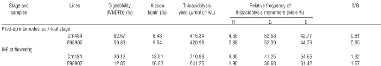
Table 2 - Digestibility, lignin content and composition of Cm484 and F98902 internodes at two developmental stages. Each value represent the mean of two assays with individual values varying by < 3% from the mean.
Stage and Lines Digestibility Klason Thioacidolysis Relative frequency of S/G samples (IVNDFD) (%) lignin (%) yield (µmol g-1 KL) thioacidolysis monomers (Mole %)
H G S
Piled-up internodes at 7-leaf stage
Cm484 62.67 8.48 415.34 4.65 52.58 42.77 0.81
F98902 59.83 9.54 420.98 2.88 52.39 44.73 0.85
INE at flowering
Cm484 30.12 13.91 710.93 4.09 41.25 54.66 1.32
F98902 12.85 16.83 641.25 1.90 36.68 61.42 1.67
H: p-hydroxyphenyl; G: guaiacyl; S: syringyl. IVNDFD: In Vitro Neutral Detergent Fiber Digestibility (Méchin et al, 2005); INE : INternodes under Ear
and D-xylose were used to quantify the concentra-tion of sugars.
Lignin histochemical staining
100 µm thick internode sections were made, from INEs and INSs at flowering and one-month after flow-ering stages, with a vibratome (LEICA VT 1000S) and stained with phloroglucinol for 30 seconds (Nakano and Meshitsuka 1992). Sections were observed using an inverted microscope (Leitz DMRIBE, Leica Micro-systems, Wetzlar, Germany) and images were regis-tered using a CCD camera (Color Coolview, Photonic Science, Milham, UK).
Quantitative measurements of lignified cell types
To calculate the relative proportion of the different cell types, 2 portions of INS sections (100 µm thick) from three plants at flowering stage were stained with phloroglucinol and scanned (6400 dpi). Each tissue type was manually delimited and the surface area of each one was calculated with Image PRO-Plus software (Media Cybernetics, Silver Spring, MD). To evaluate the size distribution of vascular bundles, measurements were performed as described in
Tamasloukht et al (2011).
Morphometric characteristics of PeriVascular Scle-renchyma (PVS)
Measurements of the Vascular Bundles (VB) in the rind were performed on 130 µm thick phloroglucinol stained cross-sections of INS at flowering stage. The PVS was manually delimited for the measurement of cell wall density and cell surface area. For each VB in the rind, the total surface area and the lumen area of PVS were calculated using the Image PRO-Plus software (Media Cybernetics, Silver Spring, MD). The relative cell wall area was calculated by subtracting the lumen area from total surface area. Cell wall
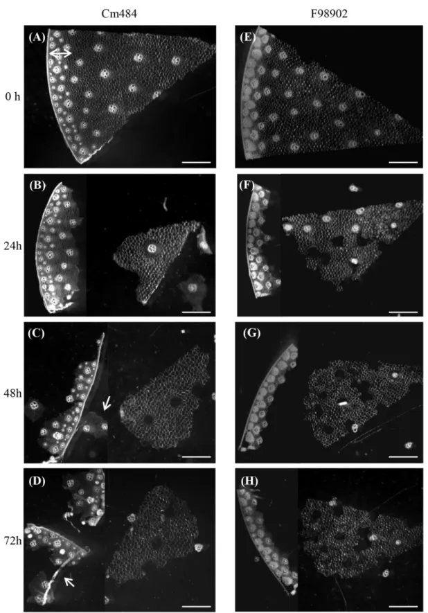
thick-Figure 1 - In section enzymatic degradation of Cm484 and F98902 internodes. a-d: 100 µm cross sections of Cm484 were in-cubated with commercial Celluclast for 24h to 72h. e-h: 100 µm cross sections of F98902 were also inin-cubated with commercial Celluclast for 24 to 72h. Double arrows indicate the rind in a. Arrows indicate patches of rind parenchyma in e and remaining VB and SES after parenchyma degradation in g. The degradation was carried out at a temperature of 40°C on three independent internodes (INE) collected at flowering stage for each line. Bars : 2 mm.
ness was measured between two adjacent cells ex-hibiting the same size. A total of 100 measurements were realized to determine cell wall thickness.
In section enzymatic cell wall hydrolysis
Enzymatic hydrolysis was carried out using cel-lulase from Trichoderma reesei ATCC 26921, with a manufacturer-stated activity of 700 endoglucanase units (EGU)/g (SIGMA-ALDRICH, C2730, Steinheim, Germany). The enzyme was prepared in 500 mM so-dium acetate buffer at pH 5.0 and used at a dilution of 1/100. Hydrolysis was performed on 100µm thick cross-sections from INEs at flowering stage. The cross-sections were placed in 1 ml of the enzymatic solution and incubated at 40°C. After 24h, 48h, and 72h, samples were removed from the incubator for observations.
Affymetrix array hybridization
RNA was extracted from 6 INEs collected at the flowering stage, and pooled into two samples for analysis. Hybridization experiments were performed as described in (Cossegal et al, 2008). All raw and normalized data are available through the CATdb da-tabase (Gagnot et al, 2008) and from the Gene Ex-pression Omnibus (GEO) repository at the National Center for Biotechnology Information (NCBI) (Barrett et al, 2007) accession number : GSE 18857.
Fourier-Transformed InfraRed spectroscopy (FTIR)
Infrared spectra were collected between 1800 and 800 cm-1 at 4 cm-1 intervals using a spectrom-eter (Tensor 27, Bruker) coupled with a microscope (Hyperion 2000, Bruker). Each spectrum resulted from the co-addition of 500 scans. The microscope
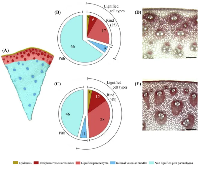
Figure 2 - Relative proportion of different cell types in Cm484 and F98902 internodes. a: Color-coded cell types of a typical in-ternode cross section, the rind region (reddish color) containing closely packed VB is distinguishesd from the pith region(blue color) which contains sparce VB. b and c: Relative proportion (%) of the different cell types in internodes of Cm484 and F98902 lines, respectively. d and e: Focus on the rind portion of phloroglucinol-HCl stained INS cross section of Cm484 and F98902 respectively. 3 plants were collected and 2 portions of each internode sections have been measured. Bars: 200 µm
was equipped with a transparent knife edge aperture and a computer-controlled x/y sample stage. The 15x Schwarzschild IR objective and a 15x15 mm aperture were selected to record infrared spectra of parenchy-ma, PVS and SES cell. Spectra were obtained from cross sections (thickness: 10 mm) of INEs at flower-ing stage (Cm484 and F98902). Sections were pre-treated with amylase and protease. The spectra were baseline corrected and normalized (unit vector nor-malization) according to Robert et al (2005).
Laser Capture Microdissection (LCM)
At flowering stage, INE of three plants for each line were fixed in 70% ethanol. The basal region was cut into 1/8 portions and one portion from each plant was randomly chosen to be rehydrated and cross-sectioned to 50-70 µm using the Leica VT 100S vi-bratome. Sections were mounted on metal-framed PEN (PolyEthylene Naphthalate) membrane slides (Arcturus, LCM0521). Samples were air-dried and kept until they were used for LCM. The dried sam-ples were placed on the Arcturus XT™ microdissec-tion system (Arcturus Bioscience), and brought into focus. CapSure® Macro LCM caps (MDS Analytical Technologies, LCM0211) were then placed over the cross-section. After focusing the section, PVS were delimited and first cut using the diode pumped solid-state UV laser (355 nm). This step had to be repeated at least three times to avoid contamination by sur-rounding cells when PVS were captured with the in-frared laser. The following parameters were used: UV laser power = 100%, all filters out; UV cutting speed = 425; IR laser power = 65 mW. The amount of PVS captured represented an approximate surface area of 4 mm² for each line.
Results
Cm484 and F98902: two maize lines displaying contrasted digestibility and lignin chemistry
When grown under greenhouse conditions, Cm484 was significantly shorter at the flowering stage than F98902 (Table 1). This difference in height was mainly due to aerial internode number (15 in F98902 and 11 in Cm484). The diameter of the INE was similar in both lines. Cm484 flowered 15 days earlier than F98902.
Cell wall chemistry was carried out at the 7-leaf and flowering stages (Table 2). At the 7-leaf stage, no significant differences in digestibility between the two lines were observed. At flowering stage, the Cm484 INE was significantly more digestible than F98902 as indicated by higher IVNDFD (In vitro Neutral Deter-gent Fiber Digestibility) values. The lignin content of Cm484 INE was 3 points lower than that of F98902. Thioacidolysis indicated that Cm484 lignin was char-acterized by a high proportion of p-Hydroxyphenyl (H) units at both stages of development. At flower-ing stage, F98902 lignin was enriched in Syrflower-ingyl (S) units compared to Cm484. Finally, ferulic acid con-tent was not significantly different between the two
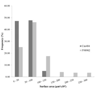
Figure 3 - Size distribution of VB in the rind parenchyma of Cm484 and F98902 internodes. 100µm thick cross-sections of INE collected at the flowering stage, were stained with pholoruglucinol and scanned. Surface area from 100 VB located in the rind parenchyma were measured using the Image PRO-Plus software (Media Cybernetics, Silver Spring, MD). Values for each class size are expressed as a percent-age of the total VB.
lines at either developmental stage (data not shown). Quantification of the major cell wall monosaccha-rides showed that both lines contained less arabi-nose and galactose at flowering as compared to the 7-leaf stage (Supplementary Table 1). However, no significant differences in monosaccharides content were observed between Cm484 and F98902 at either stage of development.
In section cell wall degradability of internodes revealed more efficient fragmentation of Cm484 compared to F98902 tissues
At the whole plant level, cell wall chemistry and digestibility measurements showed clear differences between Cm484 and F98902 lines. In order to study cell type-specific dynamics of wall degradation, in section digestion assays with Celluclast enzyme were performed on INE at flowering stage (Figure 1). Within 24 h, no significant differences in tissue diges-tion were observed between the two lines (Figure 1b, f). The pith parenchyma separated from the rind and some of the internal vascular bundles became de-tached in F98902. After 48h (Figure 1c, g), the rind of Cm484 began to fragment (Figure 1c, see arrow) whereas the rind of F98902 remained intact. After 72h (Figure 1d, h), in Cm484 there were patches of rind parenchyma that were almost completely degraded, leaving only portions of the epidermis with SES and some vascular bundles undigested (Figure 1d see ar-row). On the contrary, in F98902, the rind remained largely intact (Figure 1h). These results indicate a more extensive tissue fragmentation in Cm484 inter-nodes compared to F98902.
Cell type patterning differs in internodes of Cm484 and F98902
To analyze cell type patterning in F98902 and Cm484 internodes, the relative surface area of the different cell types was quantified in the oldest inter-node, INS, where lignification was completed (Figure 2a-e). The total proportion of lignified cell types in the rind (parenchyma, peripheral vascular bundles and the epidermis) is significantly higher in F98902 than in Cm484 (43% vs. 25%, respectively). This is due to a greater proportion of lignified parenchyma and peripheral vascular bundles. These data prompted us to compare vascular bundle (VB) size and density within the rind. When considering peripheral VB size distribution, 95% were smaller than 100,000 µm2 in Cm484 whereas in F98902, the size of VB was more variable, with a tendency towards larger bundles (≤ 300,000 µm2) (Figure 3). Peripheral VB density was higher in Cm484 than in F98902, 1.6 and 0.9 VB mm-² respectively. To further characterize peripheral VB, we measured cell wall thickness, cell density and the relative cell wall area of PVS. Interestingly, although the cell wall thickness was the same between F98902 and Cm484 (1.75 ± 0.34 µm vs 1.16 ± 0.35 µm), the
Figure 4 - Lignin staining of Cm484 and F98902 internodes at two developmental stages. a and b: 100µm thick cross section of Cm484 and F98902 respectively at flowering stage. c and d: 100µm thick cross section of Cm484 and F98902 at one month post-flowering stage. Phloroglucinol staining was performed on cross sections of INE from plants grown under greenhouse conditions at the flowering stage. The red color reveals the presence of lignin. P : Parenchyma, PVS : PeriVascular Sclerenchyma, SES : Sub-Epidermal Sclerenchyma, X : Xylem. Bars : 200 µm
cell density was greater in F98902 than in Cm484, 2,460 and 1,800 cells mm-², respectively. Conse-quently, this resulted in an overall greater relative proportion of cell wall surface area /total surface area in F98902 compared to Cm484, 0.57% and 0.33% respectively. Together, these results underline the significant differences in lignified cell type patterning between the two lines.
Delayed lignification and specific lignin composi-tion of PVS (PeriVascular Sclerenchyma) between Cm484 and F98902
In order to follow the chronology of lignification of the different cell types, phloroglucinol-stained cross sections of Cm484 and F98902 INE were observed at the flowering stage and one month post-flowering (Figure 4). At the flowering stage, xylem vessels, SES and 1-3 PVS cell layers stained red with phloroglu-cinol in both lines (Figure 4a, b). One month later, a striking difference in the lignification pattern was observed between F98902 and Cm484. PVS was strongly stained in F98902 whereas in Cm484, only weak staining was observed (Figure 4c, d). At this stage, the rind parenchyma of F98902 was weakly lignified whereas it remained unlignified in Cm484.
Figure 5 - Lignin content and composition of PVS in Cm484 and F98902 lines. a: FTIR spectra of PVS. Peaks of interest, 1,604 cm-1 and 1,740 cm-1 representing lignins and 1,045 cm-1 representing xylans, are marked with arrows. Cm484 line is represented
by full lines and F98902 by dotted lines. b: Laser capture microdissection of PVS. Isolation of PVS was carried out on 50 µm thick cross-sections using UV and IR laser beams. c: Thioacidolysis analysis of micro-dissected PVS. H, p-hydroxyphenyl; G, guaiacyl; S, syringyl. All experiments were performed on INE from three plants grown in greenhouse conditions and collected at the flowering stage.
Since several differences in PVS were observed between F98902 and Cm484 (ie greater surface area and cell density, difference in the timing of lignifica-tion), we subsequently focused our effort in char-acterizing this cell type at the cellular level. Fourier transform infrared (FTIR) microspectroscopy of PVS from Cm484 and F98902 indicated more lignin (wave-length: 1,604-1,740 cm-1) and less xylan (wavelength: 1040 cm-1) in F98902 than in Cm484 (Figure 5a). Thio-acidolysis was then performed on laser capture mi-cro-dissected PVS (Figure 5b). In PVS of Cm484, the lignin monomeric composition had a relatively higher proportion of H units whereas PVS of F98902 was more S-rich (Figure 5c). These results are in agree-ment with thioacidolysis data performed on whole INE, suggesting that the lignin composition and pro-portion of PVS influences global lignin composition in these two lines.
Differential expression of phenylpropanoid genes and associated Transcription Factors
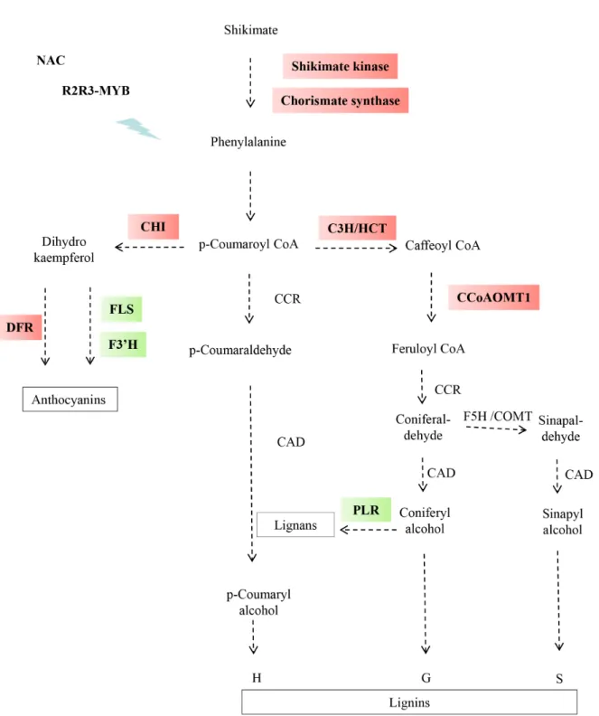
To understand the mechanisms underlying the differences in cell wall chemistry and tissue pattern-ing between F98902 and Cm484, transcriptomic analysis was performed on INE at flowering stage using the 18K Affymetrix Maize Genome Array. The complete data set is available at NCBI under acces-sion GSE 18857. A total of 2,496 genes were differ-entially expressed between the two lines. 1,183 were up-regulated and 1,313 down-regulated in F98902 compared to Cm484. Differentially expressed genes associated with phenylpropanoid metabolism are shown in Figure 6. Upstream, genes including kimate kinase, the entry point enzyme into the shi-kimate pathway and chorismate synthase, are more highly expressed in F98902, suggesting enhanced channelling of phenylpropanoid metabolites in this line. Further downstream in the phenylpropanoid pathway, Hydroxycinnamoyl-CoA Shikimate/Quinate Hycroxycinnamoyl Transferase (HCT), p-coumarate 3-hydroxylase CYP98A3 (C3H) and Caffeoyl-CoA 3-O-methyltransferase 1 (CCoAOMT1) from the
lignin branch of the pathway were more highly ex-pressed in F98902 than in Cm484. Interestingly, all of these genes, were already up-regulated in F98902 at the 7-leaf stage (data not shown). Similarly, the expression of anthocyanin biosynthetic genes such as Chalcone isomerase (CHI) and Dihydroflavonol-4-reductase (DFR) was also up-regulated in F98902. Conversely, genes involved in flavonoid metabo-lism such as Flavonol Synthase (FLS) and Flavonoid 3’-monooxygenase (TT7) as well as Pinoresinol Re-ductase 1 (PLR), a key enzyme of lignan synthesis, were down-regulated in F98902.
Several TFs that regulate cell wall biosynthesis in different plant species have been reported (Zhong et al, 2011; Zhong et al, 2008). Among the most studied, R2R3-MYB TFs have been shown in some cases to be negative regulators of lignification (Fornalé et al, 2010; Fornalé et al, 2006; Sonbol et al, 2009). Eight MYB factors were differentially expressed between the two lines (Supplementary Table 2). Among them, three belonging to the R2R3-MYB family are signifi-cantly down-regulated in F98902. Upstream, NAC TFs are involved in fiber differentiation and second-ary cell wall formation (Zhong et al, 2006; Zhong et al, 2007). Among the 18 NAC genes deposited on the 18K Affymetrix Maize Genome Array, one was up-reg-ulated in Cm484 and one down-regup-reg-ulated. ANAC073 (SND2) which was down-regulated in Cm484 plays a role in secondary cell wall formation in Arabidopsis (Zhong et al, 2008). More recently, WRKY TFs which are normally associated with disease response have also been shown to be involved in the lignification of parenchyma cells in Arabidopsis (Wang et al, 2010). Three WRKYs were down-regulated in F98902 lines (Supplementary Table 2). Finally, 22 Zinc finger genes were differentially expressed in Cm484 vs. F98902 (Supplementary Table 2). In the plant kingdom, Zinc finger genes comprise a large family and play impor-tant roles in various physiological processes ( Cift-ci-Yilmaz and Mittler, 2008). In relation to cell wall formation, it has been shown that some Zinc finger proteins are up-regulated during the formation of tension wood in poplar (Andersson-Gunneras et al, 2006). Moreover, it has been shown that the family belonging to the CCCH type is the targets of WRKY TFs (Wang et al, 2010).
Discussion
Tissue fragmentation: a key step in cell wall de-gradability
Although cell wall digestibility in maize has been extensively studied over the past decades at the whole plant level, the current view of the different factors determining forage digestibility at cellular level remains fragmentary (Boon et al, 2005; Boon et al, 2008; Engels and Schuurmans, 1992; Jung and Casler, 2006; Lopez et al, 1993; Scobbie et al, 1993). In section digestibility studies presented herein show that, although the initial first steps of tissue
degra-dation were indistinguishable between the two lines, cell wall degradation, independently of the inherent digestibility of the lines, does not appear to occur in a spatially random manner. In both genotypes, the pith parenchyma cells at the lignified/non lignified inter-face and the bundle sheath layer surrounding each inner vascular bundle were the first cell types to be degraded (Figure 1b, f). These results are in agree-ment with those obtained by (Jung and Casler, 2006) who analyzed the degradation in internode sections of three maize hybrids in the presence of rumen gas-tric fluid. This suggests that the cell wall degrada-tion process is similar with commercial and ruminant microbial enzymes. The most striking differences between the two lines occurred at a later stage of degradation (72h) with a clear fragmentation of the rind in Cm484 compared to F98902 (Figure 1d, h). This could be related to differences in cell type pat-terning (higher sclerenchyma/parenchyma ratio in the peripheral rind of F98902), thereby limiting enzyme accessibility. In the future, it would be of interest to investigate the kinetics of in vitro enzyme adhesion to these different cell types in maize internodes.
Delayed lignification of PVS in Cm484 line is cor-related with down-regulation of lignification genes and up-regulation of R2R3-MYB TFs
In maize, lignification is spatiotemporally regulat-ed not only at the whole plant level, but also within a given internode (Guillaumie et al, 2007b; Scobbie et al, 1993). Moreover, at the cellular level, all cell types that are destined to lignify do not undergo lignifica-tion simultaneously or at the same rate (Joseleau and Ruel, 1997). For example, in maize internodes, ves-sels and fibers undergo secondary wall lignification earlier than parenchyma (Joseleau and Ruel, 1997). Herein, by combining cytological and biochemical ap-proaches we show that the chronology of lignification of PVS is an important factor in discriminating Cm484 and F98902 lines. In Cm484, even one month after flowering, which corresponds to the harvest stage for silage maize, the PVS is only very weakly lignified compared to F98902. This result cannot be explained by differences in internode elongation since the delay in lignification was not systematically observed in all cell types. For example, SES was lignified to the same extent in Cm484 and F98902 internodes at both time points. Interestingly, laser capture micro-dissected PVS of Cm484 were more H unit-rich compared to F98902 PVS. Typically, H units are the first to be laid down, followed by G and then S units (Terashima et al, 1993). The fact that H units are proportionately more abundant in Cm484 suggests a more “juvenile” lignin in this line. In keeping with the lower lignin con-tent and a delay in lignification in Cm484, transcrip-tomic data showed a down-regulation of HCT, C3H and CCoAOMT1 genes. These genes have previously been shown to be involved in G/S unit flux (Day et al, 2009; Nakashima et al, 2008). Moreover, three MYB factors were more highly expressed in Cm484 than in
Figure 6 - Differential gene expression of shikimate and phenylpropanoid pathways in Cm484 and F98902 INEs. A decrease or increase in gene expression in F98902 is indicated by green or red boxes respectively. Shikimate kinase (AY105920.1), Choris-mate synthase (AY105171.1), Hydroxycinnamoyl transferase (HCT1, CF045093) p-coumarate 3-hydroxylase (C3H, CO527025), Caffeoyl-CoA O-methyltransferase, (CCoAOMT1, AI691472), Chalcone isomerase (CHI, CD437816), Dihydroflavonol-4- reduc-tase (DFR, BM073419), Flavonol Synthase (FLS, CO529883), Flavonoid 3’-monooxygenase (TT7 AY072297.1) and Pinoresinol Reductase 1 (PRR1, AW400260). RNA was extracted from 6 INEs collected at the flowering stage, and pooled into two samples for analysis.
F98902. These MYB genes belong to the R2R3-MYB factor family, whose members have been character-ized as negative regulators of lignification (Fornalé et al, 2010; Sonbol et al, 2009). For example, the over-expression of the maize ZmMYB42 gene in Arabi-dopsis led to the down-regulation of several genes involved in lignin and flavonol biosynthesis (Sonbol et al, 2009). The overexpression of ZmMYB31 in Arabi-dopsis also resulted in the down-regulation of several genes along the monolignol biosynthetic pathway, whereas anthocyanin biosynthetic genes were in-duced (Fornalé et al, 2010). The expression pattern of R2R3-MYB factor and phenylpropanoid pathway genes in Cm484 is in keeping with these studies: three R2R3-MYB TFs were up-regulated, lignin and flavonoid biosynthetic genes were repressed, and genes involved in anthocyanin production induced. Thus, these three R2R3-MYB TFs, which are closely related but not identical to ZmMYB31 and ZmMYB42, represent new candidate genes for improving digest-ibility in maize.
Digestibility: more a complex developmental bio-logic process than just cell wall chemistry
Several studies on Arabidopsis mutants indicated that vascular cell type patterning is under genetic control (Fukuda, 2004). For example, the Arabidopsis mutant, avb1 (amphivasal vascular bundles), exhibits a monocot instead of a dicot vascular architecture (Zhong et al, 1999). This gain of function mutation in IFL/REV, a class III HD-Zip gene, is essential for em-bryo patterning, meristem initiation and homeostasis, and vascular development (Zhong and Ye, 2004). In our studies, many lines of evidence converge to-wards differences in cell type patterning between Cm484 and F98902. They include i), vascular bundle organization in terms of both size and number, ii), rind parenchyma thickness, and iii), PVS cell density and hence the relative proportion of PVS cell wall- surface area/ total cell surface area. F98902 is characterized by fewer, yet larger vascular bundles whereas Cm484 contained numerous vascular bundles with propor-tionately less PVS. Interestingly, the mutation in the cell wall-related COBRA-like protein in the maize mutant, Brittle stalk2, resulted in changes in stem anatomy (Ching et al, 2006; Sindhu et al, 2007). The wild-type stems produce small VB similar to Cm484, whereas the bk2 resembled F98902. Similarly, For-nalé et al (2011) reported that transgenic maize plants down-regulated for CAD exhibited increased number of VB with less-developed sclerenchyma. Variations in vascular bundle number have also been reported in several rice mutants. One of them, nrl1, for nar-row and rolled leaf mutant (Wu et al, 2010) is affected in a cellulose synthase-like D4 gene. Interestingly, in Cm484, one of the most down-regulated gene was a cellulose synthase-like D2 gene (see full dataset No accession GSE 18857). Another is nal1, for
nar-row leaf gene (Qi et al, 2008), mutated in a gene of
unknown function, that provokes a severe defect in
polar auxin transport. nal1 culms exhibited an overall increase in VB number, especially in the peripheral ring, as well as an increase amount of parenchyma cells, reminiscent of the Cm484 lignified cell type pat-terning. Significant differences in the transcriptional regulation of hormone metabolism, and in particular auxin, were observed between the two lines (Supple-mentary Table 3), suggesting the importance of auxin signaling in cell type patterning in maize. Together, cytological and transcriptomic data suggest that di-gestibility should be considered in a developmental biologic context as opposed to just a cell wall con-text. It would be of interest to define the precise mo-ment in plant developmo-ment when lignified cell type patterning in maize is determined. This information could facilitate the identification of QTLs (and their underlying genes) that control the proportion of ligni-fied tissues, vascular bundle size and number as a novel approach to improving cell wall digestibility in maize.
Acknowledgements
This work was supported by the Association In-terprofessionnelle du Développement des Semences du Sud-Ouest, the Institut National de la Recherche Agronomique and the Centre National de la Recher-che Scientifique. We would like to thank the people from Biogemma and Syngenta greenhouses, Michel Pagniez, Fabienne Mezzasalma and Sylvie Loras, and Véronique Rois respectively. We also sincerely thank Frédéric Legée and Laurent Cézard (UMR 1318 AgroParisTech-INRA) for the lignin analysis. The LCM equipment was financed by the Region Midi-Pyré-nées.
References
Andersson-Gunneras S, Mellerowicz EJ, Love J, Segerman B, Ohmiya Y, Coutinho PM, Nilsson P, Henrissat B, Moritz T, Sundberg B, 2006. Biosyn-thesis of cellulose-enriched tension wood in Pop-ulus: global analysis of transcripts and metabo-lites identifies biochemical and developmental regulators in secondary wall biosynthesis. Plant J 45: 144-165
Barrett T, Troup DB, Wilhite SE, Ledoux P, Rudnev D, Evangelista C, Kim IF, Soboleva A, Tomashevsky M, Edgar R, 2007. NCBI GEO: mining tens of mil-lions of expression profiles--database and tools update. Nucleic Acids Res (Database issue) 35: D760-765
Beaugrand J, Cronier D, Thiebeau P, Schreiber L, Debeire P, Chabbert B, 2004. Structure, chemical composition, and xylanase degradation of exter-nal layers isolated from developing wheat grain. J Agric Food Chem 52: 7108-7117
Boon EJMC, Engels FM, Struik PC, Cone JW, 2005. Stem characteristics of two forage maize (Zea
mays L) cultivars varying in whole plant
fermenta-tion characteristics and anatomical and chemical features within a single internode. Njas-Wagen J Life Sc 53: 87-109
Boon JJ, Struik PC, Tamminga S, Engels FM, Cone JW, 2008. Stem characteristics of two forage maize (Zea mays L) cultivars varying in whole plant digestibility. III. Intra-stem variability in anatomy, chemical composition and in vitro rumen fermen-tation. NJAS 56: 101-122
Cherney DJR, Mertens DR, Moore JE, 1990. Mor-phology, fiber composition and mean particle diameter relationships in ground barley and oat forages at different ages. Anim Feed Sci Tech 31: 65-78
Ching A, Dhugga KS, Appenzeller L, Meeley R, Bo-urett TM, Howard RJ, Rafalski A, 2006. Brittle stalk 2 encodes a putative glycosylphosphati-dylinositol-anchored protein that affects mechan-ical strength of maize tissues by altering the com-position and structure of secondary cell walls. Planta 224: 1174-1184
Ciftci-Yilmaz S, Mittler R, 2008. The zinc finger net-work of plants. Cell Mol Life Sci 65: 1150-1160 Cossegal M, Chambrier P, Mbelo S, Balzergue S,
Martin-Magniette ML, Moing A, Deborde C, Guy-on V, Perez P, Rogowsky P, 2008. TranscriptiGuy-onal and metabolic adjustments in ADP-glucose pyro-phosphorylase-deficient bt2 maize kernels. Plant Physiol 146: 1553-1570
Courtial A, Jourda C, Arribat S, Balzergue S, Huguet S, Reymond M, Grima-Pettenati J, Barrière Y, 2012. Comparative expression of cell wall related genes in four maize RILs and one parental line of variable lignin content and cell wall degradability. Maydica 57: 56-74
Day A, Neutelings G, Nolin F, Grec S, Habrant A, Cronier D, Maher B, Rolando C, David H, Chab-bert B, Hawkins S, 2009. Caffeoyl coenzyme A O-methyltransferase down-regulation is associ-ated with modifications in lignin and cell-wall ar-chitecture in flax secondary xylem. Plant Physiol Biochem 47: 9-19
Dembinsky D, Woll K, Saleem M, Liu Y, Fu Y, Bor-suk LA, Lamkemeyer T, Fladerer C, Madlung J, Barbazuk B, Nordheim A, Nettleton D, Schnable PS, Hochholdinger F, 2007. Transcriptomic and proteomic analyses of pericycle cells of the maize primary root. Plant Physiol 145: 575-588
Engels FM, Schuurmans JLL, 1992. Relationship be-tween structural development of cell-walls and degradation of tissues in maize stems. J Sci Food Agr 59: 45-51
Fornalé S, Capellades M, Encina A, Wang K, Irar S, Lapierre C, Ruel K, Joseleau JP, Berenguer J, Puigdomènech P, Rigau J, Caparrós-Ruiz D, 2011. Altered lignin biosynthesis improves cel-lulosic bioethanol production in transgenic maize plants down-regulated for cinnamyl alcohol dehy-drogenase. Mol Plant 5: 817-830
Fornalé S, Shi X, Chai C, Encina A, Irar S, Capellades M, Fuguet E, Torres JL, Rovira P, Puigdomenech P, Rigau J, Grotewold E, Gray J, Caparros-Ruiz D, 2010. ZmMYB31 directly represses maize lignin genes and redirects the phenylpropanoid meta-bolic flux. Plant J 64: 633-644
Fornalé S, Sonbol FM, Maes T, Capellades M, Puig-domenech P, Rigau J, Caparros-Ruiz D, 2006. Down-regulation of the maize and Arabidopsis thaliana caffeic acid O-methyl-transferase genes by two new maize R2R3-MYB transcription fac-tors. Plant Mol Biol 62: 809-823
Fukuda H, 2004. Signals that control plant vascular cell differentiation. Nat Rev Mol Cell Bio 5: 379-391
Gagnot S, Tamby JP, Martin-Magniette ML, Bitton F, Taconnat L, Balzergue S, Aubourg S, Renou JP, Lecharny A, Brunaud V, 2008. CATdb: a public access to Arabidopsis transcriptome data from the URGV-CATMA platform. Nucleic Acids Res (Database issue) 36: D986-990
Goto M, Morita O, Chesson A, 1991. Morphological and anatomical variations among barley cultivars influence straw degradability. Crop Sci 31: 1536-1541
Guillaumie S, Pichon M, Martinant JP, Bosio M, Goff-ner D, Barrière Y, 2007a. Differential expression of phenylpropanoid and related genes in brown-midrib bm1, bm2, bm3, and bm4 young near-iso-genic maize plants. Planta 226: 235-250
Guillaumie S, San-Clemente H, Deswarte C, Martinez Y, Lapierre C, Murigneux A, Barrière Y, Pichon M, Goffner D, 2007b. MAIZEWALL. Database and developmental gene expression profiling of cell wall biosynthesis and assembly in maize. Plant Physiol 143: 339-363
Joseleau JP, Ruel K, 1997. Study of lignification by noninvasive techniques in growing maize inter-nodes. An investigation by Fourier transform in-frared cross-polarization-magic angle spinning 13C-nuclear magnetic resonance spectroscopy and immunocytochemical transmission electron microscopy. Plant Physiol 114: 1123-1133 Jung HG, Casler MD, 2006. Maize stem tissues:
Im-pact of development on cell wall degradability. Crop Sci 46: 1801-1809
Lopez S, Murison SD, Travis AJ, Chesson A, 1993. Degradability of parenchyma and sclerenchyma cell-walls isolated at different developmental stages from a newly extended maize internode. Acta Bot Neerl 42: 165-174
McCann MC, Defernez M, Urbanowicz BR, Tewari JC, Langewisch T, Olek A, Wells B, Wilson RH, Carpita NC, 2007. Neural network analyses of infrared spectra for classifying cell wall architec-tures. Plant Physiol 143: 1314-1326
Méchin V, Argillier O, Rocher F, Hébert Y, Mila I, Pol-let B, Barrière Y, Lapierre C, 2005. In search of a maize ideotype for cell wall enzymatic
degradabil-ity using histological and biochemical lignin char-acterization. J Agric Food Chem 53: 5872-5881 Migné C, 1995. Localisation immunocytochimique
des constituants pariétaux de l’entre-noeud api-cal de la tige de maïs avant et après dégradation dans le rumen. Thèse Université Blaise Pascal, Clermont-Ferrand
Mouille G, Robin S, Lecomte M, Pagant S, Hofte H, 2003. Classification and identification of Arabi-dopsis cell wall mutants using Fourier-Transform InfraRed (FT-IR) microspectroscopy. Plant J 35: 393-404
Nakano J, Meshitsuka G, 1992. The detection of lig-nin, pp 23-61. In: Methods in lignin chemistry. Lin SY, Dence CW eds. Springer-Verlag, Berlin Nakashima J, Chen F, Jackson L, Shadle G, Dixon
RA, 2008. Multi-site genetic modification of monolignol biosynthesis in alfalfa (Medicago
sa-tiva): effects on lignin composition in specific cell
types. New Phytol 179: 738-750
Nakazono M, 2003. Laser-Capture Microdissection, a tool for the global analysis of gene expression in specific plant cell types: Identification of genes expressed differentially in epidermal cells or vas-cular tissues of maize. Plant Cell 15: 583-596 Qi J, Qian Q, Bu Q, Li S, Chen Q, Sun J, Liang W,
Zhou Y, Chu C, Li X, Ren F, Palme K, Zhao B, Chen J, Chen M, Li C, 2008. Mutation of the rice
Narrow leaf1 gene, which encodes a novel
pro-tein, affects vein patterning and polar auxin trans-port. Plant Physiol 147: 1947-1959
Robert P, Marquis M, Barron C, Guillon F, Saulnier L, 2005. FT-IR investigation of cell wall polysac-charides from cereal grains. Arabinoxylan infrared assignment. J Agric Food Chem 53: 7014-7018 Ruel K, Berrio-Sierra J, Derikvand MM, Pollet B,
Thevenin J, Lapierre C, Jouanin L, Joseleau JP, 2009. Impact of CCR1 silencing on the assembly of lignified secondary walls in Arabidopsis
thali-ana. New Phytol 184: 99-113
Schertz KF, Rosenow DT, 1977. Anatomical variation in stalkinternodes of sorghum. Crop Sci 17: 628-631
Scobbie L, Russell W, Provan GJ, Chesson A, 1993. The newly extended maize internode: a model for the study of secondary cell wall formation and consequences for digestibility. J Sci Food Agric 61: 217-225
Sindhu A, Langewisch T, Olek A, Multani DS, Mc-Cann MC, Vermerris W, Carpita NC, Johal G, 2007. Maize Brittle stalk2 encodes a COBRA-like protein expressed in early organ development but required for tissue flexibility at maturity. Plant Physiol 145: 1444-1459
Sonbol FM, Fornale S, Capellades M, Encina A, Touri-no S, Torres JL, Rovira P, Ruel K, Puigdomenech P, Rigau J, Caparros-Ruiz D, 2009. The maize ZmMYB42 represses the phenylpropanoid path-way and affects the cell wall structure,
compo-sition and degradability in Arabidopsis thaliana. Plant Mol Biol 70: 283-296
Tamasloukht B, Wong Quai Lam MS, Martinez Y, Tozo K, Barbier O, Jourda C, Jauneau A, Border-ies G, Balzergue S, Renou JP, Huguet S, Marti-nant JP, Tatout C, Lapierre C, Barrière Y, Goffner D, Pichon M, 2011. Characterization of a
cinnam-oyl-CoA reductase 1 (CCR1) mutant in maize:
effects on lignification, fibre development, and global gene expression. J Exp Bot 62: 3837-3848 Terashima N, Fukushima K, He LF, Takabe K, 1993.
Comprehensive model of the lignified plant cell wall, pp. 133–163. In: Cell Wall Structure and Digestibility. Ralph J ed. American Society of Agronomy Inc, Madison, WI
Thomas J, Guillaumie S, Verdu C, Denoue D, Pichon M, Barrière Y, 2010. Cell wall phenylpropanoid-related gene expression in early maize recom-binant inbred lines differing in parental alleles at a major lignin QTL position. Mol Breeding 25(1): 105-124
Twidwell EK, Johnson KD, Patterson JA, Cherney JH, Bracker CE, 1990. Degradation of switchgrass anatomical tissue by rumen microorganisms. Crop Sci 30: 1321-1328
Wang H, Avci U, Nakashima J, Hahn MG, Chen F, Dixon RA, 2010. Mutation of WRKY transcription factors initiates pith secondary wall formation and increases stem biomass in dicotyledonous plants. Proc Natl Acad Sci USA 107: 22338-22343 Wilson JR, 1993. Isolates of cell types from sorghum
stems: Digestion, cell wall and anatomical char-acteristics. J Sci Food Agric 63: 407-417
Wu C, Fu Y, Hu G, Si H, Cheng S, Liu W, 2010. Iso-lation and characterization of a rice mutant with narrow and rolled leaves. Planta 232: 313-324 Zhong R, Demura T, Ye ZH, 2006. SND1, a NAC
domain transcription factor, is a key regulator of secondary wall synthesis in fibers of Arabidopsis. Plant Cell 18: 3158-3170
Zhong R, Lee C, McCarthy RL, Reeves CK, Jones EG, Ye ZH, 2011. Transcriptional activation of secondary wall biosynthesis by rice and maize NAC and MYB transcription factors. Plant Cell Physiol 52: 1856-1871
Zhong R, Lee C, Zhou J, McCarthy RL, Ye ZH, 2008. A battery of transcription factors involved in the regulation of secondary cell wall biosynthesis in Arabidopsis. Plant Cell 20: 2763-2782
Zhong R, Richardson EA, Ye ZH, 2007. Two NAC do-main transcription factors, SND1 and NST1, func-tion redundantly in regulafunc-tion of secondary wall synthesis in fibers of Arabidopsis. Planta 225: 1603-1611
Zhong R, Ye ZH, 2004. Amphivasal vascular bundle 1, a gain-of-function mutation of the IFL1/REV gene, is associated with alterations in the polarity of leaves, stems and carpels. Plant Cell Physiol 45: 369-385
Zhong RQ, Taylor JJ, Ye ZH, 1999. Transformation of the collateral vascular bundles into amphivasal vascular bundles in an Arabidopsis mutant. Plant Physiol 120: 53-64