HAL Id: hal-03147727
https://hal.archives-ouvertes.fr/hal-03147727
Submitted on 20 Feb 2021
HAL is a multi-disciplinary open access
archive for the deposit and dissemination of
sci-entific research documents, whether they are
pub-lished or not. The documents may come from
teaching and research institutions in France or
abroad, or from public or private research centers.
L’archive ouverte pluridisciplinaire HAL, est
destinée au dépôt et à la diffusion de documents
scientifiques de niveau recherche, publiés ou non,
émanant des établissements d’enseignement et de
recherche français ou étrangers, des laboratoires
publics ou privés.
INFLUENCE OF PLASMA-DEPOSITED COATING
OF SILVER NANOPARTICLES EMBEDDED IN A
SiOC:H MATRIX ON THE ADSORPTION OF
FIBRONECTIN
Laurine Martocq, Pascale Chevallier, Morgane Laurent, Gaetan Laroche, K
Makasheva
To cite this version:
Laurine Martocq, Pascale Chevallier, Morgane Laurent, Gaetan Laroche, K Makasheva. INFLUENCE
OF PLASMA-DEPOSITED COATING OF SILVER NANOPARTICLES EMBEDDED IN A SiOC:H
MATRIX ON THE ADSORPTION OF FIBRONECTIN. 28th Annual Conference of Biomaterials in
Medicine and Veterinary Medicine, Oct 2019, Rytro, Poland. pp.53. �hal-03147727�
INFLUENCE
OF
PLASMA-DEPOSITED COATING OF SILVER
NANOPARTICLES EMBEDDED IN
A
SiOC:H
MATRIX
ON
THE
ADSORPTION OF FIBRONECTIN
LAURINE MARTOCQ1,2,3*,PASCALE CHEVALLIER2, MORGANELAURENT2,GAETAN LAROCHE1,2,KREMENA MAKASHEVA3.
1LABORATOIRE D’INGENIERIE DE SURFACE,DEPARTEMENT DE
GENIE DES MINES, DE LA METALLURGIE ET DES MATERIAUX,
UNIVERSITE LAVAL,QUEBEC,CANADA
2C
ENTRE DE RECHERCHE DU CHU DE QUEBEC,HOPITAL ST
FRANÇOIS D’ASSISE,QUEBEC,CANADA
3LAPLACE,U
NIVERSITE DE TOULOUSE,CNRS,UPS,INPT,
TOULOUSE,FRANCE
*E-MAIL: LAURINE.MARTOCQ.1@ULAVAL.CA
Introduction
At any given time, out of 100 hospitalized patients, 7 in developed and 10 in developing countries will acquire at least one health care-associated infection (HCAI). The development of HCAIs is mainly due to the material’s surface colonization by microorganisms. Adherent bacteria organize themselves to form a biofilm, once the extracellular polymeric matrix made of proteins, polysaccharides and lipids is formed [1]. The biofilm structure renders the bacteria highly resistant to existing antibacterial agents as antibiotics, and accordingly, there is a growing need to develop new strategies to inhibit this bacterial structuration. This project takes advantages of the known antibacterial potential of silver nanoparticles (AgNPs) [2]. However, to achieve a long-lasting delivery of Ag+ ions, a fast oxidation of the AgNPs should be avoided.
Protection of the AgNPs was ensured through coating with a plasma-deposited organosilicon matrix [3,4]. This study will focus on the adsorption of fibronectin (FN) on plasma-deposited organosilicon (SiOC:H) surfaces containing AgNPs, as protein adsorption is the first step towards bacterial biofilm formation.
Materials and Methods
An axially-asymmetric radiofrequency capacitive plasma process was used to deposit a SiOC:H matrix and AgNPs on quartz substrates. The matrix was obtained using a gas mixture of hexamethyldisiloxane (HMDSO) and argon, while the AgNPs were synthesized by sputtering of the upper silver electrode in an argon plasma [3,4]. Optical Emission Spectroscopy (OES) was used to control the plasma deposition process. A droplet of 10 µL of human FN solution in HEPES buffer at a concentration of 200 µg/mL, was deposited on the plasma-deposited SiOC:H surfaces with or without AgNPs. The adsorption lasted for 1h and was followed by 3 successive washings in nanopure water with vortex and drying. Atomic Force Microscopy (AFM), X-ray Photoelectron Spectroscopy (XPS) and dynamic contact angle measurements were used to characterize the surfaces and the FN conformation after adsorption.
Results and Discussion
OES spectra revealed the presence of Ag during the plasma deposition of the SiOC:H containing AgNPs (data not shown) while no Ag was detected for the matrix alone. XPS spectra (Table 1) clearly put in evidence the presence of Ag in the samples containing AgNPs. In addition, the apparition of N1s feature, characteristic of proteins, showed the presence of adsorbed FN while no N1s peak was detected on the fresh plasma-deposited SiOC:H and
SiOC:H+AgNPs thin films. The FN adsorption is further confirmed through the decrease of %Si and %Ag concentration with the amount of adsorbed FN on the surfaces. Moreover, the difference of %N between the FN/SiOC:H and FN/SiOC:H+AgNPs suggested either a modification in the FN organization or in quantity of adsorbed FN influenced by the AgNPs. In fact, the first hypothesis is that the proteins could adopt an organization which can hide or show some functional groups depending on the surface where they adsorb. The depth of penetration of XPS being only of 5 nm and the FN size of 70 nm, these functional groups could be more or less detected. The second hypothesis is that the AgNPs have an effect on the amount of adsorbed FN due to some interactions.
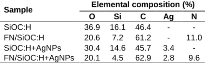
Table 1: Elemental composition obtained by XPS survey
Sample Elemental composition (%)
O Si C Ag N
SiOC:H 36.9 16.1 46.4 - -
FN/SiOC:H 20.6 7.2 61.2 - 11.0 SiOC:H+AgNPs 30.4 14.6 45.7 3.4 - FN/SiOC:H+AgNPs 20.1 4.5 62.9 2.8 9.6 AFM images also confirmed the presence of adsorbed FN by modification of the roughness (Fig. 1). Moreover, the difference of roughness between FN/SiOC:H (2) and FN/SiOC:H+AgNPs (4) showed that the AgNPs have an influence on the FN adsorption leading to a conformation modification of the proteins when they are brought in contact with the surface.
Fig. 1: Average roughness and AFM images (4 μm x 4 μm) of: (1) SiOC:H, (2) FN/SiOC:H, (3) SiOC:H+AgNPs and (4) FN/SiOC:H+AgNPs
Conclusions
XPS spectra and AFM images confirmed the presence of Ag and adsorbed FN on the surfaces. These analyses also suggested a modification of the protein conformation or in the quantity of proteins after adsorption on the surface of thin organosilicon films containing AgNPs. In addition, FTIR studies will be performed to evaluate the secondary structure of the adsorbed protein. Finally, ELISA tests will be used to identify the availability of the specific sites (RGD) of the protein.
Acknowledgments
Authors would like to acknowledge NSERC, CFQCU and APR 2016 ADAGIO project, Toulouse, France for their financial support.
References
[1] M. Habash, and G. Reid, J. Clin. Pharmacol. 39 (1999) 887-98.
[2] M. Rai, et al., Biotechnol. Adv. 27 (2008) 76-83. [3] B. Despax, and P. Raynaud, Plasma Process. Polym. 4 (2007) 127-134.
[4] K. Makasheva, et al., IEEE Trans. Nanotechnology 15 (2016) 839-848. SiOC:HFN/SiOC:H SiOC:H+A gNPs FN/SiOC:H +AgNPs 0 1 2 3 4 5 Aver age r ou gh ness (n m)