Surgical Endoscopy
Ultrasound andInterventional Techniques 0 Springer-Verlag New York Inc. 2001
Trocar and Veress needle injuries during laparoscopy
M. Schafer,1'2 M. Lauper,i L. Krahenbiltill'2Swiss Association for Laparoscopic and Thoracoscopic Surgery (SALTS), Aarberg Hospital, Aarberg, Switzerland
2 Department of Visceral and Transplantation Surgery, University of Zurich, Ramistrasse, CH-8091 Zilfich, Switzerland
Received: 25 January 2000/Accepted in final form: 27 June 2000/Online publication: 12 December 2000
Abstract
Background: Inadvertent lesions of the intraabdominal
or-gans and vessels caused by trocars and Veress needles are rare but serious complications of laparoscopic surgery. Es-tablishing the pneumoperitoneum is believed to be the most dangerous step.
Methods: The Swiss Association for Laparoscopic and
Tho-racoscopic Surgery (SALTS) prospectively collected the data on 14,243 patients undergoing various standard lapa-roscopic procedures between 1995 and 1997. This database was investigated with special regard to intraabdominal com-plications caused by trocars and Veress needles.
Results: There were 22 trocar and four needle injuries
(in-cidence, 0.18%). Nineteen lesions involved visceral organs; the remaining seven were vessel injuries. The small bowel was the single most affected organ (six cases), followed by the large bowel and the liver (three cases each). All vascular lesions, except for one laceration of the right iliac artery, occurred as venous bleeding of either the greater omentum or the mesentery. Fourteen trocars were inserted under di-rect vision. Nineteen trocar injuries were recognized intra-operatively; diagnoses of two small bowel and one bladder injuries were made postoperatively. Needle injuries were all diagnosed intraoperatively. Only five injuries could be re-paired laparoscopically; the remaining lesions were rere-paired openly. Four patients underwent an open reoperation, and another patient needed five reoperations. There was one death (4.0%).
Conclusions: Trocar and needle injuries are rare
complica-tions of laparoscopy. However, if not recognized intraop-eratively and repaired immediately, they induce increased morbidity and mortality. Both open and closed establish-ment of the pneumoperitoneum are related to a potential danger of perforating lesions, but inserting the first trocar under direct vision allows early recognition and immediate repair.
Presented at the annual meeting of the Society of American Gastrointes-tinal Endoscopic Surgeons (SAGES), Atlanta, Georgia. USA 1 April 2000
Correspondence to: L. Krahenbiihl
Key words: Laparoscopy — Complications — Trocar — Veress needle — Open vs closed — Pneumoperitoneum — Prospective study
The set-up phase of laparoscopy with the establishment of the pneumoperitoneum and creation of the working space is associated with the danger of perforating injuries caused by trocars and the spring-loaded (Veress) needle [3]. In par-ticular, blind insertions of the Veress needle and/or the first trocar near the umbilicus are related to the inadvertent per-foration of vessels and parenchymatous and hollow viscus organs. If those injuries remain unrecognized at the time of the initial laparoscopy, they generally cause severe compli-cations and may contribute to an increase in overall mor-bidity. Nonetheless, many surgeons prefer to use the closed technique to establish the pneumoperitoneum because it is believed to be less time-consuming and to prevent gas leak-age. The open (Hasson) technique has been reported to be safer and is therefore increasingly favored by many sur-geons [10]. However, even blunt introduction of the first trocar under direct vision cannot fully prevent every iatro-genic injury [3, 8, 17]. According to reports in the literature, the incidence of trocar and needle injuries is low, ranging from 0.03% to 0.2%; however, these complications may be underreported, and the true incidence may be even higher [3, 5, 6, 9, 11, 17].
The main purpose of this prospective study was to in-vestigate the intraabdominal complications of trocar and Veress needle injuries during standard intra- and extraperi-toeneal laparoscopic procedures performed at a large num-ber of different institutions in Switzerland.
Patients and methods
Since 1995, the Swiss Association for Laparoscopic and Thoracoscopic Surgery (SALTS) has prospectively collected data from patients undergo-ing various laparoscopic procedures at 97 institutions that perform general surgery (university. county and district hospitals, and surgeons in private practice) in Switzerland. This database represents probably >65% of all
276
laparoscopic procedures performed annually in Switzerland. Due to the national health care system, there is no systematic registration of all lapa-roscopies performed annually. For every patient, > 130 single items-including personal records, ASA classification, indication for surgery, pre-operative investigations, intrapre-operative findings, pre-operative complications, conversion rate, postoperative morbidity, reoperation rate, and mortality-were recorded on a specially designed computerized worksheet. Follow-up of the patients was performed either by the surgical institution or the general practitioner.
The data for 14,243 laparoscopic procedures performed between Janu-ary 1995 and December 1997 were analyzed with special attention to needle and trocar injuries. All patients with these complications were iden-tified. Their medical records, operative reports, and an additional question-naire, which was sent to the referring surgical institution or the general practitioner to obtain further details and follow-up, were then carefully reviewed. If necessary, the institutions or general practitioner were visited by one of us to collect the missing data.
The definition of a trocar (TI) or needle injury (NI) included any perforation or laceration of intra- and retroperitoneal structures and organs, such as vessels, parenchymatous organs, small and large bowel, bladder, mesentery, and greater omentum. Vascular injuries of the abdominal wall (e.g., epigastric vessels) were excluded.
In the closed technique, the abdominal wall is lifted by the operator and assistant. The peritoneal cavity is punctured blindly with the Veress needle, followed by both an aspiration and a drop test, which are performed rou-tinely. Then the Veress needle is connected to the insufflator. Open intro-duction of the first trocar means that after the fascia is incised and lifted up with stay sutures, the peritoneum is opened and the blunt trocar is inserted. Results are expressed as mean ± standard deviation (SD) and range values.
Results
Patients and surgical procedures
There were 14,243 patients (6084 male and 8159 female; sex ratio M/F, 0.7) with a mean age of 51.4 years (range, 6-95 ys) at the time of the operation. Most of the patients (90.3%) presented in a low-risk condition according to their ASA 1 and 2 classifications.
Cholecystectomy was the most frequently performed main procedure (59.4%), followed by herniotomy (17.6%) and appendectomy (11.8%). Although fundoplication and colon resection are established procedures, they were rarely performed. Adhesiolysis was performed predominantly as an additional procedure to one of the other main interven-tions. Four of five laparoscopic procedures were elective operations, whereas the remaining procedures (20.3%) were performed on an emergency basis. The patients' baseline data and the various types of surgical procedures are sum-marized in Table 1.
Twenty-five patients (eight male and 17 female) with a mean age of 58.7 years (range, 29-89; y, TI, 57.5; NI, 65.0 y) were found to have had an intraabdominal complication due to a trocar or needle injury. Since one patient had two trocar-related lesions, a total of 26 TI and NI (incidence, 0.182%) were analyzed. The characteristics of that patient group are shown in Table 2. Fifteen patients (60%) had had a least one previous abdominal operation; the other 10 either had had no prior abdominal surgery (nine patients) or their status was unknown (one patient). Three TI and one NI occurred after access to the peritoneal cavity had been at-tempted through a preexisting abdominal scar. Although most of the patients were moderately obese (mean BMI, 29.6 and 26.9 kg/m2), the BMI ranged widely, from 19.0 to
45.4 kg/m2.
Table 1. Patients and surgical procedures
No. of patients 14,243 M/F 6084/8159 Sex ratio 0.7 Age (yr) Mean 51.4 Range 6-95 Patient risk (%)
Low-risk (ASA 1 and 2) 90.3 High-risk (ASA 3 and 4) 9.7 Surgical procedures Main procedure (%) Cholecystectomy 8452 (59.4) Appendectomy 1687 (11.8) Hemiotomy 2511 (17.6) Fundoplication 272 (1.9) Colon resection 352 (2.5) Others 969 (6.8) Additional procedure Adhesiolyis 1133 Elective operation (%) 79.3 Emergency operation (%) 20.3 Patients with trocar and needle injuries (%) 25 (0.182) ASA, American Society of Anesthesiologists
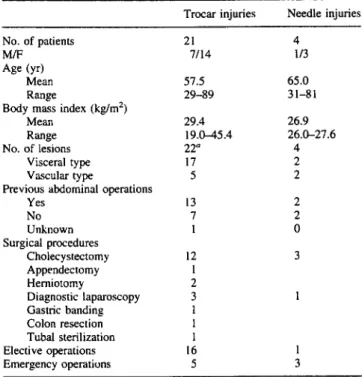
Table 2. Characteristics of patients with trocar and Veress needle injuries Trocar injuries Needle injuries
No. of patients 21 4
M/F 7/14 1/3
Age (yr)
Mean 57.5 65.0
Range 29-89 31-81
Body mass index (kg/m2)
Mean 29.4 26.9
Range 19.0-45.4 26.0-27.6
No. of lesions 22° 4
Visceral type 17 2
Vascular type 5 2
Previous abdominal operations
Yes 13 2 No 7 2 Unknown 1 0 Surgical procedures Cholecystectomy 12 3 Appendectomy 1 Hemiotomy 2 Diagnostic laparoscopy 3 1 Gastric banding 1 Colon resection 1 Tubal sterilization 1 Elective operations 16 1 Emergency operations 5 3
One patient had two injuries
The surgical procedures performed comprised 15 cho-lecystectomies (60%), four diagnostic laparoscopies (16%), and two transabdominal endoscopic hernia repairs (8%). The remaining four procedures (appendectomy, gastric banding, colon resection, tubal sterilization) were each per-formed once. TI occurred in 16 cases (76.2%) during elec-tive laparoscopies, and five TI were incurred during emer-gency laparoscopic procedures. However, three of the four NI occurred during emergency operations. Only four TI and NI were caused by inexperienced surgeons (<50 laparosco-pies); the remaining 22 injuries were caused by laparoscopic
Table 3. Trocar injuries
Affected visceral organs (n = 17) Stomach Small bowel 6 Large bowel 2 Greater omentum 2 Liver 3 Bladder, penis 2 Unknown
Affected vascular structures (n = 5)
Vessel of the mesentery 2
Vessel of the greater omentum 2
Vessel of the falciform ligament Trocar placement
Umbilicus 8
Median—epigastric 2
Median—suprapubic 6
Left upper quadrant 2
Left lower quadrant Right lower quadrant Unknown Trocar size 5-mm 6 10-mm 12 12-mm Unknown Trocar shape Conical 9 Sharp 9 Unknown 3
Trocar insertion under direct vision 14
Hasson technique (first trocar) 3
Insertion under camera control (second, third trocar) II
Blind trocar insertion 8
Umbilicus (first trocar) 6
Other localization
Lesion recognized intraoperatively 18
Lesion not recognized intraoperatively 4
Small bowel 2
Large bowel 1
Bladder
surgeons with a large (51-100 laparoscopies) or even a very large (>100 laparoscopies) experience.
Trocar injuries (Table 3)
There were 17 lesions of visceral organs (77.3%) and five vascular lesions (22.7%). Hollow viscus organs were the most frequently affected (nine of 17 lesions). The small bowel was the most injured single organ (six cases), fol-lowed by the liver (three cases), the large bowel (two cases), the urogenital organs (two cases), and the greater omentum (two cases). There was one transmural laceration of the gastric wall, and one final lesion was not specified. Vascular lesions were restricted to venous bleeding; these occurred in the greater omentum (two cases) and the small bowel mes-entery (two cases). Finally, there was one vascular lesion of a smaller vessel in the falciform ligament.
The insertion of umbilical trocars caused eight lesions, which were all of the visceral type. Six trocars were inserted blindly; the other two trocars were introduced openly (Has-son technique). Another six lesions occurred during trocar insertion into the suprapubic area. Two further TI were caused by epigastric trocar placement. The remaining five trocars were placed in four different abdominal locations
Fig. 1. Localization of trocar and needle injuries. Trocar and needle place-ment in the patient's midline, particulary at umbilical and suprapubic sites, is related to an increased risk of perforating lesion. In this series, 19 of 26 lesions (73.1%) occurred after midline insertions of trocars and needles. TI. trocar injury; NI, needle injury.
(Fig. 1). The most frequently used trocar sizes were either 10-mm (12x) or 5-mm (sixx). Another TI was caused by a 12-mm trocar. For one TI, the trocar size was unknown. It was only partially possible to evaluate the trocar shape; neither so-called safety trocars nor the type (i.e., disposable vs reusable trocars) were specially investigated. However, in our series there were nine sharp (pyramidal introducer) and nine conical (non-cutting) trocars. For three TI, the trocar shape could not be determined. Eighth trocars (38.1%) were inserted blindly after creation of the pneumo-peritoneum as first trocars; another 10 trocars (14 TI, 61.9%) were inserted under direct vision (second or third working trocar), and the remaining three trocars were in-serted using the Hasson technique.
Although 18 lesions were recognized immediately after their occurrence, one perforation of the transverse colon was only found as an additional lesion in a patient who needed conversion due to a vascular lesion of the mesentery. The remaining three TI were diagnosed after a delay of 48-72 h. There were two perforations of the small bowel and one leakage of the bladder that were not discovered during the initial laparoscopic procedure.
Veress needle injuries (Table 4)
Only four intraabdominal NI (two visceral and two vascular lesions) were found in the whole patient group. There was one perforation of the transverse colon and one lesion that created an opening of the small bowel mesentery. Another patient had severe bleeding due to perforation of the right iliac artery. The other vascular lesion was due to an omental vein laceration. In all cases but one, the Veress needle had been introduced at the umbilicus (see Fig. 1). All four le-sions were evident intraoperatively and were treated
imme-Table 4. Veress needle injuries Table 5. Treatment, conversion rate, and postoperative morbidity Affected visceral organs
Large bowel
Mesentery of the small bowel Affected vascular structures
Vessel of the greater omentum Major retroperitoneal vessel Needle placement
Umbilicus 3
Right upper quadrant
Lesion recognized intraoperatively 4 Lesion not recognized intraoperatively 0
diately. Only the laceration of the small bowel mesentery was not repaired during the initial laparoscopy.
Treatment and postoperative morbidity
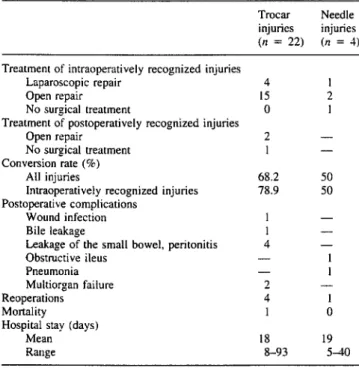
As shown in Table 5 and Table 6, only five of 23 (21.7%) intraoperatively recognized TI and NI were repaired lapa-roscopically, whereas in 18 cases (78.3%) conversion to an open approach was needed. One patient had a tear of the mesentery caused by an NI that was recognized intraopera-tively, but no repair was done. He developed an obstruc-tional ileus of the small bowel during the early postopera-tive course and underwent an open reoperation. Intraopera-tively, an internal herniation of the small bowel through the mesenteric opening was found. Three patients who under-went a laparoscopic repair had a trocar-induced bleeding complication; hemostasis was achieved with either sutures, clips, or monopolar current. Two other patients had a su-perficial lesion of the anterior gastric wall and a tear of the greater omentum, respectively. The lesions were closed with laparoscopically applied sutures.
After conversion to an open approach, the repair of TI and NI was successful in all patients, except for two who had an insufficient closure of their small bowel perforation. Both patients developed severe peritonitis within 48-72 h of the initial laparoscopy. These two patients needed addi-tional surgical interventions; in particular, one patient needed five reoperations.
Among the three TI discovered postoperatively, there were two small bowel perforations and one bladder injury. Development of severe peritonitis prompted an open reop-eration, and the small bowel leakages were found. In both cases, after segmental small bowel resection and abdominal lavage, the further course was uneventful. The bladder lac-eration was treated conservatively by inserting an urinary catheter for 10 days. An 86-year-old female patient with preexisting liver cirrhosis, portal hypertension, and the his-tological finding of a gallbladder carcinoma (pT2) suffered a TI of the greater omentum that was repaired without any problems. However, during the postoperative course, she developed multiorgan failure and died.
Although the hospital stay ranged widely from 5 to 93 days, conversion to open repair prolonged the hospital stay for most of the patients. The mean hospital stay was 18 and 19 days for TI and NI, respectively.
Discussion
The aim of this prospective study was to assess the inci-dence, management, and outcome of trocar- and
needle-Trocar injuries (n = 22) Needle injuries (n = 4) Treatment of intraoperatively recognized injuries
Laparoscopic repair 4 1
Open repair 15 2
No surgical treatment 0 1
Treatment of postoperatively recognized injuries
Open repair 2
No surgical treatment Conversion rate (%)
All injuries 68.2 50
Intraoperatively recognized injuries 78.9 50
Postoperative complications
Wound infection 1
B ile leakage 1
Leakage of the small bowel, peritonitis 4 Obstructive ileus
Pneumonia
Multiorgan failure 2
Reoperations 4
Mortality 1 0
Hospital stay (days)
Mean 18 19
Range 8-93 5-40
induced intraabdominal injuries incurred during standard laparoscopic procedures. There were 26 TI and NI (inci-dence, 0.182%) found among the 14,243 laparoscopies per-formed by the SALTS members between 1995 and 1997.
Establishing the pneumoperitoneum and inserting the working trocars remain the most crucial steps of laparosco-py. At least three different techniques are currently used to obtain laparoscopic access to the abdominal cavity. How-ever, none of them entirely obviate the possibility of intra-operative complications caused by trocars and needles [3, 8]. The closed technique is probably the most widely used method. The peritoneal cavity is punctured blindly with the Veress needle, followed by insufflation of the carbon diox-ide [13]. Thereafter, the first trocar, which is often placed near the umbilicus, is introduced blindly into the peritoneal cavity. Alternatively, the first trocar can be inserted blindly without prior creation of the pneumoperitoneum [4, 18]. However, this modified closed technique has rarely been adopted by general surgeons in Switzerland due to the in-creased risk of bowel perforations.
In contrast, the open (Hasson) technique is characterized by the open introduction of the first trocar under direct vision [10]. The pneumoperitoneum is subsequently estab-lished through the blunt-tipped trocar. Although the open technique is a very safe method to enter the peritoneal cav-ity. perforating lesions have also been described by different authors, as well as in this series [7, 17]. Those injuries may be related to the lysis of intraabdominal adhesions and per-forations with the instruments used to create the mini-laparotomy, whereas direct injuries from the blunt-tipped trocar appear to be rather less important.
The closed technique using the Veress needle—which is believed to be less time-consuming, less invasive, and better at preventing gas leakage at the first trocar entry site—is preferred by the majority of surgeons [3]. However, at least two comparative studies have failed to show any time-saving advantages for closed laparoscopy, and in
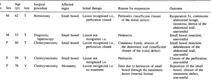
experi-Table 6. Reasons for reoperation and outcome Pt. no. Sex Age (yr) Inst. Surgical procedure Affected
organ Initial therapy Reason for reoperation Outcome
I M 62 T Herniotomy Small bowel Lesion recognized i.o.,
perforation closed
Peritonitis (insufficient closure of the initial defect)
Reoperation 5x, continuous abdominal lavage, ileostoma, hernia of the abdominal wall, uneventful
2 M 53 T Diagnostic
laparoscopy
Small bowel Lesion not recognized i.o.
Peritonitis Small bowel resection,
uneventful 3 F 60 T Cholecystectomy Small bowel Lesion recognized i.o.,
perforation closed
Cutaneous fistula. abscess of the abdominal wall (insufficient closure of the initial defect)
Small bowel resection, debridement of the abdominal wall, uneventful
4 F 59 T Cholecystectomy Small bowel Lesion not
recognized i.o.
Peritonitis Closure of the perforation,
uneventful
5 F 76 V Cholecystectomy Mesentery Lesion recognized i.o.
no treatment
Ileus due to herniation of small bowel through the mesenteric lesion (internal hernia)
Reposition of the small bowel, closure of the mesenteric defect, uneventful Pt. no., patient number; M, male; F, female: Inst., offending instrument: T, trocar; V, Veress needle; i.o.. intraoperatively
enced hands, the leakage of carbon dioxide can be prevented by using a careful dissection technique [2, 16].
The overall incidence of vascular and visceral injuries during laparoscopy, which includes TI, NI, and other intra-operative injuries caused by different instruments (electro-cautery hook, scissors, and graspers), ranges from 0.05% to 1.05% and from 0.048% to 0.35%, respectively [3, 5, 6, 17]. The wide range of reported incidences may reflect the use of different definitions. Furthermore, there is a tendency to underreport such complications in retrospective and multi-center studies. In our series, the overall incidence of intra-abdominal complications caused by trocars and Veress needles was 0.182%; vascular injuries occurred in 0.049% and visceral injuries in 0.133% of cases. There were 12 lesions after blind insertion of the first trocar (following establishment of the pneumoperitoneum) or the Veress needle; the remaining 14 lesions occurred after the open introduction of either the first trocar (Hasson technique) or the other working trocars under visual control.
There have been no prospective randomized trials com-paring open vs closed establishment of the pneumoperito-neum. However, two prospective randomized trials compar-ing Veress needle vs direct trocar insertion without prior pneumoperitoneum showed significant advantages for the latter technique [4, 12]. Due to the very low incidence of injuries, a large number of patients would be required to reach statistically significant differences. Therefore, most studies are either retrospective series, reviews of the pub-lished data, or smaller case series [3]. Wherry et al. reported on three access-related major bleeding complications among 10 vascular lesions (30%) [17]. Furthermore, 11 of 29 visceral injuries (51.7%) were caused by open-inserted trocars. Other, mostly smaller series also found intraab-nominal complications after open trocar insertion, espe-cially in cases of umbilical trocar placement [5, 6]. In con-trast, in their series and literature review, Bonjer et al. and Sigman et al. clearly showed an increased incidence of vas-cular and visceral injuries after closed vs open establish-ment of the pneumoperitoneum [3, 16].
Introduction of both the first trocar and the Veress
needle near the umbilicus and at the suprapubic site is as-sociated with a higher rate of perforating injuries [5, 18]. In the current series, 17 of 26 TI and NI were related to one of these two locations. Furthermore, epigastric trocar place-ment contributed to another two lesions. All of these com-plications (19 lacerations, 73.1%) were due to a midline access, which seems to be more dangerous than lateral tro-car or needle insertion. Lifting the abdominal wall in obese patients often increases the distance between the skin and the abdominal fascia because of the loosely attached sub-cutaneous adipose tissue. The Veress needle or trocar then requires increased force to enter the peritoneal cavity, thus running a risk of intraabdominal injuries. At the same time, thin patients with bowel and major retroperitoneal vessels close to the abdominal wall are also at risk of a perforating complication. Prior abdominal surgery, which is often per-formed through a midline laparotomy, leads to adhesions of the small and large bowel to the anterior abdominal wall, and blind insertion may cause inadvertent perforation of the adjacent hollow viscus organs. Dilated bowel loops are a common problem in cases of emergency laparoscopy and cases of adhesion- and cancer-related (peritoneal carcino-matosis) bowel obstruction, so open access is probably safer than blind puncture of the abdominal cavity in these cir-cumstances [1].
As far as we could tell, neither the type, size, or shape of the trocar contributed to an increased risk of perforating lesions. The use of conical rather than sharp trocars may reduce the number of abdominal wall injuries, but intraab-doininal lesions cannot be prevented by any type of trocar [5]. In contrast to the findings of Bonjer et al. and Zaraca et al., the open access method used in the current series failed to show any superiority over the closed establishment of the pneumoperitoneum [3, 19].
The outcome of TI and NI is strongly related to the type of anatomical structure that is injured (visceral or vascular), the immediate recognition of the lesion, and the meticulous repair, which usually necessitates a conversion to laparoto-my. Hematoma formation in the abdominal cavity or the retroperitoneal space is the main clinical sign of any
vas-280
cular injury, and its occurrence facilitates immediate recog-nition of the injury. However, the exact localization of the predominantly venous bleeding sources in the mesentery or the greater omentum may be hindered by an increased ab-sorption of light in the presence of blood, excessive adipose tissue, and moving bowel loops. Laparoscopic or open he-mostasis is achieved using bipolar current, clips, or sutures. Major vascular injuries in the retroperitoneal space are dan-gerous incidents, and rapid conversion is mandatory to achieve hemostasis and avoid further complications (e.g., ischemic complications, thrombosis). As with vascular in-juries, lacerations of parenchymatous organs, such as the liver and spleen, are generally indicated by intraabdominal bleeding, and thus are discovered rapidly. Smaller lesions restricted to the surface of the injured organ can be managed laparoscopically.
Special attention should be paid to small bowel injuries because their recognition is often delayed; subsequently, the morbidity is markedly increased [14, 15, 18]. If there is only a slight leakage without obvious extravasation, the lacera-tion is likely to remain unrecognized intraoperatively. Mov-ing small bowel loops and the greater omentum tend to hide the defect and may even close the leakage temporarily. Moreover, it can be assumed that minor puncture injuries will close spontaneously without causing further complica-tions. The postoperative development of peritonitis is an indication of severe perforating bowel injuries, and emer-gency reoperation is called for. In general, careful closure of the defect or segmental resection of the injured bowel re-quires either a conversion or an open reoperation. Septic complications due to insufficient closure at the site of bowel leakage may be deleterious for the patient. In our series, two small bowel injuries went unrecognized during the initial surgical procedure; both patients underwent open reopera-tion. Another two patients who had had primary closure of small bowel lesions developed intraabdominal abscess and enterocutanous fistula formation due to releakage of im-properly closed small bowel defects in the further postop-erative course.
Conclusions
Trocar and needle injuries are rare complications of lapa-roscopy (incidence, 0.18%). Unexpected hematoma forma-tion indicates vascular lesions, and immediate recogniforma-tion of the injury facilitates the achievement of hemostasis. The diagnosis of visceral lesions—in particular, perforation of the small bowel—remains difficult; thus, delayed recogni-tion is associated with increased morbidity. Careful access to the peritoneal cavity remains one of the most crucial steps in laparoscopic surgery. In cases of severe hemodynamic changes during the early postoperative course, bleeding complications due to TI and NI should be excluded.
More-over, the development of sepsis and peritonitis 24-72 h postoperatively after laparoscopy is highly suspicious of bowel perforation, which must be excluded by emergency reintervention.
References
1. Bailey IS, Rhodes M, O'Rourke N, Nathanson L, Fielding G (1998) Laparoscopic management of acute small bowel obstruction. Br J Surg 85: 84-87
2. Ballem RV, Rudomanski J (1993) Techniques of pneumoperitoneum. Surg Laparosc Endosc 3: 42-43
3. Bonjer HJ, Hazebroek EJ, Kazemier G, Giuffrida MC, Meijer WS, Lange JF (1997) Open versus closed establishment of pneumoperito-neum in laparoscopic surgery. Br J Surg 84: 599-602
4. Byron JW, Markenson G, Miyazawa K (1993) A randomized com-parison of Verres needle and direct trocar insertion for laparoscopy. Surg Gynecol Obstet 177: 259-262
5. Champault G, Cazacu F, Taffinder N (1996) Serious trocar accidents in laparoscopic surgery: a French survey of 103,852 operations. Surg Laparosc Endosc 6: 367-370
6. Deziel DJ, Millikan KW, Economou SG, Doolas A, Ko ST, Airan MC (1993) Complications of laparoscopic cholecystectomy: a national sur-vey of 4,292 hospitals and an analysis of 77,604 cases. Am J Surg 165: 9-14
7. Hanney RM, Carmalt HL, Merrett N, Tait N (1999) Use of the Hasson cannula producing major vascular injury at laparoscopy. Surg Endosc 13: 1238-1240 DOI: 10.1007/s004649901235
8. Hanney RM, Carmalt HL, Merrett N, Tait N (1999) Vascular injuries during laparoscopy associated with the Hasson technique. J Am Coll Surg 188: 337-338
9. Hashizume M, Sugimachi K (1997) Needle and trocar injury during laparoscopic surgery in Japan. Surg Endosc 11: 1198-1201 DOI:
10.1007/s004649900568
10. Hasson HM (1971) A modified instrument and method for laparosco-py. Am J Obstet Gynecol 110: 886-887
11. Mac CC, Lecuru F, Rizk E, Robin F, Boucaya V, Taurelle R (1999) Morbidity in laparoscopic gynecological surgery: results of a prospec-tive single-center study. Surg Endosc 13: 57-61 DOI: 10.1007/ s004649900898
12. Nezhat FR, Silfen SL, Evans D, Nezhat C (1991) Comparison of direct insertion of disposable and standard reusable laparoscopic trocars and previous pneumoperitoneum with Veress needle. Obstet Gynecol 78: 148-150
13. Reddick EJ, Olsen DO (1989) Laparoscopic laser cholecystectomy: a comparison with mini-lap cholecystectomy. Surg Endosc 3: 131-133 14. Reich H (1992) Laparoscopic bowel injury. Surg Laparosc Endosc 2:
74- 78
15. Sadeghi-Nejad H, Kavoussi LR, Peters CA (1994) Bowel injury in open technique laparoscopic cannula placement. Urology 43: 559-560 16. Sigman HH, Fried GM, Garzon J, Hinchey El, Wexler MJ, Meakins JL, Barkun JS (1993) Risks of blind versus open approach to celi-otomy for laparoscopic surgery. Surg Laparosc Endosc 3: 296-299 17. Wherry DC, Marohn MR, Malanoski MP, Hetz SP, Rich NM (1996)
An external audit of laparoscopic cholecystectomy in the steady state performed in medical treatment facilities of the Department of De-fense. Ann Surg 224: 145-154
18. Woolcott R (1997) The safety of laparoscopy performed by direct trocar insertion and carbon dioxide insufflation under vision. Aust N Z J Obstet Gynaecol 37: 216-219
19. Zaraca F, Catarci M, Gossetti F, Mulieri G. Carboni M (1999) Routine use of open laparoscopy: 1,006 consecutive cases. J Laparoendosc Adv Surg Tech A 9: 75-80