ORIGINAL ARTICLE
Gene array of primary human osteoblasts exposed to enamel
matrix derivative in combination with a natural bone mineral
Richard J. Miron&Dieter D. Bosshardt&
Yufeng Zhang&Daniel Buser&Anton Sculean
Received: 1 February 2012 / Accepted: 16 April 2012 / Published online: 3 May 2012 # Springer-Verlag 2012
Abstract
Objectives The application of an enamel matrix derivative (EMD) for regenerative periodontal surgery has been shown to promote formation of new cementum, periodontal liga-ment, and alveolar bone. In intrabony defects with a com-plicated anatomy, the combination of EMD with various bone grafting materials has resulted in additional clinical improvements, but the initial cellular response of osteoblasts coming in contact with these particles have not yet been fully elucidated. The objective of the present study was to evaluate the in vitro effects of EMD combined with a natural bone mineral (NBM) on a wide variety of genes, cytokines, and transcription factors and extracellular matrix proteins on primary human osteoblasts.
Material and methods Primary human osteoblasts were seed-ed on NBM particles pre-coatseed-ed with versus without EMD and analyzed for gene differences using a human osteogenesis gene super-array (Applied Biosystems). Osteoblast-related
genes include those transcribed during bone mineralization, ossification, bone metabolism, cell growth and differentia-tion, as well as gene products representing extracellular matrix molecules, transcription factors, and cell adhesion molecules.
Results EMD promoted gene expression of various osteo-blast differentiation markers including a number of collagen types and isoforms, SMAD intracellular proteins, osteopon-tin, cadherin, alkaline phosphatase, and bone sialoprotein. EMD also upregulated a variety of growth factors including bone morphogenetic proteins, vascular endothelial growth factors, insulin-like growth factor, transforming growth fac-tor, and their associated receptor proteins.
Conclusion The results from the present study demonstrate that EMD is capable of activating a wide variety of genes, growth factors, and cytokines when pre-coated onto NBM particles.
Clinical relevance The described in vitro effects of EMD on human primary osteoblasts provide further biologic support for the clinical application of a combination of EMD with NBM particles in periodontal and oral regenerative surgery.
Keywords EMD . Emdogain . Enamel matrix proteins . Bone grafting materials . Natural bone mineral . Gene array
Introduction
Enamel matrix derivative (EMD) has been shown to pro-mote periodontal regeneration by inducing formation of cementum, periodontal ligament (PDL), and alveolar bone which is clinically evidenced by probing depth reduction, attachment gain, and radiographic defect fill [1–3]. The major components of EMD are amelogenins, a family of hydrophobic proteins derived from different splice variants
R. J. Miron
:
D. D. Bosshardt:
A. Sculean (*)Department of Periodontology, Dental School, University of Bern, Freiburgstrasse 7,
3010 Bern, Switzerland
e-mail: [email protected] R. J. Miron
:
D. D. Bosshardt:
D. Buser Department of Oral Surgery and Stomatology, School of Dental Medicine, University of Bern, Freiburgstrasse 7,3010 Bern, Switzerland Y. Zhang
The State Key Laboratory Breeding Base of Basic Science of Stomatology (Hubei-MOST) and Key Laboratory of Oral Biomedicine Ministry of Education, School and Hospital of Stomatology, Wuhan University,
237 Luoyu Road,
and controlled by post-secretory processing from a single gene that account for more than 95 % of the total protein content [4]. These proteins self-assemble into supramolecu-lar aggregates that form an insoluble extracellusupramolecu-lar matrix that function to control the ultrastructural organization of the developing enamel crystallites [4]. Other proteins found in the enamel matrix include enamelin, ameloblastin (also called amelin or sheathlin), amelotin, apin, and various proteinases [5, 6]. The rationale for the clinical use of EMD is the observation that enamel matrix proteins are deposited onto the surface of developing tooth roots prior to cementum formation [7].
Although histological studies in animals and humans have provided evidence for periodontal regeneration and substantial clinical improvements following the use of EMD, concerns have been expressed regarding the vis-cous nature of EMD, which may not be sufficient to prevent flap collapse in periodontal defects with a com-plicated anatomy [8, 9]. In order to overcome this po-tential limitation and improve the clinical outcomes, various combinations of EMD and different types of grafting materials have been used [1–3, 10–16]. The combination of EMD with a natural bone mineral of bovine origin (NBM) has provided additional periodontal regeneration and substantial clinical improvements when compared to either NBM alone or EMD alone [1, 3,
10–14]. Recently, we have demonstrated that the combi-nation of EMD with NBM particles enhances osteoblast and PDL cell proliferation and differentiation in vitro [17]. The aim of the present study was to investigate more deeply the initial behavior of primary human osteo-blasts exposed to this combination by assessing a wide variety of osteoblast cytokines, growth factors, differenti-ation markers, and extracellular matrix molecules using a commercially available super-array.
Materials and methods
Surface coating with EMD
EMD was prepared according to Institut Straumann AG standard operating protocols as previously discussed [18]. Thirty milligrams of EMD was dissolved in 3 ml of sterile 0.1 % acetic acid at 4 °C. For experiments, stock EMD was diluted 100× in 0.1 M carbonate buffer at 4 °C to a working concentration of 100μg/ml in order to maintain physiological pH. One milliliter of EMD solution was poured onto 100 mg of NBM particles (BioOss, Geistlich Pharma AG, Wolhusen, Switzerland) in 24-well culture dishes and incubated overnight at 4 °C. Following incu-bation, dishes were rinsed twice with 1 ml phosphate buffered saline twice.
Human primary osteoblast isolation and differentiation
Human bone chips from a single donor were cultured according to an explant model [19] under a protocol ap-proved by the Ethics Committee, Katon Bern, Switzerland as previously described [20]. Primary human osteoblasts were detached from the tissue culture plastic using trypsin solution (Invitrogen, Basel, Switzerland). Cells used for experimental seeding were from passages 4–6. Osteoblasts were seeded at a density of 50,000 cells in 24-well culture plates (Falcon) for experimental seeding.
Super-array of osteogenic potential
The initial expression of osteoblast-related genes was exam-ined after culture of cells for 24 h. Total RNA was isolated using TRIZOL reagent and RNAeasy Mini kit (QIAGEN, Basel, Switzerland). A TaqMan® Human Osteogenesis 96-well Plate Super-array (4414096, Applied Biosystems, Rotkreuz, Switzerland) was employed for the analysis. Osteoblast-related genes include those transcribed during bone mineralization, ossification, bone metabolism, cell growth, and differentiation. The gene products represent extracellular matrix molecules, transcription factors, and cell adhesion molecules among others. Real-time RT-PCR was performed according to manufacturer’s protocol using 20 μl final reaction volume of TaqMan®’s One step Master Mix kit (Applied Biosystems) as previously described [21]. RNA quantification was performed using a Nanodrop 2000c (Thermo Scientific, Waltham, MA, USA) and 100 ng of total RNA was used per sample well. Gene fold increase represent data from NBM particles pre-coated with EMD versus NBM particles that were left uncoated.
Statistical analysis
Gene array analysis was performed for both control (n04) and experimental groups (n04). Means and standard devia-tions (SE) were calculated, and the statistical significance of differences among each group were examined by student t test between both groups (*, p values<0.05).
Results
Osteoblast regulation of transcription factors and differentiation parameters
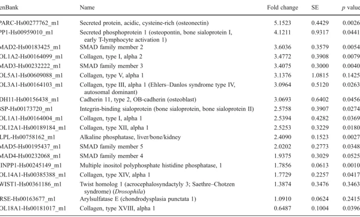
Analysis of gene array data revealed an increase of blast differentiation genes across a wide variety of osteo-blast differentiation markers and secreted proteins (Table1). Specifically, many collagen types and isoforms were upregulated including collagen type I alpha 2 (3.4772 fold,
p<0.0079), collagen type V alpha 1 (3.1376, p<0.1425), collagen type 1, alpha 1 (2.5394, p<0.0369), collagen type X11 alpha 1 (2.5253, p<0.0180), and collagen type XIV, alpha 1 (1.7729, p<0.0417). Intracellular proteins responsible for transducing extracellular signals (SMAD family) were also upregulated by EMD when compared to control uncoated samples (upregulation varied between 1.9375 and 3.6036 fold). EMD also upregulated a number of osteoblast differentiation markers including osteonectin (5.1523 fold, p<0.0026), osteopontin (4.1211, p<0.0441), cadherin 11 (3.0693, p<0.0456), bone sialoprotein (2.5758, p<0.0274), and alkaline phosphatase (2.4090, p<0.0027).
Osteoblast regulation of osteoblast growth factors
An array of growth factors essential for osteoblast differenti-ation were also quantified using real-time RT-PCR (Table2). In general, EMD had a positive impact on the release of bone morphogenetic proteins (BMPs) including BMP1 (6.6867 fold increase, p<0.0033), BMP2 (4.2685, p<0.0028), BMP6 (3.7190, p<0.0028), and BMP4 (3.0751<0.0226). EMD also upregulated vascular endothelial growth factor A and B (VEGF-A, VEGF-B, 4.8375, p<0.0231 and 2.3501, p<0.0104, respectively), fibroblast growth factor 1 (FGF1, 3.4801, p<0.0132), insulin-like growth factor 2 (IGF2,
Table 1 Gene fold increase in osteoblast differentiation markers, transcription factors, and extracellular matrix proteins
GenBank Name Fold change SE p value
SPARC-Hs00277762_m1 Secreted protein, acidic, cysteine-rich (osteonectin) 5.1523 0.4429 0.0026 SPP1-Hs00959010_m1 Secreted phosphoprotein 1 (osteopontin, bone sialoprotein I,
early T-lymphocyte activation 1)
4.1211 0.9317 0.0441
SMAD2-Hs00183425_m1 SMAD family member 2 3.6036 0.3579 0.0054
COL1A2-Hs00164099_m1 Collagen, type I, alpha 2 3.4772 0.3908 0.0079
SMAD3-Hs00232222_m1 SMAD family member 3 3.4075 0.3000 0.0040
COL5A1-Hs00609088_m1 Collagen, type V, alpha 1 3.1376 1.0815 0.1425 COL3A1-Hs00164103_m1 Collagen, type III, alpha 1 (Ehlers–Danlos syndrome type IV,
autosomal dominant)
3.0964 0.5120 0.0263 CDH11-Hs00156438_m1 Cadherin 11, type 2, OB-cadherin (osteoblast) 3.0693 0.6402 0.0456 IBSP-Hs00173720_m1 Integrin-binding sialoprotein (bone sialoprotein, bone sialoprotein II) 2.5758 0.3907 0.0274 COL1A1-Hs00164004_m1 Collagen, type I, alpha 1 2.5394 0.4282 0.0369 COL12A1-Hs00189184_m1 Collagen, type XII, alpha 1 2.5253 0.3229 0.0180 ALPL-Hs00758162_m1 Alkaline phosphatase, liver/bone/kidney 2.4090 0.1523 0.0027
SMAD5-Hs00195437_m1 SMAD family member 5 2.0202 0.2773 0.0348
SMAD4-Hs00232068_m1 SMAD family member 4 1.9375 0.3029 0.0525
MINPP1-Hs00245149_m1 Multiple inositol polyphosphate histidine phosphatase, 1 1.7856 0.0613 0.0010 COL14A1-Hs00385388_m1 Collagen, type XIV, alpha 1 1.7729 0.2257 0.0417 TWIST1-Hs00361186_m1 Twist homolog 1 (acrocephalosyndactyly 3; Saethre–Chotzen
syndrome) (Drosophila)
1.3874 0.3476 0.3463 ARSE-Hs00163677_m1 Arylsulfatase E (chondrodysplasia punctata 1) 1.0910 0.0624 0.2415 COL18A1-Hs00181017_m1 Collagen, type XVIII, alpha 1 0.6487 0.1004 0.0396
Table 2 Gene fold increase in osteoblast growth factors and cytokines
GenBank Name Fold change SE p value
BMP1-Hs00241807_m1 Bone morphogenetic protein 1 6.6867 0.6575 0.0033 VEGFA-Hs00900054_m1 Vascular endothelial growth factor A 4.8375 0.8922 0.0231 BMP2-Hs00154192_m1 Bone morphogenetic protein 2 4.2685 0.3577 0.0028 BMP6-Hs00233470_m1 Bone morphogenetic protein 6 3.7190 0.2980 0.0028 FGF1-Hs00265254_m1 Fibroblast growth factor 1 (acidic) 3.4801 0.4692 0.0132 BMP4-Hs00370078_m1 Bone morphogenetic protein 4 3.0751 0.4782 0.0226 VEGFB-Hs00173634_m1 Vascular endothelial growth factor B 2.3501 0.2348 0.0104 IGF2-Hs00171254_m1 Insulin-like growth factor 2
(somatomedin A)
2.1332 0.2599 0.0223 TGFB1-Hs99999918_m1 Transforming growth factor, beta 1 1.8082 0.0965 0.0036 TGFB3-Hs00234245_m1 Transforming growth factor, beta 3 1.5939 0.1298 0.0196
2.1332, p<0.0223) as well as transforming growth factor, beta 1 and 3 (TGFβ1, TGFβ3, 1.8082, p < 0.0036 and 1.5939, p<0.0196 fold, respectively). Interestingly, EMD showed an even more pronounced effect on the receptors associated with each growth factor (Table3). EMD increased epidermal growth factor receptor 17.0332 fold, TGFβ recep-tor 14.9025 fold, IGF1 receprecep-tor 6.2826 fold, BMP receprecep-tor 5.7594 fold, and FGF receptor 1 2.6171 fold (Table3).
Discussion
The results from the present study demonstrate that EMD has the ability to enhance cytokine and growth factor gene expression as well as increase osteoblast differentiation markers and transcription factors when combined onto NBM particles. Previous in vitro research has documented the role of EMD in both osteoblasts and PDL cells in various cell culture systems [22]. EMD has a significant influence on cell adhesion, cell proliferation, and cell differ-entiation of many cell types by mediating cell attachment, spreading, proliferation, and survival as well as expression of transcription factors, growth factors, cytokines, extracel-lular matrix constituents, and other molecules involved in the regulation of bone remodeling [22].
The rational for choosing osteoblasts as a primary cell source as opposed to PDL cells was to simulate in vivo situations. Although EMD stimulates periodontal regenera-tion, the cells that come in contact with bone grafting particles pre-coated with EMD are typically osteoblasts as PDL cells themselves generally attach and proliferate along the root cementum. Despite primary human osteoblasts be-ing harvested from a sbe-ingle donor, these conditions repre-sent a more realistic clinical similarity when compared to other single donor cell lines derived from various species [23,24]. Previously, many investigators have demonstrated that EMD influence osteoblast differentiation when seeded on standard tissue culture plastic using a wide variety of cell lines from various species (MG63, SaOS, MC3T3, HSC-2, mice/rat calvarial osteoblasts) [22].
In the present study, we observed that EMD enhanced many growth factors and cytokines including cadherin gene expression (Table 1). Interestingly, we have previously shown that EMD upregulates the expression of vital osteo-blast cell–cell communication and adhesion molecules N-cadherin and connexin43 (intercellular gap junction channel proteins) at early time points, which enhances the differen-tiation and mineralization activity of osteoblasts [20]. EMD also had a pronounced effect on SMAD intercellular pro-teins (Table 1). The family of SMAD signal proteins is utilized by many cell types including osteoblasts to trans-duce extracellular signals from TGFβ from the cell mem-brane to the nucleus [25]. Previously, it was observed that EMD induced rapid translocation of SMAD2 into the nu-cleus causing an increase in cell proliferation [26, 27]. Interestingly, results from our super-array revealed upregu-lation of multiple SMAD proteins, with over threefold increases in SMAD2 and SMAD3 (Table1) demonstrating a very plausible role of TGFβ for EMD-treated osteoblasts. Furthermore, TGFβ1 and TGFB receptor were increased 1.8082 and 14.9025 fold, respectively, on EMD-coated NBM particles. These results are consistent with other authors who have demonstrated that EMD increased the secretion of TGFβ1 and PDGF through intracellular cAMP [28,29].
EMD also stimulated a variety of growth factors contrib-uting to osteoblast maturation (Table2). These findings are consistent with other authors who have analyzed the effects of EMD on cells grown on cell culture plastic [30–32]. Recently, it was demonstrated that the effect of EMD on cell proliferation was mediated through binding to amelo-genins while the differentiation of progenitor cells was caused mainly by the release of BMPs [30]. Furthermore, it was shown that the receptors for BMPs played an impor-tant role in differentiation of PDL cells in response to mechanical stimulation and interleukin 1β [31]. In a previous in vitro gene expression assay on periodontal ligament cells treated with EMD on cell culture plastic, EMD upregulated growth factors PDGF, BMPs, TGFβ, and VEGF [32]. In this study, EMD was not only capable of
Table 3 Gene fold increase in osteoblast receptors associated with growth factors and cytokines
GenBank Name Fold change SE p value
EGFR-Hs00193306_m1 Epidermal growth factor receptor (erythroblastic leukemia viral (v-erb-b) oncogene homolog, avian)
17.0332 4.8205 0.0449 TGFBR1-Hs00610319_m1 Transforming growth factor, beta receptor I (activin A receptor
type II-like kinase, 53 kDa)
14.9025 3.7939 0.0351 IGF1R-Hs00609566_m1 Insulin-like growth factor 1 receptor 6.2826 1.6289 0.0477 BMPR1A-Hs00831730_s1 Bone morphogenetic protein receptor, type IA 5.7594 1.0529 0.0202 FGFR1-Hs00241111_m1 Fibroblast growth factor receptor 1 (fms-related tyrosine kinase
2, Pfeiffer syndrome)
2.6171 0.9069 0.1748 TGFBR2-Hs00559661_m1 Transforming growth factor, beta receptor II (70/80 kDa) 2.3731 0.5244 0.0791
increasing expression of osteoblast growth factors but also their respective membrane surface receptors (Table3). The role of each of these receptors on EMD-induced proliferation and differentiation requires further investigation.
Taken together, the present study has demonstrated that the addition of EMD to NBM particles improves the initial cell response of primary human osteoblast in vitro. The results provide further evidence that EMD has an influence on secreted extracellular matrix proteins, osteoblast tran-scription factors, and differentiation markers as well as growth factors and their associated receptors thus supporting the clinical use of a combination of EMD with bone grafting particles.
Conflicts of interest This work was funded by the Department of Periodontology at the University of Bern, Geistlich Pharma AG (Wolhusen, Switzerland) and Institut Straumann AG (Basel, Switzerland). No other conflict of interest exists.
References
1. Sculean A, Alessandri R, Miron RJ, Salvi G, Bosshard DD (2011) Enamel matrix proteins and periodontal wound healing and regen-eration. Clin Adv Periodontics 1:101–117
2. Gkranias ND, Graziani F, Sculean A, Donos N (2012) Wound healing following regenerative procedures in furcation degree III defects: histomorphometric outcomes. Clin Oral Investig 16:239– 249
3. Pietruska M, Pietruski J, Nagy K, Brecx M, Arweiler NB, Sculean A (2011) Four-year results following treatment of intrabony peri-odontal defects with an enamel matrix derivative alone or com-bined with a biphasic calcium phosphate. Clin Oral Investig. doi:10.1007/s00784-011-0611-2
4. Lyngstadaas SP, Wohlfahrt JC, Brookes SJ, Paine ML, Snead ML, Reseland JE (2009) Enamel matrix proteins; old molecules for new applications. Orthod Craniofac Res 12:243–253
5. Margolis HC, Beniash E, Fowler CE (2006) Role of macromolec-ular assembly of enamel matrix proteins in enamel formation. J Dent Res 85:775–793
6. Bartlett JD, Ganss B, Goldberg M, Moradian-Oldak J, Paine ML, Snead ML, Wen X, White SN, Zhou YL (2006) Protein–protein interactions of the developing enamel matrix. Curr Top Dev Biol 74:57–115
7. Hammarström L (1997) Enamel matrix, cementum development and regeneration. J Clin Periodontol 24:658–668
8. Polimeni G, Koo KT, Qahash M, Xiropaidis AV, Albandar JM, Wikesjo UM (2004) Prognostic factors for alveolar regeneration: effect of a space-providing biomaterial on guided tissue regenera-tion. J Clin Periodontol 31:725–729
9. Siciliano VI, Andreuccetti G, Siciliano AI, Blasi A, Sculean A, Salvi GE (2011) Clinical outcomes after treatment of non-contained intrabony defects with enamel matrix derivative or guid-ed tissue regeneration: a 12-month randomizguid-ed controllguid-ed clinical trial. J Periodontol 82:62–71
10. Sculean A, Windisch P, Keglevich T, Chiantella GC, Gera I, Donos N (2003) Clinical and histologic evaluation of human intrabony defects treated with an enamel matrix protein derivative combined with a bovine-derived xenograft. Int J Periodontics Restorative Dent 23:47–55
11. Yamamoto S, Masuda H, Shibukawa Y, Yamada S (2007) Combi-nation of bovine-derived xenografts and enamel matrix derivative in the treatment of intrabony periodontal defects in dogs. Int J Periodontics Restorative Dent 27:471–479
12. Lekovic V, Camargo PM, Weinlaender M, Nedic M, Aleksic Z, Kenney EB (2000) A comparison between enamel matrix proteins used alone or in combination with bovine porous bone mineral in the treatment of intrabony periodontal defects in humans. J Perio-dontol 71:1110–1116
13. Velasquez-Plata D, Scheyer ET, Mellonig JT (2002) Clinical com-parison of an enamel matrix derivative used alone or in combina-tion with a bovine-derived xenograft for the treatment of periodontal osseous defects in humans. J Periodontol 73:433–440 14. Zucchelli G, Amore C, Montebugnoli L, De Sanctis M (2003) Enamel matrix proteins and bovine porous bone mineral in the treatment of intrabony defects: a comparative controlled clinical trial. J Periodontol 74:1725–1735
15. Mrozik KM, Gronthos S, Menicanin D, Marino V, Bartold PM (2011) Effect of coating Straumann® Bone Ceramic with Emdo-gain on mesenchymal stromal cell hard tissue formation. Clin Oral Investig. doi:10.1007/s00784-011-0558-3
16. Chambrone D, Pasin IM, Chambrone L, Pannuti CM, Conde MC, Lima LA (2010) Treatment of infrabony defects with or without enamel matrix proteins: a 24-month follow-up randomized pilot study. Quintessence Int 41:125–134
17. Miron RJ, Bosshardt D, Hedbom E, Zhang Y, Haenni B, Buser D, Sculean A (2012) Adsorption of Enamel Matrix Proteins to a Bovine Derived Bone Grafting Material and its Regulation of Cell Adhesion, Proliferation and Differentiation. J Periodontol. doi:10.1902/jop.2011.110480
18. Grandin HM, Gemperli AC, Dard M (2012) Enamel Matrix Derivative: A Review of Cellular Effects In Vitro and a Model of Molecular Arrangement and Functioning. Tissue Eng Part B Rev. doi:10.1089/ten.TEB.2011.0365
19. Bennett JH, Carter DH, Alavi AL, Beresford JN, Walsh S (2001) Patterns of integrin expression in a human mandibular explant model of osteoblast differentiation. Arch Oral Biol 46:229–238 20. Miron RJ, Hedbom E, Ruggiero S, Bosshardt DD, Zhang Y, Mauth
C, Gemperli AC, Iizuka T, Buser D, Sculean A (2011) Premature osteoblast clustering by enamel matrix proteins induces osteoblast differentiation through up-regulation of connexin 43 and N-cadherin. PLoS One 6:e23375
21. Miron RJ, Oates CJ, Molenberg A, Dard M, Hamilton DW (2010) The effect of enamel matrix proteins on the spreading, proliferation and differentiation of osteoblasts cultured on titanium surfaces. Biomaterials 31:449–460
22. Bosshardt DD (2008) Biological mediators and periodontal regen-eration: a review of enamel matrix proteins at the cellular and molecular levels. J Clin Periodontol 35:87–105
23. Carinci F, Piattelli A, Guida L, Perrotti V, Laino G, Oliva A, Annunziata M, Palmieri A, Pezzetti F (2006) Effects of Emdogain on osteoblast gene expression. Oral Dis 12:329–342
24. Kapferer I, Schmidt S, Gstir R, Durstberger G, Huber LA, Vietor I (2011) Gene-expression profiles of epithelial cells treated with EMD in vitro: analysis using complementary DNA arrays. J Peri-odontal Res 46:118–125
25. Heldin CH, Miyazono K, ten Dijke P (1997) TGF-beta signalling from cell membrane to nucleus through SMAD proteins. Nature 390:465–471
26. Kawase T, Okuda K, Momose M, Kato Y, Yoshie H, Burns DM (2001) Enamel matrix derivative (EMDOGAIN) rapidly stimulates phosphorylation of the MAP kinase family and nuclear accumula-tion of smad2 in both oral epithelial and fibroblastic human cells. J Periodontal Res 36:367–376
27. Kawase T, Okuda K, Yoshie H, Burns DM (2002) Anti-TGF-beta antibody blocks enamel matrix derivative-induced upregulation of
p21WAF1/cip1 and prevents its inhibition of human oral epithelial cell proliferation. J Periodontal Res 37:255–262
28. Suzuki N, Ohyama M, Maeno M, Ito K, Otsuka K (2001) Attach-ment of human periodontal ligaAttach-ment cells to enamel matrix-derived protein is mediated via interaction between BSP-like mol-ecules and integrin alpha(v)beta3. J Periodontol 72:1520–1526 29. Lyngstadaas SP, Lundberg E, Ekdahl H, Andersson C, Gestrelius S
(2001) Autocrine growth factors in human periodontal ligament cells cultured on enamel matrix derivative. J Clin Periodontol 28:181–188
30. Kémoun P, Gronthos S, Snead ML, Rue J, Courtois B, Vaysse F, Salles JP, Brunel G (2011) The role of cell surface markers and
enamel matrix derivatives on human periodontal ligament mes-enchymal progenitor responses in vitro. Biomaterials 32:7375– 7388
31. Nokhbehsaim M, Deschner B, Winter J, Bourauel C, Rath B, Jäger A, Jepsen S, Deschner J (2011) Interactions of regenera-tive, inflammatory and biomechanical signals on bone morpho-genetic protein-2 in periodontal ligament cells. J Periodontal Res 46:374–381
32. Parkar MH, Tonetti M (2004) Gene expression profiles of periodontal ligament cells treated with enamel matrix proteins in vitro: analysis using cDNA arrays. J Periodontol 75:1539– 1546