REVIEW
The implications of selenium deficiency for wild herbivore
conservation: a review
W. T. Flueck&J. M. Smith-Flueck&J. Mionczynski&B. J. Mincher
Received: 21 October 2011 / Revised: 9 June 2012 / Accepted: 20 June 2012 / Published online: 13 July 2012 # Springer-Verlag 2012
Abstract Selenium (Se) is required at a fundamental phys-iological level in all animals. Adequate levels of Se are necessary for proper bone metabolism, iodine metabolism, immune function, reproductive success, and recruitment. Selenium is a component of enzymes which scavenge oxi-dative free radicals that would otherwise degrade cell mem-branes. Severe deficiency results in obvious symptoms such as white muscle disease in ungulates. However, more fre-quently, deficiency may be chronic and subclinical. Individuals then display no obvious signs of malady, yet performance suffers until their populations decline without
apparent cause or through proximate factors which obscure underlying primary factors. Although well known in domes-tic stock, the link between population performance and Se deficiency in wild populations has been difficult to firmly establish. Confounding factors include the role of vitamin E, which also acts as an antioxidant to mitigate the need for Se under some circumstances; changing Se requirements at changing times in animal life history; changing Se require-ments in relation to pollution levels and other factors caus-ing oxidative stress; and the non-uniform distribution of Se in its various chemical forms in the environment. The latter point is especially important to wild populations that have been reduced to remnant portions of their previous range. Here, we have reviewed the literature of Se in wildlife as well as provided an introduction to Se in physiology and Se behavior in the environment for the wildlife researcher and manager. We conclude that unrecognized Se deficiency may often impede optimal population performance, and we pro-vide recommendations for habitat analysis with regard to Se which can be used in future research. Finally, evidence that the amount of available Se in the environment is decreasing from anthropogenic causes is shown.
Keywords Biogeochemical cycle . Environmental chemistry . Iodine . Micronutrient deficiency . Nutritional ecology . Nutritional muscular dystrophy . Selenium . White muscle disease . Wild ungulates . Wildlife conservation
Introduction
Selenium (Se) has been identified as an essential micronutrient in all animals. It is required at the most fundamental physio-logical level as a component of the 21st amino acid, seleno-cysteine. Adequate levels of Se are vital to proper reproductive performance, bone metabolism, immune function, and iodine
Communicated by H. Kierdorf W. T. Flueck (*)
National Council of Scientific and Technological Research (CONICET),
Buenos Aires, Argentina e-mail: [email protected] W. T. Flueck
Fundación Bariloche, 8400 Bariloche, Argentina W. T. Flueck
Swiss Tropical and Public Health Institute, University Basel, Basel, Switzerland
W. T. Flueck
:
J. M. Smith-FlueckInstitute of Natural Resources Analysis—Patagonia, Universidad Atlantida Argentina,
C.C. 592,
8400 Bariloche, Argentina J. Mionczynski
Wildlife Consultant, 70 Three Forks Road, Atlantic City, WY 82520, USA B. J. Mincher
Aqueous Separations and Radiochemistry Department, Idaho National Laboratory,
metabolism. Yet, Se is a relatively rare element and is often present at low concentrations in soil and vegetation. It has the lowest concentration in igneous bedrock of all the nutrient elements and is unevenly distributed in the landscape and frequently at deficient levels for terrestrial herbivores. Selenium deficiencies are widespread in domestic stock and are unavoidable in some wildlife populations. This may be especially true for populations confined to high-elevation ranges or on areas with granitic bedrock or volcanic soils with low Se content or that have lost access to Se-enriched parts of their ranges such as mineral licks or low-elevation winter range. The condition may be exacerbated by increased levels of oxidative stress. Herbivores acquire their Se primarily from plants. As plants do not require Se, their levels depend on geological and climatic factors, that is, on the environmental chemistry of Se and its chemical speciation in the environment. Because our understanding of Se as a micronutrient is relatively new, many wildlife managers are unaware of the element’s importance in physiology and population dynamics. Severe deficiency in ungulates results in obvi-ous symptoms such as white muscle disease (WMD). More frequently, deficiency is chronic and subclinical. Individuals then display no obvious signs of malady, yet performance suffers until their populations decline without apparent cause. While mysterious population declines are not always due to Se deficiency, the wildlife manager should be aware of the possibility. Furthermore, animal requirements for Se are not static and depend on life stage, levels of pollution, and other factors causing oxidative stress. Therefore, this review not only presents a summary of the wildlife literature regarding Se nutri-tion but also looks at the role of Se in mammalian physiology and the environmental chemistry of this im-portant element which affects its availability to wildlife.
Discussion
Selenium in physiology Biochemical roles
Although Se was long known to have toxic affects at high doses, it was not until 1957 that it was found to have an essential physiological role in mammals, when it was shown to mitigate liver necrosis in rats (Schwarz and Foltz1957). Shortly thereafter, it was also shown to prevent muscular degeneration in lambs (Schubert et al. 1961). It is now known that Se is crucial to all aerobically respiring organ-isms and is so fundamental that the genetic code was ex-panded following the recognition of the 21st naturally occurring amino acid, selenocysteine. This Se-containing amino acid is essential in all mammals (Tate et al. 1999;
Hatfield and Gladyshev 2002). Some 35 selenoproteins have now been identified in animals and humans, all con-taining selenocysteine in their active sites (Arthur 2000; Kryukov et al.2003; Raymond and Ralston2004). A review of the selenoenzymes and other selenoproteins was provided by Stadtman (1990), and the number of functional seleno-proteins as well as the range of biological roles for individ-ual selenoproteins continue to expand (Reeves and Hoffmann2009).
The most important known functions of selenoproteins are in antioxidant defense, including the elimination of active oxygen species such as hydrogen peroxide, hydro-peroxides, and oxygen-containing free radicals—the toxic products of aerobic respiration (Shchedrina et al. 2010). These reactive oxygen species react with unsaturated lipid compounds, causing the degradation of cell membranes. Four glutathione peroxidases (GPX) as well as thioredoxin reductase and other selenoproteins act as oxidant scavengers in various tissues. Sakuma et al. (2008), for example, dem-onstrated that rats denied Se exhibited low liver Se concen-trations, low GPX activity, and higher bile hydrogen peroxide and oxidative stress levels. Similarly, 65 domestic cattle with initially 0.058 mg/kg wet weight (ww) liver Se and GPX activity of 35 IU/g hemoglobin, when treated with Se boluses, exhibited an increase in liver Se to 0.070 mg/kg (ww) and GPX activity of 263 IU/g (Hidiroglou et al.1987). Similarly, 20 domestic lambs with WMD were found to have low mean liver concentrations of 0.088 mg/kg (ww) Se and GPX activity of 22.3 IU/g vs. 0.515 mg/kg (ww) Se and 34.0 IU/g for healthy lambs (P<0.001) (Beytut et al.
2002). Selenium deficiency also has effects on the blood and blood formation. Oxidative stress causes the denaturation of the globin subunits of hemoglobin, and Se deficiency makes erythrocytes osmotically fragile, increases erythropoietin levels, and leads to inefficient erythropoiesis (Kaushal et al.2011).
Selenoprotein synthesis is important to early mammalian development, as illustrated by the findings of Serdaru et al. (2004) who reported that even during periods of Se defi-ciency, domestic cattle dams continued to supply high-Se colostrum to their calves through depletion of their own reserves. Genomic studies have identified a protein homol-ogous to selenoprotein N that is unique to vertebrates (Rederstorff et al.2006), and mutations in the gene SEPN 1 cause a form of congenital muscular dystrophy. Sequencing of the gene from patients affected with this muscular dystrophy have identified several mutations, establishing for the first time a connection between a sele-noprotein and a human genetic disorder (Rederstorff et al.
2006). Congenital muscular dystrophy causes muscle pain and rigidity of the limbs and spine. Also, at least six sele-noproteins are residents of the endoplasmic reticulum, re-vealing an important role in its function which is only
beginning to be understood. These selenoproteins are main-ly involved in protein folding and the disposition of mis-folded proteins (Shchedrina et al. 2010). Four of these selenoproteins are nearly exclusive to vertebrates (Reeves and Hoffmann2009; Shchedrina et al.2010).
Although this review highlights Se, the element also interacts with iodine by affecting its role in thyroid hormone metabolism; a brief summary of these affects is warranted here. Selenium deficiency causes secondary iodine deficien-cy in vertebrates (Zagrodzki et al.1998; Arthur et al.1999; Köhrle 2005; Duntas 2010). Iodine deficiency results in abortions, stillbirths, reduced fetal weight, weak neonates, increased neonatal mortality, prolonged gestation, and infer-tility (Kalkus 1920; Schlumberger 1955; Barry 1983; Wilson et al. 2002; Sivertsen 2004; Ebert et al. 2008). Juvenile growth—even under subclinical iodine deficiency —is impaired (Choudhury and Gorman2003), neurological development is aberrant, plus various other less specific symptoms (Potter et al.1982; Matomoros et al.2003).
Selenium is necessary for the conversion of the inactive thyroid hormone thyroxine (T4) into its more active
coun-terpart, triiodothyronine (T3), via the action of one of three
Se-containing deiodinase enzymes (Wichtel et al. 1996; Köhrle 2000). The hormone T3 is required for normal
growth and development, and for energy production and oxygen consumption in cells. Selenium supplementation in deficient cattle results in higher T3activity and higher
im-munoglobulin concentrations in colostrum (Voudouri et al.
2003; Pavlata et al.2004), and Se-deficient lambs have been shown to have low levels of T3and GPX, which are
possi-bly responsible for a high incidence of stillbirths and neo-natal deaths (Kozat 2007; Dalir-Naghadeh and Rezaei
2008). For example, Dalir-Naghadeh and Rezaei (2008) compared 35 domestic lambs suffering from the Se deficien-cy malady WMD with 30 healthy lambs and found that the mean erythrocyte GPX activity was higher in the healthy lambs (132 vs. 45 IU/g hemoglobin) and that the mean serum T4concentration was lower in healthy lambs (58 vs.
77 nmol/L). Meanwhile, serum T3concentration was higher
in the healthy group (1.9 vs.1.4 nmol/L, P<0.001). Thus, Se deficiency can cause symptoms of hypothyroidism, includ-ing extreme fatigue or mental slowinclud-ing, and may thus influ-ence even basic metabolic and cold stress survival rates (Suttle2010). The thyroid gland employs at least 11 sele-noproteins, including selenoperoxidases, to protect it from hydrogen peroxide (Arthur et al.1999; Beckett and Arthur
2005; Reeves and Hoffmann2009).
Vitamin E, also acquired by herbivores from the plants they eat, plays an important antioxidant role protecting membrane lipids by inhibiting hydroperoxides and reducing GPX requirements (McDowell1989). Both dietary vitamin E and Se protect against membrane lipid peroxidation, with vitamin E acting as a lipid-soluble, membrane-bound
antioxidant and Se in GPX as a scavenger of oxidative free radicals and hydrogen peroxide in aqueous solution (Combs and Scott1977). While the two would thus act cooperatively to protect all types of cells from reactive oxygen species, the vitamin can reduce the physiological requirement for Se and complicate the interpretation of the effects of Se deficiency. For example, Brady et al. (1978) found that when captive white-tailed deer (Odocoileus virginianus) were supple-mented with vitamin E, the incidence of WMD in their young was decreased even when the animals were main-tained on a Se-marginal (although not necessarily deficient) diet of 40 ppb. However, Dierenfeld and Jessup (1990) reported that vitamin E deficiency alone was not sufficient to cause mortality in white-tailed deer. In that work, most mortality occurred when Se was also deficient. Since the production of reactive oxygen species is increased under stressful conditions, the need for Se and vitamin E is also increased under such conditions.
Deficiency maladies
The most overt malady associated with Se deficiency is WMD, also called nutritional muscular dystrophy. Well known from domestic livestock, it has also been reported in wild populations. It typically occurs in Se-deficient young at 3–6 weeks. Sometimes called “stiff lamb disease” or “weak fawn syndrome,” it results in muscle stiffness and respiratory distress. Symptoms include slumped shoulders and a stiff-legged gait, poor growth, and high respiratory rates with nasal secretions and coughing and general unthriftiness. The skeletal and cardiac muscles may be af-fected, and myocardial necrosis may lead to sudden death (Suttle 2010). For wildlife, the decreased ability to move freely would be expected to lead to increased predation losses, as reported by Hnilicka et al. (2004). This applies particularly to neonates as females may suffer from small udder size, low milk production, and thus causing early weaning (Hnilicka et al.2004).
Subclinical deficiency has less overt effects. Since it functions at a very basic biochemical level, Se deficiency may be expressed in myriad ways, and only pronounced deficiency is lethal (Rucker et al. 2008; Rederstorff et al.
2006). Under conditions of deficiency, the less essential selenoproteins are lost first, with those involved in survival to maturity and reproduction being conserved (McCann and Ames2011).
In all animal species studied, prolonged Se deficiency impairs the reproductive performance of both males and females. It affects litter size, conception rate, embryonic mortality, birth weight, age of first breeding, neonatal mor-tality, and growth rate and also causes the retention of placentas (Allen and Ullrey 2004; Bogden et al. 2006; Kumar et al. 2009). Selenium deficient ewes in New
Zealand and Australia, for example, were found to have a 20–50 % infertility rate with high lamb losses (Suttle2010). Ren et al. (2011) showed that successful completion of meiosis in the testes of offspring depends on both maternal and dietary Se supply. For Se-deficient males, sperm count and fertility are sharply reduced even though some sperm motility may persist, and damaged DNA in the sperm also affects the health of the offspring (Pfeifer et al.2001; Allen and Ullrey 2004; Rederstorff et al. 2006). Maternal Se nutrition during gestation affects the efficiency of growth in offspring irrespective of postnatal management (Neville et al.2010).
Furthermore, Se deficiency in postnatal and adult animals impairs bone metabolism, causing osteopenia and osteoar-thritis (Moreno-Reyes et al.1998,2001; Köhrle et al.2005; Ebert and Jakob2007; Ren et al. 2007; Ebert et al.2008), including Kashin–Beck disease in humans (Downey et al.
2009). Selenium deficiency in ruminants has similarly been shown to be an underlying factor for periodontitis, mandib-ular thickening, premature tooth shedding, and reduced bone density (Andrews et al.1968; Porter et al.1970; Van Reenan1980). Examples of Se-related periodontal abnor-malities in wildlife have been reported for wild sheep (Hnilicka et al. 2004) and, apparently, for huemul deer (Flueck and Smith-Flueck2008).
Selenium is also fundamental for proper immune func-tion and infectious disease resistance, and the pathoge-nicity of several disease organisms is likely based on the sequestration of Se from host cells (Taylor et al. 1997; Kasaikina et al.2011). It significantly affects response to viral infections in its role as an antioxidant (Levander et al. 1995). The discovery of selenocysteine as the 21st amino acid led to predictions and later confirmation that a selenoprotein gene in HIV was a virally encoded homolog of GPX (Taylor et al. 1997). Similarly, the role of coxsackievirus in human Keshan disease has parallels to Se deficiency in coxsackievirus-infected mice and triggered a similar cardiomyopathy, called “viral myocar-ditis.” Intriguingly, even a “benign” strain of coxsackie-virus becomes virulent in Se-deficient mice, where it mutates into a more virulent strain that can produce myocarditis even when transferred to normal mice that are not Se-deficient (Levander et al. 1995; Taylor et al.
1997). Thus, these viral mutations are consistent rather than stochastic, and long-lived (Harthill 2011). Similarly, Se-deficient hosts were more susceptible to Listeria infections (Wang et al. 2009).
In addition to infectious diseases, Se deficiency also affects cancer prevention, aging processes, and, as discussed above, iodine metabolism (Arthur et al.1999; Hatfield and Gladyshev2002; Allen and Ullrey2004). Primary deficien-cy can be due simply to living at high elevation and be associated with multidimensional stressors including
hypoxia, cold and heat exposure, nutrition, and disease state, all of which raise oxidative stress (Iyengar and Gopal-Ayengar1988; Bakonyi and Radak2004).
Selenium deficiency can elicit epigenetic effects; margin-al deficiency can lead to up- or downregulation of certain enzymes, underscoring how sensitive organisms are to changes in Se homeostasis. For example, Mueller et al. (2010) found that target gene expression was upregulated in the intestine, possibly because it is in the first line of defense from the food side. In contrast, moderate Se defi-ciency resulted in the downregulation of genes involved in inflammation and heme biosynthesis, pointing to a distur-bance in inflammatory response (Kipp et al.2012). Selenium even influences the expression of multiple genes that code for non-selenoproteins. Selenium can prevent cysteine inser-tion into selenoproteins and thereby maximize its own enzy-matic activity. For mice maintained on a Se-deficient diet, the ratio of selenocysteine/cysteine was∼1:1, while on an adequate diet it was 9:1. Cysteine was not detected in mice selenoproteins on an enhanced diet (Turanov et al.2011). Heavy metal scavenging
The deposition of atmospheric mercury and cadmium has doubled between the beginning of the nineteenth century and 1990; from 1977 to 1990, the atmospheric concentration of mercury has increased by 1.5 % per year in the Northern Hemisphere, with the anthropogenic flux being about 2.5-fold larger than the natural one (Berger et al. 1989; Slemr and Langer 1992; Jackson1997). Consequently, increased levels of heavy metals in wild herbivores have been docu-mented (Backhaus and Backhaus 1983). The exposure of mammals to heavy metals increases the physiological need for Se because these metals may sequester Se (Sørmo et al.
2011). For example, in Se-depauperate Lake Mjøsa, Norway, Sørmo et al. (2011) found that the Se/Hg ratio in brown trout (Salmo trutta) muscle tissue was a good pre-dictor of the metallothionein concentration. Metallothionein is a cysteine-rich protein which scavenges mercury and cadmium; fish with low Se levels required more of it to scavenge mercury. Similarly, cadmium mobilization was much more efficient in Se-adequate than in Se-deficient rats (Kotyzová et al.2010). Therefore, Se can be used to coun-teract the toxicity of heavy metals (Frost 1987; Whanger
1992) through complexation with selenoprotein P (Suttle
2010) or by reaction with Se itself to produce insoluble selenides (Sørmo et al. 2011). Numerous studies in many species have shown that Se supplementation counteracts the neurotoxicity, fetotoxicity, and developmental toxicity asso-ciated with mercury exposure (Raymond and Ralston2004; Usuki et al.2011). Furthermore, exposure to methylmercury induces Se deficiency by sequestering Se within hours and affecting epigenetically the expression of Se-dependent
enzymes. The UGA codon for selenocysteine is then no longer recognized, resulting in incomplete protein synthesis (Usuki et al. 2011). The mercury–Se interaction has been implicated in low reproductive success for endangered leatherback turtles (Dermochelys coriacea; Perrault et al.
2011). In addition to heavy metals, chlorinated hydrocarbon exposure has also been implicated in raising Se require-ments (Combs and Scott1977).
In summary, subclinical Se deficiencies are generically expressed as reduced immune function, reduced systemic growth and reproductive potential, and behavioral changes due both to Se deficiency malady and, possibly, secondary impairment of iodine metabolism. These subtle affects are not easily recognized, but will affect population perfor-mance. The absence of clinical signs has long been consid-ered inappropriate as evidence for adequate mineral nutrition (Hebert and Cowan1971a). Although subclinical deficiencies are probably widespread, they are rarely recog-nized in wildlife (Robbins1983; Flueck1994; Sargison et al.1998; Mincher et al. 2008) or in extensive animal pro-duction systems (Johnson et al. 1979; Jones et al. 1987). Unfortunately, subclinical deficiency is not easily detected and requires production trials for its verification (Sargison et al.1998; Grace and Wilson2002).
Selenium in wildlife General considerations
Although overt symptoms of deficiency are seldom reported for wild populations, the selenium status of wildlife popu-lations has been studied around the globe, often due to suspicions about its subclinical effects. Typically, liver or whole-blood concentrations are sampled, although the value of comparisons between studies may be limited. It must be recognized that the mineral status of an individual animal may depend on age, sex, species, season, reproductive sta-tus, and the level of oxidative stress. As previously men-tioned, high levels of vitamin E may mitigate the need for elevated Se. Furthermore, differing sampling and analytical techniques may affect Se measurement results. For this discussion, all reported liver values have been converted to wet weight for ease of comparison, assuming that wet weight concentrations are a factor of 4 lower than dry weight (Galgan and Frank1995). Values originally referred to in parts per million are reported here as milligrams per liter, while the occasional value reported as milligrams per liter is given as reported in the original. For whole blood, values less than 0.040–0.050 mg/L result in nutritional muscular dystrophy in livestock (Flueck1994and referen-ces therein). Puls (1994) provided reference values of 0.080–0.500 mg/L plasma. When compared to livestock standards, the levels of Se in wildlife and wildlife forage
have often been reported as deficient. Table 1 summarizes the wildlife Se values reported in the examples in the fol-lowing discussion and in additional studies.
Suggested selenium reference values
There are no standard recommended values for Se in wild-life. The minimum domestic stock liver recommendations are 0.120 (Van Metre and Callan 2001) to 0.150 mg/kg (ww) (Galgan and Frank 1995), and 0.063 mg/kg (ww) was reported as critically low by McDowell et al. (1995). Puls (1994) recommended criteria of deficiency when liver Se<0.15 mg/kg (ww), 0.15–0.22 mg/kg (ww) as marginal, and >0.22 mg/kg (ww) as optimal in cattle. Pollock (2005) concluded that 0.150 mg/kg (ww) liver represented deficien-cy in white-tailed deer, and values <0.050 mg/kg (ww) are considered severely deficient by the National Veterinary Council of Sweden (Galgan and Frank 1995). Wilson and Grace (2001) reported that young red deer (Cervus elaphus) with WMD had liver Se concentrations <0.035 mg/kg (ww).
Scandinavia
In examples from Scandinavia, Vikøren et al. (2005) reported a mean liver concentration of 0.110 mg/kg (ww) for 245 hunter-killed Norwegian red deer, while Galgan and Frank (1995) reported a mean of 0.250 mg/kg (ww) and with a median of 0.150 mg/kg (ww) for 2,080 Swedish moose (Alces alces). The mean reported values are frequent-ly higher than the median values as a handful of individuals from any sample often have exceptionally high Se concen-trations. These means are marginally adequate to deficient levels based on the standards above. Norwegian moose had a mean liver Se of 0.468 mg/kg (ww), reindeer (Rangifer tarandus) had 0.218 mg/kg (ww), and roe deer (Capreolus capreolus) had 0.168 mg/kg (ww), reflecting a concentra-tion gradient due to the greater aerial deposiconcentra-tion of the element in coastal areas (Vikøren et al. 2011). This study also found that an inland moose population with compro-mised condition and reduced productivity had deficient Se levels, with some individuals having liver concentrations below the detection limit. The environmental conditions that lead to a higher coastal deposition of airborne Se are dis-cussed later; however, it can be seen that populations may exist along a gradient from source to sink areas (Flueck and Smith-Flueck 2011a). In another example, Frøslie et al. (1984) reported that moose liver values ranged from 0.080 to 0.92 mg/kg (ww), depending on concentrations in moss and the levels of aerial deposition. Although 50–60 % of the sampled Swedish moose fell below 0.100 mg/kg (ww) of liver (Galgan and Frank1995; Pollock2005), these popula-tions did not display clinical signs of deficiency.
Table 1 Selenium status for wild, unsupplemented herbivores
Species Locality N Conc. Indications Reference Liver (mg/kg, ww)
Suggested reference values: <0.15, deficient; 0.15–0.22, marginal; >0.22, adequate
Red deer Norway 245 0.11±0.09 nr Vikøren et al. (2005) Red deer Norway 0.20 nr Frøslie et al. (1984) Red deer Poland 73 0.09±0.08c nr Pilarczyk et al. (2009) Red deer Poland 23 0.095±0.018 nr Pilarczyk et al. (2011b) Red deer Poland 20 0.063±0.033 ch Jarzyńska and Falandysz (2011) Red deer Croatia 19 0.241±0.053 nr Lazarus et al. (2008)
Elk WA, USA 9 0.070±0.066 nr Fielder (1986)
Moose Sweden 2,080 0.250±0.288 nr Galgan and Frank (1995) Moose Norway 422 0.47±0.69 nr Vikøren et al. (2011) Moose Norway 0.42 nr Frøslie et al. (1984) Moosea Norway 57 1.20 nr Ytrehus et al. (1999) Moosea Norway 63 0.13 nr Ytrehus et al. (1999)
Moose MN USA
ag habitat 47 0.68c dp Custer et al. (2004)
Bog/forest 32 0.30c dp Custer et al. (2004)
Roe deer Poland 96 0.16±0.14c nr Pilarczeyk et al. (2009)
Roe deer—spring Poland 17 0.08±0.03 nr Pilarczeyk et al. (2011a) Roe deer—winter Poland 21 0.05±0.02 nr Pilarczeyk et al. (2011a) Roe deer—male Poland 10 0.093±0.025 nr Pilarczeyk et al. (2011a) Roe deer—female Poland 13 0.097±0.027 nr Pilarczeyk et al. (2011a) Roe deer Germany 11 0.27±0.07 nr Humann-Ziehank et al. (2008) Roe deer Norway 280 0.17±0.14 nr Vikøren et al. (2011) Reindeer Norway 73 0.21±0.07 nr Vikøren et al. (2011) Reindeer Norway 0.50 nr Frøslie et al. (1984) Reindeer/caribou Greenland 126 0.253 nr Aastrup et al. (2000) Mule deer WA, USA 10 0.121±0.057 nr Fielder (1986)
Mule deer SD, USA 38 0.64±0.05 ch Zimmerman et al. (2008) White-tailed deer MI, USA 8 0.24±0.02 nr Brady et al. (1978) White-tailed deer FL, USA 139 0.191c nr McDowell et al. (1995) White-tailed deer SD, USA 42 0.81±0.05 ch Zimmerman et al. (2008) White-tailed deer VA, USA 60 0.08±0.03 id Sleeman et al.2009, (2010) Pronghorn adult OR, USA 17 0.12±0.03 pr Dunbar et al. (1999) Pronghorn ID, USA 55b 0.13±0.04c wfs Stoszek et al. (1980)
Pronghorn MT, USA 55b 0.30±0.05c nr Stoszek et al. (1980)
Mt. goat WA, USA 10 0.022±0.020 nr Fielder (1986) Sheep NV, USA 38 0.173±0.029 cm Cox (2006)
Wild boar Poland 172 0.190±0.09 nr Pilarczeyk et al. (2010)
Blood (mg/L)
Suggested reference values: <0.05, blood extremely deficient; 0.08–0.50, plasma adequate
Elk WA, USA 118 0.086 nr Hein et al. (1994) Moose AK, USA 20 0.12±0.013 nr Stephenson et al. (2001) Moose AK, USA 22 0.12±0.03 pr O’Hara et al. (2001) Moose WA, USA 2 0.015 nr Hein et al. (1994) Mule deer WA, USA 6 0.081 nr Hein et al. (1994) Black-tailed deer CA, USA 135 0.037±0.030 pr Flueck (1994)
Table 1 (continued)
Species Locality N Conc. Indications Reference Mule deer—resident CA, USA 563 0.103±0.1 nr Oliver et al. (1990) Mule deer—migratory CA, USA 1,132 0.081±0.067 nr Oliver et al. (1990)
Mule deer—resident CA, USA 69 0.084 nr Dierenfeld and Jessup (1990) Mule deer—migratory CA, USA 82 0.054 nr Dierenfeld and Jessup (1990) Pronghorn OR USA
Neonate Year 1 17 0.085±0.021 pr Dunbar et al. (1999) Neonate Year 2 44 0.051±0.015 pr Dunbar et al. (1999) Adult 20 0.099±0.036 pr Dunbar et al. (1999) Mt. goat WA, USA 20 0.035±0.025 nr Fielder (1986) Mt. goat WA, USA 11 0.057±0.056 nr Fielder (1986) Mt. goat WA, USA 19 0.041±0.032 nr Robbins et al. (1985) Mt. goat Alberta, CA 7 0.150±0.110 nr Samson et al. (1989) Sheep Alberta, CA 51 0.025±0.011 nr Samson et al. (1989) Sheep Alberta, CA 8 0.120±0.030 nr Samson et al. (1989) Sheep WA, USA 13 0.09±0.06 nr Coggins (2006) Sheep WA, USA 16 0.19±0.09 nr Coggins (2006) Sheep WA, USA 17 0.21±0.07 nr Coggins (2006) Sheep WA, USA 14 0.084 pr Hein et al. (1994) Sheep OR, USA 27 0.11±0.07 nr Coggins (2006) Sheep OR, USA 25 0.11±0.08 nr Coggins (2006) Sheep WY, USA 43 0.130 pr Hinilicka et al. (2004)
Skeletal muscle (mg/kg, ww)
Red deer Poland 20 0.043±0.025 ch Jarzyńska and Falandysz (2011) Red deer Croatia 34 0.531±0.091 nr Lazarus et al. (2008)
White-tailed deer MI, USA 109 0.04±0.016c nr Ullrey et al. (1981) White-tailed deer MI, USA 8 0.07±0.01 nr Brady et al. (1978) Reindeer/caribou Greenland 127 0.104 nr Aastrup et al. (2000)
Serum (mg/L)
White-tailed deer FL, USA 174 0.013±0.001c nr McDowell et al. (1995) White-tailed deer MI, USA 32 0.11±0.005 nr Brady et al. (1978) Pronghorn OR, USA 20 0.049±0.010 pr Dunbar et al. (1999)
Hair (mg/kg)
Elk OR, USA 8 0.110±0.06 nr Fielder (1986) Moose AK, USA 23 0.50±0.20 pr O’Hara et al. (2001) Mule deer OR, USA 10 0.150±0.08 nr Fielder (1986) Mt. goat OR, USA 22 0.020±0.023 nr Fielder (1986) Mt. goat OR, USA 6 0.070±0.021 nr Fielder (1986)
Values are as originally reported or the means of pooled reported values. Reference values are suggested based on the discussion in this review ch clinically healthy, cm high incidence of capture myopathy, dp declining population, nr none reported, pr poor recruitment, wfs weak fawn syndrome, id immune depression and disease
aMedian rather than mean values
bCombined Idaho and Montana sample sizes055 c
Europe
Elsewhere in Europe, roe deer in northern Germany were reported to have mean liver Se concentrations similar to those of domestic sheep of 0.270 mg/kg (ww), although their vitamin E levels were higher (Humann-Ziehank et al.
2008). Seventy-three red deer and 96 roe deer sampled in Poland from 2003 to 2007 had mean liver Se levels of 0.090 and 0.160 mg/kg (ww), respectively, all without reported clinical signs (Pilarczyk et al.2009). Kidney concentrations were higher. Polish wild boars (Sus scofra) also had higher concentrations in kidney than in liver, with the mean of 172 liver values being 0.190 mg/kg (ww) and with spring values being higher than autumn (Pilarczyk et al.2010). In contin-ued work, Pilarczyk et al. (2011b) also demonstrated a correlation between liver Se and GPX activity (r200.73). North American mountain goats and bighorn sheep High-elevation populations of mountain goats and wild sheep may be especially susceptible to Se deficiency. In North America, Fielder (1986) found that Washington mountain goats (Oreamnos americanus) had deficient mean liver Se levels of 0.022 mg/kg as compared to elk (C. elaphus) of 0.070 mg/kg and mule deer (Odocoileus hemi-onus) of 0.121 mg/kg, both from lower altitudes. Unfortunately, Fielder et al. (1986) did not report whether the liver samples were analyzed on a wet weight basis, although the values reported probably reflect wet weight analysis. Goat forage species were also analyzed for Se content, with the highest concentration reported at 0.030 mg/kg and with most falling below the detection limit, again reflecting the gradients of Se found in the environ-ment. Hair sampling results were reported as 0.070 mg/kg for goats, 0.110 mg/kg for elk, and 0.150 mg/kg for deer. W h e n w e p l o t t e d t h e m e a n l i v e r v s . m e a n h a i r
concentrations of Fielder et al. (1986), an excellent correla-tion was obtained, although the data set is admittedly limit-ed. However, this suggests that hair sampling may provide a non-intrusive method for establishing the Se status of wild populations. It must be cautioned that external Se contami-nation on hair is not easily removed by washing prior to analysis (Morton et al.2002). This is shown in Fig.1, and it suggests that more work in this area would be valuable. Blood concentrations were not correlated with liver Se in those data.
White muscle disease was reported in British Columbia mountain goats following capture-related stress by Hebert and Cowan (1971b). These animals were not sampled for their Se content; however, they used forage species contain-ing <0.050 mg/kg Se. In Nevada, Cox (2006) reported that hunter-harvested bighorn sheep (Ovis canadensis) mean liver Se ranged from 0.168 to 0.177 mg/kg. Although not specified in that report, these are probably wet weight val-ues, and supplementation with mineral blocks did not raise this value above 0.203 mg/kg, perhaps suggesting that they were adequate. These levels are within the marginal range for cattle; however, many wildlife populations with lower values have been reported (Table 1). Robbins et al. (1985) established a linear relationship between whole-blood Se levels and GPX activity in Olympic National Park, Washington, mountain goats. Selenium in blood averaged 0.041 mg/L for 19 goats, although this value was heavily influenced by one high result of 0.147 mg/L. Interestingly, the GPX activity per unit Se was higher for goats than for domestic cattle, suggesting that goats are adapted to a low Se environment and have a lower physiological requirement for the element. Goats injected with Se supplements re-quired up to 30 days to develop peak GPX activity. In contrast, Samson et al. (1989) were not able to demonstrate that GPX activity per unit Se was higher in bighorn sheep or mountain goats than for cattle.
In Alberta bighorn sheep and mountain goats, 51 sheep had low whole-blood levels of 0.025 mg/L Se with a range of 0.005–0.045 mg/L at Ram Mountain, and eight sheep had 0.120 mg/L with a range of 0.070–0.160 mg/L at Mount Allan (Samson et al. 1989). For comparison, five captive sheep maintained on an adequate Se diet had an adequate mean concentration of 0.112 mg/L, with a range of 0.090– 0.120 mg/L. In the same report, mountain goats at Caw Ridge, Alberta, had mean blood Se of 0.150 mg/L over a range of 0.069–0.330 mg/L. Although the sheep population at Ram Mountain would be considered deficient compared to livestock, once again, no clinical signs of WMD were discern-ible. In an Oregon sheep population, Se supplementation via salt was provided on the Lostine winter range (Coggins2006). Since 1977, whole-blood Se levels in this population were the highest of all populations tested in the region, with an average of 0.390 mg/L for the period 1997–2005. Neighboring herds
Fig. 1 Mean hair vs. mean liver Se concentrations of Table 2 in Fielder et al. (1986). The samples were from hunter-killed animals, with sample size ranging from n06 for hair from goats to n010 for hair from deer and liver from goats and deer
ranged from 0.090 mg/L at Asotin, Washington, to 0.210 mg/ L at Redbird, Idaho. Lostine range lamb survival was better than in the other nearby populations. Similarly, Hnilicka et al. (2004) reported improved recruitment in Se-supplemented bighorn sheep in Wyoming, while Dean et al. (2004) reported that Wyoming bighorn lambs in captivity showed a decrease from 0.190 to 0.120 mg/L whole-blood Se when maintained on a diet containing 0.020–0.030 mg/kg Se for 2.5 months. Their GPX activity also decreased; however, no clinical signs of deficiency were identified. Meanwhile, Rosen et al. (2009) reported an absence of benefit to Se supplementation of captive bighorn sheep. However, these captive sheep already had serum Se values of 0.180–0.450 mg/L, within the range considered ade-quate. Mincher et al. (2008) documented bighorn sheep leaving normal summer range to make bimonthly 26-km, 2,000-m elevation round-trip migrations, with the purpose of visiting Se-containing mineral licks on nor-mal winter range. Also drawn by sodium chloride, this geophagia supplemented a summer range diet which was deficient in Se.
White-tailed deer
Among other North American studies, 139 white-tailed deer in southern Florida had a mean liver Se concentration of 0.169 mg/kg (ww), with a range of 0.035–1070 mg/kg (ww) over 4 years of sampling. Thirteen percent of these deer were considered critically deficient and another 55 % were marginal (McDowell et al.1995). The wide range in indi-vidual values is not uncommon, and the authors commented that“some deer either live in pockets of habitat that supply substantially more selenium…or they may exhibit different feeding behavior.” As often reported, kidney Se levels were higher than for liver, but not correlated to liver Se, and liver is thought to respond more quickly to dietary changes in Se (Oh et al.1976). When we plotted the mean serum Se data reported by McDowell et al. (1995) vs. their mean liver concentrations from seven deer populations, a linear corre-lation with r200.77 was obtained. This suggests that both blood and liver Se are better indicators than kidney for assessing animal status.
Ullrey et al. (1981) reported that muscle Se concen-trations with a mean value of 0.040 mg/kg (ww) and a range of 0.013–0.123 mg/kg (ww) were considered de-ficient in 92 % of Michigan white-tailed deer as com-pared to domestic animals that were demonstrably deficient. Comparing several ungulate species in Washington, Hein et al. (1994) found that Washington bighorn sheep, mule deer, and some elk populations were marginal in whole-blood Se, while moose were reported to be severely deficient at 0.015 mg/L; howev-er, the moose sample size was very small at n02.
Black-tailed deer
Flueck (1991) found a linear relationship (r200.91) between whole-blood Se levels and GPX activity in black-tailed deer (Odocoileus hemionus columbianus) in California. Selenium levels in unsupplemented wild deer ranged from 0.009 to 0.119 mg/L, while deer supplemented with oral boluses had levels ranging from 0.020 to 0.307 mg/L Se in whole blood. It was also reported that supplementing wild black-tailed deer with initial whole-blood Se concentrations below 0.050–0.100 mg/L increased fawn survival over two-fold (Flueck et al. 1989; Flueck 1994). In continued California work, Oliver et al. (1990) sampled 1,695 mule deer from 15 geographically distinct herds for whole-blood Se. They reported that one third of these groups had median blood Se levels <0.05 mg/L and that individual deer were found to have a concentration less than the detection limit of 0.01 mg/L. Migratory deer, which utilized high-elevation range during summer, had lower overall levels than resident deer. Dierenfeld and Jessup (1990) also reported lower blood Se for non-migratory deer, although their vitamin E levels were higher.
In contrast to Flueck (1989,1994), Brady et al. (1978) reported that supplementation of white-tailed deer with a diet containing 0.200 mg/kg Se, which had been previ-ously subsisting on a usual diet containing only 0.040 mg/kg Se, did not improve juvenile mortality rates, although vitamin E supplementation did. However, serum Se levels at the onset of the experiment were in the range 0.110–0.120 mg/L Se, which may not be deficient, and liver values were 0.240 mg/kg (ww), higher than in many other deer populations.
Impaired reproduction in moose, elk, and black-tailed deer In a study of poor neonatal survival and adult mortality of Alaskan moose, particularly females were found deficient in Se (O'Hara et al. 2001; Stephenson et al.2001). O’Hara et
al. (2001) considered whole-blood levels with a mean of 0.120 mg/L Se as marginal, for 22 animals sampled. Hair was also sampled in this study, with a value of 0.500 mg/kg (ww) for 23 females and 0.900 mg/kg (ww) for nine males. A minimum recommended level for cattle hair of 0.500 mg/ kg (ww) was given by Puls (1994). Stephenson et al. (2001) reported mean whole-blood Se in Tanana Flats moose of 0.120 mg/L, with 8 of the 20 animals sampled having <0.085 mg/L, while captive moose that displayed symptoms of WMD had whole-blood Se levels of 0.160 mg/L, but critically low levels of vitamin E. These authors concluded that neonatal moose losses due to Se/vitamin E deficiency were often unrecognized. Starkley et al. (1982) speculated that the low reproductive performance of Roosevelt elk (Cervus elaphus roosevelti) stemmed from Se deficiency
since domestic livestock in their range require Se supple-mentation. Similarly, Pine and Mansfield (1983) reported a mean whole-blood Se concentration of 0.89 mg/L with a range of 0.013–0.247 mg/L for 12 black-tailed deer in California, for a herd with poor recruitment.
American pronghorn
Selenium levels in pronghorn (Antilocapra americana) from a declining herd in Oregon were determined to be mostly marginal to low in Se content, with nine males in the fall of 1996 having a range of 0.06–0.22 mg/kg (ww), with a mean of 0.11 mg/kg (ww) in the liver, and does in the spring of 1997 having a range of 0.10–0.15 mg/kg (ww) and a mean of 0.13 mg/kg (ww) (Dunbar et al. 1999). These animals also showed no signs of clinical deficiency, which the authors attributed to adequate spring serum levels of vitamin E in the does. For comparison, Raisbeck et al. (1996) reported that four captive pronghorns maintained on feed containing 0.300 mg/kg Se had mean concentrations of 0.50 mg/kg (ww) in the liver, 0.67 mg/kg (ww) in the kidney, and 0.21 mg/kg (ww) in skeletal muscle. In central Idaho, pronghorn Se levels in a struggling herd were com-pared to levels in a healthy Montana herd. Liver Se in Idaho averaged 0.13±0.04 mg/kg (ww), and a minimum of∼20 % of neonate fawns showed evidence of “weak fawn syn-drome” (Bodie and O’Gara 1980; Stoszek et al. 1980). Only 45 km to the east in Montana, pronghorn fawns had considerably higher survival rates, and liver Se averaged 0.30±0.05 mg/kg (ww).
Patagonian huemul
In South America, the 20-year general lack of recovery of Patagonian huemul deer (Hippocamelus bisulcus) was hypothesized to be due to Se and iodine deficiencies based on low antler quality compared to the past, a high prevalence of osteopathy among adults, low recruitment rates, and overt Se deficiency in neighboring Chile (Flueck and Smith-Flueck 2011b). Among young adult Patagonian huemul deer, a conservative prevalence of osteopathy of 52 %, with a specific physiognomy of chronic alveolar osteomyelitis and osteoarthritis, sug-gested that Se–iodine deficiency was a prime etiological factor (Flueck and Smith-Flueck2008). Se deficiency in that region is likely more prevalent in high-altitude refugia occupied by remnant populations of endangered huemul. Importantly, the inaccessibility of most tradi-tional winter ranges and valley bottoms, elimination of migratory traditions, and concomitant elimination of source populations were suggested to contribute to cur-rent poor huemul recruitment.
African elephants
Africa provides another example of probable Se and iodine deficiency induced by habitat loss (Milewski2002). Knysna elephants (Loxodonta africana) living near Cape Town were previously able to balance their nutrition by foraging over a large geographic range. However, their confinement to for-ests and fynbos seems to have prevented them from acquir-ing the micronutrients required for pregnancy and lactation. Seaweed cast up after storms originally provided the neces-sary iodine and Se, but Knysna elephants no longer have access to the shores. Analyzing large mineral licks used by herbivores in Tanzania, Mills and Milewski (2007) found Se concentrations to be much higher than in topsoils and sug-gested that animals may use the taste of sodium chloride as a clue to such zones where they are likely to find greater quantities of micronutrients, including Se. This finding is in agreement with that for bighorn sheep discussed above (Mincher et al.2008).
Selenium deficiency in captive animals, capture myopathy, and other clinical cases
Selenium deficiency has frequently been reported among captive wildlife populations. With the advent of deer farm-ing, Se-responsive unthriftiness in red deer and mortality rates in fallow deer (Dama dama) have been reported (Van Reenen1980; Knox et al.1987; Von Kerckerinck zur Borg
1987). Grace and Wilson (2002) reported that young red deer with extremely low liver Se concentrations of 0.002– 0.009 mg/kg (ww) exhibited WMD and that there was a linear relationship between liver and blood Se concentra-tions (r200.86). A higher incidence of post-capture myopa-thy occurred in New Zealand red deer caught in Se-deficient areas, and WMD as well as Se-responsive symptoms such as ill thrift were observed in adult deer and fawns (Van Reenen
1980). Some reports have described overt symptoms of WMD in fallow deer, red deer, and Pere David's deer (Elaphurus davidianus; Alexander 1986). Matzke (1986) reported the prevention of necrobacillosis in fawns with Se and vitamin E treatment. A trial in red deer showed a linear relationship between blood Se and GPX. In that study, both periodic oral dosing with Se or a single injection of barium selenate significantly elevated blood Se and GPX levels throughout the trials (Mackintosh et al.1989). These reports are similar to those initially reported in domestic lambs and calves (Muth1955) and deer (Hadlow1955). White muscle disease, accompanied by decreased immune function and susceptibility to disease, was also reported for lesser kudu (Tragelaphus imberbus) at the Basle Zoo (Switzerland) as compared to Se/vitamin E-supplemented kudu at the Stuttgart Zoo (Germany) (Besselmann et al. 2008). However, the Se levels in these animals were not reported.
Clinical signs including WMD have been documented in juvenile and adult captive reindeer and other ruminants: adverse effects of deficiency included failure to conceive, stillbirths, neonatal deaths, low birth weights, retarded growth, and lameness (National Zoological Park 1972,
1974; Griner 1978; National Research Council 1983). Sleeman et al. (2009) suggested that Se deficiency may have contributed to the development of paratuberculosis in white-tailed deer, with all deer considered having marginal or deficient hepatic Se levels (mean00.086±0.03 mg/kg, ww), which may also have predisposed this population to the development of a parasitism/malnutrition syndrome (Sleeman et al. 2010). Finally, Yoshioka et al. (2000) reported hepatic lesions and cardiomyopathy similar to the human Se deficiency Keshan disease in Se-deficient farmed Sika deer (Cervus nippon). Of 25 Sika deer sampled, 18 had serum Se concentration less than the detection limit of 0.025 mg/L.
As seen from the foregoing discussion and in Table 1, many sampled wild populations have deficient or even seriously deficient Se status by comparison to livestock. However, overt symptoms of deficiency have only been occasionally described, possibly due to the difficulties of monitoring wild animals. While wildlife may have lower physiological requirements than live-stock, this has not been conclusively demonstrated. Subclinical deficiencies in wild populations are espe-cially difficult to recognize and may be common. Establishing a reference range, particularly regarding subclinical situations, requires production response tri-als which would thus tri-also include all factors interact-ing in the absolute mineral requirement. In the next section, the factors that affect the amount and bioavail-ability of Se in the natural environment, and thus the amount in forage plants used by wildlife, are examined. Selenium in the environment
Selenium as a component of habitat
Selenium can exist in four species, each of which has unique chemical behavior. As a chemical analog of sulfur, the reduced species selenide is often found as a contaminant of sulfide ores, such as pyrite or galena. These selenides (Se2−/HSe−) are non-bioavailable forms of the element found in parent rocks. Oxidation of selenides by exposure to air creates higher oxidation states and increases the bio-availability of the element. Elemental selenium (Se0) is first produced (Thompson et al.1956), and continued oxidation results in selenite (HSeO3−/ SeO32−or SeIV) and, eventually,
selenate (SeO42−or SeVI; Geering et al.1968; Lakin 1972;
Elrashidi et al.1987). The presence of certain minerals or anaerobic bacteria may reduce and sorb Se (Maiers et al.
1988; Oremland et al. 1989, 1994; Myneni et al. 1997; Refait et al. 2000). However, microbial oxidizers are also known (Sarathchandra and Watkinson1981), and a variety of mechanisms compete to establish the predominant spe-cies in an environmental setting. Available selenate and selenite taken up by plants provide the traces needed by foraging wildlife as an essential nutrient.
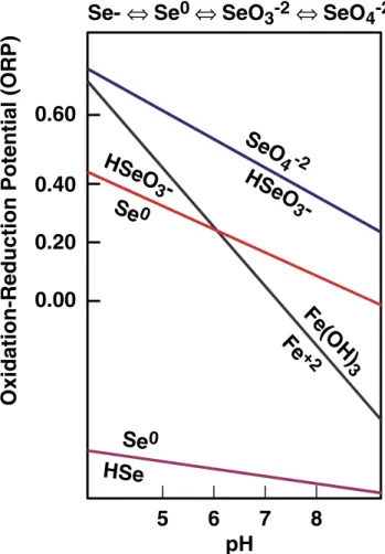
Although natural systems are seldom at equilibrium, the species of Se in soil will tend toward the predicted species in Fig.2with time (Geering et al.1968). The regions in Fig.2
were calculated based on thermodynamic considerations that have been confirmed by field measurements (Kubota and Cary1982; van Dorst and Peterson1984; Masscheleyn et al. 1990; Mincher et al. 2007). Figure 2 shows that oxidizing (dry) alkaline soil favors the available selenate and selenite species, while reducing (wet) acidic soil favors unavailable elemental Se and selenides. Low redox poten-tials occur in soil inundated with stagnant water, resulting in oxygen depletion and Se reduction (Yang et al. 2006; Mincher et al.2007). For example, Gil et al. (2004) reported t h a t l o w f o r a g e a n d d e f i c i e n t b o v i n e s e r u m S e
5
6
7
8
0.60
0.40
0.20
0.00
HSe
O
3
-Se
0
Se
0
HSe
Fe
(O
H
)
3
Fe
+2
pH
Oxidation-Reduction Potential (ORP)
Se-
⇔
Se
0
⇔
SeO
3
-2
⇔
SeO
4
-2
Se
O
4 -2
HSe
O
3
-Fig. 2 The equilibrium forms of selenium to be expected in soil at various conditions of pH and redox potential. Selenide (HSe−) and elemental Se (Se0) are bio-unavailable, while selenite (HSeO3−) and
concentrations occurred in flooded Argentina Pampas regions even though they had high soil Se.
The incorporation of selenite and selenate into plant tissues is inadvertent and occurs due to plant metabolic mechanisms designed to accumulate sulfur (Bell et al.
1992; White et al. 2004, 2007; Sors et al. 2005). The application of sulfur fertilizer actually decreases the uptake of Se by dilution (Allaway1970; Hopper and Parker1999) because sulfate (SO42−) competes with selenate (SeO42−)
uptake by plants (Dhillon and Dhillon2000; White et al.
2004; Sors et al. 2005; Mackowiak and Amacher 2008). Sulfur fertilization of soils has even led to the occurrence of Se deficiency disease in livestock (Schubert et al. 1961; Allaway1970).
Selenate remains in the inorganic form in roots, shoots, and leaves, while selenite is the species readily transformed into the amino acids selenocysteine and selenomethionine (Brown and Shrift 1981). A fraction of organoselenium compounds is also volatilized from plant roots (Zieve and Peterson1984; Zayed et al.1998). This is lost from the soil. The form of Se, rather than the total concentration in soil, determines the amount of uptake by plants and, thus, the availability to foraging wildlife.
A selenium cycle in nature
Following the discovery of microbial Se respiration and its importance as a trace nutrient in animals, the existence of a natural biological Se cycle was proposed by Shrift (1964). A modern view of the Se cycle includes inorganic processes and may be termed the biogeochemical Se cycle (Mosher and Duce1987). In this cycle, shown in Fig.3, the weath-ering of rock produces the soluble forms of Se that are incorporated into higher plants as selenate and amino acids. Biologically produced volatile forms such as dimethylsele-nide are lost as gas (Fleming and Alexander1972; Barkes and Fleming1974; Chao et al.1976; Doran and Alexander
1976; Reamer and Zoller 1980; Zieve and Peterson 1984; Karlson and Frankenberger 1989; Zayed et al. 1998; de Souza et al.1999; Zhang and Frankenberger2000). In fact, the abundance of Se in coastal locations is due to the marine biological production of gaseous Se followed by atmospher-ic deposition (Duce et al. 1975; Lag and Steinnes 1978; Steinnes et al. 1992; Fernandez-Martýnez and Charlet
2009). Mosher and Duce (1987) estimated values from major anthropogenic and natural sources including volca-noes, crustal weathering, and sea spray, reporting a total atmospheric Se flux of (13–19)×109
g/year, with 60 % of this thought to be of natural origin. This gaseous Se is eventually oxidized and adsorbed to aerosols, and the result-ing particulate forms later deposited on the Earth's surface (Atkinson et al.1990). Atmospheric deposition may then be
the most important source of Se in deficient areas (Haygarth et al.1991,1993a).
Loss of Se may result from plant cropping by foraging wildlife, accompanied by non-random spatial mortality pat-terns. For alpine species such as bighorn sheep, lambing usually occurs at high elevation with the young animals being in the nutrient accumulation stage of growth. Migration to low-elevation range later in the year with subsequent winter-related mortality results in the export of Se and other minerals from the summer range. Haygarth et al. (1991) calculated that high stocking rates of domestic lambs followed by removal from pasture represented significant losses of Se. Flueck (2009) provid-ed an analogous example of local depletion in the element phosphorous on summer range due to high winter range mortality rates. Finally, the forms of Se excreted by ani-mals are generally unavailable for plant uptake (Butler and Peterson 1963; Peterson and Spedding 1963; Doran and Alexander 1976; Olson et al. 1976; Joblin and Pritchard 1983). Thus, while plants convert the element to more available forms, animals convert Se mainly to unavailable forms and/or export it from key habitats. Furthermore, at higher elevations, selenite and selenate
Fig. 3 Simplified view of the natural cycling of Se in the environment. The weathering of rock produces forms of the element that are taken up by plants and/or volatilized. Foraging wildlife consume Se in plants. The amount available for uptake depends on many environmental factors, discussed in the text
may be lost through leaching by rainfall with subsequent transport to lower elevation. There, it often becomes con-centrated in flats and flood plains (Webb and Atkinson
1965; Carter et al. 1970; Sivertsen2004; Steinhöfel et al.
2004). An extreme example of the concentration of Se by leaching is provided by the waterfowl poisoning experi-enced at Kesterson Reservoir, in California, USA (Presser and Ohlendorf 1987), related to farm runoff.
Anthropogenic effects on the selenium cycle
The combustion of fossil fuels is a source of atmospheric Se of the same order of magnitude as biogenic sources (Andren et al. 1975; Mosher and Duce 1987). Archived soil and vegetation from southeast England shows increasing Se concentrations in the first half of the twentieth century, followed by declining concentrations since the passage of the clean air legislation (Haygarth et al. 1993b). Thus, industrialization seems to have increased, at least temporar-ily, atmospheric Se deposition in some areas. However, for remote areas, which often contain the last remaining wildlife habitat, the situation is different. Measurements of the sulfur and Se contents of both Greenland and Antarctic ice have shown that their sulfur content has increased since industri-alization, while Se has not (Weiss et al.1971). Similarly, the influence of continental sources on sulfur concentrations in the northern Pacific is greater than for Se (Mosher et al.
1987). Unlike biogenic Se, anthropogenic Se is not trans-ported to remote locations because combustion-produced SeO2 is readily reduced to unavailable elemental Se and
deposited locally. Therefore, while total Se emissions to the environment have increased, the net result is a Se dilu-tion effect due to competidilu-tion for plant uptake by increased levels of sulfur.
To this may be added the very effective sequestration of Se by heavy metals such as cadmium, mercury, or lead (Allaway et al. 1967; Combs and Combs 1986; Frost
1987). These metals are also released continuously to the environment by fossil fuel combustion. Acid precipitation was suggested to liberate heavy metals including mercury and cadmium in Sweden, thus reducing Se bioavailability to wildlife (Borg1987). Wild bison in Canada were also found to have deficient Se levels accompanied by elevated cadmi-um and mercury as compared to farmed bison (Macneil et al. 1990). Continuing declines of bighorn sheep in the Rocky Mountains have also been suggested to partially stem from anthropogenic biochemical changes in high-elevation mountains resulting in Se deficiency (Williams et al.2002; Hnilicka et al.2004).
Thus, the bioavailable Se concentration across the globe may actually be decreasing (Frost 1987; Ermakova and Jovanovic2010). Fossil fuel combustion is also acidifying the general environment through the precipitation of acidic
sulfate and nitrate on the landscape. Acidity favors the reduction of Se to the reduced, biologically unavailable forms, and many authors have reported decreased Se con-centrations in plants grown on acid-amended soils (Geering et al.1968; Goldberg and Glaubig1988; Ambe et al.1992; Tachi et al.1998; Wang et al.2000). The soil acidification process may be somewhat mitigated by natural soil buffer-ing capacity (James and Riha 1986; Clayton et al. 1991). However, the fact that these capacities can be eventually exceeded was revealed by Blake et al. (1999), who reported that soil pH at the Rothamsted Experimental Station, UK, dropped from 6.2 to 3.8 in a woodland plot and from 5.2 to 4.2 on a grassland plot between 1883 and 1999. As noted above, soil acidification also increases the solubility of the heavy metals that tie up Se as insoluble selenides. These possible implications of emissions causing acidification, a declining Se cycle, and increased exposure to heavy metals for wild ruminants were described by Flueck (1990) and Flueck and Smith-Flueck (1990).
Lastly, increasing rates of anthropogenic biomass export due to agricultural production are also disturbing the natural Se cycle. In the natural cycle, the majority of the available Se resides in biomass (Gissel-Nielsen and Hamdy 1977; Swaine 1978; Frost 1987) in plants and litter fall (Tiedemann et al. 2000; Jobbagy and Jackson 2004). Selenium accumulates in the upper soil layers and mainly in organic material. However, biomass harvesting and ex-port can remove Se and other nutrients in a short time, also leading to soil acidification which renders Se less bioavail-able (Gissel-Nielsen and Bisbjerg1970; Heylar et al.1990; Gustafsson et al.1993; Flueck1994; Jobbagy and Jackson
2004). Extensive livestock production currently occurs over 45 million square kilometers of rangeland globally. This Se export also includes the increased biomass burning during the last several centuries (Niklasson and Granstrom2000).
Implications for management and conservation
As wildlife habitat is encroached upon for multiple anthro-pogenic uses, wild animal populations lose access to impor-tant natural resources such as low-elevation range and mineral licks. This may result in loss of access to important trace minerals, including Se. If a population is suffering from severe Se deficiency, WMD would occur in deficient young at 3–6 weeks, resulting in muscle stiffness and res-piratory distress. Affected muscle has visible striations upon necropsy. The malady may result in sudden death, which obviously reduces the recruitment rates or spontaneous pop-ulation recovery. In adults, general unthriftiness, late wean-ing, and periodontal disease are common expressions. Symptoms of the deficiency may also manifest as high rates of capture myopathy or only appear after other stressful
situations, such as a severe winter. However, chronic, sub-clinical deficiency may depress population performance and be unrecognized by the wildlife manager. The following paragraphs are a guide to help managers understand under what conditions Se deficiency in wild ungulate populations is possible or even likely.
Rock of volcanic origin is typically low in Se (Carter et al. 1970; Fernandez-Martýnez and Charlet 2009) because the element is volatile at high temperatures. Soils derived from granite or basalt can be expected to be well below the world average of ∼400 ppb total Se (Kabata-Pendias and Pendias 1984), and plants growing over granitic bedrock will likely contain low concentrations. In contrast, plants in areas with sedimentary bedrock will contain higher amounts of Se (Dhillon and Dhillon2000; Mincher et al.2007). Sites at high elevation also experience losses due to leaching. For these reasons, upland range soils are often deficient (Carter et al. 1970; Ren et al. 1987; Iyengar and Gopal-Ayengar
1988; Haygarth et al. 1991; Wang and Gao2001). If defi-ciency is suspected, analysis of soil may be warranted; however, as mentioned above, forage Se concentrations are related to the soil species of Se rather than its total concen-tration. Analyses that identify Se species rather than total Se rely on specialized and expensive techniques that are not often readily available (Pickering et al.1995). Fortunately, it is possible to infer the likely species based on easily mea-sured soil parameters such as redox potential (Eh, a measure
of the oxidizing power of the soil) and pH (Geering et al.
1968). Both are measured with inexpensive probes amena-ble to field conditions. The combination of these parameters may be used to determine which Se species are most prob-able at a given site. Thus, risk factors for Se deficiency include high-elevation sites, granitic or basaltic geology, acidic soils, and high precipitation and/or stagnant, water-logged soils, especially at inland locations.
Should the measured soil Ehand pH suggest that soil Se
is in an unavailable form, it may be desirable to sample vegetation. The total concentration of Se in plants is readily measured by many laboratories, although some provide inadequate minimum detection limits. Plant species that are actually used as forage should be sampled, and the same species should be used in repetitive sampling campaigns. Different species may accumulate Se at different rates. Some plants bioaccumulate Se to high concentrations, but are seldom used as forage. The best-known example is Astragalus bisulcatus which may contain Se in leaves and seeds at thousands of milligrams per kilogram (Rosenfeld and Beath1964; Ulrich and Shrift1968; Sors et al.2005). Selenium accumulators have a characteristic odor produced by dimethyldiselenide. Foraging wildlife and/or livestock may actually be poisoned by eating these species.
For livestock, forage values of <0.1 ppm on a dry weight basis are considered deficient (Allaway and
Hodgson 1964). It is commonly assumed that wildlife requirements would be lower, although minimum requirements are unknown and may vary between spe-cies. The literature reviewed here suggests that forage species concentrations <0.05 ppm may indicate a risk factor. As we have already shown, a wide range of Se values may exist in individual animals, probably at least in part indicating that they feed on various spe-cies of plants or at various locations.
If some risk factors above are indicated, then sampling of the animal population itself may be warranted. Selenium in liver is the commonly evaluated metric, and as already discussed, blood and hair Se levels have sometimes been shown to be correlated with liver Se. Muscle tissue has also been used. In either case, individual animals need to be sacrificed, and this may not be practical for unhunted or endangered populations. A tissue sample of adequate size should be determined after consultation with the selected laboratory to ensure that enough material is collected to give adequate detection limits and should be kept cool, prefera-bly frozen, and protected from cross-contamination. The sampling of hair could be conducted without the need to sacrifice animals; however, to our knowledge, a rigorous study showing the relationship between hair and liver Se has not yet been done.
Furthermore, it is difficult to determine what levels consti-tute deficiency. Based on the literature reviewed here, we have provided recommendations in Table1. Should the conclusion be made that a population is performing poorly due to Se deficiency, the wildlife manager has little choice but to pro-vide supplementation where that is possible. Mineral salt blocks with Se supplements have long been used in domestic stock nutrition and have been used to improve recruitment in wild populations. Care in the placement of blocks should be used to mitigate the possibility that unnatural congregations of supplemented animals might occur, which could introduce other management problems such as predation or disease. If deficiency was produced by loss of access to specific habitat components, it may be preferable to restore that access rather than to provide supplements. Unfortunately, habitat restora-tion is not always a practical alternative.
Packard’s (1946) classical study on bighorn sheep eluci-dated the reasons for poor population performance by con-sidering pre-Columbian distribution and migratory behavior, market hunting by settlers, extirpations of popu-lations at low elevations, arrival of livestock, and character-istics of the high-elevation habitat where sheep still remained. He found that sheep previously used winter ranges with sedimentary bedrock and mineral licks, whereas current herds were forced to remain at high elevations. Anthropogenically caused range contraction of ungulates are heavily skewed toward loss of low-elevation ranges (Channell and Lomolino2000; Laliberte and Ripple2004).
Thus, loss of traditional seasonal movements has resulted in many sedentary wildlife populations and associated season-al deficiencies in forage quseason-ality. This has been considered an important cause of declining herds and one of the largest problems challenging long-term persistence (Risenhoover et al.1988). In situations where residual wild populations fail to respond to management or protection, the possibility that subclinical deficiency of Se or other trace elements is a factor should be considered.
Conclusions
The decline of wild ungulate populations is a worldwide recurrent problem, and the multifactorial nature of ecol-ogy, combined with the difficulties of studying elusive wild animals, tends to reveal only proximate factors like clinical disease or predation events. Underlying causes of population declines that may be more important, such as mineral and forage nutrition, are sometimes over-looked. Micronutrient deficiencies, including for Se, are especially challenging to understand because they result in a large gradient of possible effects without providing a clear and specific signature when subclini-cal. However, during this phase, much damage may be occurring because a slow decline in remnant populations may result in local extinctions which are difficult to document or counter. In such cases, expensive invest-ments in habitat, restriction of hunting regulations, or predator control will probably fail to recover the affect-ed population. The literature reviewaffect-ed here suggests that subclinical nutritional Se deficiency may be common, especially at alpine locations.
From a mineral nutritional view, the most productive sites for wild ruminant herbivores are valley floors, riparian areas, flood plains, and sedimentary beds containing deeper soils and natural mineral licks. As has been shown, Se is leached to lower elevations by natural processes and has inherently higher concentrations in sedimentary soils. In addition, milder climate and other factors result in food intake of higher mineral density and low oxidative stress on animals. When access to such nutrient hot spots is no longer possible, or vertically transmitted migration behavior to get there has been eliminated through anthropogenic forces, the remaining animals are forced to live in a nutri-tionally marginal habitat. When a population in such cir-cumstances fails to respond to management designed to increase its numbers, mineral nutrition deficiencies particu-larly of Se should be considered by the wildlife manager.
Acknowledgments We greatly appreciate the many constructive comments from three anonymous reviewers.
References
Aastrup P, Riget F, Dietz R, Asmund G (2000) Lead, zinc, cadmium, mercury, selenium and copper in Greenland caribou and reindeer (Rangifer tarandus). Sci Total Environ 245:149–159
Alexander TL (1986) Management and diseases of deer. Veterinary Deer Society, London
Allaway WH (1970) Sulphur–selenium relationships in soils and plants. Sulphur Inst J 6:3–5
Allaway WH, Cary EE, Ehlig CF (1967) The cycling of low levels of selenium in soils, plants and animals. In: Muth OH, Oldfield JE, Weswig PH (eds) Selenium in biomedicine. AVI Publishing, Westport, pp. 273–296
Allaway WH, Hodgson JF (1964) Symposium on nutrition, forage and pastures: selenium in forages as related to the geographic distribu-tion of muscular dystrophy in livestock. J Anim Sci 23:271–277 Allen ME, Ullrey DE (2004) Relationships among nutrition and
repro-duction and relevance for wild animals. Zoo Biol 23:475–487 Ambe S, Chen SY, Ohkubo Y, Kobayashi Y, Iwamoto M, Ambe F (1992)
Application of the radioactive multitracer technique to a study of adsorption of metal ions onγ-Fe2O3. Chem Lett 21:1059–1062
Andren AW, Klein DH, Talmi Y (1975) Selenium in coal-fired steam plant emissions. Environ Sci Technol 9:856–858
Andrews ED, Hartley WJ, Grant AB (1968) Selenium-responsive diseases of animals in New Zealand. NZ Vet J 16:3–17 Arthur JR (2000) The glutathione peroxidases. Cell Mol Life Sci
57:1825–1835
Arthur JR, Beckett GJ, Mitchell JH (1999) The interactions between selenium and iodine deficiencies in man and animals. Nutr Res Rev 12:55–73 Atkinson R, Aschmann SM, Hasegawa D, Thompson-Eagle ET,
Frankenberger WT Jr (1990) Kinetics of the atmospherically important reactions of dimethyl selenide. Environ Sci Technol 24:1326–1332
Backhaus B, Backhaus R (1983) The cadmium-content of roe deer in the Egge mountains. Zeits Jagdwiss 29:213–218
Bakonyi T, Radak Z (2004) High altitude and free radicals. J Sports Sci Med 3:64–69
Barkes L, Fleming RW (1974) Production of dimethylselenide gas from inorganic selenium by eleven soil fungi. Bull Environ Contam Tox 12:308–311
Barry TN (1983) Iodine. In: Grace ND (ed) The mineral requirements of grazing ruminants. Occasional Publication No. 9. New Zealand Society of Animal Production, Hamilton
Beckett GJ, Arthur JR (2005) Selenium and endocrine systems. J Endocrinol 184:455–465
Bell PF, Parker DR, Page AL (1992) Contrasting selenate sulfate interaction in selenium accumulating and nonaccumulating plant species. Soil Sci Soc Am J 56:1818–1824
Berger K, Mohry H, Schuster H (1989) Geoökologische Stoffflüsse und Konsequenzen anthropogener Aktivitäten in der Landschaft. VEB Deutscher Verlag f Grundstoffindustrie, Leipzig
Besselmann D, Schaub D, Wenker C, Völlm J, Robert N, Schelling C, Steinmetz H, Clauss M (2008) Juvenile mortality in captive lesser kudu (Tragelaphus imberbis) at Basle Zoo and its relation to nutrition and husbandry. J Zoo Wildl Med 39:86–91
Beytut E, Karatas F, Beytut E (2002) Lambs with white muscle disease and selenium content of soil and meadow hay in the region of Kars, Turkey. Vet J 163:214–217
Blake L, Goulding WT, Mott CJB, Johnston AE (1999) Changes in soil chemistry accompanying acidification over more than 100 years under woodland and grass at Rothamsted Experimental Station, UK. Euro J Soil Sci 50:401–412
Bodie WL, O'Gara BW (1980) A description of “weak fawn syn-drome” in pronghorn antelope. Proc Bien Pronghorn Antelope Workshop 9:59–70