ORIGINAL ARTICLE
Whole genome and transcriptome amplification: practicable
tools for sustainable tissue biobanking?
Adriana von Teichman&Martina Storz&Susanne Dettwiler&
Holger Moch&Peter Schraml
Received: 29 March 2012 / Revised: 24 August 2012 / Accepted: 3 September 2012 / Published online: 25 September 2012 # Springer-Verlag 2012
Abstract The use of whole genome amplification (WGA) and whole transcriptome amplification (WTA) techniques enables the enrichment of DNA and RNA from very small amounts of tissue. Here, we tested the suitability of WGA and WTA for tumor tissue biobanking. DNA and RNA from 13 standardized and seven non-standardized frozen and 12 formalin-fixed, paraffin-embedded (FFPE) clear cell renal cell carcinoma specimens (>9 years old) served to test the robustness of the WGA and WTA products by reidentifying von Hippel–Lindau (VHL) gene mutations known to exist in these samples. The enrichment of DNA and RNA from frozen tissue was up to 1,291-fold and 423-fold, respectively. The sizes and yields (10-to 73-fold) of the amplified DNA obtained from the 12 FFPE samples were generally lower. The quality of the RNA from the FFPE samples was too low to reliably perform WTA. Our results demonstrate that frozen tumor tissue is very suitable for WGA and WTA. All 20 VHL mutations were verified with WGA. Notably, we were able to show that 18 of the 20 (90 %) VHL mutations are also transcribed. In FFPE tumor tissue, 8 of 12 cases (67 %) showed the expected mutations after the first WGA. Accurate WTA with FFPE material is sophisticated and strongly depends on the modification and degradation status of the fixed tissue. We conclude that for sustainable tissue bio-banking, the use of WGA and WTA is a unique opportunity to provide researchers with sufficient amounts of nucleic acids, preferably from limited frozen tissue material.
Keywords WGA . WTA . Tissue biobank .VHL gene .
Mutation analysis
Introduction
Tumor tissue biobanks represent an ideal platform for trans-lational research. Large tumor tissue collections exist in pathology institutes at universities in which thousands of tissue samples are stored. In general, tumor tissue samples are formalin-fixed and paraffin-embedded (FFPE). The vol-ume of archived tissue samples is limited by the size of the paraffin block and varies between a few cubic millimeter
(fine needle biopsies) and 1–2 cm3
.
Due to formalin fixation, the molecular structures in FFPE material become modified, degraded, and
cross-linked [1] and are of limited value, particularly when
high-throughput technologies, such as DNA microarrays, deep sequencing, or mass spectrometry, are to be applied. As a result, frozen tumor tissue samples not required for diagnostic purposes are routinely collected in parallel in many pathology institutes.
According to the recommendations of the TuBAFrost
consortium [2], approximately 0.5 cm3is the ideal size for
collecting frozen tumor tissue samples in a tumor tissue biobank. This is also in line with our standardized operating procedure for freezing and processing tumor tissues which was established at the Central Tissue Biobank at the
Uni-versity Hospital of Zurich [3].
Depending on the organ and the surgical resection spec-imen that a tumor originated from, residual tissue fragments available for the biobank may be very small. Furthermore, it is expected that ongoing optimization of surgical dissection techniques and cancer screening tests will lead to a further increase of micro-biopsy samples in the biobanks. Subse-quently, although the demand for tumor tissue in the cancer research community is rising, the tissue material available for research projects will become more and more limited in the near future. In addition, once tumor samples have been depleted, these samples are no longer available for further
A. von Teichman
:
M. Storz:
S. Dettwiler:
H. Moch:
P. Schraml (*)Institute of Surgical Pathology, University Hospital Zurich, Schmelzbergstrasse 12,
8091 Zurich, Switzerland e-mail: [email protected]
studies. As a consequence, biobank managers are forced to look for suitable strategies to help prevent the wasting of valuable tumor tissue material. The amplification of whole genomes and transcriptomes of tumor tissue samples may be such an approach.
Several companies offer commercially available kits which allow the amplification of the human genome and
transcriptome up to over 1,000-fold [4–6]. In this study, we
evaluated the suitability of a whole genome amplification (WGA) and a whole transcriptome amplification (WTA) kit for tumor tissue biobanking using tissue from FFPE and frozen clear cell renal cell carcinoma (ccRCC) specimens. The GenomePlex® WGA2 kit (Sigma-Aldrich) is based on the random fragmentation of genomic DNA and conversion of the resulting small fragments to PCR-amplifiable DNA molecules flanked by universal priming sites. In the whole transcriptome amplification kit (WTA2, Sigma-Aldrich), RNA undergoes a single-step conversion into cDNA frag-ments flanked by universal priming sites followed by a subsequent PCR amplification of the resulting cDNA library using universal oligonucleotide primers. The accuracy and robustness of the WGA and WTA products were tested using RCC samples with known von Hippel–Lindau (VHL)
mutations [7].
Materials and methods
Tissue and nucleic acid extraction
Twenty frozen and 12 FFPE unrelated ccRCC specimens
with knownVHL mutations [7] were selected for this study
to test whether WGA and WTA are applicable to tissues that were treated differently and of a different age. Thirteen of the 20 samples were frozen according to a standardized
procedure [3] which was established at our institute in
2006 and were expected to serve as “positive controls”.
The remaining seven tissues were collected before 2006 using non-standardized protocols. The 12 FFPE ccRCC
specimens were between 9 and 19 years of age. TheVHL
mutations are listed in Tables 1 and 2. Hematoxylin and
eosin-stained (HE) sections were reviewed by one patholo-gist (H.M.). All tumor samples contained at least 70 % tumor cells. Magnified HE sections of three tumors are
shown in Fig.1. DNA and RNA were isolated from one
and five 20 μm frozen sections, respectively, using the
DNeasy Blood and Tissue and the RNeasy Mini Kits (Qiagen, Hilden, Germany), respectively. DNA and RNA from FFPE tissue were isolated from three punched tissue cylinders (diameter 0.6 mm) using the EZ1 DNA Tissue kit (Qiagen)
and an RNA extraction protocol [8], respectively. The
concen-trations of the obtained nucleic acids were measured using the Nanodrop (Thermo Fisher Scientific, Waltham, MA, USA).
Whole genome and transcriptome amplification
The GenomePlex® WGA2 and WTA2 tissue kits (Sigma-Aldrich, St. Louis, MO, USA) were used to amplify the genomes and transcriptomes of both frozen and FFPE tissue. We used between 8 and 38 ng of DNA and RNA from frozen tissue, and 100 ng of DNA from FFPE material as input for the WGA and the WTA, respectively. The quality of the amplified DNA was analyzed on 1.5 % agarose gels. The amplified products were purified using the MinElute PCR Purification Kit (Qiagen), and the concentrations were measured using the Nanodrop.
VHL mutation analysis
PCR of the threeVHL exons in the amplified genomes was
performed as previously described [9]. One hundred
nano-grams of amplified DNA was used as template.
Using the Primer3 online program, new primers were
designed to analyze the VHL mutations in the amplified
transcriptomes. One primer pair (forward: 5′-gagtacggccct
gaagaaga-3′; reverse: 5′-ggcacaaataattcagtttggtt-3′; 350 bp)
spanned exons 1 and 2. The second primer pair (forward: 5′-acacgatgggcttctggtta-3′; reverse: 5′-tcaatctcccatccgttgat-3′; 271 bp) spanned exons 2 and 3. One hundred nanograms of amplified cDNA was used as template.
Sequencing of the PCR products was performed by the dideoxy chain-termination method using the BigDye® Ter-minator v1.1 Cycle Sequencing kit (Applied Biosystems). Forward and reverse primers used for the PCR were also used for sequencing. Cycle sequencing products were ana-lyzed using the AbiPrism 3100 Genetic analyzer (Applied Biosystems).
A second PCR was performed if no amplified product or
no VHL mutation was obtained. If the result was not in
accordance with the expected result, the original DNA or RNA was subjected to a second or third WGA or WTA,
respectively. The products underwentVHL-PCR and DNA
sequencing as described above.
Results
Whole genome amplification
Between 8 and 35 ng of DNA was used as template for the whole genome amplification of frozen ccRCC tissue. The sizes of the resulting PCR products ranged between 100 and
1,000 bp (Fig.2a). A DNA yield between 2.98 and 17.36μg
of DNA was obtained. Seventeen samples had more than
10μg of DNA which is equivalent to a 300- to 1,291-fold
enrichment. There were no significant differences in the amplified DNA sizes and the yields between tissue samples
T able 1 WGA and WT A results with frozen ccRCC tissue Y ear of sample VHL mutation
Starting amount DNA
(ng)
DNA yield (µg) OD 260/280 Fold enrichment
Result
Starting amount RNA
(ng) cDNA yield (µg) OD 260/280 Fold enrichment Result 1992 c.492_51 1del/p.Gln164Glnfs 1 1 1 1.40 1.93 1,037 Correct (1st WGA) 15 4.2 2.10 275 S eq u en ce n .a . (1 st W T A ) No mutation (2nd WT A) 2004 c.263_264GG > CT/p.T rp88Ser 19 2.98 1.88 157 Correct (1st WGA) 38 10.1 1.99 267 Correct (1st WT A) 2004 c.400G > T/p.Glu134X 17 5.48 1.89 322 Correct (1st WGA) 30 8.8 1.94 298 No mutation (1st WT A) Correct (2nd WT A) 2004 c.300_308del/p.Thr- 100_Pro103delinsThr 18 12.1 1 1.91 673 Correct (1st WGA) 35 7.1 1.94 202 Correct (1st WT A) 2005 c.473T > G/p.Leu158Arg 14 6.06 1.92 433 Correct (1st WGA) 20 7.2 1.96 369 Correct (1st WT A) 2005 c.484T > C/p.Cys162Ar g 1 5 13.27 1.93 885 Correct (1st WGA) 18 6.2 2.1 1 343 Correct (1st WT A) 2005 c.512delA/p.L ys171Serfs 1 1 10.32 1.93 938 Correct (1st WGA) 32 9.2 2.00 288 Correct (1st WT A) 2006 c.255_256GC > T A/ p.LeuPro85_86LeuThr 10 1 1.38 1.94 1,138 Correct (1st WGA) 22 9.1 1.90 423 Correct (1st WT A) 2006 c.234delT/p.Asn78Asnfs 8 10.33 1.92 1,291 Correct (1st WGA) 25 9.5 2.1 1 374 Correct (1st WT A) 2006 c.232_250del/p.Asn78fs 35 10.49 1.9 300 Correct (1st WGA) 22 4.6 1.98 21 1 Correct (1st WT A) 2006 c.599_600delGG/p.Arg200fs 26 1 1.99 1.91 461 No mutation (1st WGA) 31 9.7 1.97 309 Correct (1st WT A) Correct (2nd WGA) 2006 c.562_565del/p.Leu188fs 18 12.00 1.92 667 Correct (1st WGA) 22 8.6 1.95 386 Correct (1st WT A) 2006 c.240T > A/p.Ser80Arg 15 1 1.22 1.89 748 Correct (1st WGA) 21 6.8 1.96 330 No PCR product (1st WT A) Correct (2nd WT A) 2006 c.277G > C/p.Glu93Ar g 2 0 1 1.21 1.91 561 Correct (1st WGA) 31 8 1.96 255 Correct (1st WT A) 2006 c.208G > T/p.Glu70X 20 1 1.41 1.9 571 Correct (1st WGA) 31 7.9 1.99 253 No PCR product (1st WT A) Correct (2nd WT A) 2007 c.266T > C/p.Leu89Pro 14 14.20 1.98 1,014 No mutation (1st WGA) 21 7.3 2.01 340 Correct (1st WT A) Correct (2nd WGA) 2007 c.421_430dup/p.Gly144Glufs 18 13.26 1.99 737 No mutation (1st WGA) 38 8.9 1.97 237 S eq u en ce n .a . (1 st W T A ) Correct (2nd WGA) No mutation (2nd WT A) 2007 c.484T > /p.Cys162Arg 19 17.36 1.89 914 No mutation (1st WGA) 28 9 2.08 319 Correct (1st WT A) Correct (2nd WGA) 2007 c.458T > C/p.Leu153Pro 19 13.74 1.96 723 No mutation (1st WGA) 38 1 1.8 2.02 315 No mutation (1st WT A) Correct (2nd WGA) Correct (2nd WT A) 2007 c.564-565GG > A T/p.LeuGlu188_189LeuX 19 16.63 1.98 875 No mutation (1st WGA) 38 14.9 2.01 397 Sequence n.a. (1st WT A) Correct (2nd WGA) Correct (2nd WT A) n.a. not analyzable
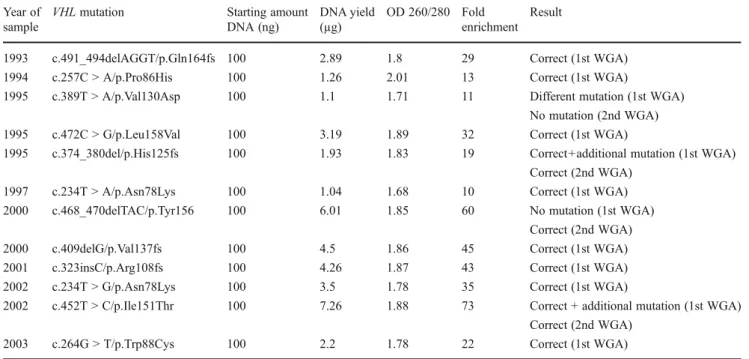
Table 2 WGA results with FFPE ccRCC tissue Year of
sample
VHL mutation Starting amount DNA (ng) DNA yield (µg) OD 260/280 Fold enrichment Result
1993 c.491_494delAGGT/p.Gln164fs 100 2.89 1.8 29 Correct (1st WGA) 1994 c.257C > A/p.Pro86His 100 1.26 2.01 13 Correct (1st WGA)
1995 c.389T > A/p.Val130Asp 100 1.1 1.71 11 Different mutation (1st WGA) No mutation (2nd WGA) 1995 c.472C > G/p.Leu158Val 100 3.19 1.89 32 Correct (1st WGA)
1995 c.374_380del/p.His125fs 100 1.93 1.83 19 Correct+additional mutation (1st WGA) Correct (2nd WGA)
1997 c.234T > A/p.Asn78Lys 100 1.04 1.68 10 Correct (1st WGA) 2000 c.468_470delTAC/p.Tyr156 100 6.01 1.85 60 No mutation (1st WGA)
Correct (2nd WGA) 2000 c.409delG/p.Val137fs 100 4.5 1.86 45 Correct (1st WGA) 2001 c.323insC/p.Arg108fs 100 4.26 1.87 43 Correct (1st WGA) 2002 c.234T > G/p.Asn78Lys 100 3.5 1.78 35 Correct (1st WGA)
2002 c.452T > C/p.Ile151Thr 100 7.26 1.88 73 Correct + additional mutation (1st WGA) Correct (2nd WGA)
2003 c.264G > T/p.Trp88Cys 100 2.2 1.78 22 Correct (1st WGA)
A
B
Fig. 1 HE sections of two homogenous ccRCC cases with strongly dominatingVHL wildtype sequences. Tumors withVHL mutations c.458T>C/p.Leu153Pro and c.564-565GG>AT/p.Leu-Glu188-189LeuX, respectively. Details are shown in Table1
collected under standardized and non-standardized
condi-tions. The detailed results are shown in Table1.
In contrast to the frozen tissue samples, the sizes and yields of the genomic DNA amplification products obtained from the 12 FFPE ccRCC samples varied significantly
among each other and were generally lower (Fig.2b). As
recommended by the supplier, 100 ng of DNA from each of the ccRCC samples was used as input for the whole genome amplification. The DNA yields were between 1.04 and
7.26μg, which is equivalent to a 10- to 73-fold enrichment.
Details are listed in Table2.
Whole transcriptome amplification
Fifteen to 38 ng of RNA, extracted from the same 20 frozen ccRCC samples, was used for the whole transcriptome ampli-fication. The quality of the amplified cDNA was not analyzed.
Between 4.2 and 14.9μg of amplified cDNA was obtained
after the WTA, which is a 202- to 423-fold enrichment. The sizes and yields of amplified cDNA obtained from standard-ized and non-standardstandard-ized tissue sets were comparable. The
results are shown in Table1.
We also intended to perform WTA with the same 12 FFPE ccRCC samples used for WGA. However, as we were not able to amplify sufficient amounts of the two PCR
products from the original RNA extracts (Fig.3),
particu-larly the 350 bp fragment which spans theVHL exons 1 and
2, we decided to omit this analysis.
VHL mutation analysis of WGA products
The WGA products served as DNA templates for VHL
exon-specific PCRs. For each sample, only the primer pair covering the known mutated exon was used. Fourteen of the 20 (70 %) amplified DNA samples showed the correct mutation after the first WGA. The WGA of six samples using the original DNA preparation from tissues was
repeat-ed because the expectrepeat-edVHL mutation was not visible in the
DNA sequence. However, it is important to note that in the
original DNA of these six ccRCC cases, theVHL wild-type
sequence was strongly dominating and the mutated
se-quence was underrepresented. The VHL mutations of two
tumors are illustrated in Figs. 4 and 5. The results of the
WGA are shown in Table1.
The genomes of all of the 12 ccRCC FFPE samples were successfully amplified. After the first WGA, eight (67 %) of these showed the expected mutations. Two cases showed the
expected mutations and an additionalVHL mutation. Of the
last two cases, one had no mutation and the other had a
different VHL mutation. After repeating the WGA with the
original DNA samples of the four tumors that showed dis-crepant results, three had the correct mutation and one had no
mutation. The results are shown in Table2. An example of a
VHL mutation identified in the original DNA and after WGA
of one ccRCC is shown in Fig.4a and b, respectively.
VHL mutation analysis of WTA products
To see whether aVHL gene affected with a mutation is also
transcribed in ccRCC, the WTA products of the 20 frozen ccRCC were subjected to a PCR which specifically
ampli-fies VHL expressed alleles. Surprisingly, 13 of 20 (65 %)
VHL mutations were confirmed as transcribed after the first WTA and five (25 %) after the second PCR or the second WTA. Six of the 20 analyzed ccRCCs had frameshift muta-tions which probably lead to a loss of function of the VHL protein (pVHL). The frameshift mutations identified in the genomic DNA of the two ccRCC samples could not be verified in the corresponding WTA products. The
tran-scribedVHL mutations of two tumors are shown in Figs.4c
and5c. The results are listed in Table1.
Discussion
In this study, we demonstrate that WGA and WTA are well suited to sufficiently and accurately enrich DNA and RNA from frozen tumor tissue. The whole amplification of genomes and transcriptomes of nucleic acids extracted from FFPE tumor tissues, however, has limitations. Below, we address some critical points which are of general importance if these methods are used to amplify DNA and RNA from
B
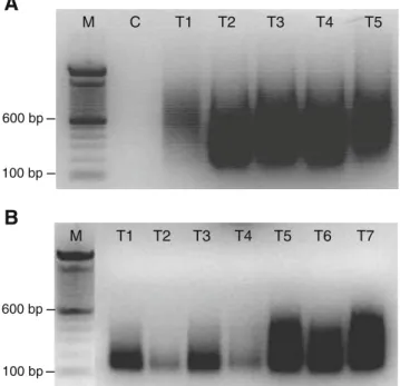
M T3 T4 T5 T6 T7A
600 bp T1 T2 M C T1 T2 T3 T4 T5 100 bp 100 bp 600 bpFig. 2 A 1.5 % agarose gel with WGA products of DNA isolated from five frozen ccRCCs (a) and of DNA isolated from seven FFPE ccRCCs (b).M, 100 bp DNA ladder; C, negative control (no DNA template); T1, tumor 1, etc
cancer tissue. For tumor tissue biobanking, these technolo-gies may help to permanently provide sufficient amounts of high quality nucleic acids from limited and valuable tumor tissue material for research purposes.
MutatedVHL alleles are commonly transcribed in ccRCC
It is widely accepted that in ccRCC,VHL inactivation
fol-lows the two-hit mechanism with loss of the chromosomal
3p arm and a mutation of VHL in the remaining allele.
Recently, we showed thatVHL mutations may exert
differ-ent impacts on pVHL’s functionality [7] provided that the
mutated gene is expressed. Several data exist about pVHL
expression in ccRCC [10–15], but the expression status of
mutatedVHL alleles has not been reported to date. Here, we
were able to demonstrate that most of the VHL mutated
alleles are transcribed, regardless of their mutation type. It is, consequently, conceivable that the location and the type
Exon 1 Exon 2 Exon 1 Intron 1 Exon 1 Intron 1
B
A
C
Fig. 4 VHL mutation c.300_308del/p.Thr100_Pro103delinsThr in the original DNA of one ccRCC patient (a), after WGA (b), and after WTA (c). The mutation site is denoted by anarrow. The boundaries between exon 1/intron 1 and exon 1/exon 2 are indicated
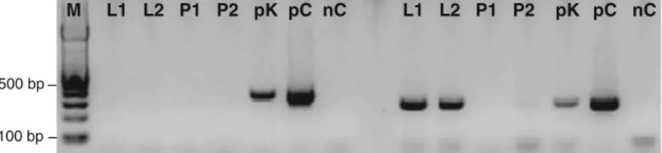
M L1 L2 P1 P2 pK pC nC L1 L2 P1 P2 pK pC nC
500 bp
100 bp
Fig. 3 Reverse transcription-PCR analysis of the 350 bp (exon 1/2) and the 271 bp (exon 2/3)VHL fragments. M, 100 bp DNA ladder; L1 andL2, FFPE normal liver; P1 and P2, FFPE normal prostate; pK,
WTA amplified cDNA control from frozen normal kidney;pC, plas-mid containing the wholeVHL coding sequence (positive control); nC, negative control (no cDNA template)
of aVHL mutation may have dramatic effects on the
multi-adaptor functions of pVHL [16] as well as on tumor behavior
and response to targeted therapies. Although evidence of mutant pVHL expression in ccRCC has not been reported, previous immunohistochemistry data indicate the presence of
pVHL in the majority of ccRCC [10–15].
WGA of DNA extracted from frozen tumor tissue
The DNA obtained after WGA was up to 1,300-fold enriched which is in the range described by the supplier.
After the first WGA, the expectedVHL mutation was
pres-ent in 14 of 20 samples but abspres-ent in six samples. After repeating the WGA from original tissue DNA, all six cases
showed the correctVHL mutation. In all 20 samples, even in
the six ccRCC cases in which theVHL mutated sequences
were significantly underrepresented, the sizes of the result-ing sequence peaks of both the wild-type and the mutated VHL sequences obtained after WGA were comparable with
those of the original, unamplified DNA (see Fig.4). As the
tumor specimen used for the WGA and sequence analyses consisted of more than 70 % tumor cells, we believe that the
observed underrepresention of VHL mutations in the six
ccRCC is a matter of genetic heterogeneity of the tumor cells. Intratumoral heterogeneity was described in a former
study in which VHL deletions were analyzed [17]. We
conclude that in tumor types that are more heterogeneous than RCC, underrepresented gene sequence alterations may frequently be missed after WGA if whole sections are used for DNA extraction. To minimize this problem, DNA should be extracted from micro-dissected or punched tumor areas rather than from whole tumor sections.
WGA of DNA extracted from FFPE tumor tissue
Although the starting amount of DNA extracted from FFPE ccRCC tissue was 100 ng, the enrichment of DNA was about tenfold lower when compared to WGA with DNA extracted from frozen tumor tissue. WGA with 10 ng of DNA resulted in significantly less amplification product (data not shown). Two-thirds of the samples had the correct mutation after the first WGA. Discrepant results were obtained from the four remaining cases that had either
different, additional, or no VHL mutations. After a second,
separate WGA, three of these cases showed the correct mutation and only one case (with a different mutation after the first WGA) had no mutation. It is known that the frequency of damaged bases can vary considerably due to the fixation time and the modifying effects of formalin on
DNA in tissue [18,19]. Therefore, the results obtained from
a mutation analysis should be interpreted with care. In a
previous comprehensiveVHL mutation study, we could not
validate the results from approximately 10 % of the
formalin-fixed ccRCC samples [7]. At least two
indepen-dent rounds of PCR and sequencing were performed to distinguish between real and artificial mutations.
A previous study showed that FFPE tissue is less accurate than frozen material for determining mutations in tumors
[20]. If WGA is to be performed with DNA from FFPE
tumor material, we suggest that two separate parallel WGA
A
Exon 3 Intron 2
B
Exon 3 Intron 2
Exon 3 Exon 2
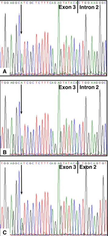
C
Fig. 5 VHL mutation c.484T>C/p.Cys162Arg in the original DNA of one ccRCC patient (a), after WGA (b), and after WTA (c). The sequence is shown in reverse and the mutation site is denoted by anarrow. The boundaries between exon 3/intron 2 and exon 3/exon 2 are indicated
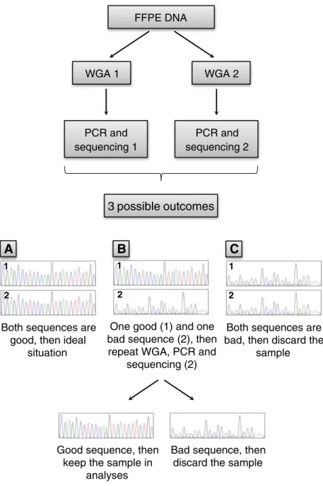
rounds using DNA from original tissue are carried out to select bad from good quality tissue specimens. As a quality control of the whole genome amplified DNA, any exonic sequence can be PCR-amplified and sequenced. If the two obtained sequences are well analyzable and identical, then the integrity of the WGA DNA is given. In case of a discrepancy between the sequences, a third WGA, PCR, and sequencing analysis should be included and compared with the first two sequences. If this third WGA sequence corresponds to one of the others, these two may be used for further molecular analyses. If, however, three different results are obtained, this tissue sample should not be con-sidered for further analyses. The proposed strategy for test-ing the quality of WGA products is schematically illustrated
in Fig.6.
To further minimize PCR artifacts, the use of non-cross-linking fixatives may be an alternative to buffered formalin as the quality of the nucleic acids seems to be similar to those
derived from frozen tissue [21,22]. A further option may be to
treat DNA isolated from FFPE tissue with uracil-DNA glyco-sylase to reduce uracil lesions which are the major cause of
sequence artifacts [23].
WTA of RNA extracted from frozen tumor tissue
The enrichment of amplified cDNA varied only between 200- and 400-fold, indicating a uniform WTA of all 20 RNA samples. With the exception of two ccRCC samples,
which hadVHL frameshift mutations in the original DNA,
the expectedVHL mutations were found to be transcribed in
FFPE DNA WGA 1 WGA 2 PCR and sequencing 1 PCR and sequencing 2
3 possible outcomes
A
Both sequences are good, then ideal
situation
One good (1) and one bad sequence (2), then repeat WGA, PCR and
sequencing (2) 1 2 1 2 1 2
Both sequences are bad, then discard the
sample
Bad sequence, then discard the sample Good sequence, then
keep the sample in analyses
B
C
Fig. 6 Proposed strategy to test the quality of DNA from WGA products derived from FFPE tissue. The original DNA is used for additional WGA rounds
the remaining 18 ccRCC samples. To our knowledge, there
are no studies to date demonstrating thatVHL mutations are
expressed at the RNA level in sporadic ccRCC, regardless of the mutation type.
In 7 of 20 cases, theVHL mutation could not be evaluated
after the first WTA. After repeating the WTA, the VHL
mutations were found in five cases but were not visible in two samples. Not only technical problems (no PCR product of
theVHL fragment or non-analyzable sequence) but also VHL
mutations, which can lead to the downregulation or even to the loss of RNA expression, may explain the discrepant find-ings. These results demonstrate that whole transcriptomes from frozen tissues are linearly amplified and are applicable for gene expression, specific gene mutation, as well as RNA-Seq analyses.
WTA of RNA extracted from FFPE tumor tissue
As already outlined for DNA, the quality of the RNA extracted from FFPE material is similarly low and,
there-fore, of limited value for molecular analysis [24]. The
am-plification of sequences becomes increasingly problematic with the size of the PCR product. To ensure that only cDNA was being amplified, we worked with primer pairs that
spanned twoVHL exons. The sizes of the PCR fragments
were, however, too large to amplify sufficient amounts of DNA for sequence analysis. As it was suggested in other
studies [1,25,26], our results imply that a successful
WTA-based mutation analysis of FFPE tissue is strongly depen-dent on the chosen size of the PCR fragment to be analyzed and the age of the tissue sample.
WGA and WTA for economic tumor tissue biobanking?
WGA and WTA technologies can help to provide sufficient amounts of high quality nucleic acids from valuable tumor tissue material for research purposes in tumor tissue bio-banking. By using fixed and non-fixed ccRCC nucleic acids
with known VHL mutations, we demonstrate that nucleic
acids from frozen tumor tissue are most suitable for WGA and WTA. The severity of the modification and degradation of nucleic acids in FFPE tumor tissue often differs and thus hampers the accurate amplification. Additional rounds of WGA or WTA and validation experiments lead to increased costs. In Switzerland, one WGA costs approximately 10 Swiss Francs and one WTA is about eight times more expensive. The routine use of WGA and WTA for large tumor tissue biobanks, which receive thousands of tissue samples annually, would cause an enormous financial
bur-den on a biobank’s budget, in terms of manpower,
consum-ables, freezers, space, and logistics. In contrast, research projects usually focus on analyzing a few dozen up to several hundred tissue samples of one specific tumor type,
which represents only a tiny part of the whole inventory of a large tumor tissue biobank. The use of WGA and WTA should be restricted to those tissue samples, preferably frozen ones, required for approved research projects. This would help to keep the costs within an affordable limit. Furthermore, to financially fund tumor tissue biobanks, it will be of utmost importance for cancer research scientists to include costs for tissue biobanking, such as sampling, storing, and processing as well as the costs for WGA and WTA in grant applications if they intend to work with DNA and RNA from cancer tissue.
Conclusions
Our data indicate that both WGA and WTA are feasible with nucleic acids extracted from frozen and FFPE tissue. Never-theless, the use of the latter has certain limitations and the results should be interpreted with caution. Although standard-ized procedures are required to guarantee the high quality of frozen tissue collections, it seems that non-standardized pro-tocols formerly used for snap freezing native tissue samples do not negatively influence the outcome of WGA and WTA.
Acknowledgments This study was supported by the Zurich Cancer League, the Foundation for Research in Science and the Humanities at the University of Zurich, and the Swiss National Science Foundation (3238BO-103145).
Conflict of interest There is no conflict of interest to declare.
References
1. Lassmann S, Kreutz C, Schoepflin A, Hopt U, Timmer J, Werner M (2009) A novel approach for reliable microarray analysis of microdissected tumor cells from formalin-fixed and paraffin-embedded colorectal cancer resection specimens. J Mol Med 87:211–224
2. Morente MM, Mager R, Alonso S, Pezzella F, Spatz A, Knox K, Kerr D, Dinjens WN, Oosterhuis JW, Lam KH, Oomen MH, van Damme B, van de Vijver M, van Boven H, Kerjaschki D, Pammer J, Lopez-Guerrero JA, Llombart Bosch A, Carbone A, Gloghini A, Teodorovic I, Isabelle M, Passioukov A, Lejeune S, Therasse P, van Veen EB, Ratcliffe C, Riegman PH (2006) TuBaFrost 2: standardising tissue collection and quality control procedures for a European virtual frozen tissue bank network. Eur J Cancer 42:2684–2691
3. Steu S, Baucamp M, von Dach G, Bawohl M, Dettwiler S, Storz M, Moch H, Schraml P (2008) A procedure for tissue freezing and processing applicable to both intra-operative frozen section diag-nosis and tissue banking in surgical pathology. Virchows Arch 452:305–312
4. Clement-Ziza M, Gentien D, Lyonnet S, Thiery JP, Besmond C, Decraene C (2009) Evaluation of methods for amplification of picogram amounts of total RNA for whole genome expression profiling. BMC Genom 10:246
5. Gonzalez-Roca E, Garcia-Albeniz X, Rodriguez-Mulero S, Gomis RR, Kornacker K, Auer H (2010) Accurate expression profiling of very small cell populations. PLoS One 5:e14418
6. Navin N, Hicks J (2011) Future medical applications of single-cell sequencing in cancer. Genome Med 3:31
7. Rechsteiner MP, von Teichman A, Nowicka A, Sulser T, Schraml P, Moch H (2011) VHL gene mutations and their effects on hypoxia inducible factor HIFalpha: identification of potential driver and passenger mutations. Cancer Res 71:5500–5511
8. Bode B, Frigerio S, Behnke S, Senn B, Odermatt B, Zimmermann DR, Moch H (2006) Mutations in the tyrosine kinase domain of the EGFR gene are rare in synovial sarcoma. Mod Pathol 19:541–547 9. von Teichman A, Comperat E, Behnke S, Storz M, Moch H, Schraml
P (2011) VHL mutations and dysregulation of pVHL- and PTEN-controlled pathways in multilocular cystic renal cell carcinoma. Mod Pathol 24:571–578
10. Kivela AJ, Parkkila S, Saarnio J, Karttunen TJ, Kivela J, Parkkila AK, Bartosova M, Mucha V, Novak M, Waheed A, Sly WS, Rajaniemi H, Pastorekova S, Pastorek J (2005) Expression of von Hippel-Lindau tumor suppressor and tumor-associated carbon-ic anhydrases IX and XII in normal and neoplastcarbon-ic colorectal mucosa. World J Gastroenterol 11:2616–2625
11. Osipov V, Keating JT, Faul PN, Loda M, Datta MW (2002) Expression of p27 and VHL in renal tumors. Appl Immunohisto-chem Mol Morphol 10:344–350
12. Schraml P, Hergovich A, Hatz F, Amin MB, Lim SD, Krek W, Mihatsch MJ, Moch H (2003) Relevance of nuclear and cytoplas-mic von Hippel Lindau protein expression for renal carcinoma progression. Am J Pathol 163:1013–1020
13. Corless CL, Kibel AS, Iliopoulos O, Kaelin WG Jr (1997) Immu-nostaining of the von Hippel-Lindau gene product in normal and neoplastic human tissues. Hum Pathol 28:459–464
14. Sakashita N, Takeya M, Kishida T, Stackhouse TM, Zbar B, Takahashi K (1999) Expression of von Hippel–Lindau protein in normal and pathological human tissues. Histochem J 31:133–144 15. Los M, Jansen GH, Kaelin WG, Lips CJ, Blijham GH, Voest EE
(1996) Expression pattern of the von Hippel-Lindau protein in human tissues. Lab Invest 75:231–238
16. Frew IJ, Krek W (2008) pVHL: a multipurpose adaptor protein. Sci Signal 1:pe30
17. Moch H, Schraml P, Bubendorf L, Richter J, Gasser TC, Mihatsch MJ, Sauter G (1998) Intratumoral heterogeneity of von Hippel-Lindau
gene deletions in renal cell carcinoma detected by fluorescence in situ hybridization. Cancer Res 58:2304–2309
18. Agell L, Hernandez S, de Muga S, Lorente JA, Juanpere N, Esgueva R, Serrano S, Gelabert A, Lloreta J (2008) KLF6 and TP53 mutations are a rare event in prostate cancer: distinguishing between Taq polymerase artifacts and true mutations. Mod Pathol 21:1470–1478
19. Quach N, Goodman MF, Shibata D (2004) In vitro mutation artifacts after formalin fixation and error prone translesion synthe-sis during PCR. BMC Clin Pathol 4:1
20. Verhoest G, Patard JJ, Fergelot P, Jouan F, Zerrouki S, Dreano S, Mottier S, Rioux-Leclercq N, Denis MG (2012) Paraffin-embedded tissue is less accurate than frozen section analysis for determining VHL mutational status in sporadic renal cell carcinoma. Urol Oncol 30:469–475
21. Moelans CB, Oostenrijk D, Moons MJ, van Diest PJ (2011) Formaldehyde substitute fixatives: effects on nucleic acid preser-vation. J Clin Pathol 64:960–967
22. Turashvili G, Yang W, McKinney S, Kalloger S, Gale N, Ng Y, Chow K, Bell L, Lorette J, Carrier M, Luk M, Aparicio S, Huntsman D, Yip S (2011) Nucleic acid quantity and quality from paraffin blocks: defining optimal fixation, processing and DNA/RNA extraction techniques. Exp Mol Pathol 92:33–43
23. Do H, Dobrovic A (2012) Dramatic reduction of sequence artefacts from DNA isolated from formalin-fixed cancer biop-sies by treatment with uracil-DNA glycosylase. Oncotarget 3:546–558
24. Klopfleisch R, Weiss AT, Gruber AD (2011) Excavation of a buried treasure—DNA, mRNA, miRNA and protein analysis in formalin fixed, paraffin embedded tissues. Histol Histopathol 26:797–810
25. Huijsmans CJ, Damen J, van der Linden JC, Savelkoul PH, Hermans MH (2010) Comparative analysis of four methods to extract DNA from paraffin-embedded tissues: effect on downstream molecular applications. BMC Res Notes 3:239
26. Ribeiro-Silva A, Zhang H, Jeffrey SS (2007) RNA extraction from ten year old formalin-fixed paraffin-embedded breast cancer samples: a comparison of column purification and magnetic bead-based tech-nologies. BMC Mol Biol 8:118