REVIEW
Adult neurogenesis in the mammalian brain
Simon M.G. BRAUN1,2, Sebastian JESSBERGER (
✉
)1,21
Brain Research Institute, Faculty of Medicine, University of Zurich, 8057 Zurich, Switzerland
2
Neuroscience Center Zurich, University of Zurich and ETH Zurich, Switzerland
© Higher Education Press and Springer-Verlag Berlin Heidelberg 2013
Abstract New neurons are generated throughout life in distinct areas of the mammalian brain. This process, called adult neurogenesis, has challenged previously held concepts about adult brain plasticity and opened novel therapeutic avenues to treat certain neuro-psychiatric diseases. Here, we review the current knowledge regarding the fate and potency of neural stem cells (NSCs), as well as the mechanisms underlying neuronal differentiation and subsequent integration. Furthermore, we discuss the functional significance of adult neurogenesis in health and disease, and offer brief insight into the future directions of the adult neurogenesisfield.
Keywords adult neurogenesis, hippocampus, stem cell, memory, neuropsychiatric disease
Introduction
Mammalian organisms are generated from a single cell that gives rise to tissues and organs that need to be sustained throughout life. During embryonic development, a fertilized egg divides into embryonic stem cells, which undergo mitosis and lead to the formation of three germ lines: the ectoderm, the endoderm and the mesoderm. From these three germ lines, all tissues and organs can be generated. This developmental process is made possible by rounds of stem cell divisions and their subsequent differentiation into the various cell types that compose the body. Following development, certain tissues retain their capacity to generate new cells from a population of somatic stem cells, also termed adult stem cells. This regenerative capacity preserves tissue function and homeostasis as differentiated cells are replaced continuously throughout life (Hall and Watt, 1989). Somatic stem cells reside in various tissues within specific niches where they can symmetrically divide into two stem cells or asymmetrically divide into a stem cell and a progenitor cell (Potten and Loeffler, 1990). The lineage committed progeni-tor cells can in turn undergo several rounds of division followed by a multistep differentiation process, giving rise to the different cell types that make up a tissue. Thus, somatic
stem cells are defined as self-renewing and multipotent cells (Morrison et al., 1997). One of the most well studied populations of adult stem cells can be found in the blood. Throughout the life of an individual, hematopoietic stem cells that reside in the bone marrow give rise to all blood cell types, allowing for the regeneration of blood cells (Abramson et al., 1977). Intestinal stem cells can also be found in the adult and are essential for tissue homeostasis as they continuously produce cells that line the surface of the small and large intestines (Potten and Loeffler, 1987). For almost a century, it was thought that no somatic stem cells resided in the brain. However, it is now well established that neural stem cells exist in the adult mammalian brain and that they can give rise to neurons throughout life, in a process termed adult neurogenesis (Gage, 2000).
Adult neurogenesis
The father of modern neuroscience, Santiago Ramón y Cajal, who pioneered investigations on the cellular architecture of the mammalian brain, stated that no newborn neurons were generated in the central nervous systems of higher vertebrates after embryonic development (Ramón y Cajal, 1928). This finding played a central role in the field of neuroscience as it was thought that no newborn cells could integrate into the complex neural networks that make up the central nervous system. Thus, it was very widely accepted that the brain lacked any substantial regenerative capacity. Altman and colleagues obtained the first experimental evidence for the
Received January 31, 2013; accepted March 28, 2013 Correspondence: Sebastian JESSBERGER
E-mail: jessberger@hifo.uzh.ch DOI 10.1007/s11515-013-1263-1
existence of newborn neurons in the adult in the early 1960s. Using3
H-thymidine labeling of mitotic cells in the adult rat brain, they were able to detect by autoradiography, dividing cells that had differentiated into cortical neurons (Altman, 1962). However, this strikingfinding went largely unnoticed by the scientific community, until the discovery of new detection methods using immunostaining of mitotically active cells labeled with another thymidine analog, 5-bromo-2'-deoxyuridine (BrdU), to label newborn neurons in the mouse brain (del Rio and Soriano, 1989; Kuhn et al., 1996). The newly identified neural stem cells (NSCs) were then isolated from adult mice and their ability to self renew and differentiate into neurons and glia was confirmed in vitro (Reynolds and Weiss, 1992). Eriksson and colleagues then confirmed that these cells did not merely exist in rodents but could also be detected in the hippocampus of humans (Eriksson et al., 1998).
It is now well established that adult neurogenesis occurs in most mammals within two distinct regions of the brain (Fig. 1), the dentate gyrus (DG) of the hippocampus (Kuhn et al., 1996) and the subventricular zone (SVZ) lining the lateral ventricles (Lois and Alvarez-Buylla, 1994). Within these specific niches, adult neural stem cells self renew and give rise to proliferating progenitors that differentiate into all three neural cell types: neurons, astrocytes and oligodendrocytes (Gage, 2000).
Adult neurogenesis in the SVZ
The subventricular zone is a prominent neurogenic region during embryogenesis and its neurogenic potential is sustained throughout life, albeit at much lower levels. In the adult SVZ, relatively quiescent neural stem cells, termed type B cells, line the lateral ventricles and give rise to actively proliferating neural progenitors, called type C cells (Doetsch et al., 1999). These transient amplifying progenitors primarily become neuroblasts (type A cells) and migrate out of the SVZ
along the rostral migratory stream (RMS) toward the olfactory bulb (Lois and Alvarez-Buylla, 1994). The immature neurons then differentiate into olfactory GABAer-gic granule interneurons, dopaminerGABAer-gic periglomerular inter-neurons or glutamatergic juxtaglomerular inter-neurons, and integrate into the local neuronal circuits (Carleton et al., 2003; Brill et al., 2009). The process of cell migration from the ventricles to the bulb takes several days as the neuroblasts migrate in chains through a dense glial tube (Lois et al., 1996).
Although many thousands of neuroblasts are generated each day in the SVZ, only a small fraction survives to become fully functional olfactory neurons. It is thought that newborn cells play an important role in olfactory learning and memory (Rochefort et al., 2002; Imayoshi et al., 2008). Type B stem cells also retain the potential to asymmetrically divide into astrocytes as well as oligodendrocytes, although it remains unclear whether the same stem cell pool generates both neurons and glia (Morrens et al., 2012). The balance between neurogenesis and gliogenesis can be altered in response to injury, as it has been shown that demyelination can induce oligodendrocyte differentiation of neural progenitor cells in the SVZ (Jablonska et al., 2010). The process of SVZ adult neurogenesis has been extensively studied, and many markers that label the different cell types have been identified, as described in Fig. 2. Finally, it is important to note that recent findings suggest that SVZ neurogenesis is largely absent from adult humans and restricted to the early stages of infancy (Sanai et al., 2011).
Adult neurogenesis in the hippocampus
The hippocampus is a cortical structure found in both hemispheres of the mammalian brain. It is located in the temporal lobe and along with the limbic cortex, amygdala, septum and fornix, it forms the limbic system. Hippocampal neurons from the dentate gyrus (DG) and the cornu ammonis (CA) form a trisynaptic circuitry that plays a central role in certain forms of learning and memory (Milner et al., 1998). The main input into the hippocampus comes from the entorhinal cortex, which can excite granule cells of the DG via the perforant path, as well as directly excite CA3 pyramidal cells. Granule cells of the DG extend dendrites into the molecular layer where they receive glutamatergic input. The cell bodies of the granule cells form the dense layer of the DG and their axons project into the hilus toward CA3 pyramidal neurons. The hilus is composed of interneurons, unmyelinated mossyfibers as well as myelinated mossy cells. In turn, CA3 pyramidal neurons connect to CA1 neurons via Schaffer collateral fibers, which then connect back to the entorhinal cortex to complete the trisynaptic loop (Amaral et al., 2007).
Adult hippocampal neural stem cells give rise to granule cells of the DG in a well-characterized multistep process. First, relatively quiescent NSCs residing in the subgranular
Figure 1 Neurogenic regions in the adult mouse brain. Neural stem cells (NSCs) reside in the dentate gyrus (DG) of the hippocampus as well as in the subventricular zone (SVZ) of the lateral ventricles (NSC niches shown in green). In the DG, NSCs give rise to mature granule cells. In the SVZ, progenitor cells actively proliferate and give rise to neuroblasts that migrate along the rostral migratory stream (RMS) toward the olfactory bulb (OB), where they differentiate into olfactory interneurons (neuroblast migratory path shown in red).
zone (SGZ) of the DG divide either symmetrically or asymmetrically. During a symmetric division, two radial glia-like cells are generated; these cells are known as type 1 or radial NSCs. During an asymmetrical division, NSCs can either self renew and give rise to an NSC and a progenitor or terminally divide into two progenitors (Bonaguidi et al., 2011; Encinas et al., 2011). Secondly, the progenitors, known as type 2 or non-radial NSCs, can give rise to actively proliferating neuroblasts also called type 3 cells. The neuronally committed neuroblasts then begin to branch out processes as they migrate up into the granule cell layer. Finally, over a period of three weeks the newborn neurons project out a large dendritic arbor into the molecular layer and an axon into the hilus that terminates in the CA3 (van Praag et al., 2002; Zhao et al., 2006). Over a period of 8 weeks, the cells become fully mature DG granule cells that functionally integrate into the local circuitry (Toni et al., 2008).
Several methods exist to identify the different cell types generated during the course of neurogenesis. Proliferating NSCs can be labeled by injection of nucleotide analogs or by stereotactic injection of retroviruses expressing fluorescent proteins (van Praag et al., 2002). Both these techniques take advantage of the fact that within the neurogenic niches, progenitors and neuroblasts make up the majority of dividing cells. In addition, transgenic mouse lines, such as NestinGFP, can be used to study NSCs labeled with the fluorescent protein GFP, expressed under control of the Nestin promoter (Yamaguchi et al., 2000). Alternatively, immunohistochem-ical analysis can be performed to label the different cell types which express a wide range of markers as described in Fig. 3. Over the last few decades, numerous studies of the SVZ and DG have led to the detailed characterization of adult neurogenesis in these two niches. However, we are only just beginning to understand why adult neurogenesis is restricted to the SVZ and DG in adult mammals. Future studies, aiming to understand the differences between the two neural stem cell niches, may hold the potential to“unlock” this regenerative process in other brain regions.
Mechanism regulating adult neurogenesis
In the human hippocampus, NSCs give rise to up to a thousand new neurons everyday and throughout life (Knoth et al., 2010). However, as the cells mature, only a small fraction survives and integrates into the local circuitry. The mechan-isms that regulate the quiescence, proliferation, differentiation and survival of cells during adult hippocampal neurogenesis have been extensively studied and will be described in the following section.
Mechanisms responsible for stem cell maintenance and quiescence
To ensure lifelong neurogenesis, adult NSCs are required to differentiate into neuronal progenitors, while maintaining a pool of multipotent stem cells. This balance between stem cell maintenance and differentiation is regulated by numerous cell intrinsic and extrinsic factors. The transcription factor Sox2 has been shown to be critical for this process as it is highly expressed in quiescent NSCs and is downregulated upon differentiation (Suh et al., 2007). Furthermore, conditional deletion of Sox2 in Nestin expressing NSCs leads to a marked reduction in the number of type 1 and type 2 cells in the adult hippocampus (Favaro et al., 2009). Downstream targets of Sox2 include Wnt and Shh (sonic hedgehog), two important regulators of NSC proliferation. It has been shown that Sox2 activation of Shh is required for stem cell quiescence and that loss of Sox2 in NSCs can be rescued at least partially by adding Shh to in vitro cultures (Favaro et al., 2009). Interestingly, Sox2 can also inhibit transcriptional activation of Wnt in NSCs, a signaling molecule that is required to drive
Figure 2 Stages of adult neurogenesis in the SVZ. Type B neural stem cells that line the lateral ventricles (LV), reside in the subventricular zone (SVZ), express Sox2 (SRY sex determining region Y-box 2, blue), GFAP (glialfibrillary acidic protein, red) and Nestin, and have a process that projects into the ventricle. The type B cells can asymmetrically divide into another B cell and a type C progenitor cell. The actively proliferating type C cells express markers such as Ascl1 (achaete-scute homolog 1) and give rise to neuroblasts (type A) that migrate to the olfactory bulb (OB) and are positive for DCX (doublecortin) and Dlx2 (distal-less homeobox 2).
NSC proliferation in vivo (Lie et al., 2005). Recent studies have shown that neuronal activity can influence Wnt levels to regulate stem cell quiescence (Jang et al., 2013), and that deletion of the Wnt inhibitor Dickkopf1 can activate quiescent NSCs to restore hippocampal neurogenesis in aged mice (Seib et al., 2013).
Another major pathway implicated in the maintenance of many embryonic and somatic stem cell populations is the Notch signaling pathway. Hes5 (hair and enhancer of split 5), a target gene and reporter of canonical Notch signaling is selectively expressed in quiescent type 1 NSCs (Lugert et al., 2010). Also, deletion of RBPJk (recombining binding protein suppressor of hairless), the transcriptional activator in the Notch signaling cascade (Ables et al., 2010; Ehm et al., 2010), in adult hippocampal NSCs, leads to an increase in neuronal differentiation and an eventual depletion of the stem cell pool. Interestingly a similar phenotype was observed when BMP (bone morphogenic protein) signaling was inhibited in adult hippocampal NSCs, both in vivo and in
vitro (Mira et al., 2010). Furthermore, ablation of proliferat-ing progenitor cells by administration of AraC (cytosine-β-D-arabinofuranoside), an anti-mitotic drug, has been shown to induce the activity of quiescent NSCs, allowing for the repopulation of the stem cell niche (Doetsch et al., 1999; Seri et al., 2001).
Finally, in vitro cultures of adult NSCs require EGF (epidermal growth factor) and FGF2 (fibroblast growth factor 2) to be maintained as multipotent proliferating cell cultures, and their removal leads to neuronal and glial differentiation (Reynolds and Weiss, 1992; Palmer et al., 1995).
One current limitation in thefield of adult neurogenesis is the lack of markers that specifically label quiescent neural stem cells. Due to the potential glial origin of radial NSCs, many of the known markers are also expressed in astrocytes that can be found throughout the brain. Developing markers and assays that allow investigators to specifically test the functional role of bona fide neural stem cells would represent a major advancement for thefield.
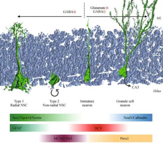
Figure 3 Stages of adult neurogenesis in the DG. Relatively quiescent type 1 NSCs express Sox2 and GFAP, and are labeled with GFP in the NestinGFP and Spot14GFP transgenic mouse lines. Non-radial type 2 NSCs also express Sox2 and NestinGFP and give rise to neuroblasts that actively proliferate and express MCM2 (minichromosome maintenance complex component 2) and Tbr2 (T-box brain 2) as well as the immature neuron marker DCX. The newborn neurons continue to express DCX and Prox1 for 2–3 weeks as they mature into granule cells that express Prox1, NeuN and Calbindin. During the early stages of neurogenesis, newborn neurons receive excitatory GABA input. At three weeks of age, newborn neurons switch to inhibitory GABA and excitatory glutamatergic input, and project axons toward CA3 neurons.
Mechanisms regulating stem cell proliferation
Proliferation of neural progenitor cells and neuroblasts in the DG greatly influences the total number of newborn neurons generated each day. The mechanisms that are responsible for regulating this process are of particular interest as targeting these can result in increased levels of neurogenesis. One key environmental factor that has been shown to positively regulate progenitor proliferation is running. In both mice and humans, it has been shown that running increases cell proliferation and newborn neuron numbers, resulting in an increased hippocampal volume (van Praag et al., 1999; Erickson et al., 2011). Also, several secreted factors are known to increase neural stem and progenitor cell prolifera-tion, including Wnt (Lie et al., 2005), BDNF (brain derived neurotrophic factor) (Scharfman et al., 2005), IGF2 (insulin growth factor 2) (Bracko et al., 2012) and VEGF (vascular endothelial growth factor) (Jin et al., 2002). In line with its positive influence on NSC proliferation, it has been shown that administration of IGF2 in the adult rat hippocampus significantly enhances memory consolidation (Chen et al., 2011). A similar improvement in memory function is also observed after running, correlating enhanced neurogenesis with improved hippocampal function (van Praag et al., 1999). Much of what we know about the molecular mechanisms that regulate stem cell proliferation has come from studies of transcription factor regulation of the cell cycle and growth factor signaling. However, it has recently been shown that the metabolic state of a stem cell can influence the balance between quiescence and proliferation. Studies in hemato-poietic stem cells have shown that the Lkb1 gene can influence the cell cycle by regulating cellular energy metabolism (Nakada et al., 2010). Interestingly, adult NSCs require high levels of fatty acid synthase (FASN)-dependent lipogenesis for proper proliferation (Knobloch et al., 2013), whereas relatively quiescent NSCs display low levels of this metabolic process. Spot14, an inhibitor of this pathway that is selectively expressed in relatively quiescent NSCs, mediates the switch between high and low levels of de novo lipogenesis.
Mechanisms regulating neuronal differentiation and survival
Although thousands of neuroblasts are generated every day in the mammalian brain, only a small fraction survive and fully mature into granule cell neurons (Tashiro et al., 2007). Early on during neurogenesis, transcription factors such as NeuroD1 (neuorgenic differentiation 1) (Gao et al., 2009) and Prox1 (prospero-related homeobox 1) (Lavado et al., 2010; Karalay et al., 2011) are required for neuroblast survival and differentiation. Deletion of these pro-neural genes in adult NSCs leads to a significant decrease in newborn neuron numbers within the DG. At later stages of neurogenesis, Cdk5 (cyclin dependent kinase 5) (Jessberger
et al., 2008), Disc1 (disrupted-in-schizophrenia 1) (Duan et al., 2007) and the Rho GTPase Cdc42 (cell division control protein 42) (Vadodaria et al., 2013), as well as several other genes (Zhao et al., 2008) are required for neuronal maturation as their deletion in NSCs leads to aberrant process extension and dendritic length defects respectively.
Certain environmental factors are known to enhance adult neurogenesis. For example, mice housed in enriched environments display significantly more newborn neurons than their littermates housed in standard cages (Kempermann et al., 1997). This increase in adult neurogenesis is attributed to an increase in neuronal survival rather than a boost in proliferation (van Praag et al., 1999).
Moreover, newborn neurons require synaptic input to survive and fully integrate into the existing hippocampal circuitry. It has been shown that newborn cells display heightened excitability and enhanced synaptic plasticity (Schmidt-Hieber et al., 2004; Wang et al., 2005; Marín-Burgin et al., 2012) compared to more mature cells, which allows them to develop mature spines and form novel synapses in the adult brain (Toni et al., 2007). There is a critical period between 1 and 1.5 months in which newborn neurons exhibit enhanced long-term potentiation (LTP) (Ge et al., 2007), a property that is important for encoding new memories.
Excitatory and inhibitory input in newborn cells
Neurotransmitters mediate excitatory and inhibitory input to cells during all stages of adult neurogenesis, from the radial NSC to the mature granule cell. Recent work has shown that radial NSCs respond tonically to GABA (gamma-aminobu-tyric acid) released by parvalbumin expressing interneurons. This excitatory input regulates the balance between quies-cence and activation of radial NSCs in response to neuronal activity (Song et al., 2012). In addition, type 2 NSCs and progenitors also express GABAA receptors and respond to
excitatory GABA input. Their depolarization induces the expression of genes such as NeuroD1, which in turn drives neuronal differentiation (Tozuka et al., 2005). As the newborn neurons differentiate and grow dendritic spines, they receive both inhibitory GABAergic synaptic input from local interneurons, and excitatory glutamatergic synaptic input from the entorhinal cortex. It has been shown that the survival and functional integration of newborn neurons into the adult hippocampal circuitry requires NMDA (N-methyl-D-aspar-tate) receptor mediated response to glutamate (Tashiro et al., 2006). Finally, hyperpolarized mature granule cells that display enhanced LTP, project mossy fibers into the hilus toward CA3 neurons, forming neuronal circuits required for memory formation as well as pattern separation (Deng et al., 2010).
Whereas embryonic neurogenesis mainly occurs in a series of distinct steps, adult neurogenesis is characterized by the presence of all different maturation stages at any given time.
Thus, understanding the signaling pathways that allow both pro- and anti- proliferative cues as well as differentiation and integration cues to co-exist in the same niche, may offer new insight to improve neural stem cell transplants.
Functions of adult neurogenesis
Learning and memory
The hippocampus is a brain structure that plays a central role in the formation of episodic and spatial memories. Patients with hippocampal lesions can suffer from anterograde amnesia, a condition characterized by an inability to form new memories while maintaining the ability to recall events that occurred before the lesion (Squire, 2009). The densely packed granule cell layer of the DG plays a critical role in encoding, consolidation and retrieval of new memories. Adult NSC derived mature granule cells integrate into this DG circuitry and display unique properties that have been shown to contribute to the memory process. Importantly, increased levels of neurogenesis have been correlated with improved memory function, as mice with increased levels of neurogen-esis, induced by running or enriched environments, per-formed better in spatial learning tasks (Kempermann et al., 1997; van Praag et al., 1999). However, these effects may be attributed to neurogenesis independent functions that are also stimulated by running or enriched environments, therefore extensive studies have since been performed to determine neurogenesis specific functions. Using a transgenic mouse model, Deng and colleagues were able to lower neurogenesis levels by expressing the HSV thymidine kinase gene under the control of the Nestin promoter, allowing for Ganciclovir mediated cell ablation in proliferating neural stem cells. With this model, they were able to show that reduced neurogenesis levels led to defective longterm retention of spatial memory and impaired extinction of conditioned contextual fear, again suggesting an important role for immature neurons in hippocampus-dependent learning and memory (Deng et al., 2009).
Recent studies have focused on more specific hippocampal tasks, revealing a central role for adult neurogenesis in pattern separation. This function is defined as the ability to differentially encode small or weak changes derived from increasingly similar or interfering inputs and is particularly important for the accuracy of memory encoding (Clelland et al., 2009). Using focal irradiation and virus mediated inhibition, it has been shown that mice with reduced levels of adult hippocampal neurogenesis show impaired spatial memory, characterized by an inability to correctly perform pattern separation tasks (Clelland et al., 2009). Thisfinding was further supported by studies showing that enhanced levels of neurogenesis are sufficient to improve pattern separation. In these experiments, neurogenesis was boosted by impairing immature neuron cell death through the
inducible ablation of the pro-apoptotic gene Bax (bcl2 associated x) in NSCs (Sahay et al., 2011). Taken together, these results indicate that adult neurogenesis is important for proper pattern separation, i.e., the ability to correctly discriminate between highly similar memories. However, despite the general acceptance of this function, several studies have been published revealing potential inconsistencies in this theory (Aimone et al., 2011).
The recent development of optogenetic techniques has proven to be an exciting new tool for the field of neuroscience. This neuromodulation technique is character-ized by the expression of light sensitive channels in neurons (Channelrhodopsin or Halorhodopsin), allowing for light inducible excitation or inhibition of neuronal activity (Fenno et al., 2011). Using retrovirus-expressed optogenes, Gu and colleagues were able to selectively silence 4 week old neurons in the DG of adult mice, resulting in impaired retrieval of hippocampal-dependent memory (Gu et al., 2012). These results, along with other ablation experiments (Arruda-Carvalho et al., 2011), suggest that newborn neurons function in a time-dependent manner, contributing to memory formation at around 4 weeks, before maturing into the hippocampal circuitry where they become important for memory retrieval.
Olfaction
Thousands of new neurons are born everyday in the olfactory bulb of most mammals. This constant supply of neurons is essential to maintain the structure of the OB, as blocking SVZ neurogenesis leads to a decrease in OB interneuron numbers over time (Imayoshi et al., 2008). Studies have shown that ablating neurogenesis also has functional implications for olfaction. Impaired neurogenesis affects the threshold of olfactory detection as well as certain forms of olfactory memory (Nissant and Pallotto, 2011). Although these findings have been questioned by contradicting reports, these discrepancies are probably due to the variety of methods used to ablate stem cells which could explain the different behavioral phenotypes (Lazarini and Lledo, 2011).
There is increasing evidence that newborn neurons of different ages have specific functions within the adult brain. Novel tools such as optogenetics and other inducible systems may offer new insight into the relationship between the birth and maturation of newborn neurons and their contribution to the formation of new memories. In addition, these tools could be used to determine whether newborn neurons of the same age function together or independently.
Adult neurogenesis in disease
Impaired adult neurogenesis has been shown to play a critical role in many diseases affecting the central nervous system. Diseases that have been associated with altered neurogenesis
include among others, affective disorders, age-related cogni-tive decline, stroke, epilepsy, and neurodegeneracogni-tive diseases such as Alzheimer’s disease. Although abnormal neurogen-esis may not be the major cause of these brain disorders, it is believed that certain symptoms associated with these diseases are dependent on the generation of newborn neurons. Therefore targeting processes that regulate neurogenesis may provide novel therapeutics for treating certain brain disorders.
Neurodegenerative disorders
Neurodegenerative diseases are characterized by a progres-sive neuronal cell death in various regions of the adult brain, often induced by the aggregation of toxic proteins. Numerous studies have shown abnormal levels of adult neurogenesis in transgenic mouse models of neurodegenerative disease, as well as behavioral phenotypes consistent with impaired neurogenesis. In Parkinson’s disease models such as the α-synuclein overexpressing mouse line, adult neurogenesis levels are reduced in both the DG and SVZ (Winner et al., 2004). Similarly, NSC proliferation is reduced in animal models of Huntington’s disease expressing mutant Huntingtin protein, which display impaired olfactory function (Lazic et al., 2004). In Alzheimer’s disease models, the effects on neurogenesis appear to be dependent on the different mouse models as both increased and decreased levels of newborn neurons have been observed. Interestingly, in the triple mutant (APP, PSEN1 and tau) Alzheimer’s disease mouse model, NSC proliferation was reduced and associated with the presence of amyloid-β plaques in hippocampal neurons (Rodríguez et al., 2008). Furthermore, Alzheimer’s patients suffer from memory impairment and cognitive decline, functions associated with neurogenesis in the hippocampus. However, it is important to note that neurodegeneration is not at all restricted to the neurogenic niches, but can be found throughout the brain. Also, the relatively low number of newborn neurons generated in the adult brain is insufficient to replace the vast number of degenerating neurons. Thus, studying adult neurogenesis in the context of neurodegenera-tion rather offers insight into the mechanisms responsible for the disease rather than an immediate therapeutic target. Studying neural stem cell proliferation, maturation, survival and integration in the context of disease, has improved our understanding of the causes of neuronal degeneration. Affective disorders
Shortly after the discovery of adult neurogenesis, studies identified a correlation between the beneficial effects of antidepressant treatment and increased levels of NSC proliferation (Malberg et al., 2000). This finding generated much enthusiasm in thefield, as the cellular mechanisms of antidepressant treatment remain largely unknown despite their considerable use over the last decades for treating
millions of depressed individuals throughout the world. Interestingly, chronic but not acute treatment with selective serotonin reuptake inhibitors (SSRIs) induces neurogenesis, consistent with the delayed beneficial effect of antidepres-sants (Malberg et al., 2000). Furthermore, it was shown that adult neurogenesis is required to mediate the behavioral effects of antidepressants, as blocking neurogenesis by irradiation of the hippocampus prevents these effects. However, inhibiting neurogenesis is not sufficient to induce a depressive or anxious behavior in mice, suggesting that low levels of neurogenesis are not the cause of affective disorders (Santarelli et al., 2003). It is thought that SSRI treatment increases levels of the neurotrophic factor BDNF, which in turn stimulates NSC proliferation. The subsequent increase in new granule cells is believed to modulate the hypothalamic-pituitary-adrenal (HPA) axis, leading to changes in mood. Recent studies have shown that ablating neurogenesis, using transgenic mice expressing thymidine kinase in NSCs, leads to a depressive-like behavior in response to stress (Snyder et al., 2011). These results suggest that new neurons may function in buffering stress levels to modulate the effects of stress on emotional behavior.
Stroke and epilepsy
Strokes are the second most common cause of death worldwide. Interestingly, neurogenesis levels are stimulated after an ischemic insult in both the SVZ and DG. Furthermore, it has been shown in animal models that in response to a stroke, SVZ progenitors are able to migrate to a lesion site in the striatum and differentiate into neurons (Arvidsson et al., 2002). This finding suggests that adult NSCs contribute to brain repair in response to damage, even outside the neurogenic niche.
Epileptic seizures are also potent inducers of neurogenesis in the DG and SVZ (Parent et al., 1997). However, newborn granule cells generated following seizures display aberrant processes and although they stably integrate into the hippocampal circuitry, it is believed that they disrupt its proper function (Jessberger et al., 2007b; Pun et al., 2012). Blocking seizure-induced neurogenesis with valproic acid protects animals from subsequent cognitive decline asso-ciated with epilepsy (Jessberger et al., 2007a).
The clinical relevance of targeting adult neurogenesis for the treatment of neuro-psychiatric diseases remains to be determined as modulating neurogenesis levels will likely not be sufficient to cure patients. However, targeting NSCs that reside in the human brain to harness their regenerative capacity may be of benefit to patients. The aim of this approach is to instruct NSCs to proliferate, migrate to damaged brain areas and differentiate into the required neuronal and glial cell types. Although still very far from being clinically applicable, this approach is very appealing as it avoids many of the limitations associated with transplanting cells.
Conclusion
The discovery of adult neurogenesis has greatly influenced our understanding of the mammalian brain. Studying the mechanisms responsible for neural stem cell activity in a tissue made up of billions of post-mitotic cells has changed our views on brain plasticity. We are beginning to understand the functional roles of newborn neurons and their contribution to memory. Future studies will be required to determine the potential of adult neural stem cells for brain repair.
Compliance with ethics guidelines
Simon Braun and Sebastian Jessberger declare that they have no conflict of interest. This manuscript is a review article and does not involve a research protocol requiring approval by the relevant institutional review board or ethics committee.
References
Ables J L, Decarolis N A, Johnson M A, Rivera P D, Gao Z, Cooper D C, Radtke F, Hsieh J, Eisch A J (2010). Notch1 is required for maintenance of the reservoir of adult hippocampal stem cells. J Neurosci, 30(31): 10484–10492
Abramson S, Miller R G, Phillips R A (1977). The identification in adult bone marrow of pluripotent and restricted stem cells of the myeloid and lymphoid systems. J Exp Med, 145(6): 1567–1579
Aimone J B, Deng W, Gage F H (2011). Resolving new memories: a critical look at the dentate gyrus, adult neurogenesis, and pattern separation. Neuron, 70(4): 589–596
Altman J (1962). Are new neurons formed in the brains of adult mammals? Science, 135(3509): 1127–1128
Amaral D G, Scharfman H E, Lavenex P (2007). The dentate gyrus: fundamental neuroanatomical organization (dentate gyrus for dummies). Prog Brain Res, 163: 3–22
Arruda-Carvalho M, Sakaguchi M, Akers K G, Josselyn S A, Frankland P W (2011). Posttraining ablation of adult-generated neurons degrades previously acquired memories. J Neurosci, 31(42): 15113–15127
Arvidsson A, Collin T, Kirik D, Kokaia Z, Lindvall O (2002). Neuronal replacement from endogenous precursors in the adult brain after stroke. Nat Med, 8(9): 963–970
Bonaguidi M A, Wheeler M A, Shapiro J S, Stadel R P, Sun G J, Ming G L, Song H (2011). In vivo clonal analysis reveals self-renewing and multipotent adult neural stem cell characteristics. Cell, 145(7): 1142– 1155
Bracko O, Singer T, Aigner S, Knobloch M, Winner B, Ray J, Clemenson G D Jr, Suh H, Couillard-Despres S, Aigner L, Gage F H, Jessberger S (2012). Gene expression profiling of neural stem cells and their neuronal progeny reveals IGF2 as a regulator of adult hippocampal neurogenesis. J Neurosci, 32(10): 3376–3387 Brill M S, Ninkovic J, Winpenny E, Hodge R D, Ozen I, Yang R, Lepier
A, Gascón S, Erdelyi F, Szabo G, Parras C, Guillemot F, Frotscher M, Berninger B, Hevner R F, Raineteau O, Götz M (2009). Adult generation of glutamatergic olfactory bulb interneurons. Nat
Neurosci, 12(12): 1524–1533
Carleton A, Petreanu L T, Lansford R, Alvarez-Buylla A, Lledo P M (2003). Becoming a new neuron in the adult olfactory bulb. Nat Neurosci, 6(5): 507–518
Chen D Y, Stern S A, Garcia-Osta A, Saunier-Rebori B, Pollonini G, Bambah-Mukku D, Blitzer R D, Alberini C M (2011). A critical role for IGF-II in memory consolidation and enhancement. Nature, 469 (7331): 491–497
Clelland C D, Choi M, Romberg C, Clemenson G D Jr, Fragniere A, Tyers P, Jessberger S, Saksida L M, Barker R A, Gage F H, Bussey T J (2009). A functional role for adult hippocampal neurogenesis in spatial pattern separation. Science, 325(5937): 210–213
del Rio J A, Soriano E (1989). Immunocytochemical detection of 5′-bromodeoxyuridine incorporation in the central nervous system of the mouse. Brain Res Dev Brain Res, 49(2): 311–317
Deng W, Aimone J B, Gage F H (2010). New neurons and new memories: how does adult hippocampal neurogenesis affect learning and memory? Nat Rev Neurosci, 11(5): 339–350
Deng W, Saxe M D, Gallina I S, Gage F H (2009). Adult-born hippocampal dentate granule cells undergoing maturation modulate learning and memory in the brain. J Neurosci, 29(43): 13532–13542 Doetsch F, Caillé I, Lim D A, García-Verdugo J M, Alvarez-Buylla A (1999). Subventricular zone astrocytes are neural stem cells in the adult mammalian brain. Cell, 97(6): 703–716
Duan X, Chang J H, Ge S, Faulkner R L, Kim J Y, Kitabatake Y, Liu X B, Yang C H, Jordan J D, Ma D K, Liu C Y, Ganesan S, Cheng H J, Ming G L, Lu B, Song H (2007). Disrupted-In-Schizophrenia 1 regulates integration of newly generated neurons in the adult brain. Cell, 130(6): 1146–1158
Ehm O, Göritz C, Covic M, Schäffner I, Schwarz T J, Karaca E, Kempkes B, Kremmer E, Pfrieger F W, Espinosa L, Bigas A, Giachino C, Taylor V, Frisén J, Lie D C (2010). RBPJkappa-dependent signaling is essential for long-term maintenance of neural stem cells in the adult hippocampus. J Neurosci, 30(41): 13794– 13807
Encinas J M, Michurina T V, Peunova N, Park J H, Tordo J, Peterson D A, Fishell G, Koulakov A, Enikolopov G (2011). Division-coupled astrocytic differentiation and age-related depletion of neural stem cells in the adult hippocampus. Cell Stem Cell, 8(5): 566–579 Erickson K I, Voss M W, Prakash R S, Basak C, Szabo A, Chaddock L,
Kim J S, Heo S, Alves H, White S M, Wojcicki T R, Mailey E, Vieira V J, Martin S A, Pence B D, Woods J A, McAuley E, Kramer A F (2011). Exercise training increases size of hippocampus and improves memory. Proc Natl Acad Sci USA, 108(7): 3017–3022 Eriksson P S, Perfilieva E, Björk-Eriksson T, Alborn A M, Nordborg C,
Peterson D A, Gage F H (1998). Neurogenesis in the adult human hippocampus. Nat Med, 4(11): 1313–1317
Favaro R, Valotta M, Ferri A L M, Latorre E, Mariani J, Giachino C, Lancini C, Tosetti V, Ottolenghi S, Taylor V, Nicolis S K (2009). Hippocampal development and neural stem cell maintenance require Sox2-dependent regulation of Shh. Nat Neurosci, 12(10): 1248–1256 Fenno L, Yizhar O, Deisseroth K (2011). The development and
application of optogenetics. Annu Rev Neurosci, 34(1): 389–412 Gage F H (2000). Mammalian neural stem cells. Science, 287(5457):
1433–1438
Gao Z, Ure K, Ables J L, Lagace D C, Nave K A, Goebbels S, Eisch A J, Hsieh J (2009). Neurod1 is essential for the survival and maturation
of adult-born neurons. Nat Neurosci, 12(9): 1090–1092
Ge S, Yang C H, Hsu K S, Ming G L, Song H (2007). A critical period for enhanced synaptic plasticity in newly generated neurons of the adult brain. Neuron, 54(4): 559–566
Gu Y, Arruda-Carvalho M, Wang J, Janoschka S R, Josselyn S A, Frankland P W, Ge S (2012). Optical controlling reveals time-dependent roles for adult-born dentate granule cells. Nat Neurosci, 15 (12): 1700–1706
Hall P A, Watt F M (1989). Stem cells: the generation and maintenance of cellular diversity. Development, 106(4): 619–633
Imayoshi I, Sakamoto M, Ohtsuka T, Takao K, Miyakawa T, Yamaguchi M, Mori K, Ikeda T, Itohara S, Kageyama R (2008). Roles of continuous neurogenesis in the structural and functional integrity of the adult forebrain. Nat Neurosci, 11(10): 1153–1161
Jablonska B, Aguirre A, Raymond M, Szabo G, Kitabatake Y, Sailor K A, Ming G L, Song H, Gallo V (2010). Chordin-induced lineage plasticity of adult SVZ neuroblasts after demyelination. Nat Neurosci, 13(5): 541–550
Jang M H, Bonaguidi M A, Kitabatake Y, Sun J, Song J, Kang E, Jun H, Zhong C, Su Y, Guo J U, Wang M X, Sailor K A, Kim J Y, Gao Y, Christian K M, Ming G L, Song H (2013). Secreted frizzled-related protein 3 regulates activity-dependent adult hippocampal neurogen-esis. Cell Stem Cell, 12(2): 215–223
Jessberger S, Aigner S, Clemenson G D Jr, Toni N, Lie D C, Karalay O, Overall R, Kempermann G, Gage F H (2008). Cdk5 regulates accurate maturation of newborn granule cells in the adult hippocampus. PLoS Biol, 6(11): e272
Jessberger S, Nakashima K, Clemenson G D Jr, Mejia E, Mathews E, Ure K, Ogawa S, Sinton C M, Gage F H, Hsieh J (2007a). Epigenetic modulation of seizure-induced neurogenesis and cognitive decline. J Neurosci, 27(22): 5967–5975
Jessberger S, Zhao C, Toni N, Clemenson G D Jr, Li Y, Gage F H (2007b). Seizure-associated, aberrant neurogenesis in adult rats characterized with retrovirus-mediated cell labeling. J Neurosci, 27 (35): 9400–9407
Jin K, Zhu Y, Sun Y, Mao X O, Xie L, Greenberg D A (2002). Vascular endothelial growth factor (VEGF) stimulates neurogenesis in vitro and in vivo. Proc Natl Acad Sci USA, 99(18): 11946–11950 Karalay O, Doberauer K, Vadodaria K C, Knobloch M, Berti L,
Miquelajauregui A, Schwark M, Jagasia R, Taketo M M, Tarabykin V, Lie D C, Jessberger S (2011). Prospero-related homeobox 1 gene (Prox1) is regulated by canonical Wnt signaling and has a stage-specific role in adult hippocampal neurogenesis. Proc Natl Acad Sci USA, 108(14): 5807–5812
Kempermann G, Kuhn H G, Gage F H (1997). More hippocampal neurons in adult mice living in an enriched environment. Nature, 386 (6624): 493–495
Knobloch M, Braun S M G, Zurkirchen L, von Schoultz C, Zamboni N, Araúzo-Bravo M J, Kovacs W J, Karalay O, Suter U, Machado R A, Roccio M, Lutolf M P, Semenkovich C F, Jessberger S (2013). Metabolic control of adult neural stem cell activity by Fasn-dependent lipogenesis. Nature, 493(7431): 226–230
Knoth R, Singec I, Ditter M, Pantazis G, Capetian P, Meyer R P, Horvat V, Volk B, Kempermann G (2010). Murine features of neurogenesis in the human hippocampus across the lifespan from 0 to 100 years. PLoS ONE, 5(1): e8809
Kuhn H G, Dickinson-Anson H, Gage F H (1996). Neurogenesis in the
dentate gyrus of the adult rat: age-related decrease of neuronal progenitor proliferation. J Neurosci, 16(6): 2027–2033
Lavado A, Lagutin O V, Chow L M L, Baker S J, Oliver G (2010). Prox1 is required for granule cell maturation and intermediate progenitor maintenance during brain neurogenesis. PLoS Biol, 8(8): 8 Lazarini F, Lledo P M (2011). Is adult neurogenesis essential for
olfaction? Trends Neurosci, 34(1): 20–30
Lazic S E, Grote H, Armstrong R J E, Blakemore C, Hannan A J, van Dellen A, Barker R A (2004). Decreased hippocampal cell proliferation in R6/1 Huntington’s mice. Neuroreport, 15(5): 811– 813
Lie D C, Colamarino S A, Song H J, Désiré L, Mira H, Consiglio A, Lein E S, Jessberger S, Lansford H, Dearie A R, Gage F H (2005). Wnt signalling regulates adult hippocampal neurogenesis. Nature, 437 (7063): 1370–1375
Lois C, Alvarez-Buylla A (1994). Long-distance neuronal migration in the adult mammalian brain. Science, 264(5162): 1145–1148 Lois C, García-Verdugo J M, Alvarez-Buylla A (1996). Chain migration
of neuronal precursors. Science, 271(5251): 978–981
Lugert S, Basak O, Knuckles P, Haussler U, Fabel K, Götz M, Haas C A, Kempermann G, Taylor V, Giachino C (2010). Quiescent and active hippocampal neural stem cells with distinct morphologies respond selectively to physiological and pathological stimuli and aging. Cell Stem Cell, 6(5): 445–456
Malberg J E, Eisch A J, Nestler E J, Duman R S (2000). Chronic antidepressant treatment increases neurogenesis in adult rat hippo-campus. J Neurosci, 20(24): 9104–9110
Marín-Burgin A, Mongiat L A, Pardi M B, Schinder A F (2012). Unique processing during a period of high excitation/inhibition balance in adult-born neurons. Science, 335(6073): 1238–1242
Milner B, Squire L R, Kandel E R (1998). Cognitive neuroscience and the study of memory. Neuron, 20(3): 445–468
Mira H, Andreu Z, Suh H, Lie D C, Jessberger S, Consiglio A, San Emeterio J, Hortigüela R, Marqués-Torrejón M A, Nakashima K, Colak D, Götz M, Fariñas I, Gage F H (2010). Signaling through BMPR-IA regulates quiescence and long-term activity of neural stem cells in the adult hippocampus. Cell Stem Cell, 7(1): 78–89 Morrens J, Van Den Broeck W, Kempermann G (2012). Glial cells in
adult neurogenesis. Glia, 60(2): 159–174
Morrison S J, Shah N M, Anderson D J (1997). Regulatory mechanisms in stem cell biology. Cell, 88(3): 287–298
Nakada D, Saunders T L, Morrison S J (2010). Lkb1 regulates cell cycle and energy metabolism in haematopoietic stem cells. Nature, 468 (7324): 653–658
Nissant A, Pallotto M (2011). Integration and maturation of newborn neurons in the adult olfactory bulb—from synapses to function. Eur J Neurosci, 33(6): 1069–1077
Palmer T D, Ray J, Gage F H (1995). FGF-2-responsive neuronal progenitors reside in proliferative and quiescent regions of the adult rodent brain. Mol Cell Neurosci, 6(5): 474–486
Parent J M, Yu T W, Leibowitz R T, Geschwind D H, Sloviter R S, Lowenstein D H (1997). Dentate granule cell neurogenesis is increased by seizures and contributes to aberrant network reorganiza-tion in the adult rat hippocampus. J Neurosci, 17(10): 3727–3738 Potten C S, Loeffler M (1987). A comprehensive model of the crypts of
the small intestine of the mouse provides insight into the mechanisms of cell migration and the proliferation hierarchy. J Theor Biol, 127(4):
381–391
Potten C S, Loeffler M (1990). Stem cells: attributes, cycles, spirals, pitfalls and uncertainties. Lessons for and from the crypt. Develop-ment, 110(4): 1001–1020
Pun R Y K, Rolle I J, Lasarge C L, Hosford B E, Rosen J M, Uhl J D, Schmeltzer S N, Faulkner C, Bronson S L, Murphy B L, Richards D A, Holland K D, Danzer S C (2012). Excessive activation of mTOR in postnatally generated granule cells is sufficient to cause epilepsy. Neuron, 75(6): 1022–1034
Ramón y Cajal S (1928) Degeneration and regeneration of the nervous system. Oxford University Press.
Reynolds B A, Weiss S (1992). Generation of neurons and astrocytes from isolated cells of the adult mammalian central nervous system. Science, 255(5052): 1707–1710
Rochefort C, Gheusi G, Vincent J D, Lledo P M (2002). Enriched odor exposure increases the number of newborn neurons in the adult olfactory bulb and improves odor memory. J Neurosci, 22(7): 2679– 2689
Rodríguez J J, Jones V C, Tabuchi M, Allan S M, Knight E M, LaFerla F M, Oddo S, Verkhratsky A (2008). Impaired adult neurogenesis in the dentate gyrus of a triple transgenic mouse model of Alzheimer’s disease. PLoS ONE, 3(8): e2935
Sahay A, Scobie K N, Hill A S, O’Carroll C M, Kheirbek M A, Burghardt N S, Fenton A A, Dranovsky A, Hen R (2011). Increasing adult hippocampal neurogenesis is sufficient to improve pattern separation. Nature, 472(7344): 466–470
Sanai N, Nguyen T, Ihrie R A, Mirzadeh Z, Tsai H H, Wong M, Gupta N, Berger M S, Huang E, Garcia-Verdugo J M, Rowitch D H, Alvarez-Buylla A (2011). Corridors of migrating neurons in the human brain and their decline during infancy. Nature, 478(7369): 382–386 Santarelli L, Saxe M, Gross C, Surget A, Battaglia F, Dulawa S,
Weisstaub N, Lee J, Duman R, Arancio O, Belzung C, Hen R (2003). Requirement of hippocampal neurogenesis for the behavioral effects of antidepressants. Science, 301(5634): 805–809
Scharfman H, Goodman J, Macleod A, Phani S, Antonelli C, Croll S (2005). Increased neurogenesis and the ectopic granule cells after intrahippocampal BDNF infusion in adult rats. Exp Neurol, 192(2): 348–356
Schmidt-Hieber C, Jonas P, Bischofberger J (2004). Enhanced synaptic plasticity in newly generated granule cells of the adult hippocampus. Nature, 429(6988): 184–187
Seib D R M, Corsini N S, Ellwanger K, Plaas C, Mateos A, Pitzer C, Niehrs C, Celikel T, Martin-Villalba A (2013). Loss of Dickkopf-1 restores neurogenesis in old age and counteracts cognitive decline. Cell Stem Cell, 12(2): 204–214
Seri B, García-Verdugo J M, McEwen B S, Alvarez-Buylla A (2001). Astrocytes give rise to new neurons in the adult mammalian hippocampus. J Neurosci, 21(18): 7153–7160
Snyder J S, Soumier A, Brewer M, Pickel J, Cameron H A (2011). Adult hippocampal neurogenesis buffers stress responses and depressive behaviour. Nature, 476(7361): 458–461
Song J, Zhong C, Bonaguidi M A, Sun G J, Hsu D, Gu Y, Meletis K, Huang Z J, Ge S, Enikolopov G, Deisseroth K, Luscher B, Christian K M, Ming G L, Song H (2012). Neuronal circuitry mechanism regulating adult quiescent neural stem-cell fate decision. Nature, 489 (7414): 150–154
Squire L R (2009). The legacy of patient H.M. for neuroscience. Neuron, 61(1): 6–9
Suh H, Consiglio A, Ray J, Sawai T, D’Amour K A, Gage F H (2007). In vivo fate analysis reveals the multipotent and self-renewal capacities of Sox2+ neural stem cells in the adult hippocampus. Cell Stem Cell, 1(5): 515–528
Tashiro A, Makino H, Gage F H (2007). Experience-specific functional modification of the dentate gyrus through adult neurogenesis: a critical period during an immature stage. J Neurosci, 27(12): 3252– 3259
Tashiro A, Sandler V M, Toni N, Zhao C, Gage F H (2006). NMDA-receptor-mediated, cell-specific integration of new neurons in adult dentate gyrus. Nature, 442(7105): 929–933
Toni N, Laplagne D A, Zhao C, Lombardi G, Ribak C E, Gage F H, Schinder A F (2008). Neurons born in the adult dentate gyrus form functional synapses with target cells. Nat Neurosci, 11(8): 901–907 Toni N, Teng E M, Bushong E A, Aimone J B, Zhao C, Consiglio A, van
Praag H, Martone M E, Ellisman M H, Gage F H (2007). Synapse formation on neurons born in the adult hippocampus. Nat Neurosci, 10(6): 727–734
Tozuka Y, Fukuda S, Namba T, Seki T, Hisatsune T (2005). GABAergic excitation promotes neuronal differentiation in adult hippocampal progenitor cells. Neuron, 47(6): 803–815
Vadodaria K C, Brakebusch C, Suter U, Jessberger S (2013). Stage-specific functions of the small Rho GTPases Cdc42 and Rac1 for adult hippocampal neurogenesis. J Neurosci, 33(3): 1179–1189 van Praag H, Kempermann G, Gage F H (1999). Running increases cell
proliferation and neurogenesis in the adult mouse dentate gyrus. Nat Neurosci, 2(3): 266–270
van Praag H, Schinder A F, Christie B R, Toni N, Palmer T D, Gage F H (2002). Functional neurogenesis in the adult hippocampus. Nature, 415(6875): 1030–1034
Wang L P, Kempermann G, Kettenmann H (2005). A subpopulation of precursor cells in the mouse dentate gyrus receives synaptic GABAergic input. Mol Cell Neurosci, 29(2): 181–189
Winner B, Lie D C, Rockenstein E, Aigner R, Aigner L, Masliah E, Kuhn H G, Winkler J (2004). Human wild-type alpha-synuclein impairs neurogenesis. J Neuropathol Exp Neurol, 63(11): 1155–1166 Yamaguchi M, Saito H, Suzuki M, Mori K (2000). Visualization of neurogenesis in the central nervous system using nestin promoter-GFP transgenic mice. Neuroreport, 11(9): 1991–1996
Zhao C, Deng W, Gage F H (2008). Mechanisms and functional implications of adult neurogenesis. Cell, 132(4): 645–660
Zhao C, Teng E M, Summers R G Jr, Ming G L, Gage F H (2006). Distinct morphological stages of dentate granule neuron maturation in the adult mouse hippocampus. J Neurosci, 26(1): 3–11