HAL Id: hal-03108771
https://hal.archives-ouvertes.fr/hal-03108771
Submitted on 13 Jan 2021
HAL is a multi-disciplinary open access archive for the deposit and dissemination of sci-entific research documents, whether they are pub-lished or not. The documents may come from teaching and research institutions in France or abroad, or from public or private research centers.
L’archive ouverte pluridisciplinaire HAL, est destinée au dépôt et à la diffusion de documents scientifiques de niveau recherche, publiés ou non, émanant des établissements d’enseignement et de recherche français ou étrangers, des laboratoires publics ou privés.
Distributed under a Creative Commons Attribution - NonCommercial| 4.0 International
Glycine-extended gastrin activates two independent
tyrosine-kinases in upstream of p85/p110
phosphatidylinositol 3-kinase in human colonic tumour
cells
Audrey Ferrand, Aline Kowalski-Chauvel, Julie Pannequin, Claudine
Bertrand, Daniel Fourmy, Marlene Dufresne, Catherine Seva
To cite this version:
Audrey Ferrand, Aline Kowalski-Chauvel, Julie Pannequin, Claudine Bertrand, Daniel Fourmy, et al.. Glycine-extended gastrin activates two independent tyrosine-kinases in upstream of p85/p110 phosphatidylinositol 3-kinase in human colonic tumour cells. World Journal of Gastroenterology, Baishideng Publishing Group Co. Limited, 2006, 12 (12), pp.1859-1864. �10.3748/wjg.v12.i12.1859�. �hal-03108771�
© 2006 The WJG Press. All rights reserved.
Key words: Colon cancer; Src; JAK2; Phosphatidylinositol
3-kinase; Glycine-extended gastrin
Ferrand A, Kowalski-Chauvel A, Pannequin J, Bertrand C, Fourmy D, Dufresne M, Seva C. Glycine-extended gastrin activates two independent tyrosine-kinases in upstream of p85/p110 phosphatidylinositol 3-kinase in human colonic tumour cells. World J Gastroenterol 2006; 12(12): 1859-1864
http://www.wjgnet.com/1007-9327/12/1859.asp
INTRODUCTION
Hyperproliferation of the colonic mucosa is associated with an increased risk of tumour development, and cor-responds to an early stage in the adenoma-carcinoma sequence. It is now well established that glycine-extended gastrin (G-gly), the precursor of the mature amidated gas-trin, plays an important role in colonic mucosa hyperpro-liferation. Proliferative effects of G-gly were first described in a pancreatic tumour cell line, AR4-2J[1]. Afterwards,
numerous studies have confirmed its mitogenic effects, es-pecially on colonic mucosa, and in vitro studies have shown
that G-gly is a growth factor for non transformed cell lines from colon origin or human colon cancer cells[2-5]. Trophic
effects of G-gly have also been confirmed in vivo and
MTI/G-gly transgenic mice overexpressing G-gly, as well as gastrin-deficient mice perfused with this peptide, dis-play hyperproliferation of the colonic mucosa[6].
Further-more, perfusion of G-gly into rats results in proliferation of colonic mucosal cells forming aberrant crypt foci and increases the sensitivity to azoxymethane, a colon carcino-gen[7].
The p85/p110 PI3K is a lipid kinase composed of two constitutively associated subunits: p85, the regulatory subunit; and p110, the catalytic subunit. Upon stimula-tion, PI3K phosphorylates the D3 position of phosphoi-nositides leading to second messengers, namely phosphati-dylinositol 3, 4 biphosphate and phosphatiphosphati-dylinositol 3, 4, 5 triphosphate[8]. The PI3K pathway is involved in the
regulation of many cellular processes including
prolifera-Glycine-extended gastrin activates two independent
tyrosine-kinases in upstream of p85/p110 phosphatidylinositol
3-kinase in human colonic tumour cells
Audrey Ferrand, Aline Kowalski-Chauvel, Julie Pannequin, Claudine Bertrand, Daniel Fourmy, Marlene Dufresne, Catherine Seva
Audrey Ferrand, Aline Kowalski-Chauvel, Claudine Bertrand, Daniel Fourmy, Marlene Dufresne, Catherine Seva, INSERM U.531, Groupe de Recherche de Biologie et Pathologie Digestives, IFR31, Institut Louis Bugnard, BP 84225, 31432 toulouse Cedex 4, France
Julie Pannequin, University of Melbourne Department of Surgery, Austin Campus, ARMC, Heidelberg, Victoria 3084, Australia
Supported by INSERM, the "Association pour la Recherche Contre le Cancer" Grants # 3664, # 4430, and the "Ligue Contre le Cancer"
Correspondence to: Catherine Seva, IFR31, Institut Louis Bugnard, BP 84225, Unité INSERM 531, Biologie et Pathologie Digestives, 31432 toulouse Cedex 4,
France. sevac@toulouse.inserm.fr
Telephone: +33-5-61322408 Fax: +33-5-61322403 Received: 2005-09-13 Accepted: 2005-10-26
Abstract
AIM: To investigate whether Src, JAK2 and
phosphati-dylinositol 3-kinase (PI3K) pathways are involved in the proliferation of human colonic tumour cells induced by glycine-extended gastrin (G-gly), the precursor of the mature amidated gastrin and to elucidate the molecular interaction between these three kinases in response to this peptide.
METHODS: Using the human colonic tumour cell line
HCT116 as a model, we first measured the activation of PI3K, p60-Src and JAK2 in response to G-gly by in vitro kinase assays. Then we investigated the involvement of these kinases in G-gly-induced cell proliferation by MTT test.
RESULTS: G-gly stimulation induced p60-Src, JAK2 and
PI3K activation in HCT116. The different pathways were involved in proliferation of human colon cancer cells induced by G-gly. Furthermore, we found that both Src and JAK2 were necessary to PI3K regulation by this pep-tide. However, we did not find any cross-talk between the two tyrosine kinases.
CONCLUSION: Our results suggest that the p60-Src/
PI3K and JAK2/PI3K pathways act independently to me-diate G-gly proliferative effect on human colonic tumour cells.
wjg@wjgnet.com © 2006 The WJG Press. All rights reserved. COLORECTAL CANCER
tion and survival. During the last years, many studies have shown the implication of the PI3K in colon carcinogen-esis. In particular, the PI3K has been found to play an important role in colon cancer development and progres-sion by promoting cell growth and allowing cells to escape apoptosis[9]. Activation of this pathway is also involved in
the progression of human colon adenocarcinoma[10].
Different groups including ours, have demonstrated the involvement of the phosphatidylinositol 3-kinase in G-gly functions. We have reported the activation of lipid kinase in response to this peptide in a pancreatic tumoral cell line, AR4-2J[11]. Afterwards, Hollande et al[12] have described
the involvement of the phosphatidylinositol 3-kinase in adhesion and migration of gastric epithelial cells regulated by G-gly. More recently, we described the overexpression of the regulatory subunit p85 as well as an overactivation of the downstream effector Akt, in the hyperprolifera-tive colonic mucosal epithelium of MTI/G-Gly mice[13].
Moreover, in the same study, we have demonstrated the involvement of the PI3K pathway as well as Src and JAK2 pathways in the proliferation of isolated normal murine colonic epithelial cells in response to G-gly.
Our aim here was to investigate whether these three kinases are involved in G-gly-induced proliferation of hu-man colonic tumour cells and to elucidate the molecular interaction between Src, JAK2 and PI3K in response to the peptide. In HCT116, a human colon tumour cell line, we first measured the activation of these kinases by G-gly as well as their involvement in G-gly-induced cells prolifer-ation. The results indicate that both p60-Src and JAK2 are necessary to PI3K regulation by the peptide. However, we did not observe any cross-talk between the two tyrosine ki-nases, suggesting that the p60-Src/PI3K and JAK2/PI3K pathways act independently.
MATERIALS AND METHODS
MaterialsPolyclonal anti-JAK2 antibody was purchased from Up-state Biotechnology Inc. Monoclonal p60-Src anti-body was obtained from Oncogene Science. Rabbit poly-clonal antibody specific to p85, the regulatory subunit of the phosphatidylinositol 3-kinase, was kindly provided by Drs. Y. Le Marchand-Brustel and J. F. Tanti (Nice, France). DFO, LY 294002, PP2 and AG490 were from Calbiochem. [γ-32P] ATP (7000 Ci/mmol) was from ICN. Phosphati-dylinositol was purchased from Sigma.
Cell culture
HCT116, human colonic tumour cells, were grown in DMEM containing 4.5g/L glucose-glutamax, supplement-ed with 10% fetal calf serum at 37 ℃ in a 50mL/L CO2
atmosphere. Proliferation assay
Approximately 75 000 cells/well were plated into 96-well plates. Forty-eight hours after plating, cells were treated for 48 h with G-gly (10 pmol/L) with or without inhibitors (10µmol/L). MTT colorimetric assay (MTT, Sigma) was used to measure proliferation as previously described[13].
Immunoprecipitation and Western-blot analysis
HCT116 cells were serum-starved for 18 h before peptide addition. After stimulation, the cells were lysed, and the soluble fractions containing identical levels of proteins were immunoprecipitated and analyzed by Western-blot with the indicated antibodies as described previously[14].
Band intensity was analyzed using the image analyzer Bio-com (France).
Src kinase assay
Src kinase activities were determined on anti-p60-Src-im-munoprecipitates as previously described[15]. Briefly, kinase
assay was carried out at 37 ℃ for 10 min in kinase buffer (12.5 mmol/L MnCl2, 1.25 mmol/L DTT, 25 mmol/L Hepes, pH 7.4) containing 3.75 µmol/L ATP, 10 µCi/point ATP[γ-32P]. Proteins were separated by SDS–PAGE and the gel was autoradiographied. The gels were then treated with 1N KOH at 50 ℃ for 1h and autoradiographied. Band intensity was analyzed using the image analyzer Bio-com (France).
JAK2 kinase assay
JAK2 kinase activities were determined on anti-JAK2-im-munoprecipitates as previously described[14]. Briefly, kinase
assay was carried out at 37 ℃ for 10 min in kinase buffer (10 mmol/L MnCl2, 5 mmol/L MgCl2, 0.1 mmol/L Vn2+, 10 mmol/L Tris, pH 7.4) containing 3.75 µmol/L ATP, 10 µCi/point ATP[γ-32P]. Proteins were separated by SDS– PAGE and the gel was autoradiographed. Band intensity was analyzed using the image analyzer Biocom (France). Phosphatidylinositol 3-kinase assay
Cell lysates were immunoprecipitated with the indicated antibodies. PI3K assay was performed on immunoprecipi-tates as described previously[16]. Briefly, phosphatidylinositol
used as an exogenous substrate, was prepared in 5 mmol/L HEPES and then sonicated for 15 min at 4 ℃. The sam-ples were incubated for 15 min at room temperature with phosphatidylinositol (0.2 mg/mL) and 50 µmol/L
ATP[γ-32P] (10 µCi/point). The reaction was stopped by adding
HCl 4N and lipids were extracted using agitation in chloro-forme/methanol (1/1) solution for 45 s. After centrifuga-tion, the upper phase containing the lipids was analyzed. The phospholipids were separated by thin layer chromatog-raphy and analyzed by autoradiogchromatog-raphy. Spot intensity was analyzed using the image analyzer Biocom (France). When indicated, the cells were pretreated for 1h with 1µmol/L desferrioxamine (DFO).
Statistical analysis
Statistical analysis was carried out by Student’s t test using
GraphPad Prism.
RESULTS
Glycine-extended gastrin activated phosphatidylinositol 3-kinase
We first tested whether the p85/p110 phosphatidylinositol 3-kinase (PI3K) was activated in HCT116 cells after G-gly stimulation. Cells were treated with the peptidefor varying 1860 ISSN 1007-9327 CN 14-1219/ R World J Gastroenterol March 28, 2006 Volume 12 Number 12
lengths of time. Lipid kinase assay was performed in vitro
on anti-p85 immunoprecipitates using inositol phosphate as an exogenous substrate. Lipids were then separated by thin layer chromatography. Increase in phosphatidylinosi-tol phosphorylation was detected within 3-5 min after treatment with G-gly. This stimulation similar to our pre-vious observation in pancreatic tumour cells[11], indicated
that PI3K activation was also an early event of G-gly signalling in colon cancer cells. The ferric ion chelator, desferrioxamine (DFO) could inhibit both the binding of G-gly to its receptor and biological activity[17]. To test the
specificity of the activation by G-gly, we performed PI3K assays after pretreatment of the cells with DF0 for 1h. DFO could completely inhibit PI3K activation in response to the peptide (Figure 1).
Glycine-extended gastrin activated tyrosine kinases p60-Src and JAK2
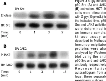
To investigate whether glycine-extended gastrin could reg-ulate p60-Src activation, serum-starved HCT116 cells were treated with G-gly for various times and lysed. Tyrosine kinase assays were performed in anti-p60-Src immunopcipitates using enolase as an exogenous substrate. Our re-sults indicated that p60-src was very rapidly and transiently
activated in HCT116 cells in response to G-gly. The activa-tion of p60-Src was detected 1 min after peptide addiactiva-tion. The stimulation was maximal at 3 min and decreased after 5 min (Figure 2A).
To test whether JAK2 was activated by G-gly, lysates from cells stimulated for various times were immunopre-cipitated with anti-JAK2 antibodies and in vitro kinase
as-says were performed on immunoprecipitates as described in “Methods”. We observed a rapid and transient increase in JAK2 autophosphorylation in response to G-gly stimu-lation. The maximal activation obtained within 5 min after peptide addition decreased toward the basal level at 15 min (Figure 2B).
Involvement of JAK2, Src and phosphatidylinositol 3-ki-nase pathways in glycine-extended gastrin-induced prolif-eration
In order to identify the role of PI3K, Src and JAK2 path-ways in the proliferation of human colonic tumour cells induced by G-gly, we measured HCT116 proliferation in the presence or absence of specific inhibitors for each pathway, LY290042, PP2 and AG490 respectively. Stimula-tion of HCT116 cells for 48h by G-gly induced a signifi-cant increase of cell proliferation (mean ± SE; 1.38 ± 0.07;
P<0.05; n = 5) (Figure 3). Treatment of the cells with PP2,
AG 490 or LY 290042 completely inhibited G-gly-induced HCT116 proliferation while inhibitors alone did not af-fect basal cell proliferation (data not shown). The result indicated that these three pathways could mediate G-gly proliferative effects not only on normal colonic epithelial cells but also on human colonic tumoral cells.
Involvement of both p60-Src and JAK2 upstream of PI3K activation in response to G-gly
In order to determine whether p60-Src was involved in PI3K activation by G-gly, we first tested whether PI3K activity was detected in association with p60-Src. After peptide treatment for various times, PI3K assays were car-ried out on anti-p60-Src immunoprecipitates. In response to G-gly, we observed an increased PI-kinase activity co-precipitated with maximal p60-Src after 5 min of peptide stimulation (Figure 4A). To test the PI3K specificity, we
IP: anti-p85 Time: 0 1 3 5 (min) PIP IP: anti-p85 PIP G-gly: - + - + DFO A B Figure 1 G-gly-induced PI3K (A) and G-gly (B) activation. HCT116 cells were stimulated with G-gly (10 pmol/L) for the indicated time. PI3K activity was determined in an immune complex kinase assay as described in Methods. The specificity of G-gly activation was accessed by pretreating the cells with ferric chelator DFO (1µmol/L). Representative autoradiograms from at least three separate experiments are shown. IP: Src Enolase IB: Src Time: 0 1 3 5 (min) IP: JAK2 P-JAK2 IB: JAK2 Time: 0 1 3 5 15 (min) A B Figure 2 G-gly-induced p60-Src (A) and JAK2 (B) activation. HCT116 cells were stimulated with G-gly (10 pmol/L) for the indicated time. p60-Src and JAK2 activities were determined in an immune complex k i n a s e a s s a y a s described in Methods. Immunoprecipitated proteins were also analysed by Western-blot using the anti-p60-Src and anti-JAK2 antibody respectively. R e p r e s e n t a t i v e autoradiogram from at least three separate experiments is shown. 150 140 130 120 110 100 90 80 G-gly - + + + + Inhibitor - - PP2 AG490 LY 290042 Cel l pr oli fer ation (%/contr ol)
Figure 3 Role of PI3K, Src or JAK2 pathways in human colonic tumour cell proliferation induced by G-gly. Serum-starved HCT116 cells were treated with G-gly for 48h in the presence or absence of the indicated inhibitor (10 µmol/L). The proliferation was determined as described in Methods. Each point represents data from triplicate and is representative of 5 independent experiments. Data are presented as means ± SE.
added the PI3K inhibitor, LY 294002, in the in vitro
li-pid kinase assay. Under these conditions, G-gly induced-PI-kinase activity was completely blocked. These results demonstrated that glycine-extended gastrin was associated with p60-Src and PI3K. In order to confirm that an Src family member could act upstream of the PI3K pathway activated by G-gly, we pretreated the cells for 1h with PP2 before G-gly stimulation. PI3K activation was inhibited in PP2 pretreated cells, indicating the role of Src in PI3K activation (Figure 4B).
We then examined the role of JAK2 in PI3K activation by G-gly. After peptide treatment for the time indicated, the cells were lysed and PI3K assays were performed on anti-JAK2 immunoprecipitates. Increased PI-kinase activity was associated with JAK2, detectable at 1 min and
maxi-mal after 5 min of peptide stimulation (Figure 5A). The inhibition of PI-kinase activity observed when LY 294002 was added in the in vitro lipid kinase assay indicated that
glycine-extended gastrin was associated with JAK2 and PI3K. To confirm the involvement of JAK2 upstream of the G-gly-induced PI3K pathway, the cells were pretreated for 1h with AG 490 prior to G-gly addition. The PI3K activation in response to G-gly was inhibited in the pres-ence of JAK2 inhibitor, indicating the role of this tyrosine kinase upstream in PI3K activation (Figure 5B).
No cross-talk between p60-Src and JAK2 pathways in response to G-gly
To investigate the possible association between p60-Src and JAK2, solubilized proteins were immunoprecipitated with an anti-JAK2 antibody and the precipitates were analyzed by immunoblot using an anti-p60-Src antibody. No p60-Src protein associated with JAK2 was detected in control cells or in cells stimulated with G-gly, whereas comparable amounts of JAK2 proteins were detected in immunoprecipitates (data not shown).
Independently of any association, we studied whether Src-family kinases were involved in JAK2 activation by G-gly. We tested the effect of PP2 on JAK2 activity. Cells were pretreated for 1h with PP2 5 min before stimulation with G-gly. JAK2 kinase assay was performed on JAK2 im-munoprecipitates. PP2 did not inhibit G-gly-induced JAK2 activation, indicating that Src kinases were not involved in upstream of JAK2 activation by G-gly (Figure 6A). Similarly, using AG490, we tested a possible role of JAK2 upstream in p60-Src activation. The presence of AG490in the incubation medium did not significantly change p60-Src activation by G-gly (Figure6B). Indeed, in this human colon tumoral cell line, activation of the two tyrosine-ki-nases, p60-Src and JAK2, in response to G-gly was entirely independent.
DISCUSSION
The p85/p110 phosphatidylinositol 3-kinase (PI3K) is a
PIP Time: 0 1 5 15 5 (min) LY 294002 IP: Src PIP Time: 0 3 5 15 5 (min) LY 294002 IP: JAK2 PIP IP: p85 G-gly - + - + PP2 PIP G-gly - + - + AG490 IP: p85 A B A B
Figure 4 Involvement of p60-Src (A) and PI3K (B) activation in response to G-gly. HCT116 cells pretreated with or without 10 µmol/L of LY290042 and anti-p85 antibody were stimulated (+) or not (-) with glycine-extended gastrin (10 pmol/L) for the indicated time. After immunoprecipitation of cell lysates with anti-p60-Src and anti-p85 antibody, precipitates were assayed for PIK activity using inositol phosphate as an exogenous substrate. Lipids were separated by thin layer chromatography and autoradiographied. The result shown is representative from five and three separate experiments.
Figure 5 Involvement of PI3K (A) and JAK2 (B) activation in response to G-gly. HCT116 cells pretreated with or without 10µmol/L of LY290042 and were 10µmol/L of AG 40, stimulated (+) or not (-) with glycine-extended gastrin G-gly (10 pmol/L) for the indicated time. After immunoprecipitation of cell lysates with anti-JAK2 and anti-p85 antibody, precipitates were assayed for PIK activity using inositol phosphate as an exogenous substrate. Lipids were separated by thin layer chromatography and autoradiographied. The result shown is representative from five separate experiments.
P-JAK2 G-gly - + - + PP2 IP: JAK2 IB: JAK2 Enolase IB: Src G-gly - + - + AG490 IP: Src A B
Figure 6 No involvement of p60-Src (A) and JAK2 (B) activation in response to G-gly. Cells pretreated with or without PP2 and AG 490 (10 µmol/L), were stimulated (+) or not (-) by G-gly (10 pmol/L) for 5 min. JAK2 and p60-Src kinase activities were determined by immune complex kinase assay using anti-JAK2 and anti-p60-Src antibodies as described in Materials and Methods. Proteins were separated by SDS-PAGE and JAK2 autophosphorylation was detected by autoradiography. A representative autoradiogram from at least three separate experiments is shown.
lipid kinase which plays an important role in human colon cancer. Its specific inhibitor, LY 294002, has been shown to inhibit cell growth and to induce apoptosis. Treatment of human colon cancer cell lines in vitro with LY 294002 or
transplanted in vivo abolishes tumour cell growth and leads
to apoptosis[9]. In addition, Philp et al[18] have identified
somatic mutations in the gene of the p85 subunit leading to a constitutive active form of PI3K. These mutations are present in primary human colon tumours and cancer cells, suggesting that PI3K is involved in human colonic tumori-genesis
Non-amidated gastrin precursors, including G-gly, are produced by colorectal cancers and exert growth factor ef-fects on these tissues[19, 20]. They are expressed in 80% - 90%
of colorectal tumours and polyps in human beings[21-24].
Identification of signalling pathways involved in colonic mucosa hyperproliferation is important for the understand-ing of tumour processes. In a previous study we have iden-tified that Src, JAK2 and PI3K pathways are overactivated in the colonic epithelium of mice overexpressing G-gly and involved in G-gly-induced proliferation of normal colonic epithelial cells isolated from control mice[13].
In the current study, we used the human colon tumour cell line, HCT116, to investigate whether these three ki-nases interact to regulate the growth of human tumoral colon cells. The results indicate that both p60-Src and JAK2 are necessary to PI3K regulation by the peptide. Theses results are in accordance with previous reports showing that JAK2 are involved upstream of the PI3K pathway of other cellular models. Pretreatment of human neutrophils with the JAK2 inhibitor, AG 490 could abol-ish the stimulation of the p85/p110 PI3K in response of the granulocyte-macrophage colony-stimulating factor. In addition, Attoub et al[25] have shown that leptin can induce
a transient elevation of the PI3K lipid products in JAK2 immunoprecipitates prepared from MDCK cells. Similarly, Src family kinases are involved upstream of the PI3K/ AKT pathway[26, 27]. Previous studies suggest that the IRS
proteins could serve as scaffolding intermediates between tyrosine kinases and PI3K[28]. This mechanism is likely
involved in PI3K activation by G-gly. In HCT116 cells we observed a basal association between JAK2 and IRS1 and the tyrosine phosphorylation of IRS-1 in response to G-gly (data not shown). In addition, we have previously re-ported that tyrosine phosphorylation of IRS-1 in pancre-atic tumour cells, leading to the rapid recruitment of the regulatory subunit of PI3K, might represent a common mechanism for PI3K activation by different factors includ-ing insulin and G-gly[11]. Obviously, we cannot exclude
other hypotheses such as interaction between the SH2 domain of p85 and phospho-tyrosine of p60-Src or JAK2. Different studies have previously described the involve-ment of Src family kinases upstream of JAK2 activation. For example, cell transformation by the oncogene v-Src[29]
or the overexpression of certain members of the Src family-kinases as Lck[30] can lead to a constitutive activation
of JAK2. However, we did not find any cross-talk between the two tyrosine kinases, suggesting that the p60-Src/PI3K and JAK2/PI3K pathways act independently. The involve-ment of two different mechanisms upstream of the lipid kinase might be a way to amplify the PI3K signal.
Howev-er, in our study, the level of PI3K activity associated with JAK2 or p60-Src was not different from that observed in anti-p85 immunoprecipitates. As for many other signalling molecules, it seems that the cellular localization of PI3K is important for the function of the enzyme. Therefore the recruitment of PI3K to different cell compartments by the two independent tyrosine-kinases allows the enzyme to play a complementary role. The finding that p60-Src and JAK2 do not associate in response to G-gly stimulation, demonstrates that the p60-Src/PI3K and JAK2/PI3K pathways act independently and indicates that they might be involved in different cell compartments. The analysis of the cellular localization of phosphatidylinositol 3-kinase, might be crucial to understand the role of this signalling molecule in different biological effects of G-gly.
Our previous studies indicate that gastrin precursors contribute to the initiation phases of colon carcinogenesis by upregulating the signalling pathways involved in cellular proliferation and cell survival. The results of this study demonstrate that G-gly is also able to regulate proliferation of tumoral colonic cells and support the idea that gastrin precursors are not only involved in the initiation steps of the colon tumour process but also involved in the later stages of tumour progression after genetic alterations, by conferring a growth advantage to the cells via the
constitu-tive activation of numerous signalling pathways.
REFERENCES
1 Seva C, Dickinson CJ, Yamada T. Growth-promoting effects of glycine-extended progastrin. Science 1994; 265: 410-412 2 Hollande F, Imdahl A, Mantamadiotis T, Ciccotosto GD,
Shulkes A, Baldwin GS. Glycine-extended gastrin acts as an autocrine growth factor in a nontransformed colon cell line.
Gastroenterology 1997; 113: 1576-1588
3 Singh P, Owlia A, Espeijo R, Dai B. Novel gastrin receptors mediate mitogenic effects of gastrin and processing intermedi-ates of gastrin on Swiss 3T3 fibroblasts. Absence of detectable cholecystokinin (CCK)-A and CCK-B receptors. J Biol Chem 1995; 270: 8429-8438
4 Iwase K, Evers BM, Hellmich MR, Guo YS, Higashide S, Kim HJ, Townsend CM Jr. Regulation of growth of human gastric cancer by gastrin and glycine-extended progastrin.
Gastroen-terology 1997; 113: 782-790
5 Stepan VM, Sawada M, Todisco A, Dickinson CJ. Glycine-extended gastrin exerts growth-promoting effects on human colon cancer cells. Mol Med 1999; 5: 147-159
6 Koh TJ, Dockray GJ, Varro A, Cahill RJ, Dangler CA, Fox JG, Wang TC. Overexpression of glycine-extended gastrin in transgenic mice results in increased colonic proliferation. J Clin
Invest 1999; 103: 1119-1126
7 Aly A, Shulkes A, Baldwin GS. Short term infusion of glycine-extended gastrin(17) stimulates both proliferation and forma-tion of aberrant crypt foci in rat colonic mucosa. Int J Cancer 2001; 94: 307-313
8 Duronio V, Scheid MP, Ettinger S. Downstream signalling events regulated by phosphatidylinositol 3-kinase activity. Cell
Signal 1998; 10: 233-239
9 Semba S, Itoh N, Ito M, Harada M, Yamakawa M. The in vitro and in vivo effects of 2-(4-morpholinyl)-8-phenyl-chromone (LY294002), a specific inhibitor of phosphatidylinositol 3’ -kinase, in human colon cancer cells. Clin Cancer Res 2002; 8: 1957-1963
10 Khaleghpour K, Li Y, Banville D, Yu Z, Shen SH. Involvement of the PI 3-kinase signaling pathway in progression of colon adenocarcinoma. Carcinogenesis 2004; 25: 241-248
11 Kowalski-Chauvel A, Pradayrol L, Vaysse N, Seva C. Tyro-sine phosphorylation of insulin receptor substrate-1 and
acti-vation of the PI-3-kinase pathway by glycine-extended gastrin precursors. Biochem Biophys Res Commun 1997; 236: 687-692 12 Hollande F, Choquet A, Blanc EM, Lee DJ, Bali JP, Baldwin
GS. Involvement of phosphatidylinositol 3-kinase and mi-togen-activated protein kinases in glycine-extended gastrin-induced dissociation and migration of gastric epithelial cells. J
Biol Chem 2001; 276: 40402-40410
13 Ferrand A, Bertrand C, Portolan G, Cui G, Carlson J, Praday-rol L, Fourmy D, Dufresne M, Wang TC, Seva C. Signaling pathways associated with colonic mucosa hyperproliferation in mice overexpressing gastrin precursors. Cancer Res 2005; 65: 2770-2777
14 Ferrand A, Kowalski-Chauvel A, Bertrand C, Pradayrol L, Fourmy D, Dufresne M, Seva C. Involvement of JAK2 up-stream of the PI 3-kinase in cell-cell adhesion regulation by gastrin. Exp Cell Res 2004; 301: 128-138
15 Daulhac L, Kowalski-Chauvel A, Pradayrol L, Vaysse N, Seva C. Src-family tyrosine kinases in activation of ERK-1 and p85/ p110-phosphatidylinositol 3-kinase by G/CCKB receptors. J
Biol Chem 1999; 274: 20657-20663
16 Kowalski-Chauvel A, Pradayrol L, Vaysse N, Seva C. Gastrin stimulates tyrosine phosphorylation of insulin receptor sub-strate 1 and its association with Grb2 and the phosphatidyli-nositol 3-kinase. J Biol Chem 1996; 271: 26356-26361
17 Pannequin J, Barnham KJ, Hollande F, Shulkes A, Norton RS, Baldwin GS. Ferric ions are essential for the biological activity of the hormone glycine-extended gastrin. J Biol Chem 2002; 277: 48602-48609
18 Philp AJ, Campbell IG, Leet C, Vincan E, Rockman SP, White-head RH, Thomas RJ, Phillips WA. The phosphatidylinositol 3’-kinase p85alpha gene is an oncogene in human ovarian and colon tumors. Cancer Res 2001; 61: 7426-7429
19 Aly A, Shulkes A, Baldwin GS. Gastrins, cholecystokinins and gastrointestinal cancer. Biochim Biophys Acta 2004; 1704: 1-10 20 Ferrand A, Wang TC. Gastrin and cancer: a review. Cancer Lett
2006; 238: 15-29
21 Ciccotosto GD, McLeish A, Hardy KJ, Shulkes A. Expression, processing, and secretion of gastrin in patients with colorectal carcinoma. Gastroenterology 1995; 109: 1142-1153
22 Nemeth J, Taylor B, Pauwels S, Varro A, Dockray GJ. Identifi-cation of progastrin derived peptides in colorectal carcinoma extracts. Gut 1993; 34: 90-95
23 Kochman ML, DelValle J, Dickinson CJ, Boland CR. Post-translational processing of gastrin in neoplastic human colonic tissues. Biochem Biophys Res Commun 1992; 189: 1165-1169 24 Smith AM, Watson SA. Gastrin and gastrin receptor
activa-tion: an early event in the adenoma-carcinoma sequence. Gut 2000; 47: 820-824
25 Attoub S, Noe V, Pirola L, Bruyneel E, Chastre E, Mareel M, Wymann MP, Gespach C. Leptin promotes invasiveness of kidney and colonic epithelial cells via phosphoinositide 3-ki-nase-, rho-, and rac-dependent signaling pathways. FASEB J 2000; 14: 2329-2338
26 Frame MC. Src in cancer: deregulation and consequences for cell behaviour. Biochim Biophys Acta 2002; 1602: 114-130 27 Penuel E, Martin GS. Transformation by v-Src: Ras-MAPK
and PI3K-mTOR mediate parallel pathways. Mol Biol Cell 1999; 10: 1693-1703
28 Yamauchi T, Kaburagi Y, Ueki K, Tsuji Y, Stark GR, Kerr IM, Tsushima T, Akanuma Y, Komuro I, Tobe K, Yazaki Y, Kad-owaki T. Growth hormone and prolactin stimulate tyrosine phosphorylation of insulin receptor substrate-1, -2, and -3, their association with p85 phosphatidylinositol 3-kinase (PI3-kinase), and concomitantly PI3-kinase activation via JAK2 kinase. J Biol Chem 1998; 273: 15719-15726
29 Murakami Y, Nakano S, Niho Y, Hamasaki N, Izuhara K. Con-stitutive activation of Jak-2 and Tyk-2 in a v-Src-transformed human gallbladder adenocarcinoma cell line. J Cell Physiol 1998; 175: 220-228
30 Yu CL, Jove R, Burakoff SJ. Constitutive activation of the Janus kinase-STAT pathway in T lymphoma overexpressing the Lck protein tyrosine kinase. J Immunol 1997; 159: 5206-5210
S- Editor Wang J L- Editor Wang XL E- Editor Liu WF 1864 ISSN 1007-9327 CN 14-1219/ R World J Gastroenterol March 28, 2006 Volume 12 Number 12