HAL Id: inserm-00784860
https://www.hal.inserm.fr/inserm-00784860
Submitted on 10 Jun 2013
HAL is a multi-disciplinary open access
archive for the deposit and dissemination of
sci-entific research documents, whether they are
pub-lished or not. The documents may come from
teaching and research institutions in France or
abroad, or from public or private research centers.
L’archive ouverte pluridisciplinaire HAL, est
destinée au dépôt et à la diffusion de documents
scientifiques de niveau recherche, publiés ou non,
émanant des établissements d’enseignement et de
recherche français ou étrangers, des laboratoires
publics ou privés.
An a contrario approach for the detection of activated
brain areas in fMRI
Camille Maumet, Pierre Maurel, Jean-Christophe Ferré, Christian Barillot
To cite this version:
Camille Maumet, Pierre Maurel, Jean-Christophe Ferré, Christian Barillot. An a contrario approach
for the detection of activated brain areas in fMRI. International Society for Magnetic Resonance in
Medicine 21st Annual Meeting & Exhibition (ISMRM), Apr 2013, Salt Lake City, United States.
pp.3260. �inserm-00784860�
1250
An a contrario approach for the detection of activated brain areas in fMRI Camille MAUMET1, Pierre Maurel1, Jean-Christophe Ferré1,2, and Christian Barillot1
1Visages Project-team / U746, University of Rennes 1, INSERM, CNRS, Inria, RENNES, Brittany, France, 2Department of Neuroradiology, CHU Rennes, Rennes,
Brittany, France
Target audience: Medical doctors, computer scientists.
Purpose: BOLD functional MRI (fMRI) is now a widespread imaging technique to study task-related activity in the brain. However, getting the areas of activation at the individual subject level is still an open issue. The standard massively univariate statistical analysis is usually performed after smoothing the data and makes use of a single p-value for final thresholding of the results [1]. In group fMRI studies, the need for compensation of cross-subjects misregistrations clearly justifies the smoothing. However, at the individual level, where neat delineations of the activated areas are of interest, the use of gaussian smoothing as a pre-processing step is more questionable. In this paper, we propose to study the ability of an a contrario approach, recently adapted for basal perfusion abnormalities detection [2], to correctly detect areas of functional activity.
Methods: In the a contrario approach [2], pre-smoothing of the data is avoided by taking into account the spatial neighborhood into the statistical analysis. Starting from a voxel-wise probability map (obtained from a standard massively univariate general linear model (GLM)), a region-based probability map is built. To this aim, a grid of shapes is defined in the image of interest. In this study, we work with grids of spheres as we have no a priori shape for the detections. The initial voxel-wise probability map is thresholded using a set of pre-defined p-values P={0.01, 0.005, 0.001} and, then, the number of overthreshold voxels is counted for each pre-defined p-value in each region (sphere). In the general a contrario formulation, the region-based probability is then estimated, assuming spatial independence of the residuals, using a binomial distribution parametrized by the number of voxels in each sphere and the pre-defined p-values. To account for potential mismodeling, the null distribution used to define the voxel-wise probabilities was estimated with a non-parametric approach in the background of the image.
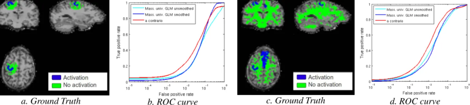
Results: 12 healthy subjects were involved in this study and underwent 3 BOLD fMRI sessions of a block motor paradigm [3]. In order to compare the performances of the standard massively univariate GLM with the proposed a contrario approach, we used Receiver-Operating-Characteristics (ROC) curves and estimated the area under the curve for false positive rates ranging from 0 to 0.1 (as only a small percentage of the voxels are expected to be active). Validation of fMRI analysis is still challenging due to the lack of ground truth. We focused on a well-studied right-hand motor paradigm in which the main activations are expected in the precentral and postcentral sulci (Left and Right) and in the Supplementary Motor Area (SMA) (Left and Right) [3]. We studied two different “ground truths”. First, as suggested in [3], we targeted the activation of the grey matter in the right hand motor area as manually delineated by an expert neuro-radiologist, while the surrounding white matter was expected to be inactive. Second, we also tested a global criterion, in which activation in both the hand motor area and part of the SMA (as defined by the AAL atlas) were activation targets, while whole-brain white matter (>70%) was expected to be inactive. We compared the standard massively
univariate GLM (no smoothing and smoothing with a gaussian FWHM kernel of 6mm3) with the a contrario approach.
Fig. 1 displays the expected active and inactive regions for a representative subject along with the associated ROC curves averaged over sessions and subjects. Overall the a contrario approach outperformed both the unsmoothed and smoothed massively univariate GLM in term of area under the curve. Two sample t-tests were performed in order to detect significant improvement at the group level (false discovery rate q < 0.05). The a contrario approach with local ground truth was significantly better than the smoothed GLM for all sessions (p = 0.018, p = 0.038, p = 0.016) and than the unsmoothed GLM for 2 sessions out of 3 (p =0.003, p = 0.005). Moreover, using the global ground truth, the a contrario approach performed significantly better than the smoothed GLM for all sessions (p = 0.003, p = 0.009, p = 0.006) and than the unsmoothed GLM for 2 sessions out of 3 (p = 0.002, p = 0.002)
a. Ground Truth b. ROC curve c. Ground Truth d. ROC curve
Figure 1 : Comparison of single-subject detections of activations between the massively univariate GLM and the a contrario approach. Local: a) Ground Truth for a representative subject, b) ROC curve; Global: c) Ground Truth, d) ROC curve.
Discussion: We proposed a new a contrario approach to detect fMRI activations. This method displayed better spatially defined activations with a more interesting trade-off between sensitivity and specificity by comparison to the standard massively univariate GLM.