HAL Id: hal-02628867
https://hal.inrae.fr/hal-02628867
Submitted on 27 May 2020
HAL is a multi-disciplinary open access
archive for the deposit and dissemination of
sci-entific research documents, whether they are
pub-lished or not. The documents may come from
teaching and research institutions in France or
abroad, or from public or private research centers.
L’archive ouverte pluridisciplinaire HAL, est
destinée au dépôt et à la diffusion de documents
scientifiques de niveau recherche, publiés ou non,
émanant des établissements d’enseignement et de
recherche français ou étrangers, des laboratoires
publics ou privés.
Copyright
E.O. da Silva, Ana-Paula Loureiro-Bracarense, Isabelle P. Oswald
To cite this version:
E.O. da Silva, Ana-Paula Loureiro-Bracarense, Isabelle P. Oswald. Mycotoxins and oxidative stress:
where are we?. World Mycotoxin Journal, 2018, 11 (1), pp.113-133. �10.3920/WMJ2017.2267�.
�hal-02628867�
1. Introduction
Mycotoxins are secondary fungal metabolites often
found as contaminants in agricultural commodities all
over the world and pose a risk for human and animal
health (Bennett and Klich, 2003; Wu et al., 2014a). More
than 400 different mycotoxins have been isolated and
chemically characterised. Those of major medical and
agricultural concern are aflatoxins, fumonisins, ochratoxins,
trichothecenes, zearalenone (ZEA) and patulin (PAT) (Wu
et al., 2014a).
The molecular mechanisms behind the toxic effects of
the major mycotoxins are established and oxidative stress
and the generation of free radicals have been shown to be
implicated in mycotoxin toxicity (Adhikari et al., 2017;
Wang et al., 2016). Indeed, the imbalance between free
radicals and the antioxidant defence systems can cause
chemical damage to DNA, proteins and lipids, as observed
upon exposure to mycotoxins (Assi, 2017).
As human and animal exposure to mycotoxins is
unavoidable, effective ways to mitigate their harmful
impacts are required. Several studies have demonstrated the
beneficial effects of antioxidant substances in the prevention
and treatment of various diseases (Li et al., 2015). In this
context, the use of natural antioxidants has been shown
to mitigate and/or prevent the toxic effects of mycotoxins
(Sorrenti et al., 2013).
The aims of this review are first to describe the cellular
mechanisms involved in the physiological control and
imbalance of free radical generation; second to summarise
the toxic effects of the major mycotoxins associated with
oxidative stress; and third, to present the main natural
antioxidants used to mitigate the toxic effects of these
mycotoxins.
Mycotoxins and oxidative stress: where are we?
E.O. da Silva
1, A.P.F.L. Bracarense
1*and I.P. Oswald
2*1
Universidade Estadual de Londrina, Laboratory of Animal Pathology, Campus Universitário, Rodovia Celso Garcia Cid,
Km 380, Londrina, Paraná 86051-990, Brazil;
2Université de Toulouse, Toxalim, Research Center in Food Toxicology,
INRA, UMR 1331 ENVT, INP-PURPAN, 31076 Toulouse, France;
[email protected]
;
[email protected]
Received: 2 November 2017 / Accepted: 11 December 2017
© 2018 Wageningen Academic Publishers
REVIEW ARTICLE
Abstract
Mycotoxins are the most common contaminants of food and feed worldwide and are considered an important risk
factor for human and animal health. Oxidative stress occurs in cells when the concentration of reactive oxygen
species exceeds the cell’s antioxidant capacity. Oxidative stress causes DNA damage, enhances lipid peroxidation,
protein damage and cell death. This review addresses the toxicity of the major mycotoxins, especially aflatoxin B
1,
deoxynivalenol, nivalenol, T-2 toxin, fumonisin B
1, ochratoxin, patulin and zearalenone, in relation to oxidative
stress. It summarises the data associated with oxidative stress as a plausible mechanism for mycotoxin-induced
toxicity. Given the contamination caused by mycotoxins worldwide, the protective effects of a variety of natural
compounds due to their antioxidant capacities have been evaluated. We review data on the ability of vitamins,
flavonoids, crocin, curcumin, green tea, lycopene, phytic acid, L-carnitine, melatonin, minerals and mixtures of
anti-oxidants to mitigate the toxic effect of mycotoxins associated with oxidative stress.
Keywords: antioxidants, mycotoxins, oxidative stress.
OPEN ACCESS
2. Oxidative stress: physiological control and
damage caused by overproduction of free
radicals
One consequence of aerobic conditions is activation of
oxidative mechanisms and the subsequent generation of
reactive oxygen species (ROS) (Droge, 2002). Cells have
developed primary and secondary enzymatic systems to
avoid ROS-induced damage (Valko et al., 2007). Superoxide
dismutase (SOD), catalase (CAT), glutathione reductase
(GR) and glutathione peroxidase (GPx) are characterised as
primary antioxidant enzymes that trigger the breakdown of
free radicals or combine toxic compounds with glutathione
(GSH). The mechanisms of action of these enzymes are
diverse. SOD breaks down the superoxide anion radical
into H
2O and O
2, whereas CAT catalyses the decomposition
of hydrogen peroxide (H
2O
2) into water and oxygen. GPx
reduces hydrogen peroxide to water and GR can regenerate
GSH (Droge, 2002). By contrast, glutathione S-transferase
(GST), a secondary detoxification enzyme, acts by binding
ROS to GSH (Hayes and Strange, 1995) or by detoxifying
lipid peroxides (Pickett and Lu, 1989).
Other mechanisms, including cysteine and GSH, are also
involved in physiological control of ROS generation (Droge,
2002). GSH interacts with multiple antioxidant enzymes,
modulating the action of GR, GPx and GST (a decrease in
GSH content reduces enzymatic activity).
A control mechanism also exists for the expression of
enzymes with antioxidant activity. Control is regulated by
antioxidant response elements (AREs) that are activated by
nuclear factor erythroid 2-related factor 2 (Nrf2) (Jin et al.,
2014). Nrf2-ARE is considered to be an important signalling
pathway associated with antioxidant activity. Cells subjected
to oxidative stress induce Nrf2 translocation to the nucleus,
thereby activating genes encoding antioxidant enzymes and
detoxifying enzymes of phase II (e.g. SOD) through ARE
binding. Interestingly, although high antioxidant induction
is associated with Nrf2 when this pathway is activated by
ROS, the response is limited because ROS also activates
a cell death-signalling pathway (Jin et al., 2014; Valko et
al., 2007).
Organelles such as peroxisomes and mitochondria provide
membrane-limited compartments specialised in redox
activities. Consumption of oxygen leads to the production of
H
2O
2, which oxidises some molecules. Furthermore, these
organelles contribute to metabolic functions as they contain
CAT, an enzyme that decomposes the H
2O
2and prevents
intracellular accumulation of this compound (Valko et al.,
2007). Cellular respiration in mitochondria creates one of
the main superoxide production sites. During this process,
ATP is produced by the electron transport chain and during
energy transduction, free radical superoxide is formed,
which has been associated with the cell pathophysiology
in several diseases (Droge, 2002; Valko et al., 2007).
Generation of oxidative stress, free radicals, and damage
to DNA, proteins and lipids
Cells in homeostasis may produce free radicals as a result of
physiological reactions (cellular respiration, for example).
A variety of exogenous factors can promote oxidative
stress and overproduction of free radicals (Young and
Woodside, 2001). Oxidative stress occurs in cells when
the production of ROS, such as such as hydroxyl radical
(HO), perhydroxyl radical (HOO
-), superoxide anion (O
2-
)
and reactive nitrogen species (RNS) including nitric oxide
(NO), exceeds the antioxidant capacity of a cell (Valko et al.,
2007). Changes in intracellular antioxidant systems or in
the production of free radicals can result in oxidative stress
(Halliwell and Whiteman, 2004). Increased ROS production
alters and/or activates several intracellular mechanisms that
promote oxidative damage to DNA, proteins and membrane
lipids. Lipid peroxidation may also lead to cell death. The
mechanisms involved in the induction of cell apoptosis
caused by the generation of ROS include activation of p53,
mitogen-activated protein kinases (MAPKs), caspases and
changes in the Bcl-2/Bax expression (Farley et al., 2006).
Cells in homeostasis are maintained in a redox state through
the association of the iron and copper redox couple.
However, in a situation of oxidative stress, when superoxide
is overproduced, the ‘free iron’ (Fe
2+) is released into the
cytoplasm. This release considerably increases the oxidative
stress, and leads to the generation of other reactive radicals
through the Fenton reaction. In this reaction, Fe
2+and H
2
O
2generate one of the most harmful radicals, the reactive
hydroxyl (Fe
2++ H
2
O
2→ Fe
3++ •OH
+OH
−). Transition
metal ions, mainly iron, have been implicated in the
generation of highly reactive radicals leading to DNA and
membrane damage. Cellular and organelle membranes are
attractive targets for oxidation due to the polyunsaturated
fatty acid residues of phospholipids (Birben et al., 2012).
Secondary ROS metabolites can be produced, including
endoperoxides (cyclisation reaction) and malondialdehyde
(MDA), the toxic final product of lipid peroxidation, which
is potentially mutagenic (Birben et al., 2012; Marnett, 1999).
The level of cytosolic calcium (Ca
2+) can be increased
by ROS generation through an influx of extracellular
Ca
2+or mobilisation of intracellular Ca
2+stores (Droge,
2002). This increase in the cytosolic Ca
2+level contributes
to the activation of protein kinase C alpha and to the
transcriptional induction of the activator protein 1
(AP-1), c-Fos and c-Jun (Maki et al., 1992). MAPK signalling
cascades are activated through a variety of membrane
receptors (receptor tyrosine kinases, protein tyrosine
kinases, receptors of cytokines and growth factors, and
heterotrimeric G protein-coupled receptors) and are
regulated by phosphorylation and dephosphorylation
on serine and/or threonine residues (Droge, 2002). The
association between oxidative stress and the generation
of free radicals can activate the MAPK pathway, mainly
c-Jun N-terminal kinase (JNK) and p38, resulting in cell
apoptosis (Allen and Tressini, 2000).
Another important free reactive radical produced
in biological systems is NO. NO is a normal cellular
metabolite with different functions in cells including
neurotransmission, maintaining vascular tone, defence,
smooth muscle relaxation and immune regulation
(Bergendi et al., 1999). Like with ROS, a nitrosative stress
occurs following overproduction of RNS and disruption
of the antioxidant system (Ridnour et al., 2004). The
mechanisms of cell damage induced by nitrosative stress
include changes in protein structure (through nitrosylation
reactions) leading to inhibition of their function (Valko et
al., 2007) and cell apoptosis. The induction of apoptosis
by NO is associated with a decrease in the concentration
of cardiolipin, an important component of the inner
mitochondrial membrane. This molecule contributes
to the optimal function of enzymatic systems involved
in mitochondrial energy metabolism. A decrease in the
cardiolipin level results in disruption of the electron
transport chain, changes in mitochondria permeability and
the release of cytochrome C into the cytosol (Droge, 2002).
Furthermore, free radicals, such as superoxide anion and
NO, are produced by phagocytic cells during the respiratory
burst occurring in the inflammatory process. Together,
these radicals can react to produce the peroxynitrite anion
(ONOO
−), a molecule that is a powerful oxidant and can
cause DNA fragmentation and lipid oxidation.
The generation of free radicals can increase the expression
of cyclooxygenase-2 (COX-2), and of arachidonic acid
metabolism, promote the upregulation of proinflammatory
cytokines, such as tumour necrosis factor (TNF),
interleukin(IL)-1, IL-6 and IL-8, thereby inducing a chronic
inflammatory response and the stimulation of more free
radicals (Reuter et al., 2010). Extensive data has shown that
oxidative stress contributes to the inflammatory process,
which, in turn, leads to overproduction of reactive radical
species thereby promoting a harmful feedback process that
increases cellular damage.
The primary function of the respiratory chain is to
use the energy produced to transfer electrons into the
mitochondrial intermembrane space. However, a small
percentage of electrons escape from the mitochondrial
space, producing superoxide (Birben et al., 2012). In normal
conditions, the production of superoxide is limited by
SOD, which transforms the anion into hydrogen peroxide
(Droge, 2002). Under oxidative stress, overproduction
of superoxide occurs via activation of nicotine adenine
dinucleotide phosphate (NADPH) and depletion of SOD
(Birben et al., 2012). Under oxidative stress, some organelles
including peroxisomes, mitochondria and endoplasmic
reticulum (ER) are affected by the overproduction of
free radicals, mainly associated with lipid peroxidation.
The peroxisome damage leads to CAT depletion and
intracellular accumulation of H
2O
2(Valko et al., 2007). The
mitochondria are an important target for injury induced
by oxidative stress caused via endogenous metabolic
processes and/or exogenous oxidative influences (Guo et
al., 2013). The mitochondrial damage to DNA caused by
oxidative stress can result in a decrease in proteins that are
important for electron transport, leading to the generation
of ROS and the dysregulation of organelles, which, in turn,
activate cell apoptotic mechanisms (Van Houten et al.,
2006). Furthermore, radicals such as NO
−and ONOO
−are
responsible for detrimental changes in the mitochondrial
respiratory chain (Sas et al., 2007). The structural changes
in mitochondrial proteins result in altered function in
which enzymatic systems of the electron-transport chain
(nicotinamide adenine dinucleotide dehydrogenase,
cytochrome-c-oxidase, and adenosine triphosphate
synthase) are the main targets of the free radicals (Van
Houten et al., 2006).
ROS can also alter mitochondrial phospholipids resulting in
lipid peroxidation, which, in turn, increases mitochondrial
membrane permeability. The mitochondrial permeability
transition pore (MPTP) can be induced by ROS generation
due the oxidation of thiol groups on the adenine nucleotide
translocator (part of the MPTP) (Valko et al., 2007).
ONOO
−can also affect mitochondrial homeostasis and
energy production by inactivating enzymatic systems and
promoting the release of mitochondrial Ca
2+(Douarre et
al., 2012). The intracellular elevation of the Ca
2+level also
changes mitochondrial membrane potential (MMP) and
induces the production of superoxide radicals, resulting in
a vicious cycle (Douarre et al., 2012) The mitochondrial
excess of Ca
2+contributes with the formation of MPTP, to
osmotic swelling and rupture of the outer mitochondrial
membrane (Douarre et al., 2012). These mitochondrial
changes caused by oxidative stress can lead to cell apoptosis
due to the release of cytochrome-c, changes in Bcl2/Bax
expression (down-regulation of the Bcl2 protein and an
increase in Bax expression), activation of MAPKs and
casp-3 (Anuradha et al., 2001; Farley et al., 2006).
The ER is an organelle that regulates protein synthesis, drug
detoxification, carbohydrate metabolism, lipid biosynthesis
and Ca
2+homeostasis. Oxidative stress and ROS generation
deregulate the ER functions and release Ca
2+into the
cytosol (Minasyan et al., 2017). The ER and mitochondria
interact physiologically and functionally at sites called
mitochondrial associated membranes. The damage to
ER caused by oxidative stress results in mitochondrial
dysfunction and cell apoptosis (Kim et al., 2008).
3. Response to mycotoxins and oxidative
stress: interaction in in vitro, in vivo and ex
vivo models
The mycotoxins aflatoxin B
1(AFB
1), deoxynivalenol (DON),
nivalenol (NIV), fumonisin B
1(FB
1), ochratoxin A (OTA),
PAT and ZEA are the main contaminants of food and feed
worldwide and have been extensively studied due their
toxic effects on the human and the animal health (Wu et
al., 2014a). Several aspects of the intracellular action of
mycotoxins have been elucidated: the induction of oxidative
stress and ROS generation have become one of the major
triggers of their lesional mechanisms, as observed in in vitro,
in vivo and ex vivo studies. The oxidative stress mechanisms
associated with AFB
1, DON, NIV, T-2 toxin (T-2), FB
1,
OTA, PAT and ZEA are summarised in Figure 1 and 2.
Aflatoxin B
1Aflatoxins are fungal metabolites mainly produced by
Aspergillus flavus and Aspergillus parasiticus (Saini and
Kaur, 2012). More than 10 forms of aflatoxins are known,
among which the main ones are AFB
1, and aflatoxins B
2(AFB
2), G
1(AFG
1), G
2(AFG
2), M
1(AFM
1) and M
2(AFM
2)
(Kumar et al., 2017). Aflatoxins are natural contaminants of
cereals (maize, rice, oats, barley and sorghum), groundnuts,
pistachio nuts, almonds, cottonseed and walnuts (Wu et
al., 2014a). Milk can be contaminated by AFM
1, which is a
principal hydroxylated-AFB
1metabolite biotransformed by
hepatic cytochrome P450 in cows fed an AFB
1contaminated
diet (Bennet and Klick, 2003).
The toxicity of AFB
1is mainly associated with the
binding of bioactivated AFB
1-8,9-epoxide to cellular
macromolecules, such as mitochondria, nuclear nucleic
acids and nucleoproteins, with cytotoxic effects (Bennet
and Klich, 2003).
Studies in vitro (Liu and Wang, 2016; Mary et al., 2012;
Wang et al., 2017) and in vivo (Abdel-Wahhab and Aly,
2003; Shi et al., 2015) demonstrated that oxidative stress
plays a major role in the toxic effects of AFB
1. The main
consequences of ROS generation induced by AFB
1are
damage to DNA (Wang et al., 2017; Zhang et al., 2015b)
and mitochondrial lesions (Liu and Wang, 2016) as
summarised in Figure 1. AFB
1uncouples mitochondrial
oxidative phosphorylation, reduces MMP and induces
mitochondrial permeability (Liu and Wang, 2016; Shi et
al., 2015). The mitochondrial alterations associated with
oxidative stress activate cytochrome C, modulate Bcl2/
Bax gene expression and activate caspase 9 and caspase 3
(Liu and Wang, 2016; Mary et al., 2017; Wang et al., 2017)
leading to cell apoptosis. Mary et al. (2017) also reported
that hepatocytes treated with AFB
1increase the expression
ROS Mitochondria Apoptosis Inflammatory response Lipid peroxidation GSH MDA DNA/protein synthesis Arachnoid acid (2) Cox-2 (5) Cytokines (1, 2, 5) Fenton reaction (3) iNOS (5) NO (1) NOX-2 (1) AFB1 (1) FB1 (2) OTA (3) PAT (4) ZEA (5) Nrf2 (1, 3) CAT (1, 3, 4, 5) GPx (1, 2, 3, 5) SOD (1-5) Ca2+ level (3) Hsp 25/70 (2) MAPKs (2, 4) p53 (1, 4, 5) Bcl-2/Bax (1-5) Cytochrome C (1, 3, 4) Cytocrome P450 (3, 4) Casp-3 (1-5)
Figure 1. Summary of the intracellular lesions associated with oxidative stress induced by the main mycotoxins that contaminate
food and feed. AFB
1= aflatoxin B
1; FB
1= fumonisin B
1; OTA = ochratoxin A; PAT = patulin; ZEA = zearalenone. The numbers
between brackets indicate the mycotoxins involved in each process.
of the p53 gene, which was associated with an increase in
cell apoptosis.
Recent studies have shown that AFB
1causes changes
in intracellular antioxidant mechanisms such as Nrf2,
SOD, GPx and CAT expression (Liu and Wang et al.,
2016; Wang et al., 2017), inhibiting antioxidant enzymes
and causing an increase in lipid peroxidation (LPO) and
a decrease in the level of GSH (Ma et al., 2015; Maurya
and Trigun, 2016). Moreover, ROS generation induced
by AFB
1modulates the inflammatory response through
up-regulation of pro-inflammatory cytokines TNF-α,
IL-1α, IL-1β and IL-6 and NO expression, by reducing
anti-inflammatory cytokine IL-4 expression, inducing
cytochrome P450 activity, increasing arachidonic acid
metabolism, and activating the NADPH oxidase
(NOX)-2 dependent signalling pathway, thereby promoting the
autophagy of pro-inflammatory macrophages M1 (An et
al., 2017; Ma et al., 2015; Meissonier et al., 2007).
Deoxynivalenol
DON is a type B trichothecene predominantly produced by
Fusarium graminearum and Fusarium culmorum (Bennet
and Klich, 2003). Exposure to DON has been associated
with alterations in the intestinal, immune, endocrine and
nervous systems in several animal species and in humans
(Maresca et al., 2013; Payros et al., 2016; Pestka, 2010a). At
a molecular level, DON causes ribotoxic stress, inducing
MAPK phosphorylation, promoting apoptosis, resulting in
changes in the inflammatory response and decreasing the
expression of cell adhesion proteins (Pierron et al., 2016;
Silva et al., 2014).
Studies in vitro (Li et al., 2014; Yang et al., 2014; Zbynovska
et al., 2013) and in vivo (Borutova et al., 2008; Osselaere et
al., 2013) established the toxic effects of DON associated
with oxidative stress and ROS generation as observed in the
Figure 2. DON alters the intracellular antioxidant defence
system in target tissues such as liver, kidney, lymphoid
organs, intestine and blood/serum as demonstrated by
an increase in MDA concentration (Li et al., 2014) and a
decrease in GSH, SOD, CAT and GPx levels (Hou et al.,
2013; Strasser et al., 2013; Zbynovska et al., 2013).
The oxidative stress signalling pathway induced by DON
has been suggested to be one of the mechanisms behind
DNA fragmentation, cell death and apoptosis (Frankic et al.,
2008; Zhang et al., 2009) as well as the inhibition of protein
synthesis and an increase in carbonyl content (Strasser
et al., 2013). Furthermore, alterations in the surface of
lysosomal membranes lead to lysosomal fragility, a decrease
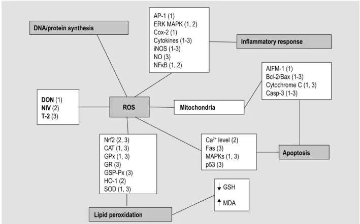
ROS Mitochondria Apoptosis Inflammatory response Lipid peroxidation GSH MDA DNA/protein synthesis AP-1 (1) ERK MAPK (1, 2) Cox-2 (1) Cytokines (1-3) iNOS (1-3) NO (3) NFкB (1, 2) DON (1) NIV (2) T-2 (3) AIFM-1 (1) Bcl-2/Bax (1-3) Cytochrome C (1, 3) Casp-3 (1-3) Ca2+ level (2) Fas (3) MAPKs (1, 3) p53 (3) Nrf2 (2, 3) CAT (1, 3) GPx (1, 3) GR (3) GSP-Px (3) HO-1 (2) SOD (1, 3)Figure 2. Summary of the intracellular lesions associated with oxidative stress induced by trichothecenes that contaminate
food and feed. DON = deoxynivalenol; NIV = nivalenol; T-2 = T-2-toxin. The numbers between brackets indicate the mycotoxins
involved in each process.
in the MMP, an increase in membrane permeability and
consequent deregulation of Bcl-2/Bax expression (leading
to release of cytochrome C and activation of caspase 3, 8,
9 and apoptosis inducing factor mitochondrion associated
1) have been associated with ROS generation induced by
DON (Kouadio et al., 2005; Li et al., 2014; Sun et al., 2015).
It has also been established that the ribotoxic stress induced
by DON can stimulate apoptosis via activation of the p38
MAPK (Pestka et al., 2008).
In addition, DON-induced oxidative stress can modulate
the inflammatory response through up-regulation of
pro-inflammatory cytokines including 1β, 2,
IL-6, IL-8, TNF-α, down-regulation of anti-inflammatory
IL-4 and IL-10, selective activation of ERK MAPK, NFκB
and AP-1, and increased and decreased expression of
intracellular proteins involved in innate immunity, such
as cyclooxygenase-2 (Cox-2) and inducible nitric oxide
synthase (iNOS), respectively (Cano et al., 2013; Graziani
et al., 2015; Pestka et al., 2010b).
Nivalenol
NIV is another type B trichothecene and is generally
a biologically active metabolite of DON, present in
agricultural commodities (Bennet and Klich, 2003). NIV
is not as prevalent as DON, but NIV showed higher acute
toxicity than DON (Alassane-Kpembi et al., 2015; Cheat
et al., 2015). Studies in vivo (Cheat et al., 2015), in vitro
(Alassane-Kpembi et al., 2015; Del Regno et al., 2015;
Marzocco et al., 2009) and ex vivo (Alassane-Kpembi et al.,
2017; Cheat et al., 2015) reported that NIV, such as DON,
induce inhibition of protein, DNA and RNA synthesis,
mitochondrial damage, cell apoptosis, decreases cellular
viability and modulate inflammatory response mainly due to
ROS generation associated with induction of oxidative stress
as demonstrated in Figure 2, affecting the gastrointestinal
tract and organs of the immune system.
The oxidative stress induced by NIV promotes ROS release
via the NADPH oxidase signalling pathway, decreases the
GSH level, alters Ca
2+homeostasis and activates nuclear
factor kappa beta (NF-κB) (Del Regno et al., 2015). This
ROS generation induces DNA and mitochondrial damage,
activation of extracellular regulated kinase (ERK) MAPK,
changes in Bcl-2 expression, up-regulation of Bax gene and
activation of caspase 3, thereby promoting cell apoptosis
(Marzocco et al., 2009). The oxidative stress induced by
NIV stimulates the antioxidant intracellular mechanisms
of defence through an increase in heme oxygenase-1
(HO-1) and activation of Nrf2 (Del Regno et al., 2015).
In addition, the NIV-induced oxidative stress modulates
the inflammatory response by activation of NF-κB,
up-regulation of pro-inflammatory cytokines such as IL-8,
IL-1α, IL-1β, IL-17A, IL-22, interferon (IFN)-α and an
increase in iNOS expression (Alassane-Kpembi et al., 2017;
Del Regno et al., 2015; Marzocco et al., 2009).
T-2 toxin
T-2 is a type A trichothecene produced by several Fusarium
species, mainly Fusarium sporotrichiodes, Fusarium poae
and Fusarium langsethiae. Studies have demonstrated
that T-2 affects the gastrointestinal tract, kidney, liver,
heart, skin, the nervous, immunological, and reproductive
systems, and embryogenic development in humans and
animals (Agrawal et al., 2012; Li et al., 2011; Meissonnier
et al., 2008).
The main molecular target of trichothecenes is the ribosomal
unit, affecting initiation of the polypeptide chain (Li et al.,
2011). Like other trichothecenes, T-2 binds and inactivates
peptidyl transferase activity resulting in inhibition of protein
synthesis and disruption of the mitochondrial morphology,
ER and other membranes (Adhikari et al., 2017). Studies in
vitro (Chen et al., 2008; Yang et al., 2016; Zhang et al., 2016)
and in vivo (Chaudhari and Lakshmana, 2010) provided
evidence that T-2-induced oxidative stress is associated
with an increase in ROS generation and DNA, protein and
lipid peroxidation leading to cell apoptosis.
The oxidative stress induced by T-2 promotes Fas
up-regulation, p53 activation, down-regulation of Bcl-2 and
up-regulation of the pro-apoptotic factor Bax causing
cytochrome C release, caspase 3 activation and cell
apoptosis (Chen et al., 2008; Zhang et al., 2018) (Figure
2). ROS generation causes a decrease in Nrf2 expression,
changes in the intracellular antioxidant enzymes GPx, GR,
SOD and CAT, promoting a decrease in GSH level and an
increase in MDA level (Wu et al., 2014b; Yang et al., 2016).
Another apoptosis signalling pathway linked to oxidative
stress induced by T-2 is through the activation of JNK1,
p38 MAPK, increase in heat shock protein (Hsp) 70
expression, increase in iNOS activity and NO release,
causing mitochondrial damage and activation of caspase
3 (Chaudhari and Lakshmana, 2010; Li and Pestka, 2008).
In addition, studies have shown that T-2 can modulate the
inflammatory response by increasing the expression of
pro-inflammatory cytokines such as TNF-α, IL-6, IL-1β
and IL-11 (Agrawal et al., 2012; Zhou et al., 2014).
Fumonisin B
1Fumonisins are a group of mycotoxins mainly produced by
Fusarium verticillioides and Fusarium proliferatum (Voss
et al., 2001). At least 15 related fumonisin compounds
have been identified so far, but FB
1is the most significant
fumonisin due to its toxicity and widespread occurrence
(Voss et al., 2007).
At cellular level, FB
1inhibits ceramide synthase, blocking
the synthesis of sphingolipids, a class of membrane lipids
that play an important role in cell signalling transduction
pathways and cell growth, differentiation, and death (Grenier
et al., 2012; Voss et al., 2007). Ceramide synthase inhibition
leads to reduced levels of ceramide and intracellular
accumulation of sphingolipids (So) and sphinganine (Sa).
These free sphingoid bases are pro-apoptotic, cytotoxic
growth inhibitors and are immunotoxic (Loiseau et al.,
2007; Voss et al., 2001, 2007).
Studies in vitro (Domijan et al., 2015; Mary et al., 2012)
and in vivo (Abbes et al., 2016; Hassan et al., 2015)
revealed the potential of FB
1to induce oxidative stress
with consequent ROS generation, cytotoxic effects and
apoptosis. The action of FB
1on ROS generation has
been considered a consequence rather than a mechanism
of its toxicity (Galvano et al., 2002; Wang et al., 2016).
However, some studies showed that FB
1was able to
increase the rate of oxidation, promote the production of
free radicals and accelerate the chain reactions associated
with lipid peroxidation in membranes (Hassan et al., 2015;
Stockmann-Juvala and Savolainen, 2008). These changes
were demonstrated in different animal models by alterations
in GPx and SOD expression, increase in MDA production
and decrease in the GSH level (Abbes et al., 2016; Domijan
et al., 2007; Poersch et al., 2014).
The increase in ROS production induced by FB
1has
also been associated with inhibition of DNA synthesis
and DNA fragmentation (Kouadio et al., 2005; Wang et
al., 2016), inhibition of protein synthesis (Domijan et al.,
2007), mitochondrial injury with consequent deregulation
of calcium homeostasis and caspase 3 activation, induction
of cytochrome P450 activity with an increase in arachidonic
acid metabolism and modulation of inflammatory response
(Abbes et al., 2016; Domijan and Abramov, 2011; Mary et al.,
2017). Some studies have demonstrated that perturbations
of the cellular redox state due the FB
1exposition can
activate MAPKs and Hsp 25/70. Both signalling pathways
can affect cell survival and are involved in the regulation of
apoptosis (Lalles et al., 2010; Rumora et al., 2007) (Figure 1).
Ochratoxin A
Ochratoxins are a group of mycotoxins produced by
filamentous fungal species such as Aspergillus and
Penicillium and occur in nature in three different isoforms:
ochratoxin A, B and C. OTA is the most pathogenic to
humans and animals, and is found in a wide range of foods
and feed, including cereals, meat, dried fruits, nuts, coffee,
wine and beer (Bennet and Klich, 2003; Limonciel and
Jennings, 2014; Malir et al., 2016).
Studies involving mammalian species in vitro (Bhat et
al., 2016; Gayathri et al., 2015; Lautert et al., 2014; Li et
al., 2015) and in vivo (Aydin et al., 2003; Tanaka et al.,
2016) showed nephrotoxic, hepatotoxic, immunotoxic,
enterotoxic, neurotoxic and teratogenic effects of OTA.
The toxicity and carcinogenic mechanisms of OTA have
been associated with induction of oxidative stress (Costa
et al., 2016), cell apoptosis (Ramyaa and Padma, 2013), cell
autophagy/mitophagy (Gan et al., 2017; Qian et al., 2017)
and protein synthesis inhibition (Mally and Dekant, 2009).
ROS generation has been reported to trigger OTA toxicity
(Zhu et al., 2017). Several oxidative stress mechanisms
elicited by OTA have been proposed through in vivo
(Abdel-Wahhab et al., 2017; Gan et al., 2017) and in vitro studies
(Bhat et al., 2016; Ramyaa et al., 2014) (Figure 1). OTA can
cause damage due to oxidative stress through the generation
of hydroxyl radicals via the Fenton reaction, via flavoprotein
NADPH-cytochrome P450 activation and inhibition of
Nrf2 activation and gene transcription. In addition, OTA
can decrease the expression of the intracellular antioxidant
enzymes GPx, CAT, SOD and GR (Abdel-Wahhab et al.,
2017; Bhat et al., 2016) as demonstrated by an increase in
MDA levels.
ROS generation increased by OTA promotes the
activation of the apoptosis signalling pathway through
the mitochondrial lipid peroxidation, promoting loss of
mitochondria membrane potential, increasing membrane
permeability (Bhat et al., 2016), activating JNK MAPKs
(Zhu et al., 2017) and affecting the ER calcium channels
with consequent release of the calcium into cytosol (Sheu et
al., 2017). These lesional mechanisms promote changes in
the Bcl-2 family, inducing the expression of Bax, facilitating
the release of cytochrome C and the activation of caspase
3 in the cytosol.
Patulin
PAT is a mycotoxin produced by several fungal species
of the genera Penicillium, Aspergillus, Paecilomyces and
Byssochlamys and is a common contaminant of apples and
its products, rotten fruit, mouldy feed and stored cheese
(Tannous et al., in press).
The toxic effects of PAT have been described in vitro
(Assunção et al., 2016; Jayashree et al., 2017; Zhang et al.,
2015a), in vivo (Boussabbeh et al., 2016b; Lu et al., 2017;)
and ex vivo (Maidana et al., 2016) mainly associated with
ROS generation and activation of p53 protein and cleaved
caspase 3 (Assunção et al., 2016; Boussabbeh et al., 2016b;
Jayashee et al., 2016; Jin et al., 2016) (Figure 1).
PAT has a strong affinity for sulfhydryl groups (Tannous et
al., in press). Therefore, the rapid ROS generation observed
in the PAT toxicity is likely due to its electrophilic attack
of the intracellular antioxidant enzymes containing the
sulfhydryl group, mainly GSH (Jin et al., 2016). PAT
decreases SOD and CAT activity, promoting an increase
in MDA levels (Zhang et al., 2015a).
ROS generation also leads to lipid peroxidation, modulation
of p38 MAPK expression, injury of cellular membranes and
consequent DNA damage (Jin et al., 2016). The activation of
p53 is initiated by ROS generation that results in an increase
in ROS generation (feedback loop) due to the increase in
p53-induced gene 3 (PIG 3) expression that induces the
inhibition of anti-oxidant enzyme CAT (Jin et al., 2016). In
addition, p53 activation induces mitochondrial damage and
caspase 3 activation leading to cell apoptosis (Boussabbeh
et al., 2016b). PAT also modulates other mechanisms
associated with apoptosis regulation: it decreases Bcl-2
expression and increases Bax, cytochrome C and P450
expression (Boussabbeh et al., 2016b; Jin et al., 2016). Some
studies have demonstrated that the generation of ROS
causes mitochondrial damage and activates caspase 3 due to
ER stress induced by PAT (Boussabbeh et al., 2015, 2016a).
Zearalenone
ZEA is a resorcylic acid lactone derived mycotoxin produced
by Fusarium fungi and is a contaminant commonly found
in unprocessed maize kernels. ZEA and its metabolites
(α- and β-zearalenol) have structural analogy to oestrogens.
The oestrogenic activity of ZEA and its derivative has been
demonstrated both in vivo (Koraichi et al., 2012) and in
vitro (Frizzell et al., 2011; Parveen et al., 2009).
ZEA toxic effects can be induced by mechanisms that are
not associated with its oestrogenic activity. ZEA affects
the integrity of DNA and mitochondria, decreases cell
proliferation and modulates the inflammatory response
(Liu et al., 2017; Marin et al., 2015). These cytotoxic and
genotoxic effects may be connected with oxidative stress
generated by ZEA (Marin et al., 2015). Some studies in vivo
(Liu et al., 2017; Marin et al., 2015) and in vitro (Hassen et
al., 2007; Qin et al., 2015) demonstrated the capacity of ZEA
to induce ROS and lipid peroxidation, causing oxidative
DNA and mitochondrial damage, apoptosis and modulation
of pro- and anti-inflammatory cytokines as observed in
Figure 1. The inhibition of protein and DNA synthesis
caused by the oxidative stress was related to fragmentation
of DNA, production of micronuclei and formation of
DNA-adduct (Abid-Essefi et al., 2004). Furthermore, the decrease
in cell proliferation could be the result of cell arrest in the
G2/M phase induced by ZEA (Abid-Essefi et al., 2003).
The generation of ROS by ZEA exposure led to an increase
in iNOS and Cox-2 expression, and up-regulation of
pro-inflammatory and down-regulation of anti-pro-inflammatory
cytokines (Marin et al., 2015). Studies in vivo (Liu et al.,
2017; Marin et al., 2015) and in vitro (Hassen et al., 2007;
Qin et al., 2015) showed that ZEA also increases MDA
levels due to the modulation of intracellular antioxidant
mechanisms: decrease in GSH levels and SOD activity,
increase in GPx and CAT activities. The latter enzymes are
involved in intracellular antioxidant activity of the hydrogen
peroxide conversion, consequently, the increase in GPx and
CAT activities could be associated with an intracellular
compensatory mechanism to scavenge ROS generation
induced by ZEA (Marin et al., 2015). Recent studies showed
that ZEA-ROS generation increased the expression of p53,
decreased MMP, promoting a decrease in anti-apoptotic
Bcl-2 gene expression, leading to Bax expression and
caspase 3 activation (Fan et al., 2017). Therefore, the
mitochondrial damage induced by the oxidative stress
due to ZEA exposure can result in cell apoptosis.
4. Antioxidants and mycotoxins: does a
protective effect exist?
Antioxidants are able to compete with other oxidisable
substrates at relatively low concentrations, and thus to
significantly delay or inhibit the oxidation of the substrates
(Diplock et al., 1998). The physiological role of antioxidants
is to prevent damage to cellular components arising as a
consequence of chemical reactions involving free radicals. In
recent years, studies have demonstrated that the generation
of oxidative stress and of free radicals, mainly ROS and
RNS, plays an important role in the development of several
diseases, including cancer (Reuter et al., 2010; Zuo et al.,
2015). Similar protective action of antioxidants, mainly of
natural origin, has been observed against the toxic effects
of several mycotoxins (Sorrenti et al., 2013).
The protective properties of antioxidants are probably due
to their ability to act as free radical scavengers, thereby
protecting DNA, cell proteins and lipids from
mycotoxin-induced damage. Many natural substances have been used
for their ability to modulate the oxidative stress caused by
mycotoxins, including ascorbate (vitamin C), tocopherol
(vitamin E), carotenoid (vitamin A) and the flavonoids
(Diplock et al., 1998; Sorrenti et al., 2013; Strasser et
al., 2013). Several studies have also demonstrated the
ability of crocin, curcumin, green tea, lycopene, phytic
acid, L-carnitine, melatonin and minerals to modulate
mycotoxin-induced oxidative stress (Meki et al., 2004;
Moosavi et al., 2016; Salem et al., 2016; Silva et al., 2014;
Verma and Mathuria, 2008; Zheng et al., 2013).
Vitamins
Vitamins, mainly vitamins A, C and E, and their precursors
act as free radical scavengers. These vitamins reduce
oxidative stress and mycotoxin-induced damage to the
cells (Strasser et al., 2013). The main effects of vitamins A, C
and E on the cellular oxidative stress induced by mycotoxins
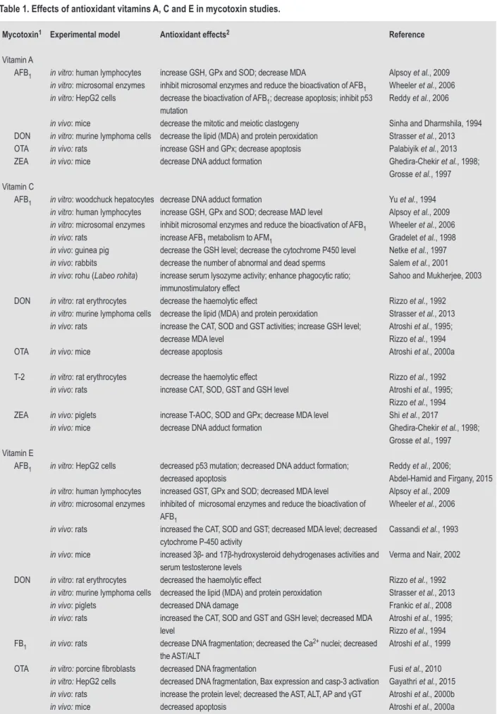
observed in in vitro and in vivo studies are listed in Table 1.
Table 1. Effects of antioxidant vitamins A, C and E in mycotoxin studies.
Mycotoxin1 Experimental model Antioxidant effects2 Reference
Vitamin A
AFB1 in vitro: human lymphocytes increase GSH, GPx and SOD; decrease MDA Alpsoy et al., 2009
in vitro: microsomal enzymes inhibit microsomal enzymes and reduce the bioactivation of AFB1 Wheeler et al., 2006
in vitro: HepG2 cells decrease the bioactivation of AFB1; decrease apoptosis; inhibit p53 mutation
Reddy et al., 2006
in vivo: mice decrease the mitotic and meiotic clastogeny Sinha and Dharmshila, 1994 DON in vitro: murine lymphoma cells decrease the lipid (MDA) and protein peroxidation Strasser et al., 2013 OTA in vivo: rats increase GSH and GPx; decrease apoptosis Palabiyik et al., 2013 ZEA in vivo: mice decrease DNA adduct formation Ghedira-Chekir et al., 1998;
Grosse et al., 1997 Vitamin C
AFB1 in vitro: woodchuck hepatocytes decrease DNA adduct formation Yu et al., 1994
in vitro: human lymphocytes increase GSH, GPx and SOD; decrease MAD level Alpsoy et al., 2009
in vitro: microsomal enzymes inhibit microsomal enzymes and reduce the bioactivation of AFB1 Wheeler et al., 2006
in vivo: rats increase AFB1 metabolism to AFM1 Gradelet et al., 1998
in vivo: guinea pig decrease the GSH level; decrease the cytochrome P450 level Netke et al., 1997
in vivo: rabbits decrease the number of abnormal and dead sperms Salem et al., 2001
in vivo: rohu (Labeo rohita) increase serum lysozyme activity; enhance phagocytic ratio; immunostimulatory effect
Sahoo and Mukherjee, 2003 DON in vitro: rat erythrocytes decrease the haemolytic effect Rizzo et al., 1992
in vitro: murine lymphoma cells decrease the lipid (MDA) and protein peroxidation Strasser et al., 2013
in vivo: rats increase the CAT, SOD and GST activities; increase GSH level; decrease MDA level
Atroshi et al., 1995; Rizzo et al., 1994 OTA in vivo: mice decrease apoptosis Atroshi et al., 2000a T-2 in vitro: rat erythrocytes decrease the haemolytic effect Rizzo et al., 1992
in vivo: rats increase CAT, SOD, GST and GSH level Atroshi et al., 1995; Rizzo et al., 1994 ZEA in vivo: piglets increase T-AOC, SOD and GPx; decrease MDA level Shi et al., 2017
in vivo: mice decrease DNA adduct formation Ghedira-Chekir et al., 1998; Grosse et al., 1997 Vitamin E
AFB1 in vitro: HepG2 cells decreased p53 mutation; decreased DNA adduct formation; decreased apoptosis
Reddy et al., 2006;
Abdel-Hamid and Firgany, 2015
in vitro: human lymphocytes increased GST, GPx and SOD; decreased MDA level Alpsoy et al., 2009
in vitro: microsomal enzymes inhibited of microsomal enzymes and reduce the bioactivation of AFB1
Wheeler et al., 2006
in vivo: rats increased the CAT, SOD and GST; decreased MDA level; decreased cytochrome P-450 activity
Cassandi et al., 1993
in vivo: mice increased 3β- and 17β-hydroxysteroid dehydrogenases activities and serum testosterone levels
Verma and Nair, 2002 DON in vitro: rat erythrocytes decreased the haemolytic effect Rizzo et al., 1992
in vitro: murine lymphoma cells decreased the lipid (MDA) and protein peroxidation Strasser et al., 2013
in vivo: piglets decreased DNA damage Frankic et al., 2008
in vivo: rats increased the CAT, SOD and GST and GSH level; decreased MDA level
Atroshi et al., 1995; Rizzo et al., 1994 FB1 in vivo: rats decrease DNA fragmentation; decreased the Ca2+ nuclei; decreased
the AST/ALT
Atroshi et al., 1999 OTA in vitro: porcine fibroblasts decreased DNA fragmentation Fusi et al., 2010
in vitro: HepG2 cells decreased DNA fragmentation, Bax expression and casp-3 activation Gayathri et al., 2015
in vivo: rats increase the protein level; decreased the AST, ALT, AP and γGT Atroshi et al., 2000b
in vivo: mice decreased apoptosis Atroshi et al., 2000a
Vitamin A has three active forms: retinol, retinal, and
retinoic acid (retinoids), which are essential for physiological
functions, including reproduction, vision, growth, and
maintenance of epithelial tissues. The antioxidative
effects of vitamin A have been associated with inhibiting
cytochrome P450-mediated metabolism of toxic substances
and preventing mutagenic epoxies from binding to DNA,
thereby forming epoxides and competing with mutagenic
epoxides in reaction with DNA (Diplock et al., 1998). The
toxic effects of AFB
1, DON, OTA and ZEA have been shown
to be reduced in interaction with vitamin A in in vitro and in
vivo models. The beneficial effects include increased levels
of antioxidant enzymes (GSH, GPx), a decrease in mycotoxin
bioactivation and in cell death (Table 1).
Vitamin C or ascorbic acid is a lactone synthesised in the
liver of many species. It is a first-line antioxidant that has
beneficial effects including protecting cell membranes,
proteins and nucleic acids from oxidation. Its biological
action and antioxidant characteristic are associated with its
ability to donate electrons. At physiological levels, vitamin C
is a powerful scavenger of oxygen-derived free radicals such
as superoxide radical anion, H
2O
2, the hydroxyl radical, and
singlet oxygen in plasma and tissues (Diplock et al., 1998). In
addition, ascorbic acid is an efficient scavenger of reactive
nitrogen oxide species, thereby avoiding nitrosative stress
and cell damage (Rock et al., 1996). Vitamin C also interacts
with GSH, reducing GSH production, which, in turn,
reduces oxidative stress. The main effects of vitamin C on
mycotoxin induced-toxicity are reducing lipid peroxidation
and increasing levels of antioxidant enzymes. These and
other effects have been described for AFB
1, DON, OTA, T-2
and ZEA (Table 1). Reduced adduct formation, decreased
apoptosis and enhancement of phagocytosis have been
reported for AFB
1and ZEA (Ghedira-Chekir et al., 1998;
Sahoo and Mukherjee, 2003).
Vitamin E refers to a group of substances that includes
tocols and tocotrienol derivatives. There are two forms of
vitamin E, γ-tocopherol and α-tocopherol. α-tocopherol is
the most biologically active form of vitamin E (Traber and
Sies, 1996) and the major function is that of a peroxyl radical
scavenger, interrupting the propagation of free radicals. In
addition, vitamin E interacts with reactive nitrogen oxide
species and singlet oxygen, thereby maintaining the integrity
of polyunsaturated fatty acids in cell membranes (Rock et al.,
1996). Vitamin E has been shown to act favourably against
seven mycotoxins (AFB
1, DON, FB
1, OTA, PAT, T-2 and
ZEA) (Table 1). Its actions are similar to those of vitamins A
and C, although decreased DNA fragmentation and damage
of DON, FB
1and OTA was also reported (Atroshi et al.,
1999; Frankic et al., 2008; Gayathri et al., 2015). In addition,
reduced Hsp 70 expression and increased lymphocyte
proliferation were described for T-2 (El Golli et al., 2006;
Jaradat et al., 2006).
Flavonoids
The flavonoids are the most common hydroxylated phenolic
substances that are synthesised by plants. Sources of
flavonoids are citrus fruits, berries, red wine and tea (Diplock
et al., 1998). The function of flavonoids is associated with its
structure, which includes a number of structurally different
subgroups, including flavonols (quercetin, kaempferol,
myricetin), flavanols (catechin and epicatechin), isoflavones
(genistein), flavones (apigenin, hesperetin), flavanones
Table 1. Continued.
Mycotoxin1 Experimental model Antioxidant effects2 Reference
Vitamin E
PAT in vitro: HepG2 cells decreased p53 activation; decreased DNA damage Ayed-Boussema et al., 2013 T-2 in vitro: chicken lymphocytes increased lymphocyte proliferation Jaradat et al., 2006
in vitro: Vero cells decreased Hsp 70 expression El Golli et al., 2006
in vitro: rat erythrocytes decreased the haemolytic effect Rizzo et al., 1992
in vivo: chicken decreased MDA level Hoehler and Marquardt, 1996
in vivo: rats increased the CAT, SOD and GST and GSH level; increase the protein level; decreased MDA level, AST, ALT, AP and γGT.
Atroshi et al., 1995, 2000a.; Rizzo et al., 1994
ZEA in vivo: mice decreased DNA adduct formation Ghedira-Chekir et al., 1998; Grosse et al., 1997
1 AFB
1 = aflatoxin B1; DON = deoxynivalenol; FB1 = fumonisin B1; NIV = nivalenol; OTA = ochratoxin A; PAT = patulin; T-2 = T-2 toxin; ZEA = zearalenone. 2 ALT = alanine transaminase; AP = alkaline phosphatase; AST = aspartate transaminase; CAT = catalase; GPx = glutathione peroxidase; GSH =
glutathione; GST = glutathione S-transferase; γGT = gamma-glutamyl transpeptidase; MDA = malondialdehyde, SOD = superoxide dismutase; T-AOC = total antioxidative capacity.
(naringenin, taxifolin) and/or anthocyanidins (cyanidin,
malvidin) (Rice-Evans and Miller, 1996). The biological
function of antioxidants is connected with their capacity
to scavenger free radicals (peroxyl radical and hydroxyl
radical), as well as chelating metals involved in the Fenton
reaction (Rice-Evans and Miller, 1996).
The flavonol quercetin is one of the most effective
polyphenolic substances linked to a reduction in the levels
of ROS and reactive nitrogen species. In previous in vivo
and in vitro studies, quercetin was shown to modulate the
effects of oxidative stress caused by T-2, AFB
1and OTA
resulting in an increase in Nrf2 expression, SOD and GPx
activity as well as total antioxidant status and GSH levels
(Abdel-Wahhab et al., 2017; Capcarova et al., 2015; Choi et
al., 2010; Ramyaa and Padma, 2013; Ramyaa et al., 2014). On
the other hand, quercetin was associated with a decrease in
ER oxidative stress, ROS generation, MAD level, P450 and
NADPH activity, cytochrome C release, casp-3 activation
cell apoptosis, Cox-2 and NO expression, TNF-α, IL-6 and
IL-2 and a decrease in DNA damage (Abdel-Wahhab et
al., 2017; Ramyaa and Padma, 2013; Ramyaa et al., 2014).
Recent studies demonstrated the antioxidant effects of
other flavonoids on oxidative stress induced by mycotoxins.
Proanthocyanidin increased Nrf2 expression, SOD, GPx,
CAT activities, GSH level, and decreased MDA content,
DNA damage and the expression of pro-inflammatory
cytokines (IL-1β, TNF-α, IL-6 and IFN-γ) in rats and mice
subjected to AFB
1(Long et al., 2016c) and ZEA (Long et
al., 2016b) diets. Cyanidin decreased DNA damage, ROS
production, lipid hydroperoxide and iNOS, and increased
HO-1 activity and non-protein thiol groups in rats, in a
pig kidney cell line (LLC-PK1), and in human fibroblasts
exposed to OTA (Sorrenti et al., 2012). Baicalein, wogonin
and hesperidin increased cell viability and decreased
genotoxicity and casp-3 activation in mice and neural
crest cells exposed to AFB
1(Nones et al., 2013; Ueng et
al., 2001). Zhong et al. (2017) reported that the apigenin
re-established MMP, increased Bcl-2 expression, decreased
Bax, p53 activation and the cytochrome C release in human
embryonic kidney cells 293 (HEK 293 cells) treated with PAT.
With the addition of silymarin to their diet, mice subjected to
FB
1contaminated feed showed decreased TNF-α expression
and casp-8 activation (Sozmen et al., 2014).
Crocin, curcumin, green tea, lycopene and phytic acid
Crocin is a major bioactive compound and is mainly
found in Gardenia jasminoides and saffron. Water and
ethanol extracts of crocin displayed antioxidant activity
against O
2-and HO radicals (Xiao et al., 2017). Curcumin
is a hydrophobic polyphenol derived from turmeric, a
compound extracted from the root of Curcuma longa L.
rhizome. Curcumin has diverse biological functions and
its structure, which is composed of methoxy groups and
phenols, is associated with its properties (Zheng et al.,
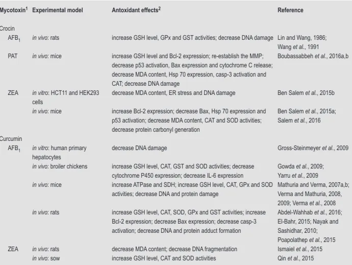
2017). The antioxidant action of crocin and curcumin on
the molecular effects of mycotoxins in vitro and in vivo are
summarised in Table 2.
Green tea is derived from Camellia sinensis leaves and
contains a wide range of bioactive compounds of which one
third are composed of polyphenols of which the majority are
flavonoids. Catechins (GTCs) are one of the main flavonoids
in green tea. GTCs have antioxidant capacity to scavenge
ROS such as O
2-, H
2
O
2and HO radicals (Cooper et al.,
2005). Lycopene is the most abundant carotenoid
(non-vitamin A) in orange fruits and vegetables, mainly tomatoes
and derived products and is responsible for their bright red
colour. Lycopene is a recognised antioxidant and has been
considered the most efficient in scavenging single oxygen
(Mordente et al., 2011). Phytic acid (IP6) is a saturated
cyclic acid commonly found in plant tissues and seeds;
and its antioxidant effect is on ROS production mainly due
its capacity to chelate iron, thereby inhibiting the Fenton
reaction (Silva and Bracarense, 2016). The antioxidant
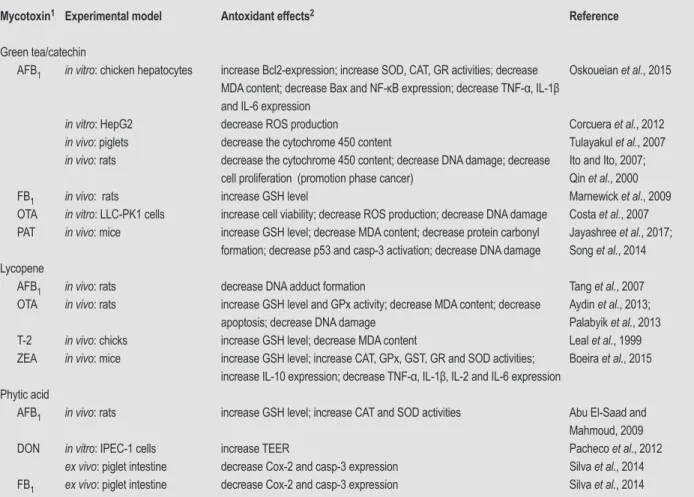
effects of green tea, lycopene and IP6 on mycotoxin-induced
oxidative stress in vitro and in vivo studies are listed in
Table 3.
L-carnitine
L-carnitine is an endogenous mitochondrial membrane
compound that plays a prominent role in facilitating the
transport of long-chain fatty acids into mitochondria and the
oxidation pathway (Adeva-Andany et al., 2017). L-carnitine
decreases oxidative stress, increases endogenous antioxidant
defence capacity, protects mitochondria against lipid
oxidation, and decreases apoptosis through the inhibition
of mitochondrial swelling and cytochrome C release
(Adeva-Andany et al., 2017). L-carnitine has been shown to decrease
ROS production, MDA level, casp-3 activation, DNA and
protein damage, and to increase MMP and GSH levels in rats
and quails subjected to AFB
1or T-2 contaminated diets (Citil
et al., 2005; Moosavi et al., 2016; Yatim and Sachan, 2001).
Melatonin
Melatonin is a hormonal product of the pineal gland
that controls reproductive functions, modulates immune
system activity, limits tumorigenesis and effectively
inhibits oxidative stress (Reiter, 1997). Antioxidant effects
of melatonin reported in rats exposed to AFB
1and OTA
contaminated diets included an increase in the GHS level
and in the activity of CAT, GPx, GSH, GST, GR and SOD
(Abdel-Wahhab et al., 2005; Meki et al., 2004; Sutken et al.,
2007), a decrease in MDA and LPO content (Abdel-Wahhab
et al., 2005; Sutken et al., 2007; Yenilmez et al., 2010) and
decreased expression of NO, Hsp 70 and casp-3 (Meki et
al., 2001, 2004).
Minerals
Several minerals are dietary constituents involved in the
antioxidant defence system, acting directly as antioxidants
or promoting detoxifying enzymes (Sorrenti et al., 2013).
Antioxidant enzymes, such as GPx and SOD require a
dietary supply of selenium (Se), copper (Cu) and zinc (Zn)
(Wang et al., in press).
Se is an essential micronutrient associated with
Se-dependent enzymes, including GPx, thioredoxin reductases,
iodothyronine deiodinases, and selenophosphate synthetases
(Wang et al., 2016). Se has been shown to increase the
antioxidant function of CAT, GSH, GPx and SOD, decrease
MDA content, increase the level of GSH and to modulate
DON-induced immunosuppression in piglet lymphocytes
and broiler chickens exposed to DON (Placha et al., 2009;
Wang et al., in press). Long et al. (2016a,b) observed that
Se increased GPx and SOD activities and Bcl-2 expression,
decreased MDA content, Bax and casp-3 expression in mice
fed with ZEA.
Zn exerts its antioxidant activity either in an acute way
or on a long-term basis. In the first form, zinc acts by
stabilising protein sulfhydryl groups against oxidation and
exchanging redox active metals (copper and iron) (Zheng
et al., 2013). In the second form, Zn induces the expression
of metallothioneins, which act as electrophilic scavengers.
Moreover, zinc is a co-factor of the SOD enzyme that
catalyses superoxide anions into less toxic O
2and H
2O
2and modulates the activity of GPx and glutamylcysteine
synthetase, through the activation of metal response
transcription factor-1 (MTF-1) (Powell, 2000). Zheng et
al. (2013) demonstrated the antioxidant effect of zinc as
being an increase in SOD activity and a decrease in ROS
generation and in DNA damage in HepG2 cells exposed
to OTA.
Table 2. Effects of antioxidant food compounds (crocin and curcumin) in mycotoxins studies.
Mycotoxin1 Experimental model Antoxidant effects2 Reference
Crocin
AFB1 in vivo: rats increase GSH level, GPx and GST activities; decrease DNA damage Lin and Wang, 1986; Wang et al., 1991 PAT in vivo: mice increase GSH level and Bcl-2 expression; re-establish the MMP;
decrease p53 activation, Bax expression and cytochrome C release; decrease MDA content, Hsp 70 expression, casp-3 activation and CAT; decrease DNA damage
Boubassabbeh et al., 2016a,b
ZEA in vitro: HCT11 and HEK293
cells
decrease MDA content, ER stress and DNA damage Ben Salem et al., 2015b
in vivo: mice increase Bcl-2 expression; decrease Bax, Hsp 70 expression and p53 activation; decrease MDA content, CAT and SOD activities; decrease protein carbonyl generation
Ben Salem et al., 2015a; Salem et al., 2016 Curcumin
AFB1 in vitro: human primary
hepatocytes
decrease DNA damage Gross-Steinmeyer et al., 2009
in vivo: broiler chickens increase GSH level, CAT, GST and SOD activities; decrease cytochrome P450 expression; decrease IL-6 expression
Gowda et al., 2009; Yarru et al., 2009
in vivo: mice increase ATPase and SDH; increase GSH level, CAT, GPx and SOD activities; decrease DNA and protein damage
Mathuria and Verma, 2007a,b; Verma and Mathuria, 2008, 2009; Verma et al., 2008
in vivo: rats increase GSH level, CAT, SOD, GPx and GST activities; increase Bcl-2 expression; decrease Bax expression; decrease casp-3 activation; decrease DNA and protein adduct formation
Abdel-Wahhab et al., 2016; El-Bahr, 2015; Nayak and Sashidhar, 2010; Poapolathep et al., 2015 ZEA in vivo: rats decrease MDA content; decrease DNA fragmentation Ismaiel et al., 2015
in vivo: sow increase GSH level, CAT and SOD activities Qin et al., 2015
1 AFB
1 = aflatoxin B1; PAT = patulin; ZEA = zearalenone.
2 ATPase = adenosine triphosphatase; CAT = catalase; GPx = glutathione peroxidase; GSH = glutathione; GST = glutathione S-transferase; GST = glutathione
S-transferase; MDA = malondialdehyde; MMP = mitochondrial membrane potential; SDH = succinate dehydrogenase; SOD = superoxide dismutase.
Mixtures
Several studies have demonstrated that mixtures of natural
substances reduce the oxidative stress lesions caused by
mycotoxins. The combination of L-carnitine, vitamin
E, selenium, melatonin, coenzyme Q10 and tamoxifen
(Abidin et al., 2013; Atroshi et al., 2000; Sutken et al.,
2007; Yenilmez et al., 2010) increased protective effects on
DNA, proteins and lipids against OTA-induced toxicity
compared to the individual effects of the compounds.
Moreover, the combination of coenzyme Q10, L-carnitine,
alpha-tocopherol and selenium, garlic and curcumin
(El-Barbary, 2016), black tea and curcumin (Alm-Eldeen et al.,
2015) displayed potent antioxidant effects against the toxic
effects of AFB
1. In T-2-induced oxidative stress experiments,
the increase in the level of GSH and the decrease in DNA
damage were more apparent in mixtures of coenzyme Q10,
L-carnitine, alpha-tocopherol and selenium (Atroshi et
al., 1999) and tamoxifen, vitamin E, and Se (Atroshi et al.,
1997, 2000).
5. Conclusions
Oxidative stress, ROS and RNS generation induced by
mycotoxins have been associated with their cytotoxic effects
on DNA, protein synthesis and mitochondria. These effects
have been confirmed in different assays on cell membranes,
proteins or nucleic acids, but the mechanisms involved in
the activation of the signalling pathways that results in cell
death or increased permeability for the different mycotoxins
remain uncertain. Which factors are involved in activation?
Dose, duration of exposure, and animal species are some of
the aspects that need to be investigated in addition to the
molecular characteristics of mycotoxins. In addition, most
available data were acquired in in vitro studies or mice/rat
models. New data from other animal models, especially
those of economic interest are still lacking.
Table 3. Effects of antioxidant food compounds (green tea/catechin, lycopene and phytic acid) in mycotoxins studies.
Mycotoxin1 Experimental model Antoxidant effects2 Reference
Green tea/catechin
AFB1 in vitro: chicken hepatocytes increase Bcl2-expression; increase SOD, CAT, GR activities; decrease MDA content; decrease Bax and NF-κB expression; decrease TNF-α, IL-1β and IL-6 expression
Oskoueian et al., 2015
in vitro: HepG2 decrease ROS production Corcuera et al., 2012
in vivo: piglets decrease the cytochrome 450 content Tulayakul et al., 2007
in vivo: rats decrease the cytochrome 450 content; decrease DNA damage; decrease cell proliferation (promotion phase cancer)
Ito and Ito, 2007; Qin et al., 2000 FB1 in vivo: rats increase GSH level Marnewick et al., 2009 OTA in vitro: LLC-PK1 cells increase cell viability; decrease ROS production; decrease DNA damage Costa et al., 2007 PAT in vivo: mice increase GSH level; decrease MDA content; decrease protein carbonyl
formation; decrease p53 and casp-3 activation; decrease DNA damage
Jayashree et al., 2017; Song et al., 2014 Lycopene
AFB1 in vivo: rats decrease DNA adduct formation Tang et al., 2007 OTA in vivo: rats increase GSH level and GPx activity; decrease MDA content; decrease
apoptosis; decrease DNA damage
Aydin et al., 2013; Palabyik et al., 2013 T-2 in vivo: chicks increase GSH level; decrease MDA content Leal et al., 1999 ZEA in vivo: mice increase GSH level; increase CAT, GPx, GST, GR and SOD activities;
increase IL-10 expression; decrease TNF-α, IL-1β, IL-2 and IL-6 expression
Boeira et al., 2015 Phytic acid
AFB1 in vivo: rats increase GSH level; increase CAT and SOD activities Abu El-Saad and Mahmoud, 2009 DON in vitro: IPEC-1 cells increase TEER Pacheco et al., 2012
ex vivo: piglet intestine decrease Cox-2 and casp-3 expression Silva et al., 2014 FB1 ex vivo: piglet intestine decrease Cox-2 and casp-3 expression Silva et al., 2014
1 AFB
1 = aflatoxin B1; FB1 = fumonisin B1; OTA = ochratoxin A; PAT = patulin; T-2 = T-2 toxin; ZEA = zearalenone.
2 CAT = catalase; GPx = glutathione peroxidase; GR = glutathione reductase; GSH = glutathione; GST = glutathione S-transferase; IL = interleukin; MDA
= malondialdehyde; NF-κB = nuclear factor kappa beta; ROS = reactive oxygen species; SOD = superoxide dismutase; TEER = transepithelial electrical resistance; TNF-α = tumour necrosis factor alpha