1. Introduction
Small non-avian theropods of Late Jurassic age are rare worldwide (WEISHAMPEL et al. 2004). Only a handful of them exist in Europe and until recently, these dinosaurs were best represented by two incom-plete skeletons of Compsognathus longipes (WAGNER 1861). Despite the historical significance of Comp -sognathus, the discovery of which played a key role in
the evolutionary debate of the 19thcentury (DESMOND
1982; OSTROM 1978; CHIAPPE 2007), the rather un -specialized morphology of “compsognathids” (Comp-sognathus and a series of alleged Late Jurassic-Early Cretaceous relatives – CURRIE& CHEN 2001; HOLTZ et al. 2004; HWANGet al. 2004; NAISHet al. 2004; JI et al. 2007b; GISHLICK & GAUTHIER 2007) has com-plicated interpretations of this group’s monophyly. In this paper, “compsognathids” are used without any
Anatomy of Juravenator starki (Theropoda: Coelurosauria)
from the Late Jurassic of Germany
Luis M. Chiappe, Los Angeles and Ursula B. Göhlich, Vienna With 26 figures
CHIAPPE, L. M. & GÖHLICH, U. B. (2011): Anatomy of Juravenator starki (Theropoda: Coelurosauria)
from the Late Jurassic of Germany. – N. Jb. Geol. Paläont. Abh., 258: 257– 296; Stuttgart.
Abstract: We provide a detailed study of the morphology of the holotype of Juravenator starki from
the Late Jurassic of the Solnhofen area of southern Germany. The incompletely ossified surface of multiple bones and lack of several skeletal fusions indicate that Juravenator starki is based on an immature specimen. Nonetheless, numerous unique morphologies and bone proportions distinguish this taxon from Compsognathus longipes, the only previously named non-avian theropod dinosaur from the Late Jurassic of the Solnhofen Archipelago. Yet, its skeletal anatomy is most similar to that of Compsognathus and other theropods that have often been regarded as closely related to the latter – sometimes within a monophyletic Compsognathidae. Juravenator is characterized by having a small size (~ 0.75-meter-long in the holotype) with few maxillary teeth, lack of a premaxillary-maxillary diastema, an antorbital fenestra subequal in length to orbit, an elongate scapula that is narrowest at its neck, a proportionally short humerus and high and abruptly tapered manual claws, and bow-like zygapophysial articulations in the mid-caudal vertebrae. Portions of the epidermis preserved mainly along the tail provide the only glimpse of the morphology of the skin of basal coelurosaurs, and structures newly revealed under UV light hint at the possibility of filamentous integumentary structures – akin to those interpreted as proto-feathers in other basal coelurosaurs – also covering the body of this dinosaur. The discovery of Juravenator has provided evidence of morphologies – from details of the skull to the epidermis – that are poorly known in other theropods interpreted as at or near the base of Coelurosauria, and thus contributes significantly to our understanding of the evolutionary history of this clade. The exquisitely preserved holotipic skeleton adds significantly to the meager record of small-bodied Late Jurassic theropods.
Key words: Juravenator, Theropoda, anatomy, taphonomy, preservation, Jurassic, Solnhofen
Lime-stones.
© 2010 E. Schweizerbart’sche Verlagsbuchhandlung, Stuttgart, Germany www.schweizerbart.de
DOI: 10.1127/0077-7749/2010/0125 0077-7749/2010/0125 $ 10.00
implication of the putative monophyletic nature of a group that includes Compsognathus longipes (WAGNER1861; OSTROM1978; PEYER2006), Huaxia-gnathus orientalis (HWANG et al. 2004), Sinosauro -pteryx prima (JI& JI1996; CHENet al. 1998; CURRIE & CHEN 2001), Scipionyx samniticus (DALSASSO & SIGNORE 1998), and Juravenator starki (GÖHLICH &
CHIAPPE 2006) among other non-avian theropods usually regarded at or near the base of the coeluro -saurian clade. The morphological diversity of these theropods has remained as a poorly understood chap-ter in the evolutionary history of Late Jurassic-Early Cretaceous theropod dinosaurs (CURRIE & CHEN 2001; HOLTZ et al. 2004; PEYER 2006). However,
Fig. 1. Geographic location of Juravenator starki (JME Sch 200), Compsognathus longipes, and selected specimens
of Archaeopteryx lithographica (reconstructions not to scale). Barred and mottled patterns indicate the approximate depositional areas during the Late Jurassic.
these generally unspecialized theropods provide key evidence for understanding the morphological transformations that occurred near the base of Coeluro -sauria, an extremely diverse clade of dinosaurs that includes all living birds.
In 1998, ending a decade-long collecting program (1989-1998) by the Jura-Museum Eichstätt under the direction of GÜNTER VIOHL, volunteers KLAUS -DIETER and HANS-JOACHIMWEISS (Kelkheim-Fisch-bach) unearthed a small Late Jurassic theropod from the FRANZ STARK Quarry of Schamhaupten (district of Eichstätt, Southern Franconian Alb, Bavaria, Ger-many) (Fig. 1). The WEISS brothers first split a slab containing portions of the skull and cervicals, which led them to quarry the adjacent slabs. The specimen (JME Sch 200) was brought to the Jura-Museum Eich-stätt for a multi-year preparation by PINOVÖLKL, who
revealed a nearly complete skeleton with traces of soft tissues (for more details on the history of discovery and preparation, see TISCHLINGER et al. [2006] and GÖHLICH et al. [2006]). The specimen was prelimi -narily described and named Juravenator starki by GÖHLICH& CHIAPPE(2006). Based on several synapo -morphies including the presence of a round orbit, some maxillary and dentary teeth without anterior serrations, and axial epipophyses not extending beyond the posterior rim of the postzygapophyses, GÖHLICH& CHIAPPE(2006) identified Juravenator as a coelurosaurian, and clustered this taxon with other basal coelurosaurians within Compsognathidae. As mentioned above, however, the monophyly of “comp-sognathids” and their relationships to other coelurosaurians, are far from settled. While a number of phylo -genetic studies (e.g., HWANGet al. 2004; SENTER2007;
Fig. 2. Stratigraphic position of STARK Quarry (Schamhaupten Member) and Juravenator starki (JME Sch 200).
XUet al. 2009; CHOINIEREet al. 2010) have recovered them as a monophyletic clade, taxa usually ascribed as “compsognathids” are distributed across the basal portion of the coelurosaurian tree in other studies (e. g., BUTLER & UPCHURCH 2007). Furthermore, cladistic analyses in which compsognathids are re -covered as monophyletic usually limit the taxonomic sampling of these theropods to better known taxa such as Compsognathus,Sinosauropteryx, and Huaxia -gnathus, but fail to include a variety of other taxa that have also been described as members of the same group (e. g., Mirischia asymmetrica, Sinocalliopteryx gigas) (MARTILLet al. 2000; NAISHet al. 2004; JIet al. 2007a).
The phylogenetic relationships of Juravenator to -gether with that of many other basal coelurosaurians (and the monophyly of “compsognathids”) needs to be studied in light of more comprehensive cladistic analyses. This notewithstanding, the discovery of Juravenator resulted in an exceptionally well-pre -served specimen – possibly the most complete non-avian theropod from Europe – and a startling new example of a small-bodied Late Jurassic carnivorous dinosaur. Juravenator thus provides important infor-mation for better understanding the role played by basal coelurosaurs in the evolutionary history of thero pods. The present paper provides a detailed description of both the skeletal morphology and in -tegumentary anatomy of this dinosaur.
I n s t i t u t i o n a l a b b r ev i a t i o n s . – AMNH, American Museum of Natural History (New York); BSPG, Bayerische Staatssammlung für Paläontologie und Geologie (Munich); CAGS, Chinese Academy of Geological Sciences (Beijing); GMV, Geological Museum of China (Beijing); JME, Jura-Museum Eichstätt (Eichstätt); SMNK, Staatliches Jura-Museum für Naturkunde Karlsruhe (Karlsruhe).
2. Geological setting
During the Late Jurassic, southern Germany was largely submerged by a shelf sea wedged between two large islands to the north and the deeper Tethys Ocean to the south (BARTHELet al. 1990). A series of lime-stone-filled basins developed within this shallow sea; one of them was the small (~ 35 km2) Schamhaupten
Basin (Southern Franconian Alb), in which limestones were deposited amid a large complex of dolomitic reefs (RENESTO& VIOHL1997; ZEISS2001; VIOHL& ZAPP2006, 2007) (Figs. 1-2).
The skeleton of Juravenator (JME Sch 200) is con-tained in the strongly silicified, laminated limestone
that outcrops at the FRANZ STARKQuarry of Scham-haupten. VIOHL & ZAPP(2006, 2007) identified this silicified plattenkalk as belonging to the Late Kim meridgian Schamhaupten Subformation, which is con -sidered to be part of the Painten Formation. Based on recent biostratigraphic studies (ZEISS 2001; VIOHL & ZAPP2006, 2007; SCHWEIGERT2007), the top lime -stones at Stark Quarry are dated as uppermost Kim-meridgian (Beckeri Zone, Ulmense Subzone) (Fig. 2), thus slightly older than the neighboring Solnhofen Lithographic Limestones (Solnhofener Plattenkalk) – the celebrated deposits that have yielded both Comp-sognathus and Archaeopteryx (WELLNHOFER2008).
The Schamhaupten plattenkalk (which is different than the Solnhofen plattenkalk) comprises two main types of beds: bindstones and detrital carbonates. Most articulated fossils are contained within bind -stones, while detrital carbonates most typically yield shells and isolated skeletal elements (VIOHL & ZAPP 2006) – Juravenator is embedded in a 3.5-cm-thick bindstone belonging to section layer E3 (see VIOHL& ZAPP2006: fig. 5, 2007: fig. 4). Containing more than 200 taxa, the Schamhaupten plattenkalk has preserved a great variety of fossil plants, nano- and microfossils, invertebrates, and vertebrates (VIOHL1999; VIOHL& ZAPP2006). Fish are the most abundant vertebrates but a variety of reptiles – among these, the turtles Solnhofia and Eurysternum, the sphenodontid Lepto-saurus, and Juravenator (RENESTO & VIOHL 1997; VIOHL1999, 2006, 2007) – are also well-represented. The fact that most Schamhaupten fossils are of marine origin suggests good connections to the Tethys Ocean but the remains of terrestrial taxa also indicate the existence of nearby islands.
The exceptional preservation illustrated by the richness of articulated fossils – fish, reptiles, crusta -ceans, sea urchins with in situ spines, and others – designates the Schamhaupten plattenkalk as a Kon -servat Lagerstätte, although one in which organisms in different stages of decay are also preserved. This extraordinary preservation has been recently ex -plained by the taphonomic model of VIOHL & ZAPP (2006, 2007). These authors suggest that a stratified salinity developed within the Schamhaupten Basin: its bottom zone was hypersaline, anaerobic or dys -aerobic, and thus lethal, while its upper water column was suitable for marine life. The hostile nature of the basin’s bottom suggests the absence of benthonic scavengers, althoughVIOHL& ZAPP(2006, 2007) inter preted the proliferation of microbial mats as respon -sible for the generation of bindstones by trapping
sedi-Fig. 3. Photograph under normal light of the skeleton of Juravenator starki (JME Sch 200). Dashed line on inset highlights
ment and for the extraordinary preservation of Scham-haupten by rapidly sealing the sunken corp ses.
3. Taphonomy of Juravenator
Instead of having the typical opisthotonic posture of many fossil amniotes (MARSHALL FAUX & PADIAN
2007), the skeleton of Juravenator (Figs. 3-5) exhibits two distinct torsions: at the level of the neck and most unusually, in front of the pelvis. While the skull and tail are primarily exposed in right lateral view, the torso shows its left lateral side. The pelvic bones are somewhat disarticulated – the ilia are displaced and exhibit their right sides – and the hindlimbs are splayed ventrally, each showing its laterocaudal sur -face.
The completeness and extensive articulation of the skeleton of Juravenator strongly suggests that the carcass floated for a very short period of time before it sunk into the hostile bottom of the basin soon after the animal’s death. Explaining the unusual death posture of the skeleton is, however, more problematic. Now -here in the skeleton is t-here apparent evidence of pre-dation that can explain the observed rotation of the pelvis with respect to the torso. The inferred bottom conditions point at the absence of benthonic sca -vengers that could have disrupted the skeleton and even the presence of very small marine woodlouses (Isopoda) (Fig. 6) – that probably scavenged the corpse before it settled on the basin’s bottom – can hardly explain the aforementioned rotation. All this suggests that the peculiar torsion of the skeleton of Juravenator is most likely the result of taphonomic factors. A possible scenario might be that the carcass settled into the basin’s floor with the head lying on its left side, the torso leaning towards its right side with the right forelimb tucked under the body, the pelvis in a more upright position, and the legs splayed. If so, the complete rotation between torso and pelvis may be explained by the strong compaction of the bindstones during diagenesis (VIOHL& ZAPP2006).
4. Systematic paleontology Dinosauria OWEN, 1842 Theropoda MARSH, 1881 Tetanurae GAUTHIER, 1986 Coelurosauria V. HUENE, 1914 Juravenator starki GÖHLICH& CHIAPPE, 2006 H o l o t y p e : JME Sch 200, a nearly complete and articu -lated skeleton – preserving portions of soft tissue – missing only the distal third of its tail (Figs. 3-5). The specimen was collected in two main blocks that were glued together during preparation (Fig. 3). Some parts of the skull and neck were collected as slab and counterslab and subsequently glued together and prepared from one side.
H o r i z o n a n d l o c a l i t y : Silicified, laminated limestone, Late Jurassic (Upper Malm, Late Kimmeridgian, Beckeri
Zone, Ulmense Subzone) in the local stratigraphy (ZEISS
2001; SCHWEIGERT2007) (Fig. 2). JME Sch 200 was
collec-ted from the Stark Quarry, a quarry owned by the Stark family and situated a few hundred meters west of the village of Schamhaupten (district of Eichstätt, Southern Franconian Alb), Bavaria, Germany (Fig. 1).
D i a g n o s i s : Small basal coelurosaur (a clade encom -passing Passer domesticus and all taxa sharing a more recent common ancestor with it than with Allosaurus fragi-lis) with a large skull proportionally longer than in Compso-gnathus longipes (skull: femur and skull: presacral verte-brae ratios are 1.5 and 0.47, and 1.1 and 0.25 in Juravenator and C. longipes, respectively), low number of maxillary teeth (less than 10 in Juravenator, 10 in Ornitholestes hermanni, 12 in Sinosauropteryx prima, 15 in C. longipes), absence of a premaxillary-maxillary (diastema present in Scipionyx samniticus), distinct indentation on the denti -gerous margin of the maxilla (between second and third teeth), an antorbital fenestra subequal in length to orbit (antorbital fenestra is nearly half the orbit in S. prima, Ornitholestes hermanni, and S. samniticus and subequal to the orbit in C. longipes and basal tyrannosauroids [XUet al. 2004, 2006; SERENOet al. 2009]), long scapula
(hume-rus:scapula and scapula:femur ratios are 0.63 and 0.81 in Juravenator, 0.86 and 0.60 in S. prima, and 1 and 0.54 in C. longipes, respectively), of which the narrowest portion is at the neck as opposed to near the mid-shaft, and short feet (metatarsal III: femur is 0.59 in Juravenator and approxi -mately 0.75 in S. prima and C. longipes [CURRIE& CHEN
2001]). Juravenator is also unique among other basal coe-lurosaurs in having longer and more slender teeth, a cave rostral margin of the jugal process of the postorbital, humeri with very short and triangular-shaped deltopectoral crests, manual claws that are very high proximally and that taper abruptly around their midpoints, and arched, bow-like zygapophysial articulations in the mid-caudal vertebrae. We regard these unique features as autapomorphies of Jura -venator starki.
5. Anatomical description
The specimen shows features indicating an early onto-genetic age – the surface of its bones is intensively scarred by small pits and grooves, thus revealing a pattern of incomplete periosteal formation (e. g. HORNER1997; SANZet al. 1997; CHIAPPEet al. 1998; CODORNIÚ & CHIAPPE 2004) (Fig. 7). Evidence of immaturity is also present in the lack of fusion be -tween sacral vertebrae and presence of open neuro-central sutures, visible on many caudal vertebrae (BROCHU1996). Based on the preserved length of the axial skeleton and the estimated length of the missing distal third of the tail, GÖHLICH & CHIAPPE (2006) projected the length of the holotype of Juravenator starki to 0.75-.80 m. This value was considered to be a slight underestimation by THERRIEN& HENDERSON (2007), who based on a least-square regression of
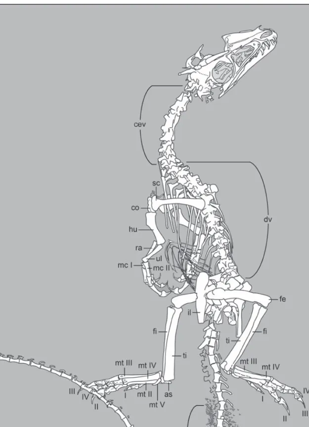
Fig. 5. Interpretive drawing of the skeleton of Juravenator starki (JME Sch 200). Abbreviations: as, astragalus; cev, cervical
vertebrae; co, coracoid; cv 44, caudal vertebra 44; dv, dorsal vertebrae; fe, femur; fi, fibula; ha, haemal arches; hu, humerus; il, ilium; mcI-II, metacarpals I-II; mt II-V, metatarsals II-V; ul, ulna; ra, radius; sc, scapula; st, soft-tissue; ti, tibia; I-IV, pedal digits I-IV.
theropod skulls calculated the length of this specimen as slightly over 1 m. The latter may well be accounted for by the relatively large skull of the holotype, but regardless of the difference between calculations based on actual skeletal length and those based on least-square regressions, the juvenile nature of the holotype indicates that the adults of this species must have substantially exceeded both projected lengths. Detailed osteological measurements are given in the Appendix.
5.1. Skull
C r a n i a l b o n e s . – The skull (Fig. 8) is large in pro-portion to the skeleton (more than 1.5 times the length of the femur) (Figs. 3-5). The rostrum is moderately long – the distance between the lacrimal and the rostrum’s tip is 53 % of the skull length – and ex -cavated by a large antorbital fossa.
The right premaxilla is preserved in lateral view and the nasal process of its counterpart is exposed dorsal to the rostral half of the former. The body of the premaxilla is rectangular, longer than it is high, and bears short maxillary (subnarial) and nasal (inter -narial) processes, which have relative lengths more like those of Compsognathus (OSTROM 1978; PEYER 2006), Scipionyx (DALSASSO& SIGNORE1998), Sino-sauropteryx (CHEN et al. 1998; CURRIE & CHEN
2001), and the basal tyrannosauroid Dilong paradoxus (XUet al. 2004) than the long processes of Huaxia-gnathus (HWANG et al. 2004) (Figs. 8-9). The nasal process of the premaxilla extends along the rostro -dorsal margin of the snout, bordering -dorsally the elliptical external nares, for about half the length of the nares (Fig. 7). The maxillary process extends cau-dally beyond the caudal end of the nasal process and it appears to be thicker than the latter, a condition similar to that in Sinocalliopteryx (JIet al. 2007a) and opposite to that in Sinosauropteryx (CURRIE& CHEN 2001). These two processes are also less caudally divergent than in Sinosauropteryx, more similar to Sinocalliopteryx (JI et al. 2007a), Huaxiagnathus (HWANG et al. 2004), Scipionyx (DAL SASSO & SIGNORE 1998), and Compsognathus (MNHM CNJ 79, MICHARD 1991) (Fig. 8). There are at least three premaxillary teeth – the last one broken at its base – but we cannot rule out the presence of a fourth and most anterior tooth (Figs. 7, 10). Similar uncertainties have been expressed for other “compsognathids”: OSTROM (1978) reported a minimum of three pre -maxillary teeth in Compsognathus longipes (BSPG AS I 563) but PEYER(2006) highlighted the presence of four alveoli in the French Compsognathus (MNHM CNJ 79). CURRIE& CHEN(2001) were not definitive whether four or five premaxillary teeth are present in Sinosauropteryx (although they believed four to be the
Fig. 6. Close-up of isopods preserved as impressions (A) or three-dimensionally (B) within the skeleton of Juravenator
number), and HWANGet al. (2004) were indeterminate between three and four. In Scipionyx – presumably also closely related to Compsognathus (DAL SASSO, pers. comm.) – there are five premaxillary teeth, but only four in the large compsognathid Sinocalliopteryx (JIet al. 2007a).
The right maxilla is exposed in lateral view; what appears to be a displaced small portion of the dorsal ramus of the left maxilla is attached to the side of the left nasal. The maxilla is largely excavated by an
antorbital fossa that contains a large antorbital fenestra and a small maxillary fenestra (Fig. 7). The maxillary region anterior to the antorbital fossa is very short – shorter than the length of the body of the pre-maxilla – a condition shared with Scipionyx (DAL SASSO & SIGNORE 1998). The extent to which this feature is related to the early ontogenetic age of the holotypes of Juravenator and Scipionyx is uncertain. However, the condition of these taxa contrasts with the extension of the maxilla in front of the antorbital fossa of Huaxiagnathus (more than three times the length of the premaxillary body; HWANG et al. 2004: fig. 2), Dilong and Sinocalliopteryx (more than twice the length of the premaxillary body; XU et al. 2004; JI et al. 2007a), Sinosauropteryx (more than 1.5 times the length of the premaxillary body; CURRIE& CHEN 2001: fig. 3A), and apparently Compsognathus, in which the region anterior to the antorbital fossa is slightly longer than the length of the premaxillary body (Fig. 8). The anterior suture of the maxilla – its contact with the premaxilla and nasal – is sinusoid. As in Huaxiagnathus and Sinosauropteryx, this margin is excluded from the external nares by the contact of the premaxilla and nasal (Fig. 8). OSTROM(1978) re -ported that the maxilla of Compsognathus formed the caudal margin of the external nares; however, this can-not be verified in either of the two specimens (BSPG AS I 563; MNHM CNJ 79). Three maxillary teeth fit the dentigerous margin anterior to the antorbital fossa – comparisons with other “compsognathids” are ham-pered by poor preservation, however two teeth appear to be present in Scipionyx and at least three teeth fit this portion of the maxilla of Sinocalliopteryx. The maxillary of Juravenator preserves an additional five teeth along the dentigerous margin beneath the thin ventral border of the antorbital fossa – the last tooth is centered along this margin (Figs. 7, 10). Thus, the maxilla of Juravenator bears at least, and possibly not more than, eight teeth. The number of maxillary teeth varies greatly among other “compsognathids” and other basal coelurosaurs: at least six are present in Sinocalliopteryx (JIet al. 2007a), seven seem to exist in Scipionyx (DAL SASSO, pers. comm.), a minimum of eight are present in both Sinosauropteryx (GMV 2124; pers. obs.) and Huaxiagnathus (HWANG et al. 2004), 10 in Ornitholestes (OSBORN1916), 13 in some basal tyrannosauroids (XUet al. 2006; SERENOet al. 2009), and 14 were reported for Compsognathus (PEYER2006) (Fig. 11). In Juravenator, the maxillary teeth are not separated from those in the premaxilla by a diastema. OSTROM(1978) reported the presence of a
Fig. 7. Detail of the surface of the left ulna-radius of
Jurave-nator starki (JME Sch 200) (A, B) as compared to that of a neonate modern bird (C, from SANZet al. 2001). Note the
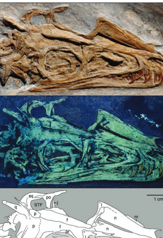
Fig. 8. Photograph of the skull of Juravenator starki (JME Sch 200) under normal (A) and ultraviolet (B) light, and
interpretive drawing (C). Abbreviations: an, angular; AF, antorbital fenestra; d, dentary; EN, external naris; f, frontal; ITF, infratemporal fenestra; j, jugal; l, lacrimal; m, maxilla; MF, maxillary fenestra; n, nasal; OR, orbit; p, parietal; pl, palatine; pm, premaxilla; po, postorbital; pr, prefrontal; pt, pterygoid; q, quadrate; qj, quadratojugal; sa, surangular; sq, squamosal; sr, sclerotic ring; STF, supratemporal fenestra. Arrows point at dentary teeth.
premaxillary-maxillary diastema in the holotype of Compsognathus longipes (BSPG AS I 563), however PEYER(2006) has argued that such a feature (absent in MNHM CNJ 79) may be a preservational artifact. Our examination of BSPG AS I 563 failed to corroborate OSTROM’s claim; as he pointed out, the ends of the premaxilla and maxilla are poorly preserved. A dia -stema is absent in Sinosauropteryx (CURRIE& CHEN 2001), Ornitholestes (OSBORN1916), Scipionyx (DAL SASSO& SIGNORE1998), and Huaxiagnathus (HWANG et al. 2004), and it is most likely that this feature was also absent in Sinocalliopteryx and Compsognathus (Fig. 11). A distinct indentation is present on the anterior dentigerous margin of Juravenator, between its second and third maxillary teeth (Figs. 8, 10). This feature appears to be somewhat exaggerated by bone weathering, but we believe it to be real and unique to Juravenator among “compsognathids”. Posterior to this indentation, the ventral margin of the maxilla in lateral view is straight throughout the extension of the tooth row, becoming slightly concave behind the last tooth. On the caudoventral corner of the maxilla, the contact between this bone and either the lacrimal or the jugal cannot be distinguished.
The maxilla does not seem to form part of the dorsal margin of the antorbital fossa; this margin appears to be formed solely by the nasal and lacrimal (Fig. 8). There is no evidence indicating that the antor-bital pneumaticity extended into the nasal – this bone appears to just form the dorsal margin of the antorbital fossa. A thin maxillary sheet – the medial lamina of the ascending ramus of the maxilla – lines the anterior fourth of the antorbital fossa. The center of this portion is punctuated by a round maxillary fenestra; however, due to poor preservation, the existence of a promaxillary fenestra cannot be determined. A narrow portion of bone extends from the base of the medial lamina of the ascending ramus caudoventrally as a long inset that defines the ventral margin of the antor-bital fenestra.
The nasals are exposed in laterodorsal (right ele-ment) and dorsal (left eleele-ment) views. These bones form the caudal and dorsocaudal margins of the external nares (Fig. 8). The internarial process of the nasal extends for about two-thirds the length of the nares. The premaxillary process of the nasal forms a broad arch, the base of which forms the caudal margin of the nares, and its tip runs between the premaxilla and the maxilla. The main body of the nasal becomes gradually wider towards the caudal end of the bone. The lateral and medial margins are straight, and
Fig. 9. Comparisons of the premaxilla and lacrimal of
Jura-venator starki (A), Sinosauropteryx prima (B, based on CURRIE& CHEN2001; C, based on GMV 2124),
Compso-gnathus longipes (D, based on PEYER2006), Ornitholestes hermanni (E, based on AMNH 619), Scipionyx samniticus (F, based on DALSASSO& SIGNORE1998), and
Huaxiagna-thus orientalis (G, based on HWANGet al. 2004).
the latter forms a long suture with its counterpart. Dorsally, the surface of the nasal is essentially flat, although the central portion of the lateral edge is slightly swollen. The caudal end of this bone contacts the frontal in a more or less transversally oriented suture, although the precise shape of this suture is not clear.
The lacrimals are exposed in both lateral (right element) and medial (left element) views. This bone has the shape of an inverted “L” and as in other “compsognathids”, it lacks any evidence of a “horn” or dorsal protuberance (Fig. 8). The inverted “L” shape of this bone more closely resembles the lacrimals of Scipionyx (DALSASSO & SIGNORE 1998) and
Comp-Fig. 10. Reconstructions of the skulls of
Juravenator starki (A), Compsognathus longipes (B, based on PEYER2006),
Scipionyx samniticus (C, based on DALSASSO& SIGNORE1998), and
Ornitholestes hermanni (D, based on AMNH 619). Hatched areas indicate missing portions of the skull. Drawings not to scale.
sognathus (PEYER2006) than that of Sinocalliopteryx (JI et al. 2007a), Sinosauropteryx (GMV 2124; pers. obs.), and basal tyrannosauroids (XU et al. 2004; SERENOet al. 2009), in which the dorsocaudal corner extends more caudally (i. e., it has a more “T”-shaped appearance) (Fig. 9). On the lateral surface, a minute swell marks the caudodorsal corner of the bone and anterior to this swell there is a small pit that we inter-pret as a pneumatopore (lacrimal fenestra), a feature widely distributed among non-avian theropods (RAUHUT 2003). If our interpretation is correct, the presence of a pneumatic lacrimal in Juravenator would distinguish this taxon from basal coelurosaurs such as Compsognathus (PEYER 2006), Sinosauro -pteryx (CURRIE& CHEN2001), Scipionyx (DALSASSO,
pers. comm.) and apparently Sinocalliopteryx (JIet al. 2007a), in which the lacrimal is apneumatic.
The nasal (horizontal) ramus of the lacrimal is sub-equal in length to the jugal (vertical) ramus; this con-dition is similar to that of Compsognathus (MNHM CNJ 79) but different from those of Scipionyx (DAL SASSO& SIGNORE1998) and Sinosauropteryx (GMV 2124) in which the nasal ramus is either shorter or longer than the jugal ramus, respectively (Fig. 9). The nasal ramus gradually tapers rostrally, ending in a sharp tip. This ramus articulates medially with the nasal, and laterally forms the dorsal margin of the caudal half of the antorbital fenestra. The jugal ramus is stout and hourglass-shaped, with expanded dorsal and ventral ends. This overall appearance of the ramus is comparable to those in Scipionyx and Compso -gnathus but clearly stouter than the slender jugal ramus of Sinosauropteryx (Fig. 9). The ventral half of the jugal ramus is laterally concave; the surface is excavated by an ample trough caudally defined by a broad and prominent rim that forms the caudal margin of the bone. Such a trough marks the caudal extension of the antorbital fossa, a condition also present in a variety of other non-avian theropods (e. g., OSTROM 1978; CURRIE & ZHAO 1993; XU et al. 2004, 2006; CARPENTERet al. 2005a; CORIA& CURRIE2006). The medial surface of the lacrimal is essentially flat. The contact between the lacrimal and the jugal is a sub -horizontal suture.
The right jugal is exposed in lateral view; a frag-ment of what appears to be the left elefrag-ment, probably in medial view, is visible in the caudal corner of the left orbit. The jugal is slender; it is dorsoventrally thinner at the center of the orbit (Fig. 8). The dorsal and ventral margins of this bone are concave and straight, respectively. Caudally, the postorbital ramus is well developed and projected dorsocaudally at a 120 degree angle with respect to the horizontal body of the jugal (i. e., the suborbital ramus). The postorbital ramus tapers to a sharp end at the midheight of the orbit. The anterior margin of this ramus forms a slightly concave contact with the jugal ramus of the postorbital. Caudally, the jugal ends in a tapering pro-cess (subtemporal ramus) shortly behind the base of the postorbital ramus. There is no apparent evidence of jugal pneumaticity. The overall morphology of the jugal agrees well with that described for Compso -gnathus (OSTROM1978; MICHARD1991), Sinosaurop-teryx (CURRIE & CHEN 2001), Scipionyx (CURRIE & CHEN2001), and Huaxiagnathus (HWANGet al. 2004). However, the subtemporal ramus of this bone is
shor-Fig. 11. Dentition of Juravenator starki (JME Sch 200).
Tooth row (A) and detail (B, C) of middle premaxillary teeth. These teeth are interpreted to occupy the second and third positions of a probably four-toothed premaxilla.
ter than those of Huaxiagnathus and Sinosauropteryx. We interpret a slender bone rimming the dorsocra-nial half of the right orbit as the prefrontal. Although it is unclear how much of this bone is actually pre -served, if our interpretation is correct, this bone pre-vents most, if not all, contribution of the frontal to the orbit (Fig. 8). Comparable ossifications are preserved rimming the orbits of Compsognathus and Scipionyx (Fig. 10).
The two frontals are preserved in dorsal view. This bone is long and dorsally flat, has a transversal contact with the nasal and a straight suture with its counterpart (Fig. 8). Caudally, the width of the frontal in -creases gradually, however, its contact with the parietal is not clear on either side – we have inter -preted a transversal line, roughly perpendicular to the interfrontal suture, as the frontoparietal contact (Fig. 8). If this is correct, the frontal participates exten -sively of the supratemporal fenestra, a condition simi-lar to that apparently present in Huaxiagnathus (HWANG et al. 2004). At the same time, the fronto -parietal suture interpreted here more closely resem-bles the sigmoid suture of the French Compsognathus (MNHM CNJ 79) than the very forked frontoparietal suture featured by OSTROM (1978) for the holotype (BSPG AS I 563).
The postorbital is exposed on lateral (right element) and medial (left element) views. This T-shaped bone has a frontal ramus slightly longer than the squamosal ramus (Fig. 8). These two rami are not at the same level; while the squamosal ramus points caudally, the frontal ramus is somewhat dorsorostrally directed. The dorsal margin of the right element, at the junction of these two rami, exhibits a small indentation. Such an indentation is absent in Scipionyx, in which the post -orbital also differs by having the frontal and squamo-sal rami essentially aligned. The tapering jugal ramus is longer than the other two rami and it gently curves rostroventrally; its convex caudal margin contacts nearly the entire length of the postorbital ramus of the jugal and its rostral margin forms the concave caudal margin of the orbit.
Portions of both squamosals are preserved. The right element is exposed laterally and partially dor sally – this bone appears to be displaced caudoven -trally – and the left element is represented only by its postorbital process exposed in medial view. The squa-mosal is a multibranched bone, and its complex morphology is best interpreted under UV light (Fig. 8). Two subvertical processes define a broad ventral notch, most probably for the articulation of the
qua-drate. The caudalmost of these projections is a pointed process that presumably contacted the paraoccipital process, although the latter is not preserved. The other process, the quadratojugal process, is a longer and also tapering projection, the tip of which is overlapped by a fragment of a bone interpreted as a remnant of the quadratojugal. A third process projects mediodorsally from the caudal margin of the squamosal and appears to abut the skull roof; this process probably forms the caudal margin of the supratemporal fenestra. A fourth process is directed rostrally. This postorbital process is best preserved on the left element, even if viewed medially. The postorbital process ends in a fork that receives the squamosal ramus of the postorbital bone and together with this ramus forms the intertemporal bar (division between supra and infratemporal fene-strae).
Less certain anatomical information is available for the quadratojugal. Only what we interpret as the tips of the squamosal and jugal processes of the right element are preserved (Fig. 8). If correctly identified, the preserved fragment of the squamosal process would suggest that the quadratojugal contacted the squamosal behind the infratemporal fenestra. Like-wise, if this interpretation is correct, it would indicate that the squamosal process of the quadratojugal of Juravenator is much thinner than the broad equivalent of Sinosauropteryx (GMV 2124; pers. Obs.). The por-tion interpreted as the jugal process is slender and together with the squamosal process gives the bone an “L” shape (Fig. 9).
The parietals are also poorly preserved, with both of them in dorsal view. The length of this bone appears to be roughly one-third the length of the frontal (Fig. 8). Dorsally, it is slightly convex; as in Compso -gnathus (OSTROM1978; MICHARD1991; PEYER2006) and Scipionyx (DALSASSO& SIGNORE1998), a sagittal crest running along the skull roof was clearly absent. The caudal margin of both parietals is defined by a thick rim that forms the nuchal crest, which appears to be stronger than the weak crest of Compsognathus (BSPG AS I 563).
A long and very poorly preserved bone situated in the caudolateral corner of the left portion of the skull is interpreted as a portion of the quadrate (Fig. 8). This bone exhibits a concave outer surface and under UV light, what appears to be a terminal articulation. Our identification of this bone is based on both its location and its elongate shape.
The vaulted, rostral portions of the palatines (vomerine processes) are among the few portions of
the palate that can be determined with certainty. These bones, preserved within the antorbital fenestra, are best seen under UV light (Fig. 8). The vomerine process of the right palatine is in place and situated within the caudal half of the fenestra. Its left counter-part is displaced, protruding towards the right side of the skull at the center of the antorbital fenestra. As in other non-avian theropods (e. g., MADSEN 1976; CURRIE& ZHAO1993; DALSASSO& SIGNORE1998; NORELL & MAKOVICKY 2004), the vomerine pro -cesses of the palatines have a strongly concave rostral margin that forms the caudal border of the choana, and a caudorostrally expanded dorsal (top) margin. These processes most likely contacted each other but this cannot be ascertained due to the displacement of the left one. A long and slender bone rostrocaudally crossing the orbit probably represents a portion of one of the pterygoids, although no morphological details of these bones are visible.
Very little anatomical information is available for the braincase, which is partially exposed in dorsal view. The supraoccipital has a pronounced midline ridge and the paroccipital processes are robust and tall. The latter project posteroventrally at approximate-ly 45 degrees, and their posterior surface extends over a single plane. A thin sliver of bone, which may re -present the stapes, anteriorly lines each paraoccipital processes.
M a n d i b u l a r b o n e s . – Only the lateral side of the right mandible is exposed and its dorsal border is over-lapped by the ventral margin of the cranium. However, portions of some mandibular teeth have been exposed by preparation of the lateral surface of the dentary. The first exposed tooth is aligned with the broken base of the last premaxillary tooth (Figs. 8, 10), and the latter covers the tip of the former. The last exposed dentary tooth lies in front of the last two maxillary teeth; however, it is not possible to determine whether the mandibular dentition ended in front (as in many non-avian theropods) or behind (as in Scipionyx) the upper tooth row. Although the number of mandibular teeth cannot be ascertained with precision, the size of the few exposed suggest a tooth count significantly smaller than the 18 teeth counted by OSTROM(1978) or the 21-22 counted by MICHARD(1991) and PEYER (2006), for Compsognathus. Assuming the mandibular tooth row to be slightly shorter than the upper tooth row, as in Compsognathus (OSTROM 1978) and most other theropods (e. g., MADSEN1976; COLBERT1989; WEISHAMPELet al. 2004), we estimate that the number
of dentary teeth did not exceed 11, a number that more closely resembles our estimate of the tooth count of Scipionyx (12-14). Likewise, the lower number of den-tary teeth of Juravenator when compared to Compso-gnathus can also be inferred by the fact that the length of the dorsal margin of its dentary is significantly shorter; OSTROM(1978) reconstructed the dorsal mar-gin of the dentary of Compsognathus as more than half the length of the skull while it is less than half the length of the skull in Juravenator (and in Scipionyx). Allometric studies on tyrannosaurids (CURRIE 2003) and other non-avian theropods (e. g., MADSEN 1976; COLBERT 1989) have failed to provide evidence in -dicating an increase in the number of teeth in relation with either size or age. However, the number of dentary teeth increases from 18 to 21-22 in Compso-gnathus, when the smaller holotype is compared to the larger French specimen (OSTROM1978; MICHARD 1991; PEYER 2006). Individual variations notwith -standing (COLBERT1989; CURRIE2003; PEYER2006) – and taking into account that the increment in tooth count in Compsognathus may suggest a similar in crement in Juravenator – we still regard this differ -ence as taxonomically important.
The lateral height of the rostral portion of the mandible is approximately two-thirds the mandibular height at the level of the orbit (Fig. 8); this ratio is about one-third in Sinosauropteryx (GMV 2124; pers. obs.), and onehalf in Scipionyx and basal tyranno -sauroids (XUet al. 2004, 2006) The lateral surface of the mandible is essentially flat along its rostral half and slightly convex more caudally. The lower jaw lacks a mandibular foramen, a condition similar to that in Compsognathus (OSTROM 1978), Huaxiagna-thus (HWANGet al. 2004), Sinosauropteryx (CURRIE& CHEN 2001), Sinocalliopteryx (JI et al. 2007a), and Scipionyx (DAL SASSO, pers. comm.). The ventral margin of the dentary is nearly straight (Fig. 8). A gently and long caudoventrally slanting suture sepa -rates this bone from the surangular and the angular. Although the postdentary portion of the mandible is partially covered by the jugal, it can be seen that the dorsoventral width of the surangular is about three times that of the angular. The ventral margin of the surangular bears a faint longitudinal rim that forms a ledge at the contact with the angular. The latter bone is narrowly exposed laterally. The rear portion of the mandible is missing beyond the surangular and angu-lar, and it is difficult to evaluate whether the caudal portions of these bones are missing as well.
C r a n i a l o p e n i n g s . – The external nares are de -fined by the premaxilla dorsally, anteriorly, and ven-trally, and the nasal posteriorly. They are subelliptical in shape with a straight ventral margin and concave dorsal margin (Figs. 8). The external nares of Jura venator end substantially in front (~ 4 mm) of the an -terior margin of the antorbital fossa. This condition seems to contrast with that of other “compso -gnathids”. The caudal end of the nares of Scipionyx (DAL SASSO & SIGNORE 1998) and Compsognathus (PEYER 2006) is aligned with the anterior margin of the antorbital fossa and HWANGet al. (2004) described the long nares of Huaxiagnathus as overlapping posteriorly the antorbital fossa (yet, this is not entirely apparent in the illustrations provided by HWANGet al. 2004).
The antorbital fossa contains what appear to be a round and small maxillary fenestra and a large antor-bital fenestra. Whether Juravenator also possessed a promaxillary fenestra is uncertain. The latter is bor -dered by the lacrimal and nasal dorsally, the maxilla rostrally and ventrally, and the lacrimal caudally; whether or not the jugal contributed to the antorbital fossa, and perhaps the fenestra, is unclear (Fig. 8). The antorbital fenestra is suboval, although its vertical caudal margin defines a nearly 90˚ angle with its ventral counterpart. The length of this fenestra is approximately threefourths the length of the antor -bital fossa – proportionally longer than in Scipionyx and Compsognathus (fenestra/fossa ratio is about 2/3) and Sinosauropteryx (fenestra/fossa ratio is between 2/3 and 1/2) (Fig. 11).
Both orbits are preserved; the left one is incom plete and the right one is slightly collapsed by the ventral displacement of the right frontal. However, it is clear that the orbit was round and bordered by equal con -tributions of the lacrimal, jugal, postorbital, and what we interpreted as the prefrontal (Figs. 8, 11). It is not clear if the frontal comprised part of the dorsal margin of the orbit, or whether a postorbital-prefrontal contact prevented a frontal contribution. Most remarkable is the fact that the length of the orbit is approximately the same as that of the antorbital fenestra, a condition similar to that of basal tyranosauroids (XUet al. 2004, 2006) but an obvious difference with Sinosauropteryx (CURRIE & CHEN 2001), Compsognathus (BIDAR et al. 1972; OSTROM1978; MICHARD1991), Ornitholestes (OSBORN1916), and Scipionyx (DALSASSO& SIGNORE 1998), in which the antorbital fenestra is substantially shorter (Fig. 11). The rostral half of the sclerotic ring is preserved within the right orbit but neither the
shape nor the number of the scleral oscicles can be determined.
Both of the temporal fenestrae are distorted. Our interpretation of the quadratojugal suggests an in -fratemporal fenestra enclosed caudally by a squa -mosal-quadratojugal bar and with a subrectangular shape, taller than wide (Fig. 8). On the left side, the skull roof and the squamosal-postorbital bar surround a large fenestra. This opening is likely the supra -temporal fenestra in dorsal view but its shape and size may be modified by the fact that the latter bar is clearly displaced and viewed medially.
D e n t i t i o n . – As stated above (see Cranial bones), three premaxillary and eight maxillary teeth are preserved in the upper jaw; the premaxilla may have carried an additional tooth but this is unlikely to be the case for the maxilla. The tooth count of the dentary is unknown (see Mandibular bones) but we estimate it to be no more than 11.
The first preserved premaxillary tooth (here inter-preted as occupying the second position) is less com-pressed than the others (Fig. 10), like the “incisiform” premaxillary teeth of other non-avian theropods (SANDER 1997). The third maxillary tooth is the lar-gest of the upper series. This tooth aligns with the rostral margin of the antorbital fossa (Figs. 8, 10). Similarly large teeth located at the rostral margin of the antorbital fossa are present in Huaxiagnathus and Compsognathus (MNHM CNJ 79). This large tooth is followed by two other large teeth, thus indicating that the largest teeth are located in the middle portion of the maxilla, similar to the condition in Scipionyx (DAL SASSO& SIGNORE1998) (Fig. 10).
All preserved teeth (from both the upper and lower jaws) lack mesial serrations. The first preserved pre-maxillary tooth also lacks distal serrations but these are present in the second preserved premaxillary tooth (the third one is highly incomplete) (Fig. 10). Among basal coelurosaurs, serrated premaxillary teeth are known in the “compsognathid” Sinocalliopteryx (JI et al. 2007a) and tyrannosauroids (XU et al. 2004), although the premaxillary teeth of many basal coelu-rosaurs (and a number of tetanurans) lack serrations (OSTROM1978; CURRIE& CHEN2001). The serrations of the premaxillary teeth of Juravenator and Sinocalli-opteryx are much finer (~ 13/mm) than those of the maxillary teeth (~ 8/mm). All other exposed teeth of Juravenator also have distal serrations. As in Compso-gnathus (BSPG AS I 563), basal tyranosauroids (XU et al. 2004, 2006; SERENO et al. 2009), and possibly
Huaxiagnathus (HWANG et al. 2004) and Sinocalli -opteryx (JIet al. 2007a), the maxillary and posterior dentary teeth of Juravenator are more recurved than the premaxillary teeth (however, minimal information is available for the dentary teeth). This appears to be a difference between these taxa and either Sinosauro pteryx or Scipionyx, where the teeth are more homo -genous in shape.
5.2. Postcranium
A x i a l s ke l e t o n . – The vertebral column is essen -tially complete and articulated from the skull to the distal third of the tail. The cervical and dorsal series are exposed primarily in dorsal view (Fig. 12), the sacral region is dislocated due to the rotation of the skeleton at this region, and the caudal series is ex -posed on right lateral view.
Fig. 12. Cervical (A) and dorsal (B) vertebral series of Juravenator starki (JME Sch 200). Arrows point to a very thin
The cervical region is not well-preserved, espe -cially its cranial half. The number of cervicals cannot be established with precision because no sternum has been preserved (thus, the separation between cervical and dorsal vertebrae cannot be determined on the basis of the articulation of ribs to this bone) and because most cervical ribs are missing. Seven verte-brae are preserved between the first one preserving a long rib of thoracic (dorsal) morphology (Figs. 3-5, 12) and a fragmentary element (presumably a cranial-most cervical) adjacent to the caudal portion of the skull. However, the possibility of an unaccounted cervical at the junction of the skull block with the body block cannot be ruled out; this region was da -maged as a result of the breakup of the entire slab in two blocks (Fig. 3). All of this suggests the presence of nine to ten cervicals including the atlas, which it is not discernable from the mass of bones exposed imme -diately behind the skull.
When visible, the cervical vertebrae have short and low neural spines (Fig. 12). However, those in the middle (for example, the third vertebra in front of the first dorsal) appear to have taller neural spines that the caudalmost elements. The diapophyses are well-developed. The lengths of these processes increase gradually from the cranialmost elements to the
caudal-most ones. Each zygapophysis (pre- and postzygapo-physes) is well-spaced with respect to its counterpart. In the caudal portion of the series, the postzygapo -physes decrease in length towards the back. The third cervical in front of the first dorsal exhibits a small, round foramen piercing the center of the centrum – this is the only possible evidence of pneumaticity in the cervical series (Fig. 12). There is no information available about the central articular surfaces.
There are at least 13 dorsal vertebrae, all of them exposed dorsally with the exception of the last one, which is exposed on its right side. The dorsal verte-brae have well-developed transverse processes that slightly angle posteriorly (Fig. 12). The neural spines of these vertebrae are generally poorly preserved. A few vertical ridges crushed against the center of some centra are interpreted as remains of neural spines but these provide minimal anatomical information. Nonetheless, the craniocaudal extension of the neural spines of the posterior dorsals appears to be large; in some instances these ridges are preserved projecting caudally between the postzygapophyses. The morpho-logy of the neural spine is clearer in the last dorsal vertebra. In this vertebra, the caudal margin of the neural arch and the posterior portion of the neural spine are exposed in lateral view, apparently over
Fig. 13. Photograph (A) and interpretive drawing (B) of the pelvic region of Juravenator starki (JME Sch 200). Abbre
-viations: af, antitrochanteric facet; bf, brevis fossa; dv, dorsal vertebra; fe, femur; il, ilium; is, ischium; pp, pubic pedicel; pu, pubis; sv, sacral vertebra; r and l refer to the right and left element.
lapping the anteriormost portion of the left preaceta-bular wing. Although the neural spine of this vertebra is not exposed completely, the observable portion shows the distal expansion characteristic of the neural spines of the posterior dorsals of Compsognathus (OSTROM1978, PEYER2006), Huaxiagnathus (HWANG et al. 2004), Scipionyx (DALSASSO, pers. comm.), Sino -sauropteryx (CURRIE& CHEN2001), Sinocalliopteryx (JI et al. 2007a), and the Brazilian compsognathid Mirischia (NAISH et al. 2004). The caudal articular surface of the last dorsal centrum is flat, thus sug-gesting that dorsal vertebrae had amphiplatyan centra. The exposure of the vertebrae prevents us from de -termining whether there is any evidence of pneu-maticity.
The number and general morphology of the sacral vertebrae is also problematic (Fig. 13). The sacrum appears to be disarticulated and is partially covered by other bones. A number of unidentifiable bones are also exposed in this area. The fact that none of the bones identified as sacral centra are coossified to one another suggests that the sacrum was not fused at the time the animal died – the complete lack of fusion between sacral vertebrae may be another feature sug-gesting an early ontogenetic age for the holotype of Juravenator. A laterally exposed centrum located immediately caudal to the thirteenth dorsal vertebra is regarded as part of the first sacral vertebra. The cen-trum is substantially larger than that of the last dorsal and it has a subquadrangular shape. The interpretation of this centrum as that of the first sacral is based on the fact that (1) it already lies within the realm of the ilia and (2) its lateral surface is broadly recessed, resembling the condition of a centrum lying between the two ilia (a vertebra more clearly identified as sacral). The latter centrum is crushed against the medial surface of the postacetabular wing of the left ilium. The lateral surface of this centrum is distinctly recessed and a ridge defines the periphery of the cen-trum. A disarticulated centrum of similar morphology lies ventral to the postacetabular wing of the left ilium. The lateral recess of this centrum is very distinct and its caudal articular surface is rounded and flat. It is unclear whether this centrum corresponds to another sacral vertebra or to the first caudal. A gap between the articulated portion of the tail and the sacral cen-trum crushed against the medial surface of the posta-cetabular wing suggests that the disarticulated cen-trum beneath the left ilium could belong to the first caudal (Fig. 13). However, if this is accepted, the cen-trum of the first caudal would have greatly differed
from those of the succeeding tail vertebrae. Compari-sons with “compsognathids” and other basal coeluro-saurs are problematic because pelvic bones often cover the sacral vertebrae. Nonetheless, the subquadrangular shape of the first sacral vertebra of Jura -venator and the fact that its sacrals are substantially bigger than both dorsal and caudal vertebrae resem-bles the condition present in Scipionyx. However, these features contrast with those in Mirischia, in which the sacrals are smaller than the dorsals (centra are shorter and more depressed) and their centra are not laterally recessed (NAISHet al. 2004).
All preserved caudals are articulated and exposed in right lateral view (Figs. 3-5, 14). The tail includes 44 vertebrae preserved in articulation but it is unclear whether the centrum of the first caudal is missing (see above). Comparisons of the number of caudal verte-brae and relative tail length with other basal coeluro-saurs are hampered by the incomplete nature of the tail of most taxa (e. g., OSTROM 1978; DALSASSO & SIGNORE1998; KIRKLANDet al. 1998; DEKLERKet al. 2000; HWANGet al. 2004; NAISHet al. 2004). How -ever, meaningful comparisons can be established with the tail of the holotype of Sinosauropteryx (NIGP 127586, CHENet al. 1998; CURRIE& CHEN2001). The nearly complete tail of NIGP 127586 preserves 64 caudal vertebrae. In this specimen, the last 20 verte-brae represent approximately 44 % of the total length of the tail and the entire length of the tail is approxi-mately 170 % of the snout-vent length (measured from the tip of the snout to the caudal end of the ilium). The preserved portion of the tail of Juravenator is approxi-mately 125 % of the snout-vent length but when the proportions observed in Sinosauropteryx (i. e., that the last 20 vertebrae constitute 44 % of the tail) are extra-polated to the Schamhaupten fossil, the length of the tail of Juravenator is estimated to represent about 180 % of the snout-vent length. This extrapolation is warranted by the fact that the ratio between the com -bined length of the first 43 caudal vertebrae and the snout-vent length of NIGP 127586 is approximately 118 %, a value slightly shorter than the one observed in Juravenator. All of this suggests that the tail of Juravenator was probably as long, if not longer, than the tail of Sinosauropteryx – the proportionally longest known tail within theropods (CHENet al. 1998).
The length of the caudal centra remains more or less constant (< 1mm in difference) until the 17th
caudal, when the length of the centrum starts increa-sing (Fig. 14). At the level of the 27th caudal, the
it remains as long with minor variance until the 35th
caudal. Distal from this point, the length of the caudal centra becomes gradually shorter, but the last pre -served vertebra is still longer than any of the first seventeen elements of the tail. This pattern is similar to that of Huaxiagnathus, in which the length of the caudal centra remains more or less constant until the 14thcaudal and then increases to a higher plateau for
the subsequent 11 caudals (only 25 caudals are pre -served in this taxon [HWANG et al. 2004]). It also approaches that of the compsognathid Sinocalli -opteryx (JIet al. 2007a) in which the longest caudals are between the 20thand the 23rdvertebrae. This trend,
however, is markedly different from that of Sinosaur-opteryx, in which the length of the centra increases until the sixth caudal but then decreases until the end of the tail (CURRIE& CHEN2001). It is also different from the one observed in Compsognathus (BSPG AS I 563 and MNHN CNJ 79 preserve approximately
15 [OSTROM1978] and 30 [MICHARD1991] elements, respectively) and Scipionyx (less than 10 vertebrae preserved) in which the length of the centra gradually increases throughout the preserved portion of the tail.
The centra are elongated and morphologically very similar throughout the entire length of the tail (Fig. 14). The height of the centrum decreases gradually throughout the tail in such a way that in the proximal caudals the mid-height of the centrum is approximate-ly half the length of the centrum (with an average of 55 % for the first five preserved caudals) and in the distal caudals the height is nearly one-quarter of the central length (with an average of 27 % for the first five preserved caudals). The general depression of the caudal centra towards the tail’s distal end is shared by Compsognathus (PEYER2006) but not by Huaxia -gnathus, in which the caudal centra remain approxi-mately the same relative height throughout the 25 preserved elements of its tail (HWANGet al. 2004). The
Fig. 14. Caudal vertebral series (A) of Juravenator starki (JME Sch 200) with close-ups of the distal (B), middle (C),
neural spines are long and caudally oriented in the first three caudals of Juravenator – the neural spine of the third caudal extends back to the middle of the fourth caudal. The neural spine of the latter vertebra is shorter but still somewhat inclined; its morphology transitions with the shorter and vertically oriented neural spines of the subsequent vertebrae. The neural spine of the fifth caudal is dorsally expanded and
fan-shaped, and this condition appears to remain present in more distal neural spines (however, the morphology of some of these processes is not clear in every verte-bra). The transverse processes are poorly preserved. They appear to be well-developed in the proximal cau-dals but their presence in more distal caucau-dals cannot be determined. The poor preservation of the transverse processes and limited preparation in areas covered by soft tissue (see Soft tissue below) complicates the assessment of the transition point. However, remnants of a moderately tall neural spine are visible down to the 14thcaudal. It is difficult to determine if a neural
spine of any significant height was present in the 15th
caudal but it seems clear that the neural spines were reduced from the 16th caudal onwards. Thus, we
regard the transition point to be around the 14th or
15th caudal vertebrae (Fig. 14), an estimate more
or less comparable to that provided for Compso -gnathus (MICHARD1991), Huaxiagnathus (HWANGet al. 2004), and other basal coelurosaurs (HOLTZ et al. 2004). None of the centra exhibit evidence of pneu -maticity.
The proximal zygapophyses are not well-preserved. However, it seems apparent that they were short. The morphology of the zygapophyses can be better ob -served in the middle and distal portion of the tail. Although still difficult to determine, the morphology of the zygapophyses of the middle caudals (roughly between the 16th and the 35th element) is strikingly
unique (Figs. 14, 15). These zygapophyses are tall and slender, relatively short, and remarkably arched, forming a “bow” over the intercentral articulation. The exact contribution of the pre- and postzygapophyses to this bow-like articulation is unclear but both zygapophyses seem to participate in this joint. The morphology of the zygapophyses becomes more con-ventional from the 36thcaudal onwards, when the
pre-zygapophyses gradually become straighter and longer, overlapping substantially (but less than half) the length of the preceding centrum.
The chevrons are elongate and rod-shaped through-out the tail (Fig. 14). There are 25-26 chevrons in articulation with the first 25-27 vertebrae – the last ones are very small and difficult to see but a chevron appears to be present between the 31st and the 32nd
caudal. The length of the chevrons decreases gra-dually towards the distal part of the tail. The last few chevrons are approximately one-third the length of the proximal ones (e. g., the 6th chevron is 8.8 mm long
and the 22ndchevron is 3.0 mm long). While the
pro-ximal chevrons tend to be caudally bowed, the middle
Fig. 15. Comparisons of middle caudals (vertebrae 19 and
20) of Juravenator starki (A), Compsognathus longipes (B, based on PEYER2006), Sinosauropteryx prima (C, based
on CURRIE & CHEN2001), and Huaxiagnathus orientalis
(D, based on photographs of CAGS-IG02-301 provided by S. Hwang). Abbreviations: ha, haemal arch.
ones are straight. These bones are articulated ob -liquely until the 20th chevron but they lay parallel to
the vertebral central onwards. The morphology and orientation of the chevrons of Juravenator is com -parable to that of Huaxiagnathus (HWANGet al. 2004), Compsognathus (MICHARD 1991), Sinocalliopteryx (JI et al. 2007a), and as far as it can be compared, Scipionyx (DALSASSO& SIGNORE1998). The chevrons of Huaxiagnathus and Compsognathus differ somewhat from the more spatulated ones of Sinosauro -pteryx (CURRIE& CHEN2001) – the middle chevrons of the latter taxon are also hooked as opposed to straight as in Juravenator.
Only a few cervical ribs are preserved, although the one corresponding to the penultimate element of the series is best exposed. These long and extremely thin ribs exhibit the characteristic “hair-like” condition (Fig. 12) of the cervical ribs of Sinosauropteryx (CURRIE & CHEN 2001), Compsognathus (PEYER 2006), Sinocalliopteryx (JIet al. 2007a), and Scipionyx (DAL SASSO, pers. comm.). Twelve pairs of thoracic ribs are preserved in near articulation, possibly repre-senting the complete ribcage (Figs. 3-5, 12). The ribs
are composed of a single element (i. e., without sternal segments) and they show no evidence of ossified un -cinate processes. They become thinner and shorter towards the caudal portion of the ribcage. Although those from the left cranial half are straight, the gently curved outline of the ribcage is better insinuated on the right side.
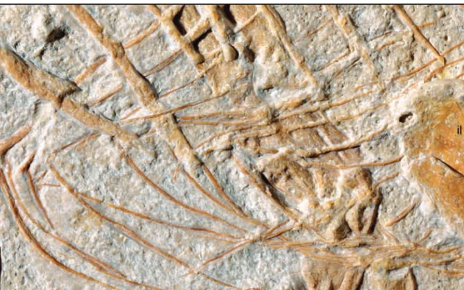
A full set of partially articulated gastralia is pre -served in front of the pelvis (Figs. 3-5, 16). There are more than 13 rows of gastralia, a condition appro -aching the 15 or so described for Sinocalliopteryx (JI et al. 2007a). The first row is the thickest and a pair of single gastralia forms it. These elements connect at their medial end forming a short cranial process. Two pairs of filament-like segments articulated in a zigzag pattern form subsequent rows of gastralia. Like in all other theropods (CLAESSENS2004), each of these pairs is formed by two overlapping elements. When visible, the medial segments are longer than the lateral one, a condition similar to that reported in Sinocalliopteryx (JI et al. 2007a) and Huaxiagnathus (HWANG et al. 2004).
Appendicular skeleton. – Both scapulae are pre -served in approximately their life-orientation – nearly perpendicular to the vertebral column and with the blade diagonally crossing the rib cage (Figs. 3-5, 17). The left scapula is entirely exposed in lateral view. Its counterpart is beneath several ribs, the left scapular blade, and the vertebral column, and only portions of its medial surface can be seen between these bones. The scapula is a slender and elongate bone, and the blade expands gradually towards its dorsal end (Fig. 17). Its length is approximately ten times its width at mid-shaft and 1.5 times the length of the humerus (Fig. 18) – while the former proportion is comparable to Sinosauropteryx and Compsognathus, the latter is much greater than that in Compsognathus (scapula: humerus is 1 in BSPG AS I 563; OSTROM1978) but similar to that in Sinosauropteryx (Fig. 19). The pro-portion of the length of the scapula to that of the humerus is 1.3 in Sinocalliopteryx, a ratio thus inter-mediate between Juravenator and Compsognathus (JI et al. 2007a). Unlike these taxa, Scipionyx has a stouter scapula – with an estimated length of about seven times the width of the mid-shaft – even if the proportion of this bone with respect to the humerus is comparable to that of Compsognathus. The scapula of Juravenator differs from that of Sinosauropteryx as well as from that of other “compsognathids” by the fact that the narrowest portion of the scapula is at the neck as opposed to near the mid-shaft – in this respect the scapula of Juravenator resembles more that of the basal coelurosaurs Nqwebasaurus (DE KLERK et al. 2000) and Tyrannosaurus [BROCHU 2003; but not basal tyrannosauroids such as Dilong (XUet al. 2004) in which the scapula is very robust. The lateral surface of the scapula is virtually flat, although it is more concave in cross-section along the ventral third of its blade (its basal portion). Based on what is exposed of the right scapula, the medial surface (at least in its basal portion) appears to be convex. In lateral view, the scapular blade is curved caudally; its cranial and caudal margins are convex and concave, respectively (Fig. 17). In this respect, Juravenator also differs from other basal coelurosaurs (e. g., Scipionyx, Sinosauro -pteryx, Sinocallio-pteryx, Compsognathus, Nqweba-saurus, ornithomimids, tyrannosauroids), where the scapular blade is straight or less visibly curved (Fig. 19). The scapula is more abruptly expanded ventrally to form the glenoid and the coracoidal articular sur -face. The former is well-exposed on the left side and directed caudally as in “compsognathids” (CURRIE& CHEN2001; HWANGet al. 2004) and basal theropods
(MADSEN1976; CURRIE& ZHAO1993; WEISHAMPEL et al. 2004). The glenoid is a concave facet, about one-fifth of the width of the ventral end of the scapula, surrounded by a distinct rim, which is more pro -nounced laterally. The acromion is prominent – much more so than in Sinosauropteryx – and subtriangular in shape. Unlike Sinosauropteryx, Sinocalliopteryx, and Huaxiagnathus, this process projects abruptly from the scapular blade and its dorsal margin is di -stinctly concave, a condition that best approaches that of Scipionyx (DAL SASSO & SIGNORE 1998). The lateral margin of the articulation for the coracoid is partially weathered, offering no information.
Neither of the two coracoids is well exposed (Fig. 17). The left element is incomplete and only a portion of it is articulated with the left scapula. The right coracoid appears to be better preserved but the left scapula and ribs unfortunately cover it. Nonetheless, the medial surface of this coracoid exhibits a distinct concavity and a rounded, convex caudoventral margin. We interpret two tiny (approximately 5 mm long), Sshaped, and rodlike bones preserved in disarticu -lation next to the cranioventral end of the left scapula as clavicles (Fig. 17). The ventralmost of these bones exhibits a hook-like end that is possibly a preser -vational artifact. Although the same end is somewhat recurved in the dorsalmost preserved element, it is clearly not hooked as in its counterpart. We interpret this extremity to be the articulation with the scapulo-coracoid and the opposite, medially curved end as the median contact between these bones. If this is correct, these tiny bones would have formed a U-shaped, albeit not fused, “furcula”. Fused clavicles forming a furcula have been reported in a great variety of non-avian theropods (RAUHUT 2003; WEISHAMPEL et al. 2004; NESBITT et al. 2009), including basal coelurosaurs (HWANGet al. 2004; JIet al. 2007a), but unfused cla-vicles are also known for the ceratosaur Segisaurus (CAMP1936; CARRANOet al. 2005; but see RAUHUT [2003] for a different interpretation) (unfused cla-vicles are also known for the Cretaceous bird Hesper-ornis [MARSH1880] and a number of modern birds [BAUMEL & WITMER 1993]). The absence of clavi -cular fusion in Juravenator could well be ontogenetic, although the relation between furcular formation and age is not well understood in non-avian theropods (e. g., the early juvenile Scipionyx has clavicles fused into a furcula). Regardless, if our interpretation of these bones is correct, the “furcula” of Juravenator would have been much more U-shaped – with a sub-stantially narrower interclavicular angle – than those
of other non-avian theropods (e. g., NORELL et al. 1997; MAKOVICKY & CURRIE 1998; DAL SASSO & SIGNORE1998; CLARKet al. 1999; HWANGet al. 2002; RAUHUT2003).
The forelimb is 50 % the length of the hindlimb (measured along their longest digit and including their corresponding claws) (Figs. 3-5), a ratio comparable to that of Huaxiagnathus (48 % according to HWANG et al. [2004]) and Sinocalliopteryx (JIet al. 2007a) and
smaller than that of the basal tyrannosauroid Guan-long (XUet al. 2006). This proportion is substantially greater than the forelimb: hindlimb ratio of Compso-gnathus and Sinosauropteryx (39 % and 36 %, re-spectively, according to HWANG et al. [2004]). The ratio between humerus + radius and femur + tibia of Juravenator is 0.43, slightly greater than that of Huaxiagnathus (0.39; HWANG et al. 2004), Compso-gnathus (0.41 for BSPG AS I 563 [HWANGet al. 2004]
Fig. 17. Photograph (A) and interpretive drawing (B) of the shoulder girdle and clavicles of Juravenator starki (JME
Sch 200). Abbreviations: cl, clavicles; co, coracoid; sc, scapula. Note the intense pitting and grooving of the bone surface of the scapula (A). r and l refer to the right and left element.