HAL Id: hal-00186603
https://hal.archives-ouvertes.fr/hal-00186603
Submitted on 12 Nov 2007
HAL is a multi-disciplinary open access
archive for the deposit and dissemination of
sci-entific research documents, whether they are
pub-lished or not. The documents may come from
teaching and research institutions in France or
abroad, or from public or private research centers.
L’archive ouverte pluridisciplinaire HAL, est
destinée au dépôt et à la diffusion de documents
scientifiques de niveau recherche, publiés ou non,
émanant des établissements d’enseignement et de
recherche français ou étrangers, des laboratoires
publics ou privés.
Towards a Better Understanding of Photo-excited Spin
Alignment Processes Using Silole Diradicals.
Nans Roques, Philippe Gerbier, Yoshio Teki, Sylvie Choua, Petra Lesniakova,
Jean-Pascal Sutter, Philippe Guionneau, Christian Guérin
To cite this version:
Nans Roques, Philippe Gerbier, Yoshio Teki, Sylvie Choua, Petra Lesniakova, et al.. Towards a Better
Understanding of Photo-excited Spin Alignment Processes Using Silole Diradicals.. New Journal
of Chemistry, Royal Society of Chemistry, 2006, 30 (9), pp.1319-1326. �10.1039/b606593g�.
�hal-00186603�
Towards a Better Understanding of Photo-excited Spin Alignment
Processes Using Silole Diradicals
Nans Roques,
aPhilippe Gerbier,*
aYoshio Teki,*
bSylvie Choua,
cPetra Lesniakovà,
cJean-Pascal Sutter,
dPhilippe Guionneau,
eand Christian Guérin.
aReceipt/Acceptance Data [DO NOT ALTER/DELETE THIS TEXT]
5
Publication data [DO NOT ALTER/DELETE THIS TEXT] DOI: 10.1039/b000000x [DO NOT ALTER/DELETE THIS TEXT]
The synthesis of two nitroxide-based diradicals connected to a 2,3,4,5-tetraphenylsilole (TPS) unit, especially designed to present high spin photo-excited states, is reported. While the bisnitronylnitroxide (NN) silole-based diradical, TPSNN, is unstable and experiences a spontaneous fragmentation of its imidazolinic ring into a iso-butylammomium salt, the corresponding 10
bisiminonitroxide (IN), TPSIN, is stable both in solution and in the solid state. This diradical crystallizes in the triclinic P-1 space
group with a = 10.984(1), b = 11.474(1), c = 17.492(1) Å, α = 81.10(1), β = 89.01(1), and γ = 65.71(2)°. Ground state magnetic properties of TPSIN have been investigated by means of SQUID and ESR measurements: the diradical displays weak intramolecular antiferromagnetic interactions (J/kB ≈ -1 K), in agreement with its topology and with the molecular packing
observed in its crystal structure. In order to investigate the magnetic photo-excited states of TPSIN, time-resolved ESR 15
experiments (TRESR) have been performed on this radical species. Despite the presence of both an appropriate topology for the diradical and a triplet photo-excited state for the TPS coupler, no TRESR signal wasobserved for this molecule within the time-scale of the measurement. In addition to the work already published in this field, this result clearly indicates that besides the radical nature, the π-topological requirements and the need of photo-tunable spin-states for the coupler, the flexibility of the molecule also plays a crucial role in the achievement of photo-induced spin alignment processes.
20
Introduction
Control of intramolecular spin alignment and exchange interactions in purely organic spin systems are essential topics in the field of molecule-based magnetism.1, 2 Since most 25
studies are limited to the magnetic ground state, synthetic efforts are currently devoted to the design of high-spin compounds, in which an appropriate topology gives rise to ferromagnetic interactions between the spin bearing units.3-5 Therefore, topologies expected to allow antiferromagnetic 30
interactions, yielding a singlet or low spin ground state, have been generally discredited. The recent observation, for purely organic π-conjugated diradicals, of photo-excited quintet (S=2) states starting from singlet ground state has relaunched the interest in designing systems with the “wrong” topology. 6-35
13
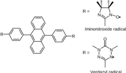
Such systems are generated through photo-induced spin alignment using the π-conjugation between dangling iminonitroxide or verdazyl radicals (S=1/2) and the photo-excited triplet (S=1) state of 9,10-diphenylanthracene (Figure 1). Excited high-spin systems arising from the radical-triplet 40
pairs have also been reported in the pioneering works of
Corvaja et al.14 and Yamauchi et al.15 However, the number of references found in the literature is fairly limited, and in almost all the studies, stable nitroxide radicals have been used as spin bearing units. For purely organic excited high-spin 45
systems, only fullerene, anthracene and pyrene derivatives have been reported as couplers. The search for novel photo-excited high-spin organic systems constructed from both different radicals and triplet couplers is therefore an important research target.
50
In this respect, we decided to investigate the potentiality of the silole to act as a photo-active magnetic coupler. With this idea, we first studied and evidenced the presence of an accessible photo-excited triplet state for the 2,3,4,5-55
tetraphenyl-1,1-dimethylsilole (TPS).16 We also reported previously the synthesis, crystal structure, and ground state magnetic properties of a series of siloles bearing two nitroxide radicals,17,18 as well as the magnetic behaviour upon light irradiation of one of them, the bis tert-butylnitroxide 60
derivative (TPSNO).16 Briefly, the ground state magnetic behaviour of TPSNO is characterized by weak intramolecular antiferromagnetic interactions leading to a singlet ground state. Surprisingly, and despite the existence of a photo-excited triplet state for the parent TPS, no higher spin state 65
was observed for the TPSNO diradical. Assuming that tert-a Laboratoire de Chimie Moléculaire et Organisation du Solide- UMR-
CNRS / UM2 5637, Université Montpellier II, C.C.007, Place E. Bataillon, 34095 Montpellier Cedex 5, France .Fax : +33.4.67.14.3852 ; Tel :+33.4.67.143.972 ; E-mail: gerbier@univ-montp2.fr
b Department of Material Science, Graduate School of Science, Osaka
City University, 3-3-138 Sugimoto Sumiyoshi-ku, Osaka 558-8585, Japan. Fax:+81-6605-8585; E-mai: teki@sci.osaka-cu.ac.jp
c Institut Charles Sadron – UPR-CNRS 22,Université Louis Pasteur, 6
Rue Boussingault - BP 40016 - 67083 Strasbourg Cedex, France. Fax : +33.3.88.414.099 ; Tel : +33.3.88.414.000 ; E-mail: turek@ics.u-strasbg.fr
d Laboratoire de Chimie de Coordination – UPR-CNRS 8241, Université
Paul Sabatier, 205 route de Narbonne, 31077 Toulouse Cedex 4, France. E-mail: sutter@lcc-toulouse.fr
e Institut de Chimie de la Matière Condensée – UPR-CNRS 9048,
Université Bordeaux I, 87, Av. Dr. Schweitzer, 33608 Pessac, France. Tel: +33.5.40.002.279 ; E-mail:guio@icmcb-bordeaux.cnrs.fr R R N N O N N N N O R = R = Iminonitroxide radical Verdazyl radical
Fig. 1 Organic spin systems derived from 9,10-diphenylanthracene in
butylnitroxide groups were not adequate for this purpose, we decided to graft nitronylnitroxide and iminonitroxide spin-bearing units on the silole ring. The choice of these groups was motivated by the fact that they have already allowed the 70
successful spin alignment in the photo-excited states of π-conjugated diradical systems derived from the 1,9-diphenylanthracene pattern.7-13 The design, synthesis, structural and electronic characterizations of a new silole-linked iminonitroxide diradical is presented here in detail. Its 75
magnetic behaviour has been investigated both in the ground and photo-excited states to explore the ability of the silole to act as a photo-active magnetic coupler.
Results
Molecular design
80
π-conjugated spin systems constructed from aromatic hydrocarbons and dangling stable radicals are ideal models to study the relationship between π topology and spin alignment in photo-excited states. The design of the delocalized π-orbital network is of the utmost importance in order to try to 85
anticipate the spin state for both the ground state and the
photo-excited state of the molecule.19 In photo-excited high-spin states the nature of the magnetic exchange coupling between two dangling radical spins through the spin coupler changes from antiferromagnetic to ferromagnetic after photo-90
excitation. The key process, which is an enhanced intersystem crossing (ISC) mechanism is directly related to the attachment of the radical species. ISC mechanism may be expected if the spin bearing units are connected through spin nodal sites to high spin bearing positions of the linker in the 95
photo-excited T1 state. Since the positions adjacent to the
silicon atom are the best spin populated ones in the T1 state, 16
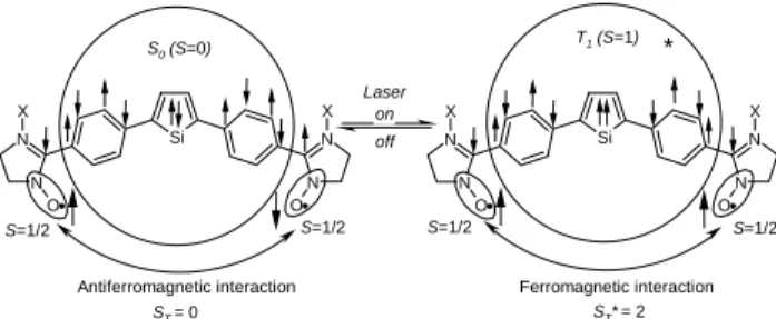
TPSNN and TPSIN (Figure 2) were designed and synthesized by considering the above-mentioned requirements.
100
Synthesis of TPSNN and TPSIN
TPSNN was prepared following the synthetic methodology reported in scheme 1.20 It involves a cross coupling reaction
between organozinc derivative 2 and the Me3Si-protected
dihydroxylamine 4, followed by acidic hydrolysis allowing 105
the removal of the protecting groups. Oxidation of 5 by phase transfer reaction using sodium periodate in water / dichloromethane afforded the crude TPSNN diradical as a very hygroscopic deep green solid in 78% yield. Unfortunately TPSNN was unstable either in concentrated 110
dichloromethane solutions or in the solid state. Concentrated TPSNN solutions yield a mixture of products in which crystallizes a white compound that has been clearly identified as a iso-butylammonium salt. Some examples of nitronylnitroxide photochemical degradation have already 115
been reported by Ullman et al.,21,22 but in all cases the 2,3-dimethylbutane pattern has been identified in the products. To our knowledge, the only skeleton rearrangement that may be invoked to explain the formation of iso-butylamine has been proposed to support the fragmentation mechanism of 120
nitronylnitroxide radicals in electrospray mass spectrometry experiments.23 As shown in Scheme 2, the fragmentation pathway upon electrospray ionization possibly involves the reductive removal of the oxygen atoms followed by a Si N N N N O O X X Si N N N N O O X X on off Laser S0 (S=0) S=1/2 S=1/2 S=1/2 S=1/2 T1 (S=1)
Antiferromagnetic interaction Ferromagnetic interaction
*
ST = 0 ST*= 2
Fig. 2 Expected photo-induced spin alignment and sign inversion of the
effective exchange between two dangling radical spins. TPSNN: X is an oxygen atom, and TPSIN: X is a lone pair. The phenyl rings present at the 3- and 4-positions and the methyl groups borne by the silicon atom have been removed by sake of clarity.
Ph N N O O N N O O Si Ph O Si Ph Ph N N O N N Si Ph Ph Si Ph Ph ZnCl ClZn N N Me3SiO OSiMe3 Br N N HO OH Br N N HO OH N HO Si Ph Ph N OH 1 2 i) ii) iii) iv) v) vi) 3 4 5 TPSNN TPSIN
Scheme 1. i) 4 Np / Li, THF; 4 ZnCl2-TMEDA; ii) 3 Me3SiCl, 3 Et3N, THF; iii) PdCl2(PPh3)2, THF; iv) HCl 0.1M; v) NaIO4, CH2Cl2/H2O; vi) NaNO2,
rearrangement of the transient tetramethylimidazolium cycle 125
into a dimethylaziridinium cation. The further cleavage of the benzylic bond yields a dimethylaziridinium radical cation that may experience a ring-opening transformation to afford the observed iso-butylamine. It is worthy of note that we have no evidence for the validity of this mechanism in our case, but it 130
has the merit to explain this very unexpected fragmentation. TPSIN has been prepared in good yield by treatment of 5 with sodium nitrite in slightly acidic conditions followed by oxidation with sodium periodate. In contrast to TPSNN, the bisimononitroxide radical TPSIN that is stable both in the 135
solid state and in concentrated solution, was purified by
preparative TLC. Orange spearhead monocrystals suitable for X-ray analysis were obtained by slow diffusion from a dichloromethane solution of TPSIN layered with pentane. 140
X-ray structure description
TPSIN crystallizes in the P-1 triclinic space group with two
molecules of TPSIN packed in the unit cell. An ORTEP view is presented in Figure 3. The silole displays a propeller-like arrangement of the four benzene rings as usually observed 145
with the others tetraarylsiloles. The dihedral angles between the mean plane of the central silole and the 2,5-benzene rings bearing the nitroxide radicals have values of 54.2(1)° and 59.8(1)° which are in the range of those previously reported for related siloles. The two iminonitroxide rings are nearly 150
coplanar with the 2,5-phenyl rings and make with them dihedral angles of 2.3(1)° and 7.4(1)° respectively. The N-O···N-O intramolecular distance is of 15.14(3) Å.
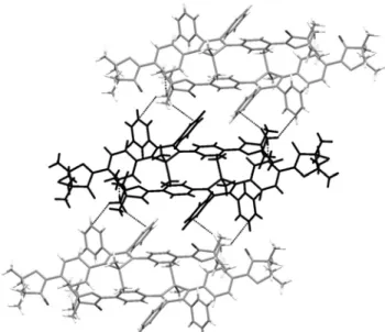
As shown in Figure 4, two C2-H2···O2-N4 weak hydrogen bonds (2.51(2) Å, 134.2(1)°) lead to the formation of a head to 155
tail silole dimer.24,25 The other nitrogen atoms of the same iminonitroxide groups are involved in the formation of two C12-H12···N3 weak hydrogen bonds (2.64(2) Å, 164.8(1)°), that are connecting the dimers together with C38-H38A···π interactions with the phenyl rings connected to C2 160
to form infinite supramolecular stair-like tapes.26 It is worthy to note that N1 and N2 containing iminonitroxide groups are not involved in any supramolecular interaction with neighbouring molecules and that the shortest N-O…N-O distance (6.78(2) Å, N2-O1···N4-O2) is observed in a dimer. 165
Packing of the tapes is assumed through C38-H38B···π and C38-H38C···π interactions with phenyl rings in 3,4-positions (four by molecule, see figure 5).
UV-Visible absorption spectra for siloles 5, TPSNN and
170
TPSIN.
The UV-visible spectra of these compounds have been measured in chloroform. Their data are summarized in Table 1. In the UV-Visible absorption spectra of siloles, it is known that the absorption maximum, ascribed to the π-π* transition 175
of the 2,5-diarylsilole π-conjugated moieties, significantly
N+ N O O N N N N N + .
Scheme 2 Proposed electrospray ionization-induced fragmentation
mechanism for nitronyl nitroxide radicals after.23
Fig. 3 ORTEP view of the molecular structure of TPSIN (thermal
ellipsoids set at 50% of probability).
Fig. 4 Side view of a dimer, showing NO···H weak hydrogen bonds
(dashed lines) and additional N···H weak hydrogen bonds yielding a stair-like arrangement of the dimers to form infinite supramolecular chains.
Fig. 5. Top view of a dimer (in black) interacting through C38-H38···π
interactions (dashed lines) with two siloles belonging to two different dimers.
+
depends on the nature of the 2,5-diaryl groups and on the nature and the position of the substituents on them. The λmax
ascribed to the π-π* transition vary from the UV region to the
visible region. For TPSNN and TPSIN, the formation of the 180
diradical species is accompanied by the appearance of two absorption bands ascribed to the Ar-NO π-π* transitions and to the N-O n-π* transitions, respectively. Small variations are observed for the siloles π-π* transitions, indicating weak differences in electronic effects between the parent 185
dihydroxylamine 5 and the corresponding imino and nitronyl-nitroxide radicals.
ESR spectra of TPSIN
ESR spectra of the nitroxide diradical TPSIN in degassed 190
dichloromethane solutions were measured in the range 4-298 K. At 298 K, it shows a characteristic thirteen lines spectrum due to hyperfine coupling between the two non-equivalent pairs of nitrogen atoms. Hyperfine coupling constants were determined to be aN1/2 = 4.61 G and aN2/2 = 2.26 G,
195
respectively. The spectrum is in agreement with a nitroxide diradical in which the exchange coupling parameter J is substantially larger than the nitrogen hyperfine coupling (⏐J⏐>>aN). At 4 K, ESR spectra gave broad signals due to
weak dipolar coupling of the unpaired electrons including ∆ms
200
= 2 transitions at about 1715 G. To determine the nature of the magnetic interactions, the intensity of the half-field transition was measured as a function of the temperature (4-30 K). As the temperature was elevated, the signal due to the triplet
increased in intensity in accordance with the Curie law, 205
indicating that both the singlet ground state and the thermally populated triplet state are nearly degenerated in TPSIN diradical (Figure 6).
The best data fit according to the Bleaney-Bowers model 210
gives a singlet-triplet energy gap ∆ET-S/kB of 0.9 K, using Eq
1 to describe the Boltzmann distribution between the two states where C is a proportionality constant.
Magnetic susceptibility of TPSIN
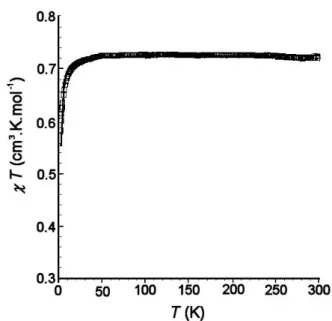
215
The static magnetic susceptibility of a polycrystalline sample of TPSIN was measured in the range 2-300 K with a SQUID susceptometer at a constant magnetic field of 5000 Gauss. The temperature dependence of the molar magnetic susceptibility (χmol) is shown in the form χmol·T vs T in Figure 7. The
220
observed χmol·T value is 0.72 cm3 K mol-1 at 300 K,
suggesting that the singlet and the triplet states are nearly statistically populated at ambient temperature and that a slight amount of monoradical is present in the sample. However, the χ·T product value is in good agreement with the theoretical 225
value of 0.75 cm3·K·mol-1 expected for two uncorrelated S=1/2 units with g = 2.0.
On decreasing the temperature, the χmol·T value remains
almost constant in the temperature range 300-10 K, and then 230
decreases to reach 0.58 cm3.K.mol-1 as the temperature is lowered down to 2 K. The temperature dependence of the χmol·T value was analysed in terms of a modified
singlet-triplet two state model where the magnetic exchange coupling constant J corresponds to an Hamiltonian of the form H = -235
2JS1S2. A purity factor f was introduced for microcrystalline
samples of the diradical used for the magnetic
Table 1 UV-visible absorption spectral data for diradicals 5, 6 and 7.a,b
Silole π→π* Ar-NO π→π* Silole n→π* NO
5 - 372 (5.05) - TPSNN 262 (5.47) 375 (5.16) 603 (3.79) TPSIN 258 (4.27) 370 (3.93) 483 (2.84) a 10-3M solutions in CHCl 3. b λmax in nm, (log ε). 0 0,1 0,2 0,3 T-1 (K-1) IESR
Figure 6. Temperature dependence of the ESR signal intensities of the
∆MS = 2 transition for diradical TPSIN.
⎥ ⎥ ⎥ ⎥ ⎦ ⎤ ⎢ ⎢ ⎢ ⎢ ⎣ ⎡ ⎟ ⎠ ⎞ ⎜ ⎝ ⎛ ∆ + = T k E T C I B ESR S -T exp 3 1 (1)
Figure 7. Temperature dependence of the molar magnetic susceptibility
(χmol) as expressed by χmol·T vs T plots for TPSIN. The experimental data
(open squares) and the theoretical behaviour (solid line, see text) are overlapping. ) / 2 exp( 3 1 ² 2 BT k J T k Ng² f B B mol = ⋅ ⋅ + −
µ
χ
(2)measurements.27 The best fit parameters were J/kB = -1.0 K,
and f = 0.96 (Eq 2). 240
Time-resolved ESR experiments
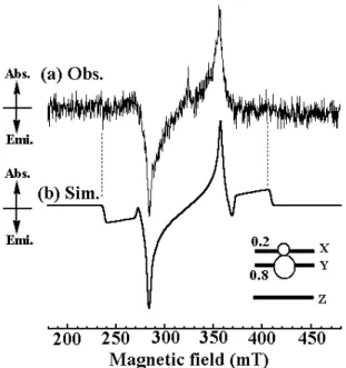
Since the existence of a photo-accessible triplet state for the silole itself remained elusive, we measured the photo-excited state of the parent TPS by time-resolved ESR. A typical TRESR spectrum of the parent TPS is shown in Figure 8(a). 245
The observed TRESR spectrum with well-resolved fine structure splitting has been unambiguously analysed to be an excited triplet state by the spectral simulation shown in Figure 8(b). The determined g value, fine-structure parameters, and relative populations of the Ms sublevels are listed in Table 2. 250
The observed TRESR spectrum shows the E/A pattern of the
dynamic electron polarization (DEP) (eeaa) (e/a: emission/absorption of microwave). This indicates that a selective intersystem crossing by the spin-orbit interaction (SO-ISC) occurs toward the spin-sublevel, Y, of the zero-field 255
wave-functions. Despite multiple attempts to observe TRESR signals characteristics of high-spin photo-excited states for TPSIN, no signal was obtained studying this diradical in contrast to the results reported when 9,10-diphenylanthracene was used as a spin coupler.8, 10, 11, 13
260
Discussion.
Structurally speaking, diradical TPSIN belongs to a class of diradicals termed as doubly disjoint by Lahti et al..28 It is mainly constituted by a non-alternating heteropentacyclic 265
system (the silole ring) in which spin bearing units are connected to the central ring through sites with low spin density: weak antiferromagnetic interactions and nearly degenerated singlet and triplet states are predicted from both valence-bond and spin polarization theory for this kind of 270
molecule.29 The intramolecular distance between the two radical centers is greater than 15 Å. In dilute solutions, this distance is too remote to allow through-space intramolecular magnetic interactions.30 As a consequence, the magnetic interactions observed following the half field signal intensity 275
by ESR traduce the existence of very weak antiferromagnetic interactions that are propagated through the skeleton of the molecule (JESR = -½(∆ET-S/kB) ≈ -0.5 K). The slight difference
observed between the JSQUID (-1.0 K) and JESR values may
traduce the existence of weak additional antiferromagnetic 280
intermolecular interactions in the solid state. The N-O···N-O shortest distances observed in a dimer (6.78 Å) are too remote to allow short contact type magnetic interactions. Magnetic interactions may be expected through hydrogen bonding considering C-H···π interactions and C-H···N or C-H···O-N 285
hydrogen-bonded motives. The well known low spin density delocalisation on methyl groups in imino or nitronylnitroxide radicals together with the low spin density delocalisation on the 3,4-phenyl rings allow to discard this hypothesis.31 Another way to explain such a slight difference is to consider 290
that in the solid state the molecular conformation of TPSIN allows better magnetic exchanges than in frozen solution, in which numerous and less favourable conformations are possible. Whatever the explanation, J values are in agreement with the values ascribed to disjoint systems, with the values 295
encountered in the case of TPSNO (-4 K) and with the ones obtained when diphenylanthracene is used as a coupler (-2.9 K).11, 17,18
Turning back to TRESR experiments, we can consider the following reasons to explain the lack of high-spin photo-300
excited states in the case of silole-based diradicals: (1) the life-time of the triplet state of the parent silole (TPS) is much shorter than the ones observed for anthracene derivatives (the triplet time of anthracene is ca. 55 ms and the triplet life-time of TPS is about few µs), and (2) the efficiency of the 305
enhanced SO-ISC is much smaller than the one encountered in diphenylanthracene-bisiminonitroxide. Concerning the last point, the SO-ISC efficiency highly depends of the nature of the spin bearing units and of the electronic structure of the triplet photo-excited spin coupler. Since TPSIN has been 310
designed to present the appropriate topology, and the molecule is built with a coupler that presents both a triplet photo-excited state and iminonitroxide radicals, it is reasonable to postulate that the enhanced SO-ISC mechanism is also operating in the present diradical. Therefore, the only 315
way to explain the lack of high-spin photo-excited state is to consider that the strong non-radiative relaxation processes from the triplet spin coupler make the triplet lifetime shorter, leading to the unsuccessful detection of high-spin photo-excited states within the time-scale of TRESR measurement. 320
Both factors arising from the molecular and electronic structures prevent the effective generation of the expected result. Therefore it is of crucial importance that the π-conjugated spin coupler pocesses a very rigid skeleton to restrict to a minimum the non-radiative deactivition channel 325
Figure 8. Time resolved ESR of TPS in EPA rigid glass 0.4 µs after a
Nd:YAG pulse laser excitation (λ = 355 nm). (a) Observed spectrum at 30 K. (b) Simulation. The spin Hamiltonian parameters used in the simulation are described in Table 2.
Table 2. Spin Hamiltonian parameters and relative polarization of Ms
sublevels in zero-field for TPS.
g value
Fine-Structure Parameters Relative
Polarizations
2.010 D = 0.076 cm-1, E = 0.002 cm-1 PX = 0.2, PY = 0.8
that may be opened if sets of molecular motion or vibrations are available. This is why rigid fused polyaromatic structures such as fullerene, porphyrine, pyrene, and of course, anthracene, are used to achieve photo-excited spin alignment.
Conclusions
330
In this article we have reported the synthesis of two nitroxide-based diradicals linked by a 2,3,4,5-tetraphenylsilole (TPS) pattern. These two molecules have been especially designed and synthesized with the aim to access to high-spin photo-excited states for both of these species. Whereas the 335
bisiminonitroxide diradical TPSIN is stable either in solution or in the solid state, its correponding bisnitronylnitroxide TPSNN experiences a spontaneous fragmentation to afford a
iso-butylammomium salt. Magnetic behaviour of TPSIN has
been analysed by means of ESR and SQUID measurements 340
combined with structural considerations. As expected from its doubly disjoint character, the TPSIN diradical displays weak intramolecular antiferromagnetic interactions with J/kB = -1.0
K. Although the combination of the silole core with iminonitroxide radicals would be expected to lead to photo-345
induced spin alignment and to high spin states, no detection of high-spin states was possible within the time-scale of TRESR measurement. Therefore, TPS-derivatives clearly provide evidences that besides the needs of a photo-tunable coupler, specific radical species, and π-topological requirements, the 350
flexibilty of the coupler skeleton also plays an important role in the achievement of photo-excited high spin states. Taking into account that the silole ring, with its unique electronic structure and its triplet photo-excited state, is all the same a good candidate to achieve the photo-induced spin alignment, 355
synthetic works are currently under progress to increase the rigidity of the whole structures by fusing the spin-bearing units to the central organometallic linker.
Experimental
360Materials and methods
The photo-excited states of the siloles were examined by Time-Resolved ESR (TRESR) at the Department of Material Science of the Osaka City University. A conventional X-band ESR spectrometer (JEOL TE300) was used in the measurements of TRESR spectra
365
without field modulation. Excitation was carried out at 355 nm light using Nd:YAG pulse laser (Continuum Surelite II-10, pulse width < 7 ns). The typical laser power used in the experiments was c.a. 2 ~ 5 mJ. EPA glass matrix was used for the TRESR experiments. The measurements were carried out at 30 K. The typical microwave
370
power was ca. 10 mW. The spectral simulation was carried out by the eigenfield/exact-diagonalization hybrid method,32-33 taking dynamic electron spin polarization (ESP) into account. The following ordinary spin Hamiltonian given in eq. (3) was used for the analysis.
H'spin = βeH.g.S + S.D.S (3) 375
The resonance field BMs↔Ms+1(θ, φ ) for each transition was directly calculated by the eigenfield method.32 The transition probabilities I
(θ, φ, ϕ) were evaluated by numerically diagonalization of the spin Hamiltonian matrix at the calculated resonance field. Since the resonance field is independent of the third Euler angle, ϕ, the above
380
procedure practically saves the computing time for the simulation. The line-shape function of the TRESR spectrum in the glass matrix is given by
g(B) = NΣ∫ dϕ ∫ dφ ∫ dθ sinθ PMs↔Ms+1(θ, φ ) I (θ, φ , ϕ) f[B –
BMs↔Ms+1(θ, φ )] (4)
385
In the simulation, the dynamic electron polarization (ESP),
PMs↔Ms+1(θ, φ ), on each spin sublevel in a zero magnetic field was given as parameters. Thus, the relative populations of the Ms sublevels were taken into account as parameters. The simulation was
carried out using a program written by the author on a personal
390
computer. The details of the simulation procedures for the high-spin TRESR spectra were described in our previous articles.
1H, 13C and 29Si NMR spectra were recorded on a Bruker Advance
200 DPX spectrometer, the FT-IR spectra on a Thermo Nicolet
395
Avatar 320 spectrometer, the UV-visible spectra on a Secomam Anthelie instrument and the MS spectra on a Jeol JMS-DX 300 spectrometer. The ESR spectra have been recorded on X-band Bruker Elexsys spectrometer. Magnetic susceptibility measurements were obtained with a Quantum Design MPMS-5S SQUID magnetometer.
400
All reactions were carried out routinely under nitrogen using standard Schlenck techniques. Solvents were distilled prior to use. THF was dried over sodium/benzophenone, and distilled under Argon. All the commercial reagents were used as received. Bis(phenylethynyl)dimethylsilane was prepared by the reaction of
405
dimethyldichlorosilane and phenylethynyllithium, which was prepared from nBuLi and phenylacetylene in ether.
Syntheses
1,3-Bis(trimethylsilyloxylamino)-2-(4-bromophenyl)-4,4,5,5-410
tetramethylimidazolidine (4). To a solution of
1,3-Dihydroxy-2-(4-bromophenyl)-4,4,5,5-tetramethylimidazolidine34 (3.15 g, 10 mmol)
in THF (30 mL) was added an excess of triethylamine (8.3 mL, 60 mmol). To the reaction mixture was added chlorotrimethylsilane (7.60 mL, 60 mmol) in THF (50 mL). After stirring at 45 °C for 20 h,
415
the solvents were evaporated to yield a residue that was treated with 200 mL of pentane and filtered. The filtrate was then evaporated in vaccuo to yield 1.92 g of 4 as white crystals (42%). M.p.: 87-89°C. IR (CHCl3, cm-1): 1373 (νN−O). 1H NMR (CDCl 3, 25°C): δ = -0.22 (s, 18H), 1.18 (s, 12H), 4.60 (s, 1H), 7.32 (d, 3JHH = 8 Hz, 2 H), 7.48 (d, 420 3J HH = 8 Hz, 2 H). 13C NMR (CDCl3, 25°C): δ = -0.36, 16.95, 24.20, 61.06, 92.37, 119.18, 121.49, 130.37, 131.71. 29Si NMR (DMSO-d 6,
25°C): δ = 22.66. HRMS (FAB+, m-nitrobenzyl alcohol matrix): m/z: calcd for C19BrH36N2O2Si2 [M+ + H]: 459.1481; found 459.1495. 425
Silole 5. A mixture of lithium (0.1 g, 14.5 mmol) and naphthalene
(1.85 g, 14.5 mmol) in THF (15 mL) was stirred at room temperature under argon for 5 h to form a deep green solution of lithiumnaphthalenide. To this mixture was added bis(phenylethynyl)dimethylsilane 1 (1 g, 3.85 mmol) in THF (10
430
mL). After stirring for 10 min, the reaction mixture was cooled to 0° C and [ZnCl2(tmen)] (tmen = N,N,N',N'-tetramethylenediamine) (3.9
g, 14.5 mmol) was added as a solid to form organozinc derivative 2. After stirring for one hour at room temperature, a solution of 4 (3.5 g, 7.65 mmol) in THF (10mL) and [PdCl2(PPh3)2] (0.14 g, 0.2 mmol) 435
were successively added. The mixture was heated under reflux and stirred for 24 h. After hydrolysis by acetic acid (35%), the mixture was extracted with Et2O several times. The combined organic layers
were washed with brine, saturated solutions of Na2CO3, dried over
MgSO4 and concentrated. The resulting residue was subjected to a 440
column chromatography (neutral silica , eluant: pentane-dichloromethane 80:20) to give 1.57g of 5 as a yellow solid (56%). Dec. temp.: 192°C. IR (KBr, cm-1): 3529, 3242 (νO−H), 1364 (νN−O). UV/Vis (CHCl3): λmax (log ε): 372 (5.05, π→π* silole). 1H NMR
(CDCl3, 25°C): δ = 0.47 (s, 6H), 1.02 (s, 12H), 1.06 (s, 12H), 4.40 (s, 445 2H) 6.83-7.06 (m, 8H), 7.09 (m, 6H), 7.25 (d, 3J HH = 8Hz, 4 H), 7.75 (s, 4H, OH). 13C NMR ([D 6]DMSO, 25°C): δ = -2.80, 17.87, 25.25, 66.89, 91.05, 126.76, 127.17, 128.48, 128.56, 129.01, 130.20, 138.78, 139.79, 140.32, 141.43, 154.29. 29Si NMR ([D 6]DMSO): δ = 8.05.
HRMS (FAB+, m-nitrobenzyl alcohol matrix): m/z: calcd for
450
C44H55N4O4Si [M+ + H]: 731.3993; found 731.3995.
TPSNN. To a solution of silole 5 (100 mg, 0.14 mmol) in 20 mL of freshly distilled dichloromethane, was added NaIO4 (130 mg, 0.6
mmol) as a solution in 20 mL of distilled water. The mixture was
455
stirred for one hour and the phases were separated. The aqueous phase was extracted with dichloromethane (3 × 20 mL). The organic layers were mixed and dried other MgSO4. The solvent was removed
under vacuum and the crude product was purified by preparative thin-layer chromatography (silicagel, eluant: pentane-ethylacetate 60:40)
460
to give 79 mg of TPSNN (58 mmol) as a very hygroscopic deep-green solid (78%). M.p.: not determined. IR (KBr, cm-1): 1363 (ν
UV/Vis (CHCl3): λmax (log ε): 262 (5.47, π→π* arylnitroxide), 375
(5.16, π→π* silole), 603 (3.79, n→π* N-O). HRMS (FAB+, m-nitrobenzyl alcohol matrix): m/z: calcd for C44H50N4O4Si [M+ + 3 H]: 465
726.3601; found 726.3594. Chemical degradation in solution. Typically, a solution of TPSNN (0.100 g, 0.14 mmol) in dichloromethane (5 mL) yields white crystals of iso-butylamine within three days at room temperature. The crystals are collected on a fritt, washed with dichloromethane and dried in air. M. p.: 175°C. IR
470 (KBr, cm-1): 3060 (ν N−H), 2922 (νC−H), 1572 (νN−C). 1H NMR (CDCl 3, 25°C): δ = 1.02 (d, 3J HH = 6.8Hz 6H), 1.63-1.75 (m, 1H), 3.25 (d, 3JHH
= 6.5Hz 2H), 7.3 (s, 2H). MS (FAB+, m-nitrobenzyl alcohol matrix): [M+]: m/z: 74 (100%).
475
TPSIN. To a solution of silole 5 (100 mg, 0.14 mmol) in 20 mL of freshly distilled dichloromethane, was added NaNO2 (38 mg, 0.6 mmol)
as a solution in 20 mL of distilled water. The mixture was stirred and a few drops of acetic acid were added to obtain a pH value near 6. After 15
480
minutes, NaIO4 (38 mg, 0.6 mmol) was added as a solid and the mixture
stirred 15 minutes more. The two phases were separated and the aqueous layer was extracted three times with 20mL of dichloromethane. The organic layers were mixed and dried other MgSO4. The solvent was
removed under vacuum and the crude product was purified by column
485
chromatography (neutral aluminum oxide, eluant: pentane-dichloromethane 80:20) to give 38 mg of TPSIN as red crystals (42%). M.p.: 182°C. IR (KBr, cm-1): 1368 (ν
N−O), 1538 (νC=N). UV/Vis (CHCl3):
λmax (log ε): 258 (4.27, π→π* arylnitroxide), 370 (3.93, π→π* silole),
483 (2.84, n→π* N-O). HRMS (FAB+, m-nitrobenzyl alcohol matrix):
490
m/z: calcd for C44H51N4O2Si [M+ + 3 H]: 695.3770; found 695.3770.
Experimental and crystal data for TPSIN. Spearhead single crystals of
approximate dimensions 0.15 x 0.08 x 0.08 mm3 were selected on
polarized microscope and mounted on a Bruker-Nonius κ-CCD diffractometer, Mo-Kα radiation (0.71073 Å). Data collection was
495
performed using mixed φ and ω scans, 179 frames of 1.5°, 285 seconds per frame and a distance crystal-detector of 30 mm. The structural determination by direct methods and the refinement of atomic parameters based on full-matrix least squares on F2 were performed using the
SHELX-97.35-36 Results: a = 10.984(1) Å, b = 11.474(1) Å, c = 17.492(1) 500
Å, α = 81.10(1)°, β = 89.01(1)°, γ = 65.70(2), V = 1982.6(9) Å3,
density(calc.) = 1.161, triclinic P-1, 97.7% completeness to theta 26.48°, 12809 collected data, 8110 independent reflections (Rint=0.032) for 605
refined parameters, Robs=0.069, wR2obs=0.147, (∆/σ)max=0.001, largest
difference peak and hole 0.46/-0.42 e.A-3, max. CCDC - 603216 contains 505
the supplementary crystallographic data for this paper. These data can be obtained free of charge from The Cambridge Crystallographic Data Centre via www.ccdc.cam.ac.uk/datarequest/cif.
Acknowledgments
510This work was supported by the French CNRS, the European Commission through the Network of Excellence MAGMANet (NMP3/CT/2005/515767), and the Grant-in-Aid for Scientific Research on Priority Area “Application of Molecular Spin” ‘Area 769, Prop. No. 15087208) from MEXT, Japan. Ph. G. and N. R. want
515
to warmly thank Prof. Philippe Turek (Université Louis Pasteur, Strasbourg, France), and Prof. Dominique Luneau (Université Claude Bernard-Lyon I, Lyon, France) for their assistance in magnetic characterization and fruitful scientific discussions.
520
Notes and references
1O. Kahn, 'Molecular Magnetism', ed. VCH, 1993.
2 P. M. Lahti, 'Magnetic properties of organic materials', Marcel
Dekker, Inc., 1999.
525
3 R. Ziessel, C. Stroh, H. Heise, F. Khöler, P. Turek, N. Claiser,
M. Souhassou, and C. Lecomte, J. Am. Chem. Soc., 2004, 126, 12604.
4 O. Benedi Borodia, P. Guionneau, H. Heise, F. H. Khöler, L.
Ducasse, J. Vidal-Gancedo, J. Veciana, S. Golhen, L. Ouahab,
530
J.-P. Sutter, Chem. Eur. J., 2005, 11, 128.
5 A. Ito, Y. Nakano, M. Urabe, T. Kato, and K. Tanaka, J. Am.
Chem. Soc., 2006, 128, 2948.
6 O. Sato, S. Hayami, Y. Einaga, and Z.-Z. Gu, Bull. Chem. Soc.
Jpn., 2003, 76, 443.
535
7 Y. Teki, T. Toichi, and S. Nakajima, Chem. Eur. J., 2006, 12,
2329.
8 Y. Teki, Polyhedron, 2001, 20, 1163.
9 Y. Teki, M. Kimura, S. Narimatsu, K. Ohara, and K. Mukai,
Bull. Chem. Soc. Jpn., 2004, 77, 95.
540
10 Y. Teki, S. Miyamoto, K. Imura, M. Naktsuji, and K. Miura, J.
Am. Chem. Soc., 2000, 122, 984.
11 Y. Teki, S. Miyamoto, M. Naktsuji, and Y. Miura, J. Am.
Chem. Soc., 2001, 123, 294.
12
Y. Teki and S. Nakajima, Chem. Lett., 2004, 33, 1500.
545
13 Y. Teki, M. Nakatsuji, and Y. Miura, Mol. Phys., 2002, 100,
1385.
14 C. Corvaja, M. Maggini, M. Prato, G. Scorrano, and M.
Venzin, J. Am. Chem. Soc., 1995, 117, 8857.
15
K. Ishii, J. Fujisawa, Y. Ohba, and S. Yamauchi, J. Am. Chem.
550
Soc., 1996, 118, 13079.
16 N. Roques, P. Gerbier, S. Nakajima, Y. Teki, and C. Guérin, J.
Phys. Chem. Solids, 2004, 64, 759.
17 N. Roques, P. Gerbier, J.-P. Sutter, P. Guionneau, D. Luneau,
and C. Guérin, Organometallics, 2003, 22, 4833.
555
18 N. Roques, P. Gerbier, U. Schatzneider, J.-P. Sutter, P.
Guionneau, J. Vidal-Gancedo, J. Veciana, E. Rentschler, and C. Guérin, Chem. Eur. J., 2006, published on Web, DOI: 10.1002/chem.200501280.
19
Y. Teki, Polyhedron, 2005, 24, 2299.
560
20 S. Yamaguchi, T. Endo, M. Uchida, T. Izumizawa, K.
Furukawa, and K. Tamao, Chem. Eur. J., 2000, 6, 1683.
21 E. F. Ullman, L. Call, and S. S. Tseng, J. Am. Chem. Soc.,
1975, 75, 1677.
22
L. Call and F. E. Ullmann, Tetrahedron Lett., 1971, 42, 3935.
565
23 C. Smith, J. Bartley, and S. Bottle, J. Mass Spectrom., 2002,
37, 897.
24 G. Desiraju, 'Crystal engineering: The Crystal as a
Supramolecular Entity: Perspectives in Supramolecular Chemistry', Wiley, 1996.
570
25 G. Desiraju, Chem. Commun., 2005, 2995. 26 M. Nishio, Cryst. Eng. Comm., 2004, 6, 130.
27 T. Matsumoto, T. Ishida, N. Koga, and H. Iwamura, J. Am.
Chem. Soc., 1992, 114, 9952.
28
P. M. Lahti and A. S. Ichimura, J. Org. Chem., 1991, 56,
575
3030.
29 D. A. Shultz, in 'Conformational exchange modulation in
trimethylenemethane-type biradicals.' ed. P. M. Lahti, New York, 1999.
30 P. Ferruti, D. Gill, M. P. Klein, H. H. Wang, G. Entine, and M. 580
Calvin, J. Am. Chem. Soc., 1970, 92, 3704.
31 C. Rancurel, H. Heise, F. H. Köhler, U. Schatzschneider, E.
Rentchler, J. Vidal-Gancedo, J. Veciana, J.-P. Sutter, J. Phys.
Chem., 2004, 108, 5903.
32
G. G. Belford, R. L. Belford, and J. F. Burkhalter, J. Magn.
585
Reson., 1973, 11, 251.
33 Y. Teki, I. Fujita, T. Takui, T. Kinoshita, and K. Itoh, J. Am.
Chem. Soc., 1994, 116, 11499.
34 L. Catala, P. Turek, J. Le Moigne, A. De Cian, and N.
Kyritsakas, Tetrahedron Lett., 2000, 41, 1015.
590
35 G. M. Sheldrick, Programs for Crystal Structure Analysis,
release 97-2, Institüt für Anorganische Chemie der Universität, Göttingen, Germany, 1998
36 L.J. Farrugia, J. Appl. Cryst., 1999, 32, 837. 595