HAL Id: hal-01086517
https://hal.archives-ouvertes.fr/hal-01086517
Submitted on 24 Nov 2014HAL is a multi-disciplinary open access archive for the deposit and dissemination of sci-entific research documents, whether they are pub-lished or not. The documents may come from teaching and research institutions in France or abroad, or from public or private research centers.
L’archive ouverte pluridisciplinaire HAL, est destinée au dépôt et à la diffusion de documents scientifiques de niveau recherche, publiés ou non, émanant des établissements d’enseignement et de recherche français ou étrangers, des laboratoires publics ou privés.
MULTILEVEL (3D) MICROFLUIDIC TECHNOLOGY
FOR AN INNOVATIVE MAGNETIC CELL
SEPARATION PLATFORM
Marc Fouet, Sébastien Cargou, Rémi Courson, Marie-Charline Blatché,
Armelle Montrose, Karine Reybier, Anne Marie Gué
To cite this version:
Marc Fouet, Sébastien Cargou, Rémi Courson, Marie-Charline Blatché, Armelle Montrose, et al.. MULTILEVEL (3D) MICROFLUIDIC TECHNOLOGY FOR AN INNOVATIVE MAGNETIC CELL SEPARATION PLATFORM. 18th International Conference on Miniaturized Systems for Chemistry and Life Sciences, Oct 2014, San Antonio, United States. pp.440-442. �hal-01086517�
MULTILEVEL (3D) MICROFLUIDIC TECHNOLOGY FOR AN
INNOVATIVE MAGNETIC CELL SEPARATION PLATFORM
M. Fouet
13*, S. Cargou
13, R. Courson
13, C. Blatche
13,
A. Montrose
23, K. Reybier
23and A.M. Gue
13 1CNRS, LAAS, Toulouse, France,
2PHARMADEV, Toulouse, France
3
Univ . de Toulouse, Toulouse France
ABSTRACT
We demonstrate a new concept of devices, which by combining 3D fluid engineering and localized mag-netic actuation enables the full integration of a cell tagging and magmag-netic separation device. We used a low cost, commercially available dry film (EMS Inc, Ohio, USA) that fits microfluidic requirements and gives the possibility to build easily 3D microfluidic structures. The labelling of blood monocytes with su-perparamagnetic particles was performed “up stream” with the aim of a microparticles 3D micromixer while cell separation was achieved “downstream” through continuous vertical separation over integrated micro-coils.
KEYWORDS: Magnetic separation, Cell tagging, Planar coils, Multilayer (3D) INTRODUCTION
Magnetophoresis is broadly used in medical and biological applications (from cellular to genomic scale) and has been frequently implemented in lab on chip. However magnetic handling tools are mostly external magnets limiting the range of functions that can be integrated: the magnetic field is consequently permanent and non-local and coupling several functions in the same chip remains quite impossible. In particular, magnetic labelling has to be performed out of the chip prior to sorting, reducing therefore the added value of the device. Here, we demonstrate a new concept of device which by combining 3D fluid engineering and localized magnetic actuation enables the full integration of a cell tagging and magnetic separation device.
MICROFABRICATION AND EXPERIMENTAL
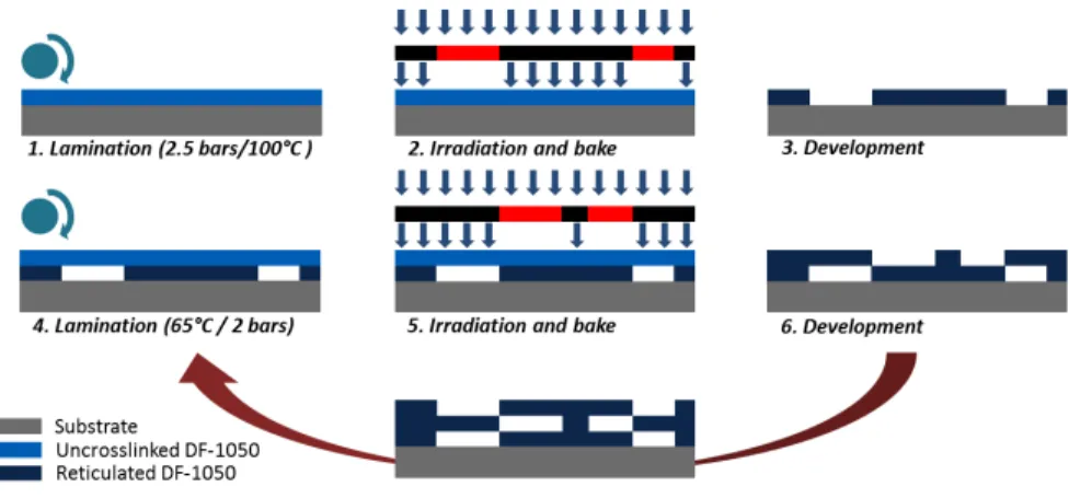
The lab on chip microfabrication was done by using a novel negative epoxy based dry film DF-1050 (EMS) whose low price is a major advantage compared to well-known epoxy photoresists: it is at least ten times cheaper than SU-8 and TMMF [1]. Microchips were fabricated following the general scheme proposed by Fulcrand et al. [2] and depicted in Figure 1.
RESULTS AND DISCUSSION
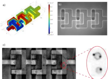
The magnetic labelling of monocytes was performed on chip. Solutions of cells and beads were intro-duced simultaneously in the micromixing module whose principle was based on baker’s transformation applied to microfluidics. This principle was firstly proposed by the theoretical work of P. Carriere [3]. Contrary to standard approaches which are more suited to molecular mixing than to particle mixing, this solution turned to be well suited to cellular labelling with an average labelling yield of 1-2 microbead/cell as shown in Figure 2.
Figure 2: a) Comsol FEM analysis of the 3D mixer device for mixing an aqueous solution of fluorescein with water. b) Top view (optical microscope) of a mixer unit (3 loops) right after micro fabrication. c) Left: trajectory of a monocyte tagged with 4 magnetic beads inside a mixer unit. Right: examples of tagged monocytes obtained after 10 units (30 loops) with 1 and 2 magnetic beads.
Downstream the magnetic actuation was driven by planar coils, which allowed a high flexibility in the de-sign and a strong magnetic field. The separation principle is given in Figure 3 a).
a) b)
Figure 3: a) Scheme of the magnetic separation stage. Sample (monocytes and beads previously mixed) and buffer are injected in superposed channels. The coils attract magnetic beads and tagged monocytes and separate them from the upper channel. b) THP1 Monocytes trapped on a planar micro coil. If the flow rate is low, beads and monocytes can be trapped on a coil instead of flowing and performing a con-tinuous separation.
The separation buffer was introduced in the lower level channel while the sample was flowing in the up-per one. Nine coils located before and on the separation stage enabled the tagged cells to be efficiently
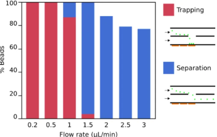
at-tracted across the superposed microchannels. Monocytes could be trapped on the micro-coil (Fig. 3 b), and the separation efficiency of magnetic beads was studied at different flow rates (Fig. 4).
Interestingly we demonstrated an abrupt transition from a “trapping mode” to a “continuous separation mode” depending on flow. The thermal safety of the device has also been checked using rhodamine B to ensure Joule heating (at a 100mA intensity) will not harm the monocytes.
Figure 4: Graph of separation efficiency of 5 µm diameter magnetic beads. Trapping occurs at low flow rates, and those tests were carried out on short periods of time so the coils are never saturated. Proper separation with a satisfying efficiency can be achieved between 1.5 and 2 µL/min (about 3 mm/s for the average velocity of the flow according the the microchannel cross section).
CONCLUSION
While our magnetofluidic devices still undergo characterization tests, we endeavor to achieve even higher integration in implementing an electrical impedance measurement based detection system. Tag-ging, separating and counting different monocytes populations would have direct applications in detecting early stages of infectious diseases, and would give a good insight of new available tools for immunology. ACKNOWLEDGEMENTS
This study was supported by the French National Research Agency (ANR) in the frame of the PIA – Nanobiotechnology project Digidiag and by the Defense Agency (DGA). It was also partly supported by the French Renatech Network
REFERENCES
[1] R. Courson, S. Cargou, V. Conédéra, M. Fouet and A.-M. Gué, “Low cost integration of multilevel lab-on-a-chip using a new generation of dry film photoresists”, Smart System Integration, Vienna (Austria), 2014. [2] R. Fulcrand, D. Jugieu, C. Escriba, A. Bancaud, D. Bourrier, A. Boukabache, and A.-M. Gué, “Development
of a flexible microfluidic system integrating magnetic micro-actuators for trapping biological species”, J.
Mi-cromech. Microeng., 19, 105019, 2009.
[3] P. Carriere, “On a three-dimensional implementation of the baker’s transformation”, Physics of Fluids, 19 (11), 118110, 2007.
CONTACT