HAL Id: hal-01822734
https://hal.sorbonne-universite.fr/hal-01822734
Submitted on 25 Jun 2018
HAL is a multi-disciplinary open access
archive for the deposit and dissemination of
sci-entific research documents, whether they are
pub-lished or not. The documents may come from
teaching and research institutions in France or
abroad, or from public or private research centers.
L’archive ouverte pluridisciplinaire HAL, est
destinée au dépôt et à la diffusion de documents
scientifiques de niveau recherche, publiés ou non,
émanant des établissements d’enseignement et de
recherche français ou étrangers, des laboratoires
publics ou privés.
Antibiotic susceptibility testing and species identification
of Nocardia isolates: a retrospective analysis of data
from a French expert laboratory, 2010-2015
David Lebeaux, Emmanuelle Bergeron, Jérémy Berthet, Juliette Djadi-Prat,
Delphine Mouniee, Patrick Boiron, Olivier Lortholary, Véronica
Rodriguez-Nava
To cite this version:
David Lebeaux, Emmanuelle Bergeron, Jérémy Berthet, Juliette Djadi-Prat, Delphine Mouniee, et al..
Antibiotic susceptibility testing and species identification of Nocardia isolates: a retrospective analysis
of data from a French expert laboratory, 2010-2015. Clinical Microbiology and Infection, Elsevier for
the European Society of Clinical Microbiology and Infectious Diseases, In press, 25 ) 4), pp.489-495.
�10.1016/j.cmi.2018.06.013�. �hal-01822734�
Original article
Antibiotic susceptibility testing and species identification of Nocardia isolates: a
retrospective analysis of data from a French expert laboratory, 2010-2015
David Lebeaux,1-5 Emmanuelle Bergeron,6 Jérémy Berthet,6 Juliette Djadi-Prat,7-8 Delphine Mouniée,6 Patrick Boiron,6 Olivier Lortholary,5 Véronica Rodriguez-Nava6
1
INSERM, U1138, LRMA, Equipe 12 du Centre de Recherche des Cordeliers, Paris, France 2 Université Pierre et Marie Curie, UMRS 1138, Paris, France
3
Université Paris Descartes, Sorbonne Paris Cité, UMRS 1138, Paris, France 4
Assistance Publique-Hôpitaux de Paris, Service de Microbiologie, Hôpital Européen Georges Pompidou, Paris, France
5
Université Paris Descartes, Sorbonne Paris Cité, AP-HP, Hôpital Necker Enfants Malades, Centre d’Infectiologie Necker-Pasteur and Institut Imagine, Paris, France
6
Research group on Bacterial Opportunistic Pathogens and Environment UMR5557 Écologie Microbienne, French Observatory of Nocardiosis, Université de Lyon 1, CNRS, VetAgro Sup, Lyon, France
7 AP-HP, Hôpital Européen Georges-Pompidou, Unité d’Épidémiologie et de Recherche Clinique, Paris, F-75015, France
8
INSERM, Centre d’Investigation Clinique 1418, module Épidémiologie Clinique, Paris, F-75015, France
Key-words: Nocardia, disk diffusion, 16S, hsp65
Corresponding author: Véronica Rodriguez-Nava, French Observatory of Nocardiosis, Research Group on
“Bacterial Opportunistic Pathogens and Environment”, UMR Ecologie Microbienne, CNRS 5557, INRA1418, UCBL, Université de Lyon, VetAgro Sup, Institute of Infectious Agents, Hospices Civils de Lyon, Institut des Sciences Pharmaceutiques et Biologiques de Lyon, France. 8 avenue Rockefeller- 69373 Lyon , France
Alternative corresponding author: David Lebeaux, Service de Microbiologie, Hôpital Européen Georges
POMPIDOU, 20 rue Leblanc, 75015 PARIS, Tel.: +33 1.56.09.39.51, Fax: +33 1.56.09.24.46. Mail: david.lebeaux@yahoo.fr or david.lebeaux@aphp.fr
This work was presented in part at the 28th European Congress of Clinical Microbiology and Infectious Diseases, Madrid, Spain, 21 to 24 April 2018
ABSTRACT
Objectives. Nocardia, a Gram-positive bacterium, is responsible for rare and severe infections. Accurate
microbiological data are essential to guide antibiotic treatment. Our primary objective was to describe species identification and results of antimicrobial susceptibility testing (AST) for Nocardia isolates analysed over a 6-year period. Secondary objectives were to study temporal trends in species distribution and AST results.
Methods. We retrospectively analysed results from Nocardia isolates sent between January 2010 and
December 2015 to a French laboratory dedicated to Nocardia (Observatoire Français des Nocardioses). Species identification was obtained by amplification and sequencing of a 600bp fragment of the 16S rRNA gene (for all isolates) and of hsp65 (when required). AST was performed using disk diffusion.
Results. We included 793 Nocardia isolates, mostly from the lungs (53.8%). The most frequent species were N.
farcinica (20.2%), N. abscessus complex (19.9%) and N. nova complex (19.5%). The proportion of N. farcinica
increased significantly over time from 13% in 2010 to 27.6% in 2014. Linezolid, amikacin, trimethoprim-sulfamethoxazole, minocycline, and imipenem were the most frequently identified active antibiotics with, respectively, 0% (0/734), 2.9% (21/730), 5.4% (40/734), 9.4% (69/734) and 19.5% (143/732) of isolates not susceptible. N. farcinica was frequently not susceptible to cefotaxime (118/148, 79.7% of the isolates), but only about 5% of N. cyriacigeorgica and N. abscessus complex isolates were not susceptible to cefotaxime.
Conclusions. In this first epidemiological study of Nocardia isolated from human samples in France, N. farcinica was the species most frequently identified and its prevalence increased over time.
INTRODUCTION
Nocardia species are Gram-positive filamentous bacteria found in a wide range of natural environments
including decaying vegetation, soil and water [1, 2]. The use of molecular microbiology, including amplification and sequencing of the 16S RNA, hsp65 and other genes, has led to the description of more than 100 species, so far [1, 3]. Nocardia can be responsible for severe opportunistic infections in immunocompromised patients and patients with chronic bronchopulmonary diseases [1, 4-6]. Infection follows bacterial inhalation or, less frequently, direct inoculation through the skin and can lead to pneumonia, brain abscesses and/or skin/soft-tissue infections [1].
The ideal initial treatment for nocardiosis should cover a broad-range of species with adequate antibiotic concentration in all involved organs [7]. However, defining the optimal treatment is difficult because of the lack of comparative and prospective clinical data. As a consequence, most antibiotic regimens currently proposed rely on microbiological data, including species identification and antimicrobial susceptibility testing (AST) [8-10]. Notably, each Nocardia species has a specific antibiotic susceptibility pattern [8-11]. Obtaining reliable species identification using molecular methods is therefore essential to guide initial antibiotic treatment.
We retrospectively collected data from a 6-year period (2010-2015) from a French laboratory dedicated to Nocardia (Observatoire Français des Nocardioses [OFN]). Our primary objective was to describe species distribution and AST results. Secondary objectives were to study temporal trends in species distribution, AST results and geographic distribution of Nocardia in France over the 6-year period.
METHODS
Study design, settings and inclusion criteria
We retrospectively reviewed results for isolates sent to the OFN for Nocardia testing between January 2010 and December 2015. The OFN is a French laboratory that has specialised in Nocardia and other Actinomycetes since 1999 [12]. French microbiology laboratories (from continental France and overseas territories) can send biological samples or bacterial isolates for molecular identification and/or AST; isolates from neighbouring countries are also analysed. The isolate density for each region of France was defined as the number of
Nocardia isolates sent to our centre per year for 100,000 population of that region; population data for 2016
were used, obtained from the Institut national de la statistique et des études économiques (INSEE) [13]. Isolates were included in the present study if they fulfilled the following criteria: i) molecular-based confirmation that the strain belonged to the genus Nocardia (see below); ii) the strain had been isolated from a human clinical sample; iii) species identification had been performed by molecular biology (see below).
Variables
The following variables were recorded: demographic data (age and patient sex), year and geographic location of the isolated strain, type of clinical sample from which the Nocardia strain had been isolated (sputum, bronchial aspirate, bronchoalveolar lavage, pleural fluid, protected-specimen brush, cerebrospinal fluid, abscess fluid, organ biopsy, blood cultures), species identified and need to amplify hsp65, results of the AST. Continuous variables are presented as means (± standard deviation) or medians (range). Categorical variables are presented as numbers and frequencies.
Microbiology
Since 1999, the OFN uses a standardised protocol for microbiological analyses of Nocardia:
i) confirmation that the bacterial strain belongs to the genus Nocardia using a 16S-based Nocardia polymerase
chain reaction (PCR) performed directly on the bacterial colony on an agar plate [14];
ii) species identification obtained by amplification and sequencing of a fragment of ~600 base pairs (bp) of the gene coding for the 16S ribosomal RNA (16S rRNA) using PCR [15]. The sequence obtained in this second step is compared to those stored in GenBank using blast alignment software (http://www.ncbi.nlm.nih.gov/blast) and
the BIBI (Bio Informatic Bacteria Identification tool: https://umr5558-bibiserv.univ-lyon1.fr/lebibi/lebibi.cgi) [16]. Identification at the species level requires 99.6% sequence similarity with the type strain of a single species. If more than 1 sequence in the database has more than 99% similarity, identification is made at the level of the complex. For N. abscessus complex or N. transvalensis complex, a 440 bp fragment of the hsp65 gene is amplified and sequenced to obtain species identification [17]. Isolates with sequence similarities < 99% are identified as Nocardia spp..
iii) AST is performed using the disk diffusion method on cation-adjusted Mueller-Hinton (CA-MH) agar plates.
Inoculums are prepared according to the Clinical and Laboratory Standards Institute (CLSI) standard M24-A2 [18, 19]. The antibiotic disks used are described in Supplementary Table 1. Results are read after 72 h of culture. For each antibiotic disk, the diameter of the inhibition zone is recorded and compared to thresholds
(Supplementary Table 1) [20]. A “non-susceptible” isolate is defined as being resistant or intermediate. Escherichia coli ATCC 25922, Staphylococcus aureus ATCC 25923 and Nocardia asteroides ATCC 19247T were
used as quality control organisms.
For trimethoprim-sulfamethoxazole, the plates are read at 80% of growth inhibition. If the inhibition zone is < 10 mm, an E-test strip is performed on a CA-MH agar plate [21]. If the trimethoprim-sulfamethoxazole minimum inhibitory concentration (MIC) obtained on the E-test strip is ≤ 2/38 µg/mL, the isolate is considered susceptible; if the MIC is ≥ 4/76 µg/mL, the strain is considered resistant [18].
Ethical aspects
This study was approved by the Comité de Protection des Personnes (CPP) Ile-de-France I Ethical board
(CPPIDF1-2015-octobre-DAP 33), the CCTIRS (Comité consultatif sur le traitement de l'information en matière de recherche dans le domaine de la santé, file 16-355 approved May 19, 2016) and the CNIL (Comité National Informatique et Liberté).
RESULTS
Description of the isolates collected
During the study period (2010-2015), 793/823 Nocardia isolates met our entry criteria and were included in the analysis. Most isolates originated from France (696/793, 88%) (Table 1). The median patient age at sampling was 66 [4-90] years. Most of the Nocardia isolates were isolated from the lungs (427/793, 53.8%) or from subcutaneous abscesses or skin (156/793, 19.7%) (Table 1).
The most frequent species identified were N. farcinica (20.2%), N. abscessus complex (19.9%), N. nova complex (19.5%) and N. cyriacigeorgica (12.9%) (Table 2). N. farcinica was the most frequently isolated species in blood cultures and brain abscesses/cerebrospinal fluid: 21/39 (54%) and 19/43 (44.2%), respectively (Supplementary
table 2). To obtain species identification for the N. abscessus and N. transvalensis complexes, hsp65 amplification and sequencing was required for 179/793 isolates (22.6%).
Antibiotic susceptibility test results
AST was performed for 736 of the Nocardia isolates (92.8%). Linezolid, amikacin, trimethoprim-sulfamethoxazole, minocycline and imipenem were the antibiotics most frequently identified as being active against Nocardia with, respectively, 0%, 2.9%, 5.4%, 9.4% and 19.5% of isolates being non-susceptible (Table
3). For the N. abscessus complex, cefotaxime and ceftriaxone were the β-lactam antibiotics most frequently
identified as being active (less than 3% of isolates were not susceptible), followed by meropenem and imipenem (7.3 and 11.8% of non-susceptible isolates, respectively) (Table 3). Conversely, N. farcinica isolates were frequently not susceptible to cefotaxime and ceftriaxone (~80% of the isolates) and meropenem (73% of isolates) but were frequently susceptible to amoxicillin/clavulanic acid or imipenem, with 20.1% and 23% of non-susceptible isolates, respectively. N. nova was more frequently not susceptible to amoxicillin-clavulanate (91.6%) than to amoxicillin (23.1%) and was infrequently not susceptible to imipenem (0.7%) and cefotaxime (20.3%). N. cyriacigeorgica was frequently not susceptible to amoxicillin-clavulanate (90.5%) and amoxicillin (87.2%) but was infrequently not susceptible to cefotaxime (7.4%) and imipenem (10.5%). N. brasiliensis was frequently not susceptible to amoxicillin (66.7%) and imipenem (85.1%) and amoxicillin-clavulanate was the most frequently active β-lactam (8.3% of N. brasiliensis isolates were not susceptible).
For carbapenem antibiotics, N. farcinica and N. cyriacigeorgica isolates were more frequently not susceptible to meropenem than to imipenem. Conversely, N. transvalensis complex and N. brasiliensis were more frequently not susceptible to imipenem than to meropenem. Non-susceptibility to different carbapenems was equally frequent for N. abscessus and N. nova complexes.
Less than 3% of the isolates were not susceptible to amikacin, with the notable exception of N. transvalensis complex in which 30.6% (15/49) were not susceptible. For gentamicin and tobramycin, more than 90% of N.
farcinica and N. transvalensis complex isolates were not susceptible; 80% of N. nova complex isolates were not
susceptible to tobramycin (Table 3).
Less than 10% of isolates were not susceptible to minocycline, but some species had higher rates of non-susceptibility, e.g., N. cyriacigeorgica (18.9%), N. farcinica (12.8%) and N. brasiliensis (12.5%).
Temporal trends
Between 2010 and 2013, the proportion of isolates identified as N. farcinica increased from 13% to 28.0%, decreasing thereafter to 19.3% in 2015 (overall Chi-square test, p=0.038) (Figure 1).
The frequency of isolates that were not susceptible to amikacin or trimethoprim-sulfamethoxazole remained
stable between 2010 and 2015 at less than 9% for both drugs (Supplementary Figure 1).
Geographic distribution of Nocardia species
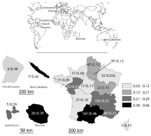
Among Nocardia isolates from France, isolate density was greater in Occitanie (the southernmost region of continental France), New Caledonia, Reunion and Guadeloupe than in other regions, with densities of 0.46, 0.42, 0.39 and 0.29 isolates/year/100,000 population, respectively (Figure 2). In continental France, isolate density was greater in the south east of the country than in other areas.
DISCUSSION
In this retrospective study, we analysed data from a French microbiology laboratory dedicated to the study of aerobic Actinomycetes. Among the 793 Nocardia isolates included, N. farcinica, N. abscessus complex, N. nova complex and N. cyriacigeorgica were the most frequently identified species, representing about three quarters of all isolates. In our sample, linezolid, amikacin and trimethoprim-sulfamethoxazole were the antibiotics that were most frequently identified as being active, each with less than 9% of isolates being non-susceptible.
The distribution of species we observed was different from that reported in recent studies from Spain (1119 isolates) and the United States of America (1299 isolates) in which N. cyriacigeorgica and N. nova complex were most frequently identified [9, 10]. Because a 16S rRNA +/- hsp65 molecular identification strategy was used in Spain and in the present report, it is unlikely that species misclassification could explain these discrepancies. Of note, our isolates of N. farcinica, the most frequently identified species in our study, were not part of an outbreak. One possible explanation for the differences in species distribution is that climatic conditions may have an impact on the epidemiology of Nocardia, as suggested by the observation that
N. brasiliensis is predominantly isolated in tropical or subtropical regions [1]. Furthermore, higher isolate density was observed in the South of France and overseas territories than in other regions, suggesting that environmental conditions may also increase the spread of Nocardia. The high densities observed in Reunion and New Caledonia also raise the possibility of an underlying genetic condition that may favour this opportunistic condition, such as alveolar proteinosis [28].
A phylogenetic analysis revealed that the 16S rRNA gene was not sufficient to obtain precise identification for some Nocardia complexes, such as abscessus or transvalensis [22]. Conversely, the 16S rRNA gene had sufficient discriminatory power for isolates belonging to the N. nova complex, N. farcinica, N.
cyriacigeorgica or brasiliensis species [9]. Although our pragmatic approach (16S rRNA +/- hsp65) appears
attractive for routine purposes, recent studies have demonstrated that the analysis of concatenated sequences of several genes, such as multilocus sequence analysis (MLSA), is likely to be more reliable for phylogenetic purposes [22]. In the near future, whole genome sequencing may also help to decipher Nocardia phylogeny.
Among the 736 Nocardia isolates that underwent AST, the antibiotics most frequently identified as active against Nocardia were linezolid, amikacin, trimethoprim-sulfamethoxazole, minocycline and imipenem. The main challenge in the field of Nocardia AST is the lack of data correlating AST results with clinical
outcomes. As a consequence, technical guidelines are derived from in vitro studies assessing the reproducibility and the technical pitfalls of each method. In our study, AST was performed by disk diffusion, even though the CLSI has stated that broth microdilution is the technical gold-standard for Nocardia [18]. However, the CLSI also acknowledged that broth microdilution may have limitations, including false-resistant results for ceftriaxone against N. brasiliensis or for imipenem against N. farcinica [18]. Sulphonamide testing with broth microdilution is also challenging and requires disk diffusion for confirmation if a resistant strain is identified [18]. Another pitfall of broth microdilution is its poor interlaboratory reproducibility, especially for some antibiotic/species combinations, such as ceftriaxone against N. cyriacigeorgica and N. wallacei or sulphonamides against N.
farcinica and N. wallacei [23]. Furthermore, testing several agents with broth microdilution is time-consuming,
especially given the lack of available commercial plates dedicated to Nocardia testing in Europe. As a consequence, alternative methods are required to routinely perform AST for Nocardia. First described in 1973 by Michael C. Bach and co-workers, antibiotic disk diffusion was further developed and analysed by Richard Wallace’s group [24, 25]. In 1997, results from a direct comparison of broth microdilution, antibiotic disk diffusion, agar dilution, E-test and the BACTEC radiometric method on 26 Nocardia isolates were published [19]. There was 100% agreement of antibiotic disk diffusion results with the study “gold standard” for amikacin, erythromycin, imipenem, minocycline and trimethoprim-sulfamethoxazole; conversely, 80 to 88% agreement was observed for ampicillin, amoxicillin-clavulanic acid, ceftriaxone and ciprofloxacin. Overall agreement for the disk diffusion method was 95.7%. Of note, this study included only a small number (9/27) of the antibiotics we tested. The discrepancies regarding β-lactam results may be related to inducible β-lactamase whose production can vary depending on the experimental conditions. The comparison of results from large-scale microbiological studies performed with broth microdilution [8, 9], E-tests [10] and disk diffusion (present study) highlights the difficulties in interpreting Nocardia AST results (see a comprehensive overview of these data on
Supplementary table 3) [8-10]. Our results using disk diffusion to assess susceptibility of
cefotaxime/ceftriaxone against N. cyriacigeorgica (low frequency of resistance) and against N. farcinica (high frequency of resistance) were similar to those from studies using other methods [8-10]. Conversely, there were significant discrepancies regarding imipenem susceptibility against N. farcinica with 67% of isolates identified as resistant by broth microdilution [9], compared to 4% by E-test [10] and 23% by disk diffusion. These results raise the question of stability issues for imipenem in broth microdilution and stress the need to interpret AST results according to the method used [26].
To our knowledge, this study is the first in which Nocardia epidemiology has been assessed over time and across geographical regions in France. The proportions of most species remained stable over time with the notable exception of N. farcinica for which the proportion increased significantly between 2010 and 2014. We recently observed that N. farcinica was more common among solid organ transplant (SOT) recipients [4, 27]. As SOT now appears to be one of the leading and increasingly common conditions favouring nocardiosis, this increase in the proportion of N. farcinica may be explained by an increase in the proportion of cases of post-SOT nocardiosis.
The main strengths of our study include the large number of isolates reliably identified at the species level using standardised methods performed by the same technical team with the same guidelines during the study period. However, there are also several limitations, including the fact that reporting nocardiosis is not mandatory in France. Thus, the isolates sent to the OFN may reflect a particular interest of the local clinical microbiology laboratory or a lack of knowledge in this field. Nevertheless, although we have described the largest sample of Nocardia isolates in France so far, it is likely that we missed some cases.
In conclusion, we provide results from the first large-scale epidemiological study of Nocardia in France.
N. farcinica was the most frequently identified species and identification rates increased over time. AST
Transparency declaration
Conflict of interest: None
Funding: David Lebeaux was supported by the following grant: Bourse Junior 2015–Société de Pathologie
Infec euse de angue Fran aise (SPILF).
Acknowledgments: The authors would like to thank Koucila Chekhrit and Clémence Bruyere for their help in data collection and Dr Karen Pickett for her editorial suggestions.
Maps were prepared with Articque, Make your Map, freely available on the internet and d-maps.com. The authors would like to thank M.C. Gaps and J.M. Ballec for their conscientious scientific assistance.
REFERENCES
[1] Brown-Elliott BA, Brown JM, Conville PS, Wallace RJ, Jr. Clinical and laboratory features of the
Nocardia spp. based on current molecular taxonomy. Clin Microbiol Rev 2006; 19(2): 259-82.
[2] Saubolle MA, Sussland D. Nocardiosis: review of clinical and laboratory experience. J Clin Microbiol
2003; 41(10): 4497-501.
[3] Euzeby JP. List of Bacterial Names with Standing in Nomenclature: a folder available on the Internet
(
http://www.bacterio.net/
). Int J Syst Bacteriol 1997; 47(2): 590-2.[4] Coussement J, Lebeaux D, van Delden C, Guillot H, Freund R, Marbus S, et al. Nocardia infection in solid organ transplant recipients: a multicenter European case-control study. Clin Infect Dis 2016; 63(3): 338-45.
[5] Minero MV, M. M, Cercenado E, Rabadan PM, Bouza E, Munoz P. Nocardiosis at the turn of the century. Medicine (Baltimore) 2009; 88(4): 250-61.
[6] Rodriguez-Nava V, Durupt S, Chyderiotis S, Freydiere AM, Karsenty J, de Montclos M, et al. A French multicentric study and review of pulmonary Nocardia spp. in cystic fibrosis patients. Med Microbiol Immunol 2015; 204(4): 493-504.
[7] Lebeaux D, Morelon E, Suarez F, Lanternier F, Scemla A, Frange P, et al. Nocardiosis in transplant recipients. Eur J Clin Microbiol Infect Dis 2014; 33(5): 689-702.
[8] Larruskain J, Idigoras P, Marimon JM, Perez-Trallero E. Susceptibility of 186 Nocardia sp. isolates to 20 antimicrobial agents. Antimicrob Agents Chemother 2011; 55(6): 2995-8.
[9] Schlaberg R, Fisher MA, Hanson KE. Susceptibility profiles of Nocardia isolates based on current taxonomy. Antimicrob Agents Chemother 2014; 58(2): 795-800.
[10] Valdezate S, Garrido N, Carrasco G, Medina-Pascual MJ, Villalon P, Navarro AM, et al. Epidemiology and susceptibility to antimicrobial agents of the main Nocardia species in Spain. J Antimicrob Chemother 2016.
[11] Wallace RJ, Jr., Steele LC, Sumter G, Smith JM. Antimicrobial susceptibility patterns of Nocardia
asteroides. Antimicrob Agents Chemother 1988; 32(12): 1776-9.
[12] Boiron P, Provost F, Chevrier G, Dupont B. Review of nocardial infections in France 1987 to 1990. Eur J Clin Microbiol Infect Dis 1992; 11(8): 709-14.
[13] Rapport INSEE 2016 -
https://www.insee.fr/fr/statistiques/2554860
-
documentation
.[14] Couble A, Rodriguez-Nava V, de Montclos MP, Boiron P, Laurent F. Direct detection of Nocardia spp. in clinical samples by a rapid molecular method. J Clin Microbiol 2005; 43(4): 1921-4.
[15] Cloud JL, Conville PS, Croft A, Harmsen D, Witebsky FG, Carroll KC. Evaluation of partial 16S ribosomal DNA sequencing for identification of Nocardia species by using the MicroSeq 500 system with an expanded database. J Clin Microbiol 2004; 42(2): 578-84.
[16] Altschul SF, Madden TL, Schaffer AA, Zhang J, Zhang Z, Miller W, et al. Gapped BLAST and PSI-BLAST: a new generation of protein database search programs. Nucleic Acids Res 1997; 25(17): 3389-402. [17] Rodriguez-Nava V, Couble A, Devulder G, Flandrois JP, Boiron P, Laurent F. Use of PCR-restriction
enzyme pattern analysis and sequencing database for hsp65 gene-based identification of Nocardia species. J Clin Microbiol 2006; 44(2): 536-46.
[18] CLSI. Susceptibility Testing of Mycobacteria, Nocardiae and Other Aerobic Actinomycetes; Approved
Standard-Second Edition. CLSI document M24-A2. Wayne, PA: Clinical and Laboratory Standards
Institute; 2011.
[19] Ambaye A, Kohner PC, Wollan PC, Roberts KL, Roberts GD, Cockerill FR, 3rd. Comparison of agar dilution, broth microdilution, disk diffusion, E-test, and BACTEC radiometric methods for antimicrobial susceptibility testing of clinical isolates of the Nocardia asteroides complex. J Clin Microbiol 1997; 35(4): 847-52.
[20]
https://www.resapath.anses.fr/resapath_uploadfiles/files/Documents/20
13_CASFM.pdf
.[21] Lowman W, Aithma N. Antimicrobial susceptibility testing and profiling of Nocardia species and other aerobic actinomycetes from South Africa: comparative evaluation of broth microdilution versus the Etest. J Clin Microbiol 2010; 48(12): 4534-40.
[22] McTaggart LR, Richardson SE, Witkowska M, Zhang SX. Phylogeny and identification of Nocardia species on the basis of multilocus sequence analysis. J Clin Microbiol 2010; 48(12): 4525-33.
[23] Conville PS, Brown-Elliott BA, Wallace RJ, Jr., Witebsky FG, Koziol D, Hall GS, et al. Multisite reproducibility of the broth microdilution method for susceptibility testing of Nocardia species. J Clin Microbiol 2012; 50(4): 1270-80.
[24] Bach MC, Gold O, Finland M. Activity of minocycline against Nocardia asteroides: comparison with tetracycline in agar-dilution and standard disc-diffusion tests and with sulfadiazine in an experimental infection of mice. J Lab Clin Med 1973; 81(5): 787-93.
[25] Wallace RJ, Jr., Septimus EJ, Musher DM, Martin RR. Disk diffusion susceptibility testing of Nocardia species. J Infect Dis 1977; 135(4): 568-76.
[26] Viaene E, Chanteux H, Servais H, Mingeot-Leclercq MP, Tulkens PM. Comparative stability studies of antipseudomonal beta-lactams for potential administration through portable elastomeric pumps (home therapy for cystic fibrosis patients) and motor-operated syringes (intensive care units). Antimicrob Agents Chemother 2002; 46(8): 2327-32.
[27] Lebeaux D, Freund R, van Delden C, Guillot H, Marbus SD, Matignon M, et al. Outcome and Treatment of Nocardiosis After Solid Organ Transplantation: New Insights From a European Study. Clin Infect Dis
2017; 64(10): 1396-405.
[28] Hadchouel A, Wieland T, Griese M, Baruffini E, Lorenz-Depiereux B, Enaud L, et al. Biallelic Mutations of Methionyl-tRNA Synthetase Cause a Specific Type of Pulmonary Alveolar Proteinosis Prevalent on Reunion Island. Am J Hum Genet 2015; 96(5): 826-31.
TABLES AND FIGURES
Table 1. Characteristics of 793 Nocardia isolates analysed at the Observatoire Français des
Nocardioses (2010-2015)
Characteristics
793
Nocardia
isolates
Patient demographical data
Age at Nocardia sampling (years) (median, range) n= 693
66 [4-90]
Male (n, %) n=743
432 (58.1)
Year of sampling (n, %)
2010
123 (15.5)
2011
130 (16.4)
2012
127 (16.0)
2013
132 (16.6)
2014
141 (17.8)
2015
140 (17.7)
Country of origin (n, %) n=791
France
696 (88.0)
Belgium
31 (3.9)
Switzerland
28 (3.5)
Netherlands
23 (2.9)
Portugal
7 (0.9)
Spain
2 (0.3)
Monaco
1 (0.1)
Lebanon
1 (0.1)
Luxembourg
1 (0.1)
Tunisia
1 (0.1)
Site and method of sampling (n, %)
Lung
427 (53.8)
Sputum
156 (19.7)
Bronchoalveolar lavage
112 (14.1)
Bronchial aspirate
99 (12.5)
Lung biopsy
20 (2.5)
Pleural fluid
20 (2.5)
Bronchial biopsy
1 (0.1)
Protected-specimen brush
1 (0.1)
Unspecified
18 (2.3)
Cutaneous biopsy or subcutaneous abscess sample
156 (19.7)
Blood culture
39 (4.9)
Brain abscess
37 (4.7)
Cerebrospinal fluid
6 (0.8)
Liver biopsy
2 (0.3)
Bone biopsy
2 (0.3)
Corneal abscess
1 (0.1)
Lymph node
1 (0.1)
Pericardial biopsy
1 (0.1)
Other abscess fluid
1 (0.1)
Table 2. Identified species for the 793 Nocardia isolates.
Nocardia species
793
Nocardia
isolates
N. farcinica
160 (20.2)
N. abscessus complex
158 (19.9)
N. abscessus sensu stricto
59
N. abscessus/N.araoensis-like
15
N. abscessus/N.arthritidis-like
6
N. arthritidis
9
N. arthritidis/gamkensis/exalbida-like
30
N. beijingensis
22
N. asiatica
4
N. testacae
2
N. abscessus complex
11
N. nova complex
155 (19.5)
N. nova sensu stricto
144
N. veterana
11
N. cyriacigeorgica
102 (12.9)
N. brasiliensis
55 (6.9)
N. transvalensis complex
51 (6.4)
N. wallacei
39
N. transvalensis sensu stricto
11
N. blacklockiae
1
N. otitidiscaviarum
13 (1.6)
N. brevicatena/paucivorans complex
11 (1.4)
N. pseudobrasiliensis
8 (1.0)
N. flavorosea
7 (0.9)
N. cerradoensis
6 (0.8)
N. mexicana
4 (0.5)
N. carnea
3 (0.4)
N. jiangxiensis
3 (0.4)
N. goodfellowii
2 (0.3)
N. asteroides
2 (0.3)
N. puris
2 (0.3)
N. higoensis
2 (0.3)
N. mikamii
2 (0.3)
N. pneumoniae
2 (0.3)
N. coubleae
1 (0.1)
N. altamirensis
1 (0.1)
N. elegans
1 (0.1)
N. neocaledoniensis
1 (0.1)
N. concava
1 (0.1)
N. rhamnosiphila
1 (0.1)
N. takedensis
1 (0.1)
N. uniformis
1 (0.1)
N. vinacea
1 (0.1)
Nocardia spp.
36 (4.5)
Table 3. Results of antibiotic susceptibility testing among 736 Nocardia isolates according to species
Antibiotic, n (%) of
non-susceptible* isolates
All
Nocardia
isolates.
(n=736)
Nocardia
abscessus
complex
(n=152)
Nocardia
farcinica
(n=149)
Nocardia
nova
complex
(n=145)
Nocardia
cyriacigeorgica
(n=95)
Nocardia
transvalensis
complex
(n=49)
Nocardia
brasiliensis
(n=48)
Amoxicillin
394 (53.8)
37 (24.3)
137 (91.9)
33 (23.1)
82 (87.2)
29 (59.2)
32 (66.7)
Amoxicillin-clavulanic acid 365 (49.7)
40 (26.3)
30 (20.1)
131 (91.6)
86 (90.5)
6 (12.2)
4 (8.3)
Ticarcillin-clavulanic acid
372 (53.8)
42 (27.6)
58 (43.6)
131 (94.2)
82 (93.2)
5 (10.9)
5 (10.9)
Piperacillin-tazobactam
457 (66.0)
39 (26.7)
129 (97.0)
103 (74.1)
83 (93.3)
31 (67.4)
17 (37.0)
Imipenem
143 (19.5)
18 (11.8)
34 (23.0)
1 (0.7)
10 (10.5)
18 (36.7)
40 (85.1)
Meropenem
227 (31.2)
11 (7.3)
108 (73.0)
6 (4.2)
41 (43.6)
8 (16.7)
28 (58.3)
Doripenem
119 (29.1)
8 (9.4)
68 (68.7)
4 (5.1)
11 (25.0)
2 (6.9)
16 (76.2)
Ertapenem
303 (42.7)
17 (11.6)
123 (89.8)
18 (12.7)
59 (62.8)
23 (48.9)
31 (64.6)
Cefotaxime
193 (26.4)
4 (2.7)
118 (79.7)
29 (20.3)
7 (7.4)
0 (0.0)
11 (22.9)
Ceftriaxone
209 (28.5)
4 (2.6)
120 (80.5)
42 (29.4)
4 (4.2)
2 (4.1)
15 (31.3)
Cefepime
281 (38.9)
12 (7.9)
134 (91.2)
33 (23.4)
33 (35.1)
8 (17.0)
27 (56.3)
Cefuroxime
98 (25.0)
3 (3.8)
64 (69.6)
7 (9.1)
1 (2.2)
1 (3.6)
9 (42.9)
Pristinamycin
82 (94.3)
22 (88.0)
19 (95.0)
13 (100.0)
7 (87.5)
7 (100.0)
2 (100.0)
Gentamicin
224 (30.7)
2 (1.3)
136 (91.9)
22 (15.5)
1 (1.1)
45 (95.7)
1 (2.1)
Tobramycin
333 (45.6)
2 (1.3)
143 (96.6)
114 (79.7)
1 (1.1)
46 (95.8)
0 (0.0)
Amikacin
21 (2.9)
1 (0.7)
2 (1.4)
0 (0.0)
1 (1.1)
15 (30.6)
0 (0.0)
Minocycline
69 (9.4)
2 (1.3)
19 (12.8)
10 (6.9)
18 (18.9)
2 (4.1)
6 (12.5)
Doxycycline
176 (42.8)
4 (4.8)
63 (63.6)
54 (67.5)
10 (21.7)
14 (48.3)
14 (66.7)
Tigecycline
202 (27.9)
11 (7.4)
66 (45.2)
66 (46.5)
15 (15.8)
23 (47.9)
2 (4.3)
Erythromycin
463 (64.1)
88 (58.7)
143 (97.3)
6 (4.3)
90 (95.7)
44 (91.7)
41 (85.4)
Linezolid
0 (0.0)
0 (0.0)
0 (0.0)
0 (0.0)
0 (0.0)
0 (0.0)
0 (0.0)
Vancomycin
465 (63.8)
91 (60.7)
46 (31.1)
108 (76.1)
81 (85.3)
43 (87.8)
45 (93.8)
Trimethoprim
530 (76.5)
63 (43.4)
125 (94.0)
137 (97.9)
47 (52.8)
46 (100.0)
43 (93.5)
Trimethoprim-sulfamethoxazole
40 (5.4)
2 (1.3)
6 (4.0)
12 (8.3)
3 (3.2)
6 (12.2)
2 (4.2)
Ciprofloxacin
520 (71.4)
131 (86.8)
62 (41.9)
139 (97.9)
95 (100.0)
5 (10.2)
42 (89.4)
Levofloxacin
409 (56.2)
105 (69.5)
41 (27.7)
128 (90.1)
81 (86.2)
3 (6.1)
18 (37.5)
Moxifloxacin
264 (36.4)
73 (48.7)
14 (9.5)
90 (63.8)
58 (61.7)
3 (6.4)
1 (2.0)
Rifampin
505 (73.0)
88 (60.7)
121 (91.0)
106 (76.3)
56 (62.9)
43 (93.5)
40 (87.0)
*Non-susceptible trains were defined as resistant or intermediate.
Other species who underwent AST were: N. otitidiscaviarum (n=11), N. brevicatena/paucivorans complex (n=9), N. pseudobrasiliensis (n=8), N.
flavorosea (n=7), N. cerradoensis (n=4), N. mexicana (n=4), N. carnea (n=3), N. jiangxiensis (n=2), N. asteroides (n=2), N. higoensis (n=2), N. mikamii (n=2), N. pneumoniae (n=2), N. goodfellowii (n=1), N. puris (n=1), N. coubleae (n=1), N. altamirensis (n=1), N. elegans (n=1), N. neocaledoniensis (n=1), N. concava (n=1), N. rhamnosiphila (n=1), N. takedensis (n=1), N. uniformis (n=1), N. vinacea (n=1), Nocardia spp.
(n=31).
21
FIGURES
1
Figure 1. Proportions of species (%) from 2010 to 2015. The proportion of N. farcinica
2
increased significantly over time (overall Chi-square test, p=0.038). Horizontal bars indicate
3
pairwise comparisons between 2010 (13%) and 2013 (28%), 2014 (27.6%) and 2015 (19.3%),
4
all with p < 0.05, Fisher’s exact test.
5
6
7
22