DOI 10.1007/s00402-006-0228-0
O R T H O P A E D I C S U R GE R Y
Free functional gracilis muscle transfer for reconstruction
of massive ectopic calci
Wcation following a lower extremity
fracture: a case report and overview of the literature
Jürgen W. Schmitt · Beata Bode · Charles E. Dumont
Received: 21 February 2006 / Published online: 21 September 2006 © Springer-Verlag 2006
Abstract We present a patient with massive posttrau-matic ectopic calciWcation in the lower extremity. The patient complained of an increasing tender soft tissue mass with signs of acute inXammation and foot dorsi-Xexion weakness following a tibial fracture for 34 years. A large radiopaque mass was resected and reconstructed using a free functional gracilis muscle transfer, resulting in recovery of stable soft tissue and foot dorsiXexion. The resected material showed highly Wbrosed soft tissue with extensive dystrophic calciWca-tions. There were no sign of recurrence at 18-month follow-up.
Keywords Soft-tissue calciWcation · Tibial fracture · Compartment syndrome · Peroneal nerve injury · Free functional gracilis muscle transfer · Surgical treatment
Introduction
The ectopic ossiWcation is a well known complication following soft tissue injury, but it is rare. It has been reported as a late complication of a compartment syndrome [1–5]. There is only one report on ectopic
calciWcation after tibial fracture combined with a nerve injury [6]. Although guidelines have been established for assessment [7, 8] and treatment [9] of compartment syndrome in the leg, little data has been collected on ectopic calciWcations as a late sequelae of compartment syndrome.
Ectopic calciWcations are commonly observed in soft tissues as a result of neoplastic or inXammatory dis-ease, injury, and aging in the absence of a systemic mineral imbalance. In damaged tissue, the body responds in a nonspeciWc manner by a generic inXam-matory response reaction. This sometimes results in calcium deposition within damaged or devitalized tis-sues.
We present a case of a massive and gradually increasing ectopic calciWcation of muscles in the lower extremity. The calciWcations developed in connection with previous injury to the lower extremity related to a compartment syndrome and nerve injury. We report successful treatment by means of block resection of the lesion and reconstruction with a free functional gracilis muscle transfer.
Case report
A 53-year-old woman presented to our outpatient clinic with acute pain and swelling in the lower extrem-ity. The onset of these symptoms followed 34 years of an asymptomatic period after injury. The patient sus-tained a closed tibiaWbula fracture and was treated by intramedullary tibial nailing when she was 19-year old. Simultaneously the patient suVered from a complex injury of the lumbosacral plexus with complete pero-neal nerve palsy. Two years later the intramedullary J. W. Schmitt (&) · C. E. Dumont
Department of Orthopedic Surgery, Balgrist Clinic, University of Zurich, Forchstrasse 340,
Zurich 8008, Switzerland
e-mail: [email protected] C. E. Dumont
e-mail: [email protected]
B. Bode
Department of Pathology, Institute of Clinical Pathology, University Hospital, Zurich 8091, Switzerland
nail was removed because of a nonunion. ORIF with cancellous bone grafting was performed. Removal of the implants was performed 5 years later. Between the last surgery and the current episode the patient com-plained of progressive limitation of the tibiotalar range of motion despite maintaining on active life style.
Clinical examination revealed a painful, tender soft tissue mass with erythema in the ventral third of the leg (Fig.1). The patient observed that the mass had grown over the last 12 months. A puncture was done and exposed a tooth paste-like white material. The ankle active range of motion was 0° of dorsiXexion to 20° of plantarXexion. Active foot extension was graded as M3 and palpation revealed voluntary muscle contraction of the proximal fourth of the tibialis anterior muscle. Sen-sory loss was conWned to the anteromedial lower leg and dorsal aspect of the foot corresponding to both superWcial and deep peroneal nerve territories.
The white blood cell (WBC) count was 10,200 cells/ mm3 and C-reactive protein (CRP) level was 18.6 mg/l. There were no abnormalities of calcium, phosphorus, and uric acid levels in the blood.
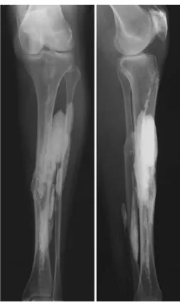
Radiographs revealed a widespread radiopaque mass in the middle third of the leg (Fig.2). The mass was located in the anterior compartment as well as the distal part of the deep posterior compartment of the
extremity. CT showed that the lesion involved the entire tibialis anterior, extensor hallucis longus, and extensor digitorum longus (data not shown). There was fatty degeneration in the posterior compartments with calciWcation of the distal tibialis posterior muscle. A magnetic resonance imaging (MRI) study revealed a mass with uniformly low signal intensity on both T1-and T2-weighted sequences (data not shown). Bone scan did not demonstrate increased uptake by the mass (data not shown).
Angiography demonstrated that the Wbularis artery and tibialis posterior artery were patent. The tibialis anterior artery was stenosed in the middle third of the leg (data not shown).
The entire muscular portion of tibialis anterior and extensor digitorum longus was substituted with an inhomogeneous chalk-like induration, which was resected en block. Necrotic parts of the tibia were also removed. There was no evidence of infection. A free functional gracilis transfer was performed with ter-mino-terminal anastomosis with the tibialis anterior artery. The saphena parva vein was used for venous anastomosis. The proximal part of the deep peroneal nerve was used at the level of proximal tibialis anterior muscle branches for the neural connection. The muscle transfer was distally connected to the tendon of tibialis anterior. A tenodesis of extensor hallucis longus and Fig. 1 Anterior view of the left leg showing tender soft tissue
mass with erythema on the anterior aspect of the tibia
Fig. 2 Preoperative AP and lateral radiograph of the left lower leg showing a large radiopaque mass in the middle third of the leg
extensor digitorum longus was performed. Postopera-tive splinting of the ankle joint in neutral was done for 6 weeks. Afterwards the patient wore a peroneal splint in order to prevent stretching of the muscle and received electrotherapy. The wounds healed primarily. Intraoperative cultures were negative. Histological investigations revealed dystrophic calciWcations of soft tissue with embedded necrotic bone (Figs.3, 4, 5).
The patient showed no signs of infection in the course of follow-up. The patient’s ability to walk improved after surgery. No clinical and radiological signs of recurrence of calciWcations were seen at 18-month follow-up. The patient was free of pain. Good soft tissue coverage exists (Fig.6). Active foot exten-sion was observed at last follow-up corresponding to M2 with electromyography-measured muscle activity
in the gracilis transfer. Recovery of ankle-joint mobil-ity was limited by contracture of the ankle joint, due to pre-operative existent soft tissue conditions. The active ankle range of motion was between-10° of dorsiXexion and 50° of plantarXexion resulting in mild stepping. The patient could stable walk without any support. She was very satisWed with both functional and aesthetic result.
Discussion
The etiology and pathophysiological mechanism of this posttraumatic ectopic calciWcation is diYcult to ascer-tain due to the tremendous delay since injury. These calciWcations cannot be explained as ossiWcation of posttraumatic hematoma. Radiologically, the tissue was homogenous and predominantly not located in direct contact with the bone. These calciWcations fol-lowed muscular Wbers and fasciae. Angiography dem-onstrated a stenosis of the tibialis anterior artery, suggesting the existence of a vascular lesion at the time of injury. The history of initial injury, paralysis, and ischemic myonecrosis, suggested these calciWcations fol-lowed a compartment syndrome. While contracture and deformities of the lower extremity are well-documented complications of compartment syndrome, dystrophic Fig. 3 Highly Wbrosed and calciWed necrotic soft tissue
(hema-toxilin and eosin staining; original magniWcation £25)
Fig. 4 Necrotic bone embedded in Wbrosed soft tissue with dys-trophic calciWcations (hematoxilin and eosin staining; original magniWcation £25)
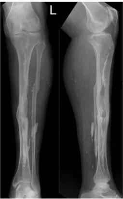
Fig. 5 Postoperative AP and lateral radiograph of the left lower leg showing residual calciWcations. No radiological signs of recur-rence of calciWcations were seen
calciWcations are rare occurrences. The pathomecha-nism of this entity as late sequelae of compartment syn-drome is not entirely clear. Certainly, the duration and severity of the initial ischemia, the extent of myonecro-sis, and the rate of Wbrous tissue regeneration in place of the necrotic tissue may, among other factors, inXu-ence its latent presentation. Interestingly the calci Wca-tions were mainly situated in the antero-lateral part of the leg, thus essentially aVecting the anterior compart-ment corresponding to the nerve injury. In the early stage of injury ankle movements may cause micro-trauma in paretic muscle Wbers, thus provoking an increased calciWcation [6]. This phenomenon has been demonstrated experimentally in rabbits [10]. The loss of normal muscle function could play a role in the development of these changes.
Histological investigation approved characteristic features of dystrophic calciWcations (Table1).
A review of the literature yielded similar cases [1–5, 13]. Reports on ectopic calciWcation after compartment syndrome are rare partly because the follow-up period has been too short to detect the calciWcation, which usually develops years after the syndrome. On the other hand, it is always diYcult to prove the existence of a past compartment syndrome in patients with established ectopic calciWcation because the patients usually are referred after considerable time delay from the initial injury.
While good results after surgery with debridement and tendon transfer have been reported [1, 14] an extensive muscle excision appears technically demand-ing and does not always result in a signiWcant improve-ment in clinical symptoms [3, 4]. Ninkovic et al. [15] proposed the transposition of the lateral gastrocnemius head to the tendons of the anterior muscle group and the concurrent transposition of its motor nerve to the intact proximal end of the deep peroneal nerve. Suc-cessful functional recovery of active dorsiXexion of the foot and toe was achieved with this method. Even though other muscle or tendon transfers like the tibi-alis posterior tendon transposition over the tibitibi-alis anterior can result in functional improvement, but they cannot cover the exposed bone resulting from removed anterior muscular compartment [16, 17].
Reinnervated free muscle transfer has proven to be invaluable in numerous reconstructive procedures e.g. soft-tissue tumor, brachial plexus and Volkmann’s con-tracture [17–22]. Several muscles potentially meet the criteria for free functional muscle transfer. Most com-monly used muscles include the gracilis, latissimus dorsi, rectus femoris, and the tensor fascia lata muscles. Few reports are available on functioning free muscle transfers in the lower extremity, where injuries have been treated mainly with tendon transfers. Wechselber-ger et al. [17] used the rectus femoris muscle for resto-ration of extension of the foot as well as soft-tissue coverage of the lower extremity in three cases with necrosis of all muscles in the anterior compartment after compartment syndrome. Doi et al. [19, 20] reported on two reinnervated free gracilis muscle trans-fers following radical excision of soft-tissue sarcoma in the extensor muscles of the lower extremity. The trans-planted muscles worked as the main activator.
Free functional gracilis muscle transfer is already an established method primarily for restoration of Xexor and extensor function in the forearm, elbow Xexion, and shoulder Xexion [21–23]. Its size, length, and shape most closely approximates that of the muscles, which provide Xexion and extension in the hand, Xexion at the elbow, as well as, dorsiXexion at the ankle. The latissi-mus dorsi latissi-muscle is an alternative to the gracilis. It has excellent strength and excursion. However, it is bulkier than the gracilis muscle, fans out at its distal insertion, and does not have a distal tendon for weave or repair making it technically more diYcult to apply to the lower leg. The Wt of the latissimus to the anterior compart-ment is not satisfactory. The gracilis and latissimus mus-cles are strap musmus-cles. Their muscle Wbers are arranged longitudinal to their direction of contraction. These muscles usually shorten between 40 and 60% of their stretched length. Thus the range of excursion of the Fig. 6 Anterior view of both legs at 1-year follow-up. After
gracilis muscle is more than 10–12 cm in adult muscles [20]. In contrast the rectus femoris and tensor fascia lata muscles are bipennate muscles. Their Wbers are ori-ented at an angle to the direction of muscle contraction. They shorten 40% of their average fascicule length. Because of their fascicular orientation the bipennate transplants provide increased strength but sacriWce excursion or range of motion [18, 24]. For restoration of foot extension and large soft-tissue reconstruction Wechselberger et al. [17] preferred the rectus femoris musculocutaneous Xap. They considered that the graci-lis muscle is not potent enough of generating suYcient force for ankle extension. Otherwise Doi et al. [19] described eVective and functional recovery of a func-tioning gracilis musculocutaneous transfer after radical excision of the anterior compartment in the lower leg.
Daigeler et al. [25] reported on evident but well tol-erated donor-site morbidity of the rectus femoris mus-cle Xap due to weakness of knee extension. The true maximal muscular capacity and maximal voluntary
contraction values were reduced by 18 and 22%, respectively. There was also a mild voluntary activa-tion deWcit. The range of moactiva-tion in the knee and hip was not inXuenced by muscle harvesting. Review of the literature showed no signiWcant functional postopera-tive morbidity in the young and acpostopera-tive patient popula-tion when the gracilis muscle was elevated [26].
Resection of degenerated muscles along with the ectopic calciWcation may be beneWcial when the necro-sis is conWned to a localised area in the leg [18], as in our patient. For restoration of function in the anterior compartment we couldnot treat the patient with tibialis posterior tendon transposition because of the resulting soft tissue defect from the resected anterior compart-ment. Furthermore the deep posterior compartment was partially aVected by fatty degeneration. In our case we preferred a free functional gracilis muscle transfer due to moderate soft tissue defect without skin defect.
The follow-up after 18 months showed a very good functional result as well as no relapse of calciWcation in Table 1 DiVerential diagnosis soft-tissue calciWcations [11, 12]
Forms of soft-tissue calciWcations Characteristic features
Dystrophic calciWcations Develop in damaged tissue secondary to minor injury, ischemic necrosis or necrotizing infectious process in the absence of a generalized metabolic disorder; usually smaller compared with tumoral calcinosis do not increase in size; may progress to ossiWcation;associated with venous insuYciency, parasitic infestation (cysticercosis, dracunculiasis, armillifer armillatus), Neoplasm [primary bone-forming tumor (osteoma, osteosarcoma), tumor necrosis], Autoimmune disease (dermatomyositis, scleroderma),
Trauma (heterotopic ossiWcation, compartment syndrome, injection granulomas) (Prevalence 95–98%)
Metastatic calciWcations Calcium deposits, related to a disturbance in calcium or phosphorus, Wne speckled Calcium, associated with hypervitaminosis D, milk–alkali (Burnett’s)-syndrome, chronic renal failure with secondary
hyperparathyreoidism; often hypercalcemia (Prevalence 1–2%) Calcium pyrophosphate dihydrate
(CPPD) crystal deposition disease (chondrocalcinosis)
Wide clinical variety, acute arthritis caused by CPPD crystal-induced inXammation, most often found in the temporomandibular joint, cervical spine and hand (tumoral form); knee and wrist (common form), massive deposits of CPPD crystals which where bordered by Wbrous connective tissue (Prevalence 1–2%) Tumoral calcinosis Massive calciWed deposits in soft-tissue often around major joints; predominant in
adolescents and young adults manifesting before age 20 years; often familiar, often occurs in African–Americans; deposits containing hydroxyapatite with lack of crystalline structures; deposits which where bordered by histiocytes and giant cells of foreign-body type, hyperphosphataemie, normocalcemia (Prevalence <1%) Fibrodysplasia ossiWcans progressiva Extreme rare, congenital, metabolic disorder of connective tissue characterized
by congenital malformation of the great toe and progressive heterotopic ossiWcation of tendons, ligaments and fasciae, development in early childhood and usually results in the premature death of the individual, induced by minor trauma to the tissue, surgery could exacerbate this condition and should
be avoided (Prevalence <1%)
Calcinosis circumscripta AVects middle-aged woman and most commonly involves the hand and wrist, secondary skin infection and Wstula formation possible, occurs in scleroderma, sclerodactyly, systemic lupus erythematosus, high association with
Raynaud’s phenomenon
Calcinosis universalis Occur mostly in children, associated frequently with scleroderma or dermatomyositis, normal serum calcium and phosphorate levels
the transfer. Though there was recovery of ankle-joint active dorsiXexion, it was limited due to strength of the gracilis transfer and preexisting contracture of the ankle joint.
Free functional gracilis muscle transfer is a viable reconstructive technique for the treatment of massive ectopic calciWcation in the leg for this rare pathology. This method can provide active muscle contraction and soft-tissue restoration without signiWcant donor site morbidity.
References
1. Early JS, Ricketts DS, Hansen ST (1994) Treatment of partmental liquefaction as a late sequela of a lower limb com-partment syndrome. J Orthop Trauma 8:445–448
2. Hyder N, Shaw DL, Bollen SR (1996) Myositis ossiWcans:cal-ciWcation of the entire tibialis anterior after ischemic injury (compartment syndrome). J Bone Joint Surg Br 78:318–319 3. Saitoh S, Hata Y, Murakami N, Seki H, Miyauchi S, Takaoka
K (2000) Ectopic calciWcation following tibial fracture: prop-erty analysis. Skeletal Radiol 29:609–612
4. Malisano LP, Hunter GA (1992) Case report. Liquefaction and calciWcation of a chronic compartment syndrome of the lower limb. J Orthop Trauma 6:245–247
5. Viau MR, Pederson HE, Salciccioli GG, Manoli A II (1983) Ectopic calciWcation as a late sequela of compartment syn-drome: reports of two cases. Clin Orthop 176:178–180 6. Osterman K, Lindholm TS (1983) Massive ectopic
calciWca-tion of the leg following nerve injury. Arch Orthop Trauma Surg 102:82–87
7. Matsen FA III (1973) Compartmental syndrome: a uniWed concept. Clin Orthop 113:8–14
8. Matsen FA III, Winquist RA, Krugmire RB (1980) Diagnosis and management of compartment syndrome. J Bone Joint Surg Am 62:286–291
9. Mubarak SJ, Owen CA (1977) Doubleincision fasciotomy of the leg for decompression in compartment syndromes. J Bone Joint Surg Am 59:184–187
10. Michelsson JE, Granroth G, Andersson LC (1980) Myositis ossiWcans following forcible manipulation of the leg. A rabbit model for the study of heterotopic bone formation. J Bone Joint Surg Am 62:811–815
11. Stewart VL, Herling P, Dalinka MK (1983) CalciWcation in soft tissues. JAMA 250:78–81
12. Resnick D (2002) Diagnosis of bone and joint disorders. 4th edn. WB Saunders, Philadelphia
13. Renwick SE, Naraghi FF, Worrell RV, Spaeth J (1994) Cystic degeneration and calciWcation of muscle: late sequelae of compartment syndrome. J Orthop Trauma 8:440–444 14. Manoli A II, Smith DG, Hansen ST Jr (1993) Scarred muscle
excision for treatment of established ischemic contracture of the lower extremity. Clin Orthop 292:309–314
15. Ninkovic M, Sucur D, Starovic B, Markovic S (1994) A new approach to persistent traumatic peroneal nerve palsy. Br J Plast Surg 47:185–189
16. Ober FR (1933) Tendon transposition in the lower extremity. N Engl J Med 209:52–55
17. Wechselberger G, Pichler M, Pulzl P, Schoeller T (2004) Free functional rectus femoris muscle transfer for restoration of extension of the foot after lower leg compartment syndrome. Microsurgery 24:437–441
18. Manktelow RT, Zuker RM, McKee NH (1984) Functioning free muscle transplantation. J Hand Surg 9A:32–39
19. Doi K, Ihara K, Kawai S (1998) Limb-sparing surgery in syno-vial sarcoma of the forearm and lower leg by reinnervated free muscle transfer. Plast Reconstr Surg 102:442–447 20. Doi K, Kuwata N, Kawakami F, Hattori Y, Otsuka K, Ihara
K (1999) Limb-sparing surgery with reinnervated free-muscle transfer following radical excision of soft-tissue sarcoma in the extremity. Plast Reconstr Surg 104:1679–1687
21. Barrie KA, Steinmann SP, Shin AY, Spinner RJ, Bishop AT (2004) Gracilis free muscle transfer for restoration of func-tion after complete brachial plexus avulsion. Neurosurg Fo-cus 16:E8
22. Chung DC, Carver N, Wei FC (1996) Results of functioning free muscle transplantation for elbow Xexion. J Hand Surg Am 21:1071–1077
23. Hattori Y, Doi K, Abe Y, Ikeda K, Dhawan V (2002) Surgical approach to the vascular pedicle of the gracilis muscle Xap. J Hand Surg Am 27:534–536
24. Gans C, Gaunt AS (1991) Muscle architecture in relation to function. J Biomech 24(1):53–65
25. Daigeler A, Dodic T, Awiszus F, Schneider W, Fansa H (2005) Donor-site morbidity of the pedicled rectus femoris muscle Xap. Plast Reconstr Surg 115:786–792
26. Vranckx JJ, Misselyn D, Fabre G, Verhelle N, Heymans O, Van den hof B (2004) The gracilis free muscle Xap is more than just a “graceful” Xap for lower-leg reconstruction. J Rec-onstr Microsurg 20:143–148