HAL Id: hal-00702251
https://hal.archives-ouvertes.fr/hal-00702251
Submitted on 29 May 2020
HAL is a multi-disciplinary open access
archive for the deposit and dissemination of
sci-entific research documents, whether they are
pub-lished or not. The documents may come from
teaching and research institutions in France or
abroad, or from public or private research centers.
L’archive ouverte pluridisciplinaire HAL, est
destinée au dépôt et à la diffusion de documents
scientifiques de niveau recherche, publiés ou non,
émanant des établissements d’enseignement et de
recherche français ou étrangers, des laboratoires
publics ou privés.
Copyright
Plants and Apicomplexan Parasites by Natural Product
Rubreserine.
Djeneb Camara, Cordelia Bisanz, Caroline Barette, Jeroen van Daele, Esmare
Human, Bernice Barnard, Dominique van der Straeten, Christophe P Stove,
Willy E Lambert, Roland Douce, et al.
To cite this version:
Djeneb Camara, Cordelia Bisanz, Caroline Barette, Jeroen van Daele, Esmare Human, et al..
Inhi-bition of p-Aminobenzoate and Folate Syntheses in Plants and Apicomplexan Parasites by Natural
Product Rubreserine.. Journal of Biological Chemistry, American Society for Biochemistry and
Molec-ular Biology, 2012, 287 (26), pp.22367-76. �10.1074/jbc.M112.365833�. �hal-00702251�
Inhibition of p-Aminobenzoate and Folate Syntheses in
Plants and Apicomplexan Parasites by Natural Product
Rubreserine
*
□SReceived for publication, March 26, 2012, and in revised form, May 9, 2012 Published, JBC Papers in Press, May 10, 2012, DOI 10.1074/jbc.M112.365833
Djeneb Camara‡1, Cordelia Bisanz§, Caroline Barette¶, Jeroen Van Daele储, Esmare Human**, Bernice Barnard**,
Dominique Van Der Straeten‡‡2, Christophe P. Stove储, Willy E. Lambert储2, Roland Douce‡, Eric Maréchal‡3, Lyn-Marie Birkholtz**, Marie-France Cesbron-Delauw§, Renaud Dumas‡, and Fabrice Rébeillé‡4
From the‡Laboratoire de Physiologie Cellulaire Végétale, Commissariat à l’Energie Atomique/CNRS UMR5168/INRA
USC1200/Université Joseph Fourier Grenoble I, Institut de Recherches en Technologies et Sciences pour le Vivant, Commissariat à l’Energie Atomique-Grenoble, F-38054 Grenoble, France, the§Laboratoire Adaptation et Pathogénie des Microorganismes, CNRS UMR 5163/Université Joseph Fourier Grenoble I, Institut Jean Roget, F-38042 Grenoble, France, the¶Centre de Criblage pour Molécules Bioactives, Institut de Recherches en Technologies et Sciences pour le Vivant, Commissariat à l’Energie Atomique-Grenoble, F-38054 Atomique-Grenoble, France, the储Laboratory of Toxicology, Ghent University, Harelbekestraat 72, B-9000 Ghent, Belgium, the **Malaria Research Group, Department of Biochemistry, University of Pretoria, Private Bag x20, Pretoria 0028, South Africa, and the‡‡Laboratory of Functional Plant Biology, Department of Physiology, Ghent University, K. L. Ledeganckstraat 35, B-9000 Ghent, Belgium
Background:pABA biosynthesis is a potential target for antifolate drugs.
Results:Rubreserine inhibits GAT-ADCS, an enzyme involved in pABA biosynthesis, and decreases the folate content in Arabidopsisand Toxoplasma.
Conclusion:Specific inhibition of pABA synthesis induces growth limitation of plants and apicomplexan parasites.
Significance:GAT-ADCS is a valuable target in eukaryotes, and rubreserine is a novel scaffold for anti-parasitic drugs.
Glutamine amidotransferase/aminodeoxychorismate thase (GAT-ADCS) is a bifunctional enzyme involved in the syn-thesis of p-aminobenzoate, a central component part of folate cofactors. GAT-ADCS is found in eukaryotic organisms auton-omous for folate biosynthesis, such as plants or parasites of the phylum Apicomplexa. Based on an automated screening to search for new inhibitors of folate biosynthesis, we found that rubreserine was able to inhibit the glutamine amidotransferase activity of the plant GAT-ADCS with an apparent IC50of about
8 M. The growth rates of Arabidopsis thaliana, Toxoplasma
gondii, and Plasmodium falciparum were inhibited by
rubreser-ine with respective IC50values of 65, 20, and 1M. The
correla-tion between folate biosynthesis and growth inhibicorrela-tion was studied with Arabidopsis and Toxoplasma. In both organisms, the folate content was decreased by 40 –50% in the presence of rubreserine. In both organisms, the addition of p-aminobenzo-ate or 5-formyltetrahydrofolp-aminobenzo-ate in the external medium restored the growth for inhibitor concentrations up to the IC50 value, indicating that, within this range of concentrations, rubreserine was specific for folate biosynthesis. Rubreserine appeared to be more efficient than sulfonamides, antifolate drugs known to inhibit the invasion and proliferation of T.
gon-dii in human fibroblasts. Altogether, these results validate the
use of the bifunctional GAT-ADCS as an efficient drug target in eukaryotic cells and indicate that the chemical structure of rubreserine presents interesting anti-parasitic (toxoplasmosis, malaria) potential.
Folates are a family of cofactors that are essential for cellular one-carbon (C1) transfer reactions. They are involved in several important metabolic pathways, such as the synthesis of nucle-otides and the methylation cycle (1–3). Folate biosynthesis can be divided into three branches (Fig. 1A) as follows: the first one is for the pterin ring synthesis; the second one is for the pABA5
synthesis, and the third one is for the assembly of these two precursors plus glutamates to form the backbone of folate derivatives (4 – 6). Blocking folate biosynthesis or turnover leads to the arrest of cell division and eventually to cell death. Antifolate drugs have been developed to exploit this feature in therapies against cancer cells and microbial or parasitic infec-tions. Biosynthesis of folate is mainly inhibited by two groups of compounds, i.e. inhibitors of dihydropteroate synthase (DHPS) and inhibitors of dihydrofolate reductase (DHFR). Inhibitors of DHFR are commonly used as therapeutic agents against cancer (7), whereas a combination of these two types of inhibitors are
*This work was supported in part the Région Rhône-Alpes (Cluster 9, “Plantacter”).
□S This article containssupplemental Fig. 1.
1Ph.D. fellowship was supported by the Région Rhône-Alpes (Cluster 9,
“Plantacter”).
2Supported by Ghent University Grant BOF09-GOA-004.
3Supported by an Agence Nationale de la Recherche PlasmoExpress grant. 4To whom correspondence should be addressed: PCV/iRTSV, CEA-Grenoble,
17 Ave. des Martyrs, F-38054 Grenoble, France. Tel.: 33-4-38-78-44-93; Fax: 33-4-38-78-50-91; E-mail: frebeille@cea.fr.
5The abbreviations used are: pABA, para-aminobenzoic acid; GAT-ADCS,
glu-tamine amidotransferase-aminodeoxychorismate synthase; ADCL, amino-deoxychorismate lyase; AS, anthranilate synthase; GMPS, GMP synthetase; GDH, glutamate dehydrogenase; THF, tetrahydrofolate; 5-FTHF, 5-formyl tetrahydrofolate; GAT, glutamine amidotransferase; HFF, human foreskin fibroblast; DHFR, dihydrofolate reductase; DHPS, dihydropteroate syn-thase; ADC, 4-amino-4-deoxychorismate.
at INRA Institut National de la Recherche Agronomique on June 13, 2018
http://www.jbc.org/
commonly used in clinical treatments against parasites of the Apicomplexa phylum, such as Plasmodium falciparum or
Toxoplasma gondii(8, 9). However, the use of these drugs is compromised by the emergence of resistance, and the currently used chemical scaffolds and protein targets are actually over-exploited. Nevertheless, the long established efficacy of folate metabolism as a clinical target is strongly encouraging to iden-tify new inhibitors acting against other enzymes of the folate pathway comprising nine enzyme activities in addition to DHPS and DHFR (Fig. 1A) (10 –12).
Among potential targets, the enzymes involved in the pABA branch of the pathway are of great interest (13). Indeed, they are absent in animals, and the only known metabolic fate of pABA is its commitment in folate synthesis. In addition, it was
recently shown in plants that the production of pABA, together with the production of pterins, is rate-limiting for the whole folate pathway (4, 5). Also, it was shown in P. falciparum that pABA was an effective salvage substrate in experiments using antifolates, suggesting that pABA metabolism might offer opportunities for chemotherapy (14). The pABA moiety is syn-thesized in two steps from chorismate (a metabolite also involved in aromatic amino acid synthesis (15)). First, choris-mate is aminated to form 4-amino-4-deoxychorischoris-mate (ADC), and ADC is then aromatized with loss of pyruvate (Fig. 1B). In many bacteria such as Escherichia coli or Bacillus subtilis, ADC synthesis requires two separate proteins, PabA (a glutamine amidotransferase) and PabB (the ADC synthase) (16). In eukaryotes, the situation appears different. Indeed, in plants and lower eukaryotes, such as yeast and Apicomplexa, ADC synthesis is catalyzed by a single bifunctional protein (Fig. 1A) containing two domains, the glutamine amidotransferase (GAT) in the N-terminal part and the ADC synthase (ADCS) in the C terminus (17). Based on sequence similarities with TrpG (the component II of anthranilate synthase), GAT-ADCS is classified as a member of the G-type group of amidotrans-ferases (18, 19). There is only one gene coding for GAT-ADCS in apicomplexan parasites and plants, and a mutation in the plant gene is embryo-defective. The ADCS domain belongs to the group of chorismate-utilizing enzymes, which also contains salicylate synthase, isochorismate synthase, and anthranilate synthase (20, 21). Until now, searches of inhibitors for this class of enzymes were only achieved using prokaryotic systems. They involved docking studies and design of chorismate analogous compounds (22–25), combinatorial chemistry approaches (26, 27), and a specific screening of a microorganism extract collec-tion using growth inhibicollec-tion of test bacteria as a marker of activity (28, 29). Several compounds were identified by these different methods but appeared to be relatively weak inhibitors of ADCS, although some of them could be quite potent against other chorismate-utilizing enzymes (22–24, 26). The most potent inhibitor of ADCS reported to date is an analog of cho-rismate (2-hydroxy-4-amino-4-deoxychocho-rismate), exhibiting a
Ki value of 38 Magainst the purified enzyme (23). To our
knowledge, the in vivo effects of these ADCS inhibitors have not been investigated.
In this report, using a purified recombinant plant GAT-ADCS as a model enzyme for bifunctional GAT-GAT-ADCS, we screened a chemical library for new inhibitors of pABA synthe-sis. We identified one compound exhibiting a Ki⬍ 10Mand
measured the impact of this molecule on a plant (Arabidopsis
thaliana) and two apicomplexan parasites (T. gondii and P.
falciparum).
EXPERIMENTAL PROCEDURES
Materials—A. thaliana (ecotype Columbia) seedlings were grown on plates containing Murashige and Skoog medium, 15% agar, plus the various molecules to be tested. Seeds were first sterilized by soaking for 15 min in a solution containing 0.095% Tween and 0.57% sodium hypochlorite before placing on the agar medium. The plates were conserved in the dark at 4 °C for 48 h and then transferred in a greenhouse (20 °C, 80% humidity, 150moles photon m⫺2䡠s⫺1, 12 h light period). The number of
FIGURE 1. Biosynthesis of THF. A, schematic representation of the pathways found in E. coli, P. falciparum, and A. thaliana. The enzymes involved are as follows: 1, GTP cyclohydrolase I; 2, NUDIX hydrolase; 3, unspecific phospha-tase; 4, dihydroneopterin aldolase; 4⬘,6-pyruvoyltetrahydropterin synthase; 5, GAT-ADCS (PabA and PabB in E. coli); 6, ADCL; 7, 6-hydroxymethyldihydrop-terin (H2Pterin) pyrophosphokinase; 8, dihydropteroate (H2Pteroate)
syn-thase; 9, dihydrofolate (H2F) synthetase; 10, dihydrofolate reductase; 11,
folyl-polyglutamate synthetase; 12, thymidylate synthase. Other abbreviations: Cho, chorismate; pABA, p-aminobenzoate; H2PterinPP,
6-hydroxymethyldihy-dropterin pyrophosphate; H4F, tetrahydrofolate; H4F-Glun, the
polyglutamy-lated form of H4F. Merged symbols represent bifunctional enzymes. The
activ-ities 9 and 11 are catalyzed by a single enzyme in E. coli and Plasmodium. In Plasmodium, the activity 6 remains to be identified. Above the broken line, the activities are absent in animals. The black stars show the two enzymes actually targeted by antifolate drugs. The white stars show the potential target studied in this work. B, details of the two-step pathway required for pABA synthesis. In all eukaryotes, the first step involves a bifunctional GAT-ADCS, whereas two separate proteins PabA and PabB are found in most (but not all) prokaryotes (19).
at INRA Institut National de la Recherche Agronomique on June 13, 2018
http://www.jbc.org/
seedlings at the two leaf stage (rosette stage) was counted after 2 weeks.
A. thaliana (ecotype Columbia) cell suspension cultures were grown and subcultured as described previously (30). For measurements of metabolites, cells were collected after 7 days of treatment, rapidly washed with distilled water, weighted, fro-zen in liquid nitrogen, and stored at⫺80 °C for later analyses.
T. gondiitachyzoites from the RH-YFP2strain (kindly
pro-vided by B. Striepen, Athens, GA) were propagated in human foreskin fibroblasts (HFF) under standard procedures as described previously (31). Invasion and proliferation assays were performed on HFF cells grown to confluence on glass coverslips in 4- or 24-multiwell plates. For the invasion assay, freshly egressed RH-YFP2parasites were incubated for 5 h with or without the different drugs. To perform synchronized inva-sion, 106parasites/well were centrifuged for 30 s at 1300 rpm
onto HFF monolayers, and wells were incubated for 15 min in a water bath at 37 °C. Wells were further washed three times with cold PBS to eliminate extracellular parasites. Infected cells were fixed in 5% formaldehyde/PBS for 30 min and stored in PBS at 4 °C until staining. To distinguish intracellular from remaining extracellular parasites, coverslips were incubated with the pri-mary antibody mAb Tg05-54 against the major Toxoplasma surface protein SAG1 (TgSAG1) and then with Texas Red-con-jugated goat anti-mouse secondary antibody (Molecular Probes). Intracellular parasites exhibiting a faint red color can easily be distinguished from extracellular parasites that are bright red. Nuclei were stained with Hoechst 33258 (Molecular Probes). The number of intracellular parasites was determined from 12 randomly selected fields per coverslip and per experi-ment with a Zeiss Axioplan 2 microscope equipped for epifluo-rescence and phase contrast. Invasion was expressed as percent of the number of intracellular parasites recorded in nontreated cells.
To assess the effect of the drugs on intracellular growth of T.
gondii(proliferation assay), HFF monolayers were infected with 105parasites/well (cf. invasion assay). Wells were then washed
three times with PBS to eliminate extracellular parasites, and drugs were added. After 24 h at 37 °C in a humidified atmo-sphere containing 5% CO2, cells were fixed and stained with
Hoechst 33258 as described above. For each drug concentra-tion, the number of parasites was determined from at least 100 individual vacuoles.
To determine folate concentrations, parasites were grown for 24 h in HFF cells in the presence or absence of 20 or 40M
rubreserine. The infected HFF monolayers were washed three times with PBS, harvested with a cell scraper, and passed four times through a 27-gauge needle to break the cells. Broken cells and released parasites were washed three times with PBS, and then parasites were purified by filtration through a 3 m Nucleopore membrane, further washed two times in PBS, con-centrated by centrifugation, and frozen at⫺80 °C until use.
Plasmodium falciparum(3D7) was maintained in human O⫹ erythrocytes as described previously (32). The in vitro anti-plas-modial activity of the rubreserine was determined using the malaria SYBR Green I fluorescence assay as described previ-ously (33, 34). In vitro ring-stage intra-erythrocytic P.
falcipa-rumparasites (1% hematocrit and 1% parasitemia) were
incu-bated with specific concentrations of rubreserine in complete P.
falciparumculture medium, with chloroquine disulfate used as a positive control (0.5M) or vehicle (1⫻ PBS) as a negative control. Fluorescence was measured after 96 h under drug pres-sure (excitation 485 nm and emission 538 nm). The data, after subtraction of background (chloroquine disulfate-treated infected RBCs and no parasite growth), were expressed as per-centage of untreated control to determine cell proliferation.
Rubreserine Preparation—Rubreserine was prepared from (⫺)-eseroline fumarate salt (Sigma). Stock solutions of esero-line (10 mM) were made in 50 mMPi(pH 8), 50 mMTris (pH 8),
or 1⫻ PBS (pH 7.5) buffers, depending on the experiment. Under these conditions, eseroline is spontaneously oxidized into rubreserine, a process completed in about 6 h at room temperature. The formation of rubreserine was controlled through the appearance of a characteristic peak of absorption at 475 nm. These stock solutions were stored at 4 °C for 48 h or at ⫺20 °C for 1 week. They were serially diluted before use.
Expression and Purification of Recombinant Arabidopsis Enzymes—AtGAT-ADCS and EcADCL proteins were ex-pressed and purified as described previously (19). Arabidopsis cDNAs encoding GMPS,-subunit of AS starting at Ala-51 to remove the plastid transit peptide, and␣-subunit of AS starting at Ala-61 were amplified by PCR and cloned into the expression vector pET28 (Novagen). E. coli BL21-CodonPlus (DE3)-RIL cells (Stratagene) were transformed and grown using the same protocol as for AtGAT-ADCS. Cells were disrupted, and His-tagged recombinant proteins were purified as described previ-ously (19).
Determination of Enzyme Activities—GAT activity can be determined by measuring the production of glutamate and ADCS activity by the production of pABA (19). Standard assays contained 100 mMTris/HCl (pH 8.0), 5 mMMgCl2, 5% v/v
glyc-erol, 0 – 0.5 mM L-glutamine, 0 – 0.02 mMchorismate, and 9g
ml⫺1of the recombinant plant enzyme. The presence of an excess of EcADCL (20 g ml⫺1, 600 nM) is required for the
production of pABA. Reactions (final volume 80l) were run in 96-well microplates (Greiner), and changes in fluorescence were continuously monitored with a microplate scanning spec-trophotometer Safir2(Tecan). To monitor glutamate
produc-tion, an excess of GDH (100g ml⫺1, 4.2 units ml⫺1) and 1 mM
NAD were added to the assay, and NADH production was fol-lowed by its emission at 450 nm (excitation 340 nm). Concen-tration of pABA was monitored by its fluorescence emission at 340 nm (excitation 290 nm) in the presence of EcADCL.
The activities of GMPS and AS (- plus ␣-subunits) were measured monitoring glutamate production. In both condi-tions, the final volume was 500 l. For GMPS, the assay medium contained the following: 200MXMP, 1 mMATP, 0.5
mM L-glutamine, 10g GMPS, 1 mM NAD, 4.2 units ml⫺1
GDH. For AS, the assay medium contained the following: 50M
chorismate, 0.5 mM L-glutamine, 10g each of ␣- and
-sub-units, 1 mMNAD, 4.2 units ml⫺1GDH and various
concentra-tions of rubreserine. The reacconcentra-tions were started by addition of glutamine, and the change in absorbance at 340 nm was moni-tored with a UV-visible spectrophotometer (Safas).
High Throughput Screening—The screening was conducted at the Center for the Screening of Bio-Active Molecules located
at INRA Institut National de la Recherche Agronomique on June 13, 2018
http://www.jbc.org/
at the Commissariat à l’Energie Atomique-Grenoble, France. The library of compounds was purchased from The Prestwick Chemical Library威. This library contains 1200 molecules selected for their high chemical and pharmacological diversity. They are marketed and 100% Food and Drug Administration-approved compounds, supplied in the library at a 10 mM
con-centration in DMSO. The final concon-centration used in the enzy-matic assay was 100M, and we verified in separate control
experiments that 2% DMSO had no effect on the activity. The enzymatic test used for primary and secondary screenings was identical to the one described above for the GAT activity. Qual-ity of the assay was assessed based on the calculation of the Z⬘ factor, as defined by Zhang et al. (35). After optimization of the assay, the Z⬘ factor was 0.76 ⫾ 0.08 (a good assay must display a Z⬘ factor of ⬎0.5). The assay was conducted as follows: 50l of a mixture containing 0.1MTris (pH 8), 5 mMMgCl2, 5%
glyc-erol, 40Mchorismate, 1 mMNAD, 0.4 unit of GDH, 0.7g of
GAT-ADCS, and 100Mof the various molecules were added
in each well (in control wells, molecules were omitted). The reaction was started by injection in each well of 30l of a solu-tion containing 1.3 mM Gln, and the fluorescence changes
(excitation 340 nm and emission 450 nm) were recorded at 0, 15, and 25 min. The most promising molecules were purchased, and their inhibitory properties were manually confirmed.
Measurements of Metabolites—Determinations of folates and pABA were essentially performed as described previously (36 –38). Briefly, the extract corresponding to 3–5⫻ 107 para-sites or 0.5 g (fresh weight) of plant material was subjected to separation using UPLC (for folates) or HPLC (for pABA), fol-lowed by tandem mass spectrometric detection on an API 4000 (Applied Biosystems, Foster City, CA), using electrospray ion-ization, in the multiple reaction monitoring mode. For folates, the final quantitative data reflect the sum of six different folate monoglutamates as follows: tetrahydrofolate (THF), 5-methyl-THF, 10-formyl-folic acid, 5,10-methenyl-5-methyl-THF, folic acid, and 5-formyl-THF (5-FTHF). [13C]Folate derivatives and 3-NH
2
-4-CH3-benzoic acid were added in the extraction buffers as
inter-nal standards for folates and pABA, respectively.
RESULTS
ADCS Assay, High Throughput Screening, and GAT-ADCS Inhibition—The search for enzyme inhibitors by global approaches requires methods to determine the activity that are accurate, robust, and compatible with the automated platforms used for screening of chemical libraries. When coupled with GDH, GAT activity was easily measurable with a UV-visible spectrophotometer (340 nm) or a fluorimeter (excitation 340 nm and emission 450 nm) monitoring NADH accumulation (19, 39). Also, it was convenient to use the same procedure to test the inhibitors on GDH alone and to discard molecules that were specific for this last activity. Based on this protocol, we designed a miniature assay easily reproducible and optimized for the identification of active drugs, with a screening coeffi-cient Z⬘ (a statistical parameter measuring the quality of the screening assay (35)) of 0.76⫾ 0.08 (see “Experimental Proce-dures”). Because all our attempts to produce active recombi-nant GAT-ADCS from apicomplexan parasites failed, we used the recombinant plant enzyme as a model for this bifunctional
system. We screened the registered Prestwick威 Chemical Library (1200 compounds) against the GAT activity of the plant GAT-ADCS. After primary and secondary screenings, we iden-tified only four molecules exhibiting IC50 values that were
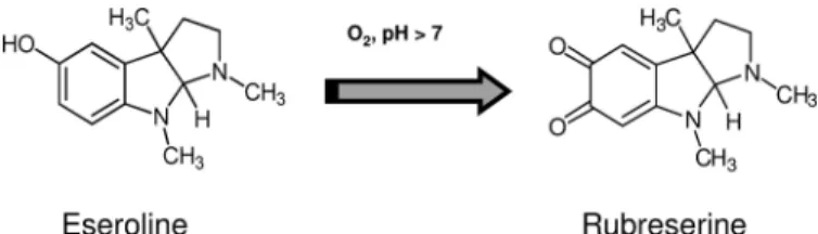
below 50 M. They were manually tested, and we found that rubreserine (1,3a,8-trimethyl-1,2,3,3a,8,8a-hexahydro-pyrrolo[2,3-b]indole-5,6-dione, an oxidative product of esero-line (40 – 42) that spontaneously forms at alkaesero-line pH (Fig. 2 andsupplemental Fig. S1)) exhibited the best inhibitory prop-erties. Eseroline itself had no effect, indicating that the carbonyl functions resulting from eseroline oxidation were essential for the inhibition. The rate constant of inhibition was rather low (about 0.1 min⫺1), and a 20-min period of incubation with rubreserine was required to obtain maximal inhibition. We observed GAT-ADCS inhibition independently of the presence or position of the His tag that was added for purification con-venience, indicating that such a tag was not involved in the inhibition process. We previously showed that the GAT activity was maximal in the presence of chorismate (the substrate of the ADCS domain) but could also operate independently (19). We observed inhibition of glutamate production in both condi-tions, i.e. with and without chorismate, and we also observed inhibition of pABA synthesis when GAT-ADCS was coupled with non limiting amounts of ADC lyase (Fig. 3, A and B). In separate control experiments, we verified that rubreserine did not impact ADC lyase activity. In all situations, rubreserine decreased the Vmand increased the Kmfor glutamine,
indicat-ing a mixed type inhibition (Fig. 3C). Apparent equilibrium constants Ki (representing the ratio [E][I]/[EI], where E is
enzyme; I is inhibitor; and S is substrate) and␣Ki(representing
the ratio [ES][I]/[ESI]) calculated from such pattern of curves (43) were estimated to be respectively 3⫾ 1 and 8 ⫾ 1Min the
different conditions, i.e. measuring either glutamate or pABA productions.
In Vivo Effects of Rubreserine on A. thaliana Growth and Folate Content—When Arabidopsis seedlings were grown on agar plates in the presence of rubreserine concentrations rang-ing from 50 to 100M, we observed a dose-dependent growth
inhibition (Fig. 4). Interestingly, when the agar plates were sup-plemented with pABA, the growth was restored for inhibi-tor concentrations up to 50Mand partially restored for higher
concentrations, the maximal effect being obtained with pABA concentrations ⱖ100 M(Fig. 4). This suggests that for low
rubreserine concentrations, at least, the growth inhibition was due to a limitation of pABA synthesis. Because pABA is required for folate biosynthesis, we also measured the potential impact of 5-FTHF on rubreserine-treated plants (Fig. 4). As shown, this folate derivative had similar effect than pABA.
FIGURE 2. Chemical structures of eseroline and rubreserine. Oxidation of eseroline into rubreserine is spontaneous and strongly increased at pH⬎7.
at INRA Institut National de la Recherche Agronomique on June 13, 2018
http://www.jbc.org/
Next, we investigated the effect of rubreserine on the folate content. We used Arabidopsis cell cultures for these experi-ments to dispose of enough material for metabolite determina-tions. For rubreserine concentrations within the range 25–100 M, the cell division came to an arrest after about 7 days. Similar
results were obtained in the presence of sulfanilamide, a well known specific inhibitor of DHPS that blocks pABA utilization in the folate pathway (Fig. 1A). After 7 days, the pools of folate in rubreserine- and sulfanilamide-treated cells (Table 1) decreased by about 40 and 60%, respectively. However, the dis-tributions of folate derivatives were not markedly modified (in all situations, the various representative pools of folates were roughly 50% 5-methyl-THF, 35% 10-formyl-THF plus 5,10-methenyl-THF, 8% 5-FTHF, 7% THF plus 5,10-methylene-THF, and 0.3% folic acid (30)). Interestingly, when 100 M
pABA were present in the culture medium, the folate content in
both control and rubreserine-treated cells were almost identi-cal (Table 1), which was clearly indicative of a protective effect of pABA. Because rubreserine inhibited the pABA branch of the folate pathway, we also attempted to measure the pABA content of Arabidopsis cells. In plants, pABA was found either as free acid or as a glucose ester conjugate. This last form (⬎80% of total pABA) is sequestered in vacuoles and does not directly contribute to folate synthesis (44, 45). This makes the determi-nation of free pABA quite difficult because, as previously shown (44), breakdown of the esterified form of folate during sample workup is very difficult to avoid and contributes, sometimes quite significantly, to the amount of free pABA. However, in the presence of 100Mrubreserine, we observed a small but
signif-icant decrease of free pABA by about 30% (Table 2). In contrast, the total amount of pABA did not markedly change, suggesting that the glucose ester conjugate in the vacuole and the free acid
FIGURE 3. Effect of rubreserine on GAT-ADCS kinetics. A, glutamine-dependent glutamate production (GAT activity alone, no chorismate) in the presence of rubreserine. GAT-ADCS was first incubated with the various concentrations of rubreserine for 20 min, and then the kinetic was started by the addition of Gln, NAD, and GDH. B, glutamine-dependent pABA production (GAT-ADCS activity, 100Mchorismate) in the presence of rubreserine and nonlimiting amount of ADCL. GAT-ADCS was first incubated with the various concentrations of rubreserine for 20 min, and then the kinetic was started by the addition of Gln, chorismate, and ADCL. rfu, relative fluorescence unit. C, reverse plot of B. Maximal rates of the recombinant enzyme were 140⫾ 40 nmol min⫺1mg⫺1for GAT activity (glutamate production, no chorismate) and 130⫾ 30 nmol min⫺1mg⫺1for GAT-ADCS activity (pABA production, with chorismate and ADCL). Curves were fitted with the hyperbolic equation of Michaelis-Menten (A and B) and linear regression (C). All our assays were made in triplicate and expressed⫾ S.D.
FIGURE 4. Effect of rubreserine on the development of Arabidopsis
seed-lings. Seedlings were grown in agar plates without (controls) or with 50, 75, or
100Mrubreserine. The estimated number of seedlings at the rosette stage after 2 weeks is expressed as % of the conditions without rubreserine. The presence in the culture medium of 200MpABA or 200M5-FTHF partially reversed the growth inhibition. Results are the average of at least three inde-pendent experiments⫾ S.D. Asterisks mark datasets showing statistical dif-ference with the condition containing rubreserine alone in a Student’s t test (p⬍ 0.05).
TABLE 1
Effect of rubreserine (Rubre) on the folate content of Arabidopsis cells and comparison with sulfanilamide (Sulfa)
Cells were cultivated for 7 days with the various compounds before folate determi-nations. The total folate contents of control cells cultivated with or without 100M
pABA were 11.8⫾ 2 and 10 ⫾ 1.5 nmol g⫺1fresh weight, respectively. Measure-ments are the average⫾ S.D. of 4–6 determinations from two to three independent set of experiments. Results for each set of experiment are expressed as % versus the corresponding control.
Conditions Total folates
% versus control Rubre 25M 60⫾ 10 Rubre 25M⫹ pABA 100M 85⫾ 9 Rubre 50M 66⫾ 3 Rubre 50M⫹ pABA 100M 116⫾ 12 Rubre 100M 54⫾ 9 Sulfa 25M 42⫾ 3 Sulfa 100M 39⫾ 6 TABLE 2
Effect of rubreserine (Rubre) on the pABA contents of Arabidopsis cells and comparison with sulfanilamide (Sulfa)
Cells were cultivated for 7 days with the various compounds before pABA determi-nation. The total and free pABA concentrations found in control cells were 8.5⫾ 2 and 1.1⫾ 0.4 nmol g⫺1fresh weight. respectively. Measurements are the average⫾ S.D. of eight determinations from four sets of experiments. Results for each set of experiments are expressed as % versus the corresponding control.
Conditions pABA Total Free % versus control Rubre 100M 90⫾ 5 73⫾ 8 Sulfa 100M 128⫾ 16 132⫾ 18
at INRA Institut National de la Recherche Agronomique on June 13, 2018
http://www.jbc.org/
form in the cytosol are not in rapid equilibrium. In sulfanil-amide-treated cells, free and total pABA slightly increased by about 30% (Table 2), an expected result taking into account that pABA utilization was blocked (46).
Effect of Rubreserine on the Proliferation of Apicomplexan Parasites—Before testing the effect of rubreserine on T. gondii, we first verified with the [3-(4,5-dimethylthiazol-2-yl)-2,5-di-phenyltetrazolium bromide] cytotoxicity assay (47) that rubre-serine concentrations up to 50Mhad no toxicity on confluent
HFF cells. Indeed, after 48 h of exposure to 50Mrubreserine,
the cell viability was still 95⫾ 5% of the control. We then eval-uated the effect of rubreserine on Toxoplasma parasites in two different situations as follows: invasion of confluent HFF by the parasites on the one hand, and intracellular development of the parasites within confluent HFF on the other hand. As shown in Fig. 5A, invasion of human cells strongly decreased when the parasites had been previously incubated with rubreserine. This effect was dose-dependent, and the number of intracellular par-asites were reduced 2-fold in the presence of 20Mrubreserine
(IC50). Likewise, when infected cells were placed in the
pres-ence of the inhibitor, the number of parasites in parasito-phorous vacuoles decreased after 24 h in comparison with untreated infected cells, which was indicative of a slowing down of the parasite’s intracellular division (Fig. 5B). In both situa-tions, rubreserine appeared much more efficient than sulfanil-amide and sulfadiazine, and this last sulfonsulfanil-amide drug was widely used in the treatment of toxoplasmosis. To correlate the inhibitory effect with folate biosynthesis, we attempted to determine the folate concentration in T. gondii cells. To our knowledge, there was no report about the intracellular concen-tration of folates within proliferating T. gondii parasites. We measured folates in parasites grown for 24 h in HFF cells placed in the presence of either 20 or 40Mrubreserine (Table 3).
Interestingly, in both conditions the total folate concentration decreased by a factor of 2. Also, similar to what was observed in plants, the distributions of folate derivatives were not signifi-cantly changed in the presence of the drug (in these cells the various representative pools of folates were as follows: 18%
5-methyltetrahydrofolate; 25% 10-formyltetrahydrofolate plus 5,10-methenyl-tetrahydrofolate; 42% 5-FTHF; 8% tetrahydro-folate plus 5,10-methylene-tetrahydrotetrahydro-folate; and 7% folic acid). In addition, for a rubreserine concentration of 20 M (IC50
value), the parasite growth was largely restored in the presence of either pABA or 5-FTHF (Fig. 6A), and the maximal protec-tive effect was obtained for concentrationsⱖ25M. Thus, for
such inhibitor concentration (20 M), the inhibitory process
largely relied on the inhibition of pABA and folate biosynthesis. However, for rubreserine concentrations higher than 20 M,
pABA and 5-FTHF had only little effect (Fig. 6B), suggesting that high concentrations of the inhibitor had other non-folate-specific actions. Because antifolate drugs (DHPS and anti-DHFR) are often used in combination, we tested the effect of rubreserine in combination with an anti-DHFR (Fig. 6C). When 20Mrubreserine and 0.4Mpyrimethamine were combined,
the inhibition was slightly but significantly increased compared with rubreserine alone. Interestingly, rubreserine alone appeared in our experimental conditions as efficient as a mix-ture combining 50Msulfadiazine and 0.4Mpyrimethamine.
Additionally, rubreserine was also tested on the in vitro pro-liferation of P. falciparum. As shown in Fig. 7, rubreserine exhibited anti-malarial properties because the intraerythro-cytic growth of the parasite was strongly inhibited by rubreser-ine, with an IC50of 1⫾ 0.04M(n⫽ 5). Whether folate
bio-synthesis is also a primary target in these organisms is currently under investigation.
Specificity of Inhibition by Rubreserine—At low rubreserine concentrations, folate biosynthesis appeared as a main target in
FIGURE 5. Effect of rubreserine (Rubre) on invasion and proliferation of T. gondii and comparison with sulfanilamide (Sulfa) and sulfadiazine (SDZ). A, invasion of human fibroblasts. The parasites were first incubated with the various drugs for 5 h and then placed in contact with HFF cells for 15 min. The number of intracellular parasites is expressed for each experiment as % of the number recorded in nontreated cells. B, intracellular growth of the parasites. Proliferation was estimated 24 h after the invasion process and expressed as the number of parasites present in the parasitophorous vacuoles. Results from invasion and proliferation experiments are the average of three to four independent experiments⫾ S.D.
TABLE 3
Effect of rubreserine (Rubre) on the folate content of T. gondii cells
Parasites were grown for 24 h in confluent HFF cells in the presence of 20 or 40M
rubreserine. The results are expressed as nmol/107parasites. Measurements are the
average⫾ S.D. of two (40MRubre), three (20MRubre), and four (control) independent set of experiments.
Conditions Total folates
nmol/107parasites No drug 10.2⫾ 2.6 Rubre 20M 3.8⫾ 1 Rubre 40M 4⫾ 2
at INRA Institut National de la Recherche Agronomique on June 13, 2018
http://www.jbc.org/
Arabidopsis and Toxoplasma. However, at high rubreserine concentrations, the growth activity could not be fully restored by the presence of pABA or 5-FTHF, raising the question of the inhibitor specificity. GAT-ADCS belongs to the family of class I glutamine amidotransferases (18, 48), which contains six other members. The GAT domain of GAT-ADCS does not share strong homologies with the GAT domains of the other mem-bers of this class, and the best scores were obtained with AS (about 28% identity) GMPS (about 13% identity) and carbamoyl phosphate synthetase II (involved in UMP synthesis, about 15%
identity). To test the effect of rubreserine on other members of this group, we attempted to produce these recombinant
Arabi-dopsisactivities. We failed to produce active recombinant car-bamoyl-phosphate synthetase, but we produced AS (a het-erodimeric protein combining the activities of - and ␣-subunits, respectively, equivalent to TrpG and TrpE in pro-karyotes (49)) and GMPS (a bifunctional enzyme with fused GAT and synthase domains (50), like GAT-ADCS). We deter-mined these activities with the same GDH-coupled assay that we used for GAT-ADCS. As shown in Table 4, rubreserine also inhibited GMPS and AS activities, although inhibition of GMPS required higher concentrations of inhibitor. Toxoplasma, but not Plasmodium, is an auxotroph for tryptophan because of the lack of AS activity (51, 52). However, the enzymes involved in the synthesis of GMP (GMPS, a bifunctional enzyme, as in plants (53)) and UMP (carbamoyl-phosphate synthetase II, a heterodimer in plants (54), but a single protein in apicomplexan
FIGURE 6. Effect of rubreserine in combination with various other compounds on the proliferation of T. gondii. The number of parasites present in the parasitophorous vacuoles after 24 h are expressed as % versus the corresponding controls (i.e. no addition, or pABA alone, or 5-FTHF alone). A, reverse effect of pABA and 5-FTHF in the presence of 20Mrubreserine (Rubre). B, reverse effect of pABA and 5-FTHF (100Meach) as a function of rubreserine concentration. C, effect of various drug combinations; the concentrations used were as follows: 20Mrubreserine; 0.4Mpyrimethamine (Pyrim); 100MpABA; 100M 5-formyltetrahydrofolate (5-FTHF); 50Msulfadiazine. Mix is a mixture containing pABA, anthranilate, UMP, and GMP at 50Meach. Asterisks indicate statistical difference in a Student’s t test (p⬍ 0.05) between the conditions rubreserine alone and rubreserine in combination with other drugs, and between the conditions sulfadiazine or pyrimethamine alone and sulfadiazine plus pyrimethamine. Results are the averages of three independent experiments⫾ S.D.
FIGURE 7. Effect of rubreserine on the growth of P. falciparum. Parasites were maintained under normal culture conditions and exposed to serial dilu-tions of rubreserine from 10 mM, and parasite proliferation was determined as a measurement of DNA content with SYBR Green I fluorescence after 96 h of exposure. Data are averaged from five independent experiments performed in triplicate (⫾ S.D.).
TABLE 4
Effect of rubreserine on the GAT activities associated with recombi-nant Arabidopsis GAT-GMPS, AS, and GAT-ADCS
Activities were estimated measuring the glutamate production through a GDH-coupled assay, as described under “Experimental Procedures.” Rubreserine concen-trations up to 50Mhad no detectable effect on GDH activity alone. Assays were made in triplicate and expressed⫾ S.D.
Enzymes Specific activity IC50
mol min⫺1mg⫺1 M
AtGAT-GMPS 0.30 25⫾ 10 AtAS (␣- ⫹ -subunits) 0.16 7⫾ 2 AtGAT-ADCS 0.40 8⫾ 2
GDH (type II from bovine liver) 44 Not measurable,⬎⬎50
at INRA Institut National de la Recherche Agronomique on June 13, 2018
http://www.jbc.org/
parasites (55)) was present in these organisms. When a mixture containing pABA, anthranilate, UMP, and GMP (50Meach)
was added to the culture medium of rubreserine-treated plants or was present in the proliferation assay of Toxoplasma, the growth recovery for both organisms was not markedly improved compared with that obtained with pABA or 5-FTHF alones (see Fig. 6B for the experiments with T. gondii).
DISCUSSION
Two main conclusions can be drawn from this study: first, our data validate for the first time the use of the bifunctional GAT-ADCS as an efficient drug target in eukaryotic cells, and second, we identified a new scaffold that inhibits plant growth and proliferation of apicomplexan parasites.
The screening test we used appeared efficient to select active compounds from Prestwick威 chemical library, and we found that an oxidative product of eseroline, rubreserine, was inhib-iting the GAT activity of AtGAT-ADCS. Eseroline and rubre-serine were identified a long time ago as metabolites of physo-stigmine (eserine), an alkaloid present in the Calabar bean (Physostigma venenosum), and previously used for its potent anticholinesterase activity. To our knowledge, rubreserine and eseroline have no current use in any medical purpose. Pharma-cological studies indicated that these two molecules were poor inhibitors of cholinesterase (56), but there was no reports indi-cating that rubreserine could affect folate biosynthesis and inhibit the growth of plants and the proliferation of parasites. Thus, we describe for this compound a new biological effect with interesting therapeutic potentialities.
When GAT-ADCS was coupled with ADC lyase, the appar-ent constant of inhibition (Ki) for pABA formation was
esti-mated to be⬍10M, which is the best constant of inhibition
obtained so far for the biosynthesis of pABA. How rubreserine affects the protein activity is not yet understood, however, and is currently under investigation. The obvious difference between the chemical structures of eseroline and rubreserine is the presence of two carbonyl functions on the aromatic ring of the latter compound. Thus, these carbonyl functions were pre-sumably at the origin of the inhibitory effect. It must be noted that many other natural quinonoid compounds were shown to display antimalarial properties (57), although the targets and modes of action were not described for most of them.
The rubreserine-dependent growth inhibitions of
Arabidop-sisand Toxoplasma were specifically associated with an inhibi-tion of folate biosynthesis for concentrainhibi-tionsⱕIC50, although
higher concentrations might also inhibit other activities. The correlation between rubreserine and folate biosynthesis was observed by direct and indirect approaches. The direct approach indicated that Arabidopsis cells exhibited folate and free pABA contents lowered by 45 and 25%, respectively, in the presence of the inhibitor. In addition, when pABA was present in the culture medium of rubreserine-treated plant cells, the folate level was almost identical to the control, indicating a pro-tective role of pABA. Likewise, the folate level in Toxoplasma cells was two times lowered by rubreserine. The impact of rubreserine on folate biosynthesis was also shown indirectly. Indeed, for rubreserine concentrations close to the IC50values, the rubreserine-dependent growth inhibitions of Arabidopsis
and Toxoplasma were for a large part reversed by the addition of pABA or 5-FTHF, indicating that the biosynthesis of pABA displayed a particular sensitivity to rubreserine and that the resulting decrease of folate biosynthesis contributed to the inhibitory process. The mode of action of rubreserine in
Plas-modiumis currently under investigation to determine to what extent folate biosynthesis is inhibited in this organism and the contribution of such an inhibition to the whole inhibitory process.
It is interesting to compare the effects of GAT-ADCS and DHPS inhibitors because both types of drugs impact pABA metabolism. Interestingly, rubreserine appeared in our experi-mental conditions much more efficient against T. gondii than sulfadiazine, a sulfonamide widely used for the treatment of severe toxoplasmosis. Indeed, the IC50 value calculated for
rubreserine was significantly lower than the IC50for
sulfadia-zine estimated from this study (⬎50M). The IC50for
sulfadi-azine may fluctuate widely depending on the parasite strain, from about 20Mto more than 200M, and is generally⬎30 M(58). Such a variation presumably illustrates the occurrence
of resistance within the numerous Toxoplasma strains. Sulfon-amide drugs are normally not used alone against parasites of the Apicomplexa phylum because of their limited activity (8). How-ever, they are potent synergizers of DHFR inhibitors (exempli-fied in Fig. 6B), which is the reason why these molecules are used in combination. Indeed, inhibition of DHPS decreases the
de novosynthesis of dihydropteroate, which, in turn, leads to reduction of dihydrofolate, the substrate of DHFR. Because the amount of dihydrofolate is decreased, the efficiency of DHFR inhibitors increases, and lower doses of these toxic molecules are required. When rubreserine was used in combination with pyrimethamine in Toxoplasma, we also observed a small but significant synergistic effect, and such an association appeared as efficient as a mixture combining sulfadiazine and pyrimeth-amine. Thus, molecules with a hexahydropyrrolo[2,3-b]indole-5,6-dione scaffold, such as rubreserine, could be interesting structures to develop novel drugs that could represent alterna-tives to sulfonamides.
Acknowledgments—We thank Bernadette Gambonnet for technical assistance, Dr. Céline Richefeu-Contesto for helpful discussions, and Drs. Stéphane Ravanel and Claude Alban for critical reading of the manuscript.
REFERENCES
1. Rébeillé, F., Ravanel, S., Jabrin, S., Douce, R., Storozhenko, S., and Van Der Straeten, D. (2006) Folates in plants. Biosynthesis, distribution, and en-hancement. Physiol. Plant. 126, 330 –342
2. Blancquaert, D., Storozhenko, S., Loizeau, K., De Steur, H., De Brouwer, V., Viaene, J., Ravanel, S., Re´beille´, F., Lambert, W., and Van Der Straeten, D. (2010) Folates and folic acid. From fundamental research toward sus-tainable health. Crit. Rev. Plant Sci. 29, 14 –35
3. Ravanel, S., Douce, R., and Rébeillé, F. (2011) in Advances in Botanical Research(Rébeillé, F., and Douce, R., eds) Vol. 59, pp. 67–106, Elsevier, Amsterdam, The Netherlands
4. Storozhenko, S., De Brouwer, V., Volckaert, M., Navarrete, O., Blanc-quaert, D., Zhang, G. F., Lambert, W., and Van Der Straeten, D. (2007) Folate fortification of rice by metabolic engineering. Nat. Biotechnol. 25, 1277–1279
at INRA Institut National de la Recherche Agronomique on June 13, 2018
http://www.jbc.org/
5. Díaz de la Garza, R. I., Gregory, J. F., 3rd, and Hanson, A. D. (2007) Folate biofortification of tomato fruit. Proc. Natl. Acad. Sci. U.S.A. 104, 4218 – 4222
6. Rébeillé, F., Alban, C., Bourguignon, J., Ravanel, S., and Douce, R. (2007) The role of plant mitochondria in the biosynthesis of coenzymes. Photo-synth. Res. 92,149 –162
7. Bertino, J. R. (2009) Cancer research. From folate antagonism to molecular targets. Best Pract. Res. Clin. Haematol. 22, 577–582
8. Nzila, A. (2006) Inhibitors of de novo folate enzymes in Plasmodium fal-ciparum. Drug Discov. Today 11,939 –944
9. Wang, P., Wang, Q., Aspinall, T. V., Sims, P. F., and Hyde, J. E. (2004) Transfection studies to explore essential folate metabolism and antifolate drug synergy in the human malaria parasite Plasmodium falciparum. Mol. Microbiol. 51,1425–1438
10. Wang, P., Wang, Q., Yang, Y., Coward, J. K., Nzila, A., Sims, P. F., and Hyde, J. E. (2010) Characterization of the bifunctional dihydrofolate syn-thase-folylpolyglutamate synthase from Plasmodium falciparum; a poten-tial novel target for antimalarial antifolate inhibition. Mol. Biochem. Para-sitol. 172,41–51
11. Nzila, A., Ward, S. A., Marsh, K., Sims, P. F., and Hyde, J. E. (2005) Com-parative folate metabolism in humans and malaria parasites (part II). Ac-tivities as yet untargeted or specific to Plasmodium. Trends Parasitol. 21, 334 –339
12. Rattanachuen, W., Jönsson, M., Swedberg, G., and Sirawaraporn, W. (2009) Probing the roles of nonhomologous insertions in the N-terminal domain of Plasmodium falciparum hydroxymethylpterin pyrophosphoki-nase-dihydropteroate synthase. Mol. Biochem. Parasitol. 168, 135–142 13. Hyde, J. E. (2005) Exploring the folate pathway in Plasmodium falciparum.
Acta Trop. 94,191–206
14. Salcedo-Sora, J. E., Ochong, E., Beveridge, S., Johnson, D., Nzila, A., Bi-agini, G. A., Stocks, P. A., O’Neill, P. M., Krishna, S., Bray, P. G., and Ward, S. A. (2011) The molecular basis of folate salvage in Plasmodium falcipa-rum. Characterization of two folate transporters. J. Biol. Chem. 286, 44659 – 44668
15. Siehl, D. (1999) in Plant Amino Acids (Singh, B. K., ed) pp. 171–204, Marcel Dekker, Inc., New York
16. Roux, B., and Walsh, C. T. (1992) p-Aminobenzoate synthesis in Esche-richia coli.Kinetic and mechanistic characterization of the amidotrans-ferase PabA. Biochemistry 31, 6904 – 6910
17. Basset, G. J., Quinlivan, E. P., Ravanel, S., Rébeillé, F., Nichols, B. P., Shi-nozaki, K., Seki, M., Adams-Phillips, L. C., Giovannoni, J. J., Gregory, J. F., 3rd, and Hanson, A. D. (2004) Folate synthesis in plants. The p-aminoben-zoate branch is initiated by a bifunctional PabA-PabB protein that is tar-geted to plastids. Proc. Natl. Acad. Sci. U.S.A. 101, 1496 –1501
18. Zalkin, H. (1993) The amidotransferases. Adv. Enzymol. Relat. Areas Mol. Biol. 66,203–309
19. Camara, D., Richefeu-Contesto, C., Gambonnet, B., Dumas, R., and Ré-beillé, F. (2011) The synthesis of pABA. Coupling between the glutamine amidotransferase and aminodeoxychorismate synthase domains of the bifunctional aminodeoxychorismate synthase from Arabidopsis thaliana. Arch. Biochem. Biophys. 505,83–90
20. Ziebart, K. T., and Toney, M. D. (2010) Nucleophile specificity in anthra-nilate synthase, aminodeoxychorismate synthase, isochorismate synthase, and salicylate synthase. Biochemistry 49, 2851–2859
21. Kerbarh, O., Bulloch, E. M., Payne, R. J., Sahr, T., Rébeillé, F., and Abell, C. (2005) Mechanistic and inhibition studies of chorismate-utilizing en-zymes. Biochem. Soc. Trans. 33, 763–766
22. Bulloch, E. M., Jones, M. A., Parker, E. J., Osborne, A. P., Stephens, E., Davies, G. M., Coggins, J. R., and Abell, C. (2004) Identification of 4-ami-no-4-deoxychorismate synthase as the molecular target for the antimicro-bial action of (6S)-6-fluoroshikimate. J. Am. Chem. Soc. 126, 9912–9913 23. Kozlowski, M. C., Tom, N. J., Seto, C. T., Sefler, A. M., and Bartlett, P. A.
(1995) Chorismate-utilizing enzymes isochorismate synthase, anthra-nilate synthase, and p-aminobenzoate synthase. Mechanistic insight through inhibitor design. J. Am. Chem. Soc. 117, 2128 –2140
24. Payne, R. J., Bulloch, E. M., Toscano, M. M., Jones, M. A., Kerbarh, O., and Abell, C. (2009) Synthesis and evaluation of 2,5-dihydrochorismate ana-logues as inhibitors of the chorismate-utilizing enzymes. Org. Biomol.
Chem. 7,2421–2429
25. Payne, R. J., Bulloch, E. M., Kerbarh, O., and Abell, C. (2010) Inhibition of chorismate-utilizing enzymes by 2-amino-4-carboxypyridine and 4-car-boxypyridone and 5-car4-car-boxypyridone analogues. Org. Biomol. Chem. 8, 3534 –3542
26. Ziebart, K. T., Dixon, S. M., Avila, B., El-Badri, M. H., Guggenheim, K. G., Kurth, M. J., and Toney, M. D. (2010) Targeting multiple chorismate-utilizing enzymes with a single inhibitor. Validation of a three-stage de-sign. J. Med. Chem. 53, 3718 –3729
27. Dixon, S., Ziebart, K. T., He, Z., Jeddeloh, M., Yoo, C. L., Wang, X., Leh-man, A., Lam, K. S., Toney, M. D., and Kurth, M. J. (2006) Aminodeoxy-chorismate synthase inhibitors from one-bead one-compound combina-torial libraries. “Staged” inhibitor design. J. Med. Chem. 49, 7413–7426 28. Riedlinger, J., Reicke, A., Zähner, H., Krismer, B., Bull, A. T., Maldonado,
L. A., Ward, A. C., Goodfellow, M., Bister, B., Bischoff, D., Süssmuth, R. D., and Fiedler, H. P. (2004) Abyssomicins, inhibitors of the para-aminoben-zoic acid pathway produced by the marine Verrucosispora strain AB-18-032. J. Antibiot. 57, 271–279
29. Keller, S., Schadt, H. S., Ortel, I., and Süssmuth, R. D. (2007) Action of atrop-abyssomicin C as an inhibitor of 4-amino-4-deoxychorismate syn-thase PabB. Angew. Chem. Int. Ed. Engl. 46, 8284 – 8286
30. Loizeau, K., De Brouwer, V., Gambonnet, B., Yu, A., Renou, J. P., Van Der Straeten, D., Lambert, W. E., Rébeillé, F., and Ravanel, S. (2008) A genome-wide and metabolic analysis determined the adaptive response of Arabi-dopsiscells to folate depletion induced by methotrexate. Plant Physiol.
148,2083–2095
31. Bisanz, C., Bastien, O., Grando, D., Jouhet, J., Maréchal, E., and Cesbron-Delauw, M. F. (2006) Toxoplasma gondii acyl-lipid metabolism. De novo synthesis from apicoplast-generated fatty acids versus scavenging of host cell precursors. Biochem. J. 394, 197–205
32. Allen, R. J., and Kirk, K. (2010) Plasmodium falciparum culture. The ben-efits of shaking. Mol. Biochem. Parasitol. 169, 63– 65
33. Bennett, T. N., Paguio, M., Gligorijevic, B., Seudieu, C., Kosar, A. D., Da-vidson, E., and Roepe, P. D. (2004) Novel, rapid, and inexpensive cell-based quantification of antimalarial drug efficacy. Antimicrob. Agents Che-mother. 48,1807–1810
34. Smilkstein, M., Sriwilaijaroen, N., Kelly, J. X., Wilairat, P., and Riscoe, M. (2004) Simple and inexpensive fluorescence-based technique for high-throughput antimalarial drug screening. Antimicrob. Agents Chemother.
48,1803–1806
35. Zhang, J. H., Chung, T. D., and Oldenburg, K. R. (1999) A simple statistical parameter for use in evaluation and validation of high throughput screen-ing assays. J. Biomol. Screen. 4, 67–73
36. Zhang, G. F., Mortier, K. A., Storozhenko, S., Van De Steene, J., Van Der Straeten, D., and Lambert, W. E. (2005) Free and total para-aminobenzoic acid analysis in plants with high performance liquid chromatography/ tandem mass spectrometry. Rapid Commun. Mass Spectrom. 19, 963–969 37. De Brouwer, V., Zhang, G. F., Storozhenko, S., Straeten, D. V., and Lam-bert, W. E. (2007) pH stability of individual folates during critical sample preparation steps in prevision of the analysis of plant folates. Phytochem. Anal. 18,496 –508
38. De Brouwer, V., Storozhenko, S., Stove, C. P., Van Daele, J., Van der Stra-eten, D., and Lambert, W. E. (2010) Ultra-performance liquid chromatog-raphy-tandem mass spectrometry (UPLC-MS/MS) for the sensitive deter-mination of folates in rice. J. Chromatogr. B Analyt. Technol. Biomed. Life Sci. 878,509 –513
39. Sahr, T., Ravanel, S., Basset, G., Nichols, B. P., Hanson, A. D., and Rébeillé, F. (2006) Folate synthesis in plants. Purification, kinetic properties, and inhibition of aminodeoxychorismate synthase. Biochem. J. 396, 157–162 40. Poobrasert, O., Chai, H., Pezzuto, J. M., and Cordell, G. A. (1996) Cyto-toxic degradation product of physostigmine. J. Nat. Prod. 59, 1087–1089 41. Yang, S. T., Wilken, L. O., and Clark, C. R. (1987) Liquid chromatographic determination of the pH-dependent degradation of eseroline. Hydrolysis product of physostigmine. J. Pharm. Biomed. Anal. 5, 383–393 42. Robinson, B. (1965) Structure of rubreserine. A decomposition product of
physostigmine. J. Pharm. Pharmacol. 17, 89 –91
43. Segel, I. H. (1975) Enzyme Kinetics, John Wiley & Sons, Inc., New York 44. Quinlivan, E. P., Roje, S., Basset, G., Shachar-Hill, Y., Gregory, J. F., 3rd,
at INRA Institut National de la Recherche Agronomique on June 13, 2018
http://www.jbc.org/
and Hanson, A. D. (2003) The folate precursor p-aminobenzoate is revers-ibly converted to its glucose ester in the plant cytosol. J. Biol. Chem. 278, 20731–20737
45. Eudes, A., Bozzo, G. G., Waller, J. C., Naponelli, V., Lim, E. K., Bowles, D. J., Gregory, J. F., 3rd, and Hanson, A. D. (2008) Metabolism of the folate precursor p-aminobenzoate in plants. Glucose ester formation and vacu-olar storage. J. Biol. Chem. 283, 15451–15459
46. Orsomando, G., Bozzo, G. G., de la Garza, R. D., Basset, G. J., Quinlivan, E. P., Naponelli, V., Rébeillé, F., Ravanel, S., Gregory, J. F., 3rd, and Hanson, A. D. (2006) Evidence for folate-salvage reactions in plants. Plant J. 46, 426 – 435
47. Mosmann, T. (1983) Rapid colorimetric assay for cellular growth and survival. Application to proliferation and cytotoxicity assays. J. Immunol. Methods 65,55– 63
48. Massière, F., and Badet-Denisot, M. A. (1998) The mechanism of glu-tamine-dependent amidotransferases. Cell. Mol. Life Sci. 54, 205–222 49. Zhang, X. H., Brotherton, J. E., Widholm, J. M., and Portis, A. R. (2001)
Targeting a nuclear anthranilate synthase␣-subunit gene to the tobacco plastid genome results in enhanced tryptophan biosynthesis. Return of a gene to its pre-endosymbiotic origin. Plant Physiol. 127, 131–141 50. Nakamura, J., Straub, K., Wu, J., and Lou, L. (1995) The glutamine
hydro-lysis function of human GMP synthetase. Identification of an essential active-site cysteine. J. Biol. Chem. 270, 23450 –23455
51. Sibley, L. D., Messina, M., and Niesman, I. R. (1994) Stable DNA transfor-mation in the obligate intracellular parasite Toxoplasma gondii by
com-plementation of tryptophan auxotrophy. Proc. Natl. Acad. Sci. U.S.A. 91, 5508 –5512
52. Roberts, C. W., Roberts, F., Lyons, R. E., Kirisits, M. J., Mui, E. J., Finnerty, J., Johnson, J. J., Ferguson, D. J., Coggins, J. R., Krell, T., Coombs, G. H., Milhous, W. K., Kyle, D. E., Tzipori, S., Barnwell, J., Dame, J. B., Carlton, J., and McLeod, R. (2002) The shikimate pathway and its branches in api-complexan parasites. J. Infect. Dis. 185, S25-S36
53. Bhat, J. Y., Shastri, B. G., and Balaram, H. (2008) Kinetic and biochemical characterization of Plasmodium falciparum GMP synthetase. Biochem. J.
409,263–273
54. Schröder, M., Giermann, N., and Zrenner, R. (2005) Functional analysis of the pyrimidine de novo synthesis pathway in solanaceous species. Plant Physiol. 138,1926 –1938
55. Fox, B. A., and Bzik, D. J. (2002) De novo pyrimidine biosynthesis is re-quired for virulence of Toxoplasma gondii. Nature 415, 926 –929 56. Ellis, S., Krayer, O., and Plachte, F. L. (1943) Studies on physostigmine and
related substances. III. Breakdown products of physostigmine; their inhib-itory effect on cholinesterase and their pharmacological action. Pharma-col. Exp. Ther. 79,309 –319
57. Kaur, K., Jain, M., Kaur, T., and Jain, R. (2009) Antimalarials from nature. Bioorg. Med. Chem. 17,3229 –3256
58. Meneceur, P., Bouldouyre, M. A., Aubert, D., Villena, I., Menotti, J., Sau-vage, V., Garin, J. F., and Derouin, F. (2008) In vitro susceptibility of various genotypic strains of Toxoplasma gondii to pyrimethamine, sulfadiazine, and atovaquone. Antimicrob. Agents Chemother. 52, 1269 –1277
at INRA Institut National de la Recherche Agronomique on June 13, 2018
http://www.jbc.org/
Renaud Dumas and Fabrice Rébeillé
Roland Douce, Eric Maréchal, Lyn-Marie Birkholtz, Marie-France Cesbron-Delauw,
Bernice Barnard, Dominique Van Der Straeten, Christophe P. Stove, Willy E. Lambert,
Djeneb Camara, Cordelia Bisanz, Caroline Barette, Jeroen Van Daele, Esmare Human,
Parasites by Natural Product Rubreserine
doi: 10.1074/jbc.M112.365833 originally published online May 10, 2012 2012, 287:22367-22376.
J. Biol. Chem.
10.1074/jbc.M112.365833
Access the most updated version of this article at doi: Alerts:
When a correction for this article is posted
•
When this article is cited
•
to choose from all of JBC's e-mail alerts
Click here
Supplemental material:
http://www.jbc.org/content/suppl/2012/05/10/M112.365833.DC1 http://www.jbc.org/content/287/26/22367.full.html#ref-list-1This article cites 56 references, 16 of which can be accessed free at
at INRA Institut National de la Recherche Agronomique on June 13, 2018
http://www.jbc.org/