HAL Id: hal-02951552
https://hal.archives-ouvertes.fr/hal-02951552
Submitted on 28 Sep 2020
HAL is a multi-disciplinary open access
archive for the deposit and dissemination of
sci-entific research documents, whether they are
pub-lished or not. The documents may come from
teaching and research institutions in France or
abroad, or from public or private research centers.
L’archive ouverte pluridisciplinaire HAL, est
destinée au dépôt et à la diffusion de documents
scientifiques de niveau recherche, publiés ou non,
émanant des établissements d’enseignement et de
recherche français ou étrangers, des laboratoires
publics ou privés.
To cite this version:
Arianne Petley-Ragan, Oliver Plümper, Benoit Ildefonse, Bjørn Jamtveit. Nano-scale earthquake
records preserved in plagioclase microfractures from the lower continental crust. Solid Earth, European
Geosciences Union, 2020, �10.5194/se-2020-146�. �hal-02951552�
Nano-scale earthquake records preserved in plagioclase
1
microfractures from the lower continental crust
2
Arianne J. Petley-Ragan
1*, Oliver Plümper
2, Benoit Ildefonse
3, and Bjørn Jamtveit
1 31Physics of Geological Processes, The Njord Centre, University of Oslo, Oslo, Norway
4
2Department of Earth Sciences, Utrecht University, Utrecht, The Netherlands
5
3Géosciences Montpellier, CNRS, University of Montpellier, Université des Antilles, Montpellier, France
6
*Corresponding to: Arianne J. Petley-Ragan ([email protected])
7
Abstract. Seismic faulting causes wall rock damage driven by both mechanical stress and thermal energy. In the
8
lower crust, coseismic damage has important implications for wall rock permeability, the progress of subsequent
9
fluid-driven metamorphic reactions, and rock rheology. Wall rock microstructures reveal high-stress conditions
10
near the slip surface during lower crustal earthquakes, however, there is limited documentation on the thermal
11
effect. Here, we present a transmission electron microscopy study of coseismic microfractures in plagioclase
12
feldspar from lower crustal granulites from the Bergen Arcs, Western Norway. Focused ion beam foils are
13
collected 1.25 mm and 1.8 cm from a 2 mm thick eclogite facies pseudotachylyte vein. Dislocation-free plagioclase
14
aggregates fill the microfractures and record a history of recovery from a short-lived high stress-temperature
(σ-15
T) state caused by seismic slip and frictional melting along the nearby fault surface. The plagioclase aggregates
16
retain the crystallographic orientation of the host rock and shape preferred orientation relative to the fault slip
17
surface. We propose that plagioclase partially amorphized along the microfractures at peak stress conditions
18
followed by repolymerization to form dislocation-free grain aggregates within the timeframe of pseudotachylyte
19
formation. The heat from the slip surface dissipated into the wall rock causing a short-lived temperature peak.
20
Subsequent cooling led to exsolution of intermediate plagioclase compositions by spinodal decomposition within
21
a few millimeters distance to the fault surface.Our findings provide microstructural evidence for the high σ-T
22
conditions that are expected in the proximity of seismic faults, highlighting the importance of micro- and
23
nanostructures for the understanding of earthquakes ruptures.
24
1 Introduction
25
During continent-continent collisions, plagioclase-rich granulite- and amphibolite-facies rocks are strong, dry and
26
prone to seismic faulting and subsequent metamorphism (Jamtveit et al., 2016). Plagioclase responds to lower
27
crustal earthquakes by microfracturing and fragmentation followed by fluid- and stress-induced recrystallization
28
(Mukai et al., 2014; Petley-Ragan et al., 2018; Soda and Okudaira, 2018). Grain size reduction by fracturing and
29
subsequent recrystallization localizes strain in the lower crust, defining a transition from brittle to crystal-plastic
30
deformation mechanisms with the potential to develop into shear zones (Svahnberg and Piazolo, 2010; Menegon
31
et al., 2013; Okudaira et al., 2016; Marti et al., 2017). Thus, recrystallization and subsequent shear may overprint
32
any microstructural record of the high-intensity stress conditions created by an earthquake. Analysis of plagioclase
33
microstructures that have not undergone extensive annealing may provide valuable insight into the stress and
34
temperature state experienced by the wall rock during a seismic event.
35
In a purely elastic model, Reches and Dewers (2005) showed that for a dynamic earthquake rupture propagating
36
at 91% of the Rayleigh wave speed wall rock stresses may approach 10 GPa within 3 mm of a propagating rupture.
Furthermore, for ambient lower crustal temperatures in the range 600-700°C, the transient temperature following
38
an earthquake may exceed 1000°C within 1 cm of the slip surface (Bestmann et al., 2012; Clerc et al., 2018). Such
39
conditions, although short-lived, are expected to drive irreversible processes within the rock record. Extensive wall
40
rock fragmentation without shear strain around amphibolite and eclogite facies faults provide some evidence for
41
the high stresses caused by the propagation of seismic ruptures (Austrheim et al., 2017; Petley-Ragan et al., 2019).
42
Recent experimental studies have reported generation of amorphous material associated with fracturing and
43
seismic slip under eclogite facies conditions (Incel et al., 2019). On the other hand, thermal radiation around
44
frictional melt veins can drive recrystallization processes and form fine-grained dislocation-free aggregates
45
(Bestmann et al., 2012; 2016). Signatures such as these are beneficial in extracting rupture and melting properties
46
of seismic faults.
47
Here we present a microstructural study of coseismic microfractures in plagioclase from granulites in the Bergen
48
Arcs of Western Norway at varying distances to a lower crustal pseudotachylyte (Fig. 1a). Microfractures
49
previously described by Petley-Ragan et al. (2018) were analyzed with a transmission electron microscope (TEM)
50
equipped with an energy dispersive X-ray (EDX) detector to observe the fine-grained aggregates at the nanoscale.
51
Our combined microstructural and chemical study aims at unravelling the thermo-mechanical evolution of
52
plagioclase during and after earthquake rupture.
53
2 Geological Setting
54
The Lindås Nappe of the Bergen Arcs of Western Norway is host to a population of seismic faults identified by
55
the presence of mm to cm thick pseudotachylytes that cut through granulite facies anorthosite (Austrheim and
56
Boundy, 1994). The pseudotachylytes contain either an eclogite-facies or amphibolite-facies mineralogy, and the
57
wall rock damage adjacent to them are spatially related to fine-grained products of the same metamorphic grade.
58
The earthquakes took place within the lower crust during the Caledonian collision at 423-429 Ma (Jamtveit et al.,
59
2019) and provoked fluid-driven amphibolitization at 600°C and 0.8-1.0 GPa (Jamtveit et al., 2018), and
60
eclogitization at 650-750°C and 1.5-2.2 GPa (Jamtveit et al., 1990; Boundy et al., 1992; Glodny et al., 2008;
61
Bhowany et al., 2017). The wall rock damage is best observed on the micro-scale due to the high density of
62
microfractures (<50 µm thick) that criss-cross the wall rock mineral phases (Fig. 1b and c).
63
3 Plagioclase wall rock damage
64
Microfractures in wall rock plagioclase are found across the island of Holsnøy, adjacent to both types of
65
pseudotachylytes, and their orientations are independent of the crystallographic orientation of the host grains. The
66
microfractures contain fine-grained aggregates (grain size <5 µm) of dominantly plagioclase and K-feldspar (Fig.
67
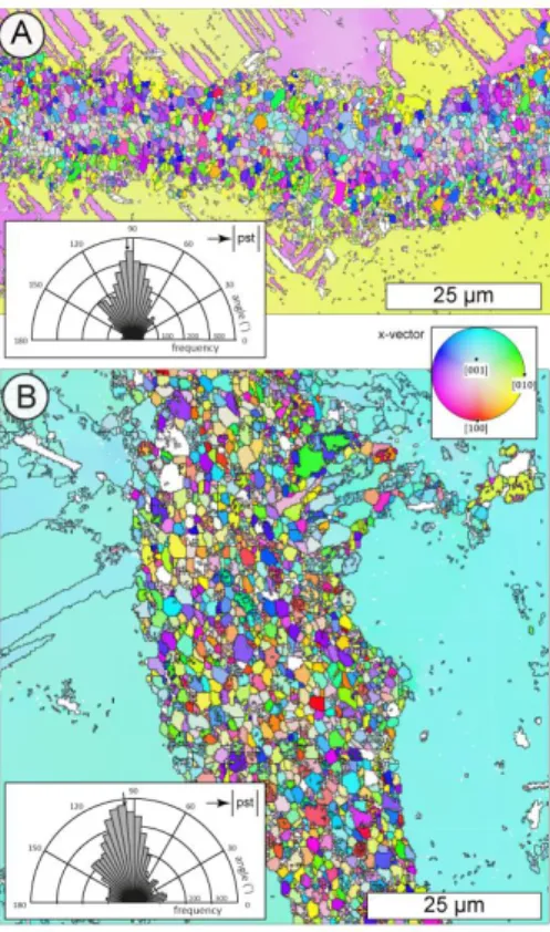
1d and e). The grains within the microfractures have a crystallographic preferred orientation (CPO) that is
68
controlled by the host plagioclase on either side of the microfracture (Fig. 2), and the K-feldspar grains have a
69
CPO that mimics that of the plagioclase grains (Petley-Ragan et al., 2018). The grains also show a strong shape
70
preferred orientation (SPO) with the long axis parallel to the pseudotachylyte wall (Fig. 2). Plagioclase
71
compositions in the ranges An25-31 and An65-83 were measured in the microfractures. These originate from a host
72
composition of An40 (Petley-Ragan et al., 2018). A similar bimodal range of plagioclase compositions were also
73
observed at garnet-plagioclase phase boundaries and in an amphibolite facies micro-shear zone at Isdal ca. 40 km
74
NE of Holsnøy (Mukai et al., 2014).
The mineralogy of the microfractures and their associated reaction products varies locally. Some contain quartz
76
and kyanite, while others are associated with intergrowths of clinozoisite, quartz and K-feldspar. Few
77
microfractures contain minor amounts of carbonates or phengite. Microfracture mineralogy is found to depend on
78
the XCO2 of the infiltrating fluid (Okudaira et al., 2016) and the orientation of the microfracture relative to the
79
principle stress (Moore et al., 2019). The detailed evolution of the microfractures is thus dependent on a multitude
80
of factors.
81
Two microfractures of dominantly plagioclase and K-feldspar previously described by Petley-Ragan et al. (2018)
82
were subject to further study with transmission electron microscopy (TEM). The grain size distributions within
83
these microfractures were characterized by electron backscatter diffraction (EBSD) (Aupart et al., 2018). The
84
microfracture from Figure 1d will hereafter be referred to as Microfracture 1 (MF1) and is located 1.25 mm away
85
from pseudotachylyte with a mean grain size of 1.73 µm2 (Aupart et al., 2018). The microfracture from Figure 1d
86
will be referred to as Microfracture 2 (MF2) and is located 1.8 cm away from the same pseudotachylyte (Fig. 1a)
87
with a mean grain size of 2.14 µm2 (Aupart et al., 2018). MF2 also contains a set of secondary fractures (Fig. 1c).
88
Both microfractures are associated with clinozoisite, quartz and kyanite growth, and only MF2 contains dolomite.
89
The lower J-index, greater misorientations and the presence of secondary fractures indicate that MF2 experienced
90
more shear deformation than MF1 (Petley-Ragan et al., 2018).
91
4 Methods
92
Mass balance calculations were performed on three microfractures by comparing the bulk microfracture
93
composition to the bulk host composition. Electron microprobe maps of the microfractures were obtained with a
94
Cameca SX100 at the University of Oslo’s Department of Geosciences. The mass balance was calculated in
95
MATLAB. Focused ion beam (FIB) foils were prepared and TEM analyses were carried out at Utrecht University.
96
The FEI Helios Nanolab G3 was used to cut FIB foils perpendicular to the length of the microfractures and
~15-97
20 µm in length in order to include both the host and microfracture constituents (Fig. 1d and e). The FEI Talos
98
200FX equipped with a high-sensitive 2D energy dispersive X-ray (EDX) system was used to obtain bright-field
99
(BF), dark-field (DF) and high angular annual dark-field (HAADF) images in scanning TEM (STEM) mode. Large
100
area EDX maps were acquired of the entire FIB foil for MF1 and parts of the FIB foil for MF2.
101
5 Results
102
Mass balance calculations based on three microfractures show that there is 5-11 times more K in the microfractures
103
compared to the host composition (Fig. 3). A bright field TEM image shows that MF1 contains dislocation-poor
104
and dislocation-free grains of dominantly plagioclase and K-feldspar defined by straight grain boundaries with
105
120° triple junctions (Fig. 4a). Few grains contain single dislocation walls within their centre. In contrast, the host
106
plagioclase is littered with free dislocations that have formed a subgrain wall made up of closely spaced
107
dislocations. Ankerite (Ca(Fe,Mg)(CO3)2), grossular-rich garnet and sphene are additional phases in MF1, with
108
apatite and rutile inclusions inside the grains, pinned along grain boundaries and concentrated along the subgrain
109
wall in the host (Fig. 4b).
110
The EDX map of MF1 displays homogeneous K-feldspar grains and plagioclase grains that are heterogeneous
111
with respect to their CaAl and NaSi content (Fig. 4b). The K-feldspar grains are clustered together creating a fabric
dominated by grain boundaries instead of phase boundaries. The irregular composition distribution of Na and Ca
113
in the plagioclase grains contradicts the backscatter electron image that suggests Ca zoning around the grains (Fig.
114
1d and 4b). Instead, the Ca-rich domains overlie areas with submicron lamellae (Fig. 5a-f). The lamellae are
115
discontinuous throughout the plagioclase grains and, locally, they are superimposed by tapered mechanical twins
116
(Fig. 5a). Other grains contain both lamellae and twins that are spatially distinct but are parallel to each other (Fig.
117
5d). In some grains, the lamellae appear slightly curved (Fig. 5c) while in others, the lamellae appear to form a
118
‘tweed’ structure (Fig. 5f). The spacing between lamellae is approximately 10-30 nm. Due to the high anorthite
119
composition obtained for plagioclase within this microfracture (An65-83; Petley-Ragan et al., 2018) this structure
120
lies within the Bøggild-Huttenlocher miscibility gap (Smith and Brown, 1988; McConnell, 2008). The intergrowth
121
is not observed within the host plagioclase.
122
MF2 is similarly dominated by dislocation-poor grains of plagioclase and K-feldspar with a number of grains
123
displaying twinning (Fig. 6a). The twins of separate grains are approximately parallel to each other and to (010)
124
of the host plagioclase (see Fig. 6 of Petley-Ragan et al., 2018), reinforcing the preservation of crystallographic
125
orientations of the host through the fracturing and recovery process. Kyanite and a K-rich micaceous phase are
126
additional phases in MF2. Apatite inclusions are present within the grains and pinned along grain boundaries. The
127
fabric is defined by 120° triple junctions with rare dislocation-rich grains that display irregular boundaries (Fig.
128
6b).
129
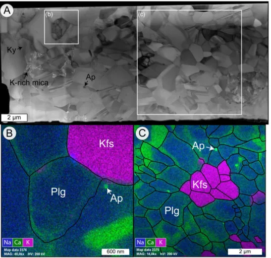
The EDX map of MF2 shows clustered homogeneous K-feldspar grains and zoned plagioclase grains (Fig. 6c)
130
creating again a grain boundary-dominated fabric. Unlike MF1, the plagioclase grains in MF2 display
Ca-131
enrichment at their grain boundaries and the submicron lamellae are absent. The Ca-rich rims are approximately
132
100-200 nm thick.
133
6 Discussion
134
The microfractures offer insight into the evolution of plagioclase feldspar that resulted from the high stress and
135
high-temperature environment created near an earthquake slip plane. The dislocation-free nature of almost all
136
grains in MF1 and MF2 suggest nearly complete annealing of the material within the microfractures (Fig. 4a and
137
6a). The grain fabric is dominated by straight phase and grain boundaries, 120° triple junctions and pinned apatite
138
inclusions suggesting the migration of grain boundaries. The inheritance of the crystallographic orientation of the
139
host plagioclase and its twins within the grains, furthermore, points towards an initial annealing process that is
140
able to transfer and preserve crystallographic information (Fig. 3). An equilibrium fabric with crystallographic
141
inheritance is generally created by dislocation creep and grain boundary migration (Passchier and Trouw, 2005).
142
However, the parallel shape preferred orientation of the grains to the pseudotachylyte wall suggests that annealing
143
was initiated while a stress or thermal field generated by the seismic slip was still present (Petley-Ragan et al.,
144
2018). This constrains the time scale of microfracture annealing to the duration of pseudotachylyte crystallization
145
and cooling (seconds to minutes). Dislocation- and grain boundary migration are too slow to have taken place
146
within this time scale, and it is additionally puzzling as to why these mechanisms were not active within the
147
dislocation-rich host. Thus, we postulate that a much more rapid recrystallization process took place prior to grain
148
boundary migration and final annealing within the microfractures, and this process must have been focused and
149
enhanced by local factors such as fluid infiltration and heat from the nearby pseudotachylyte. The resulting grain
size distributions as discussed by Aupart et al. (2018) furthermore show striking deviations from a steady-state
151
distribution.
152
6.1 Stressed wall rock plagioclase
153
Deformation experiments performed at eclogite facies conditions may offer some insight into the microstructures
154
that were present in the microfractures before complete recovery. Incel et al. (2017; 2019) observed brittle fractures
155
filled with amorphous material during deformation experiments on blueschist under eclogite facies conditions.
156
They interpret the amorphous material to result from shock loading during the propagation of a dynamic rupture.
157
Although their experiments involved a short recovery time (<1 hour) some of the amorphous material
158
recrystallized, creating idiomorphic garnet crystals with a size of ~20 nm.
159
The amorphization of plagioclase feldspar is dependent on pressure (P), temperature (T), composition (X),
160
compression rate (P/t) and pressure duration (t). Amorphization that is strongly dependent on temperature is
161
commonly referred to as heterogeneous amorphization or melting, and is a relatively slow process due to its
162
dependence on the diffusion of atoms (Wolf et al., 1990). On the other hand, amorphization that is strongly
163
dependent on pressure is referred to as pressure-induced amorphization, which may be static or dynamic,
164
depending on the compression rate (Sharma and Sikka, 1996). For the following, the pressure-induced
165
amorphization of plagioclase will be discussed. For anorthite-rich compositions (An51-100) complete
pressure-166
induced amorphization occurs P ≥ 13 GPa and T = 660°C, while albite-rich (An2) compositions are not completely
167
amorphous until P ≥ 26 GPa and T = 950°C (Daniel et al., 1997; Kubo et al., 2009; Tomioka et al., 2010).
168
Furthermore, short pressure durations result in lower degrees of amorphization (Tomioka et al., 2010) while high
169
compression rates of 101-102 GPa/s can reduce the pressure required for amorphization (Sims et al., 2019). The
170
short-lived (microseconds) high intensity (106 GPa/s) conditions in the proximity of earthquake rupture tips
171
(Reches and Dewers, 2005) may partially amorphize plagioclase feldspar (An40) in the wall rock, even if the local
172
pressure for complete amorphization is not reached. The presence of asymmetric tensile cracks on some of the
173
microfractures indicates that the propagation velocity of the microfractures approached the shear wave velocity
174
(Petley-Ragan et al., 2018) inducing similar short-lived high-intensity stresses within their vicinity. Therefore, a
175
mixture of amorphous material with remnant fragments may have been present within the microfractures
176
immediately after earthquake and microfracture rupture.
177
Repolymerization of amorphous material on the microfracture walls and remnant fragments would directly transfer
178
the crystallographic orientation of the host. Crystallographic information may also be preserved by the presence
179
of short-range atomic order within amorphous material, allowing for immediate repolymerization without the aid
180
of a fragment nucleus (Casey et al., 1993; Konrad-Schmolke et al., 2018). Repolymerization has also been
181
suggested to occur directly along crystal lattice defects where amorphous material originates (Konrad-Schmolke
182
et al., 2018). In this context, dislocations within the grains may have healed much more quickly than would be
183
expected from dislocation migration recrystallization and the fragments would have experienced healing from
184
multiple available interfaces. Other preferred areas of repolymerization were likely parallel to the minimum
185
principal stress direction, growing grains with a stress-dependent SPO. Therefore, recrystallization from an
186
amorphous material may be a likely candidate to create the observed dislocation-free fabric with a strong SPO
187
within the timeframe of pseudotachylyte formation.
6.2 Cooling within the vicinity of pseudotachylyte
189
The nano-scale intergrowth within the plagioclase grains from MF1 is here interpreted as exsolution lamellae that
190
formed as a result of rapid cooling from high temperatures within the vicinity of the pseudotachylyte. Similar
191
intergrowths were found in what is called the ‘complex feldspar’, a microstructure of fragmented plagioclase first
192
described in an amphibolite facies shear zone at Isdal, approximately 40 km east of Holsnøy (Mukai et al., 2014).
193
They interpreted the structure as fluid- and stress-induced coarsening of exsolution lamellae. Although plausible,
194
this would require that plagioclase exsolution occurred prior to the stress and thermal anomaly created by the
195
earthquake. No intergrowths are observed within the host plagioclase in the present study, and it is unlikely that
196
diffusion rates were high enough to form lamellae within the dry granulite. Our documentation of the exsolution
197
lamellae within plagioclase grains from the microfracture nearest the pseudotachylyte (Fig. 1a) suggests that the
198
thermal anomaly produced by the frictional melt vein affected the intracrystalline structure of the plagioclase grains.
199
Intergrowths form when plagioclases of intermediate composition cool from high temperature and enter a
200
miscibility gap below 800°C, exsolving into separate calcic and sodic regions (Carpenter, 1994; McConnell, 2008).
201
Although the ambient eclogite facies conditions (650-750°C) place the plagioclase within the miscibility gap, the
202
absence of fluids hinders chemical diffusion and thus exsolution. It is only until after an earthquake causes wall
203
rock damage that fluids enter the wall rock through coseismic microfractures, and these fluids are likely overheated
204
by the frictional slip (Bestmann et al., 2016). Simultaneously, the wall rock within <1 cm of the pseudotachylyte
205
experiences a thermal anomaly before rapidly cooling back to ambient conditions at rates on the order of a few °C/s
206
(Bestmann et al., 2012). NaSi-CaAl diffusivity in plagioclase at 900-1000°C is ~10-15 cm2/s (Korolyuk and Lepezin,
207
2009). Assuming that elevated temperatures lasted for up to a minute within 1 mm of the pseudotachylyte (MF1),
208
diffusion would be efficient over a distance of 25 nm, similar to the spacing of lamellae observed (Fig. 5). At
209
distances greater than 1 cm from the pseudotachylyte (MF2), the wall rock experiences minor heating to a few
210
10°C above ambient. Therefore, rapid cooling from elevated temperatures back to ambient conditions and into the
211
miscibility gap only took place within close proximity to the pseudotachylyte.
212
7 Conclusion
213
Our nanostructural observations are relevant for understanding plagioclase deformation during and after an
214
earthquake in the lower crust, prior to any subsequent shear zone development. We propose that plagioclase within
215
the microfractures experienced partial amorphization at peak pressures coeval with earthquake propagation and
216
microfracturing in the wall rock. Repolymerization on microfracture walls, remnant fragments, dislocations and
217
from short-range atomic ordering in the direction parallel to the minimum principal stress formed a strong CPO
218
and SPO in the grains. Repolymerization and recrystallization within the timeframe of pseudotachylyte formation
219
explain the presence of dislocation-free grains, as has been interpreted for similar structures observed in quartz
220
(Bestmann et al., 2012). In close proximity to the pseudotachylyte, wall rock temperatures reached 900-1000°C
221
before rapidly cooling back to ambient eclogite facies conditions and into the plagioclase miscibility gap. This
222
caused exsolution of intermediate plagioclase compositions and formation of nano-scale lamellae. We hypothesize
223
that the lamellae described here are a unique signature of rapid cooling within plagioclase-rich wall rock in the
224
vicinity of pseudotachylyte. A study of a larger number of plagioclase microfractures at varying distances to
225
pristine pseudotachylyte would provide more information and constraints on the occurrence of these intergrowths.
The dependence of plagioclase microstructures on temperature and cooling rate and their sensitivity to fluid
227
interaction offers a new tool for unraveling the history of wall rocks and their associated earthquakes.
228
Data and Sample Availability
229
Raw electron backscatter diffraction and geochemical data are available on Open Science Framework at
230
osf.io/g36m7/. Rock samples are available through A. P.-R. and FIB foils are available through O. P.
231
Author Contribution
232
B. J. designed the project. A. P.-R. collected the samples, obtained and analyzed the EBSD and geochemical data.
233
B. I. helped collect and interpret the EBSD data. O. P. cut the FIB foils, and obtained and interpreted the TEM
234
images. A.P.-R., O. P. and B. J. were part of discussions. A. P.-R. and B. J. wrote the manuscript.
235
Competing Interests
236
The authors declare that they have no conflict of interest.
237
Acknowledgements
238
This project was supported by the European Research Council (ERC) Advanced Grant Agreement 669972,
239
“Disequilibrium Metamorphism” (“DIME”) to B. J., and the Natural Science and Engineering Research Council
240
(NSERC) of Canada Postgraduate Scholarship Doctoral (PGS-D) 489392 to A. P.-R. O. P. has been supported by
241
an ERC Starting Grant “nanoEARTH” (852069). We thank H. Austrheim for field guidance on Holsnøy and
242
hospitality in Western Norway. We thank X. Zhong for help with the mass balance calculations, F. Barou for
243
assistance with EBSD measurements and M. Erambert for help on the electron microprobe.
244
References
245
Austrheim, H. and Boundy, T. M.: Pseudotachylytes generated during seismic faulting and eclogitization of the
246
deep crust, Science, 265, 82-83, http://www.jstor.org/stable/2884364, 1994.
247
Austrheim, H., Dunkel, K. G., Plümper, O., Ildefonse, B., Liu, Y., and Jamtveit, B.: Fragmentation of wall rock
248
garnets during deep crustal earthquakes, Sci. Adv., 3, 1-8, https://doi.org/10.1126/sciadv.1602067, 2017.
249
Bestmann, M., Pennacchioni, G., Nielsen, S., Göken, M., and de Wall, H.: Deformation and ultrafine dynamic
250
recrystallization of quartz in pseudotachylyte-bearing brittle faults: A matter of a few seconds. J. Struct.
251
Geol., 38, 21-38, https://doi.org/10.1016/j.jsg.2011.10.001, 2012.
252
Bestmann, M., Panncchioni, G., Mostefaoui, S., Göken, M. and de Wall, H.: Instantaneous healing of
micro-253
fractures during coseismic slip: Evidence from microstructure and Ti in quartz geochemistry within an
254
exhumed pseudotachylyte-bearing fault in tonalite, Lithos, 254-255, 84-93,
255
https://doi.org/10.1016/j.lithos.2016.03.011, 2016.
256
Bhowany, K., Hand, M., Clark, C., Kelsey, D. E., Reddy, S. M., Pearce, M. A., Tucker, N. M., and Morrissey, L.
257
J.: Phase equilibria modelling constraints on P-T conditions during fluid catalysed conversion of granulite
258
to eclogite in the Bergen Arcs, Norway, J. Metamorph. Geol., https://doi.org/10.1111/jmg.12294, 2017.
Boundy, T.M., Fountain, D.M., and Austrheim, H.: Structural development and petrofabrics of eclogite facies
260
shear zones, Bergen Arcs, western Norway: implications for deep crustal deformational processes, J.
261
Metamorph. Geol., 10, 2, 127-146, https://doi.org/10.1111/j.1525-1314.1992.tb00075.x, 1992.
262
Carpenter, M. A.: Mechanisms and kinetics of Al-Si ordering in anorthite: I. Incommensurate structure and domain
263
coarsening, Am. Mineral., 76, 1110-1119, 1991.
264
Casey, W. H., Westrich, H. R., Banfield, J. F., Ferruzi, G. and Arnold, G. W.: Leaching and reconstruction at the
265
surfaces of dissolving chain-silicate minerals, Nature, 366, 253-256, https://doi.org/10.1038/366253a0,
266
1993.
267
Daniel, I., Gillet, P., McMillan, P. F., Wolf, G. and Verhelst, M. A.: High-pressure behavior of anorthite:
268
Compression and amorphization. J. Geophys. Res., 102, 10313-10325.
269
https://doi.org/10.1029/97JB00398, 1997.
270
Glodny, J., Kühn, A., and Austrheim, H.: Geochronology of fluid-induced eclogite and amphibolite facies
271
metamorphic reactions in subduction-collision system, Bergen Arcs, Norway, Contrib. Mineral. Petr.,
272
156, 1, 27-48, https://doi.org/10.1007/s00410-007-0272-y, 2008.
273
Incel, S., Hilairet, N., Labrousse, L., John, T., Deldicque, D., Farrand, T., Wang, Y., Renner, J., Morales, L. and
274
Schubnel, A.: Laboratory earthquakes triggered during eclogitization of lawsonite-bearing blueschist.
275
Earth Planet. Sci. Lett., 459, 320-331, https://doi.org/10.1130/G45527.1, 2017.
276
Incel, S., Labrousse, L., Hilairet, N., John, T., Gasc, J., Shi, F., Wang, Y., Andersen, Renard, F., Jamtveit, B. And
277
Schubnel, A.: Reaction induced embrittlement of the lower continental crust, Geology, 47, 3, 235-238,
278
https://doi.org/10.1130/G45527.1, 2019.
279
Jamtveit, B., Austrheim, H., and Putnis, A.: Disequilibrium metamorphism of stressed lithosphere, Earth Sci. Rev.,
280
154, 1-13. https://doi.org/10.1016/j.earscirev.2015.12.002, 2016.
281
Jamtveit, B., Bucher-Nurminen, K. and Austrheim, H.: Fluid controlled eclogitization of granulites in deep crustal
282
shear zones, Bergen Arcs, Western Norway, Contrib. Mineral Petr., 104, 184-193,
283
https://doi.org/10.1007/BF00306442, 1990.
284
Jamtveit, B., Moulas, E. Andersen, T. B., Austrheim, H., Corfu, F., Petley-Ragan, A. and Schmalholz, S. M.: High
285
pressure metamorphism caused by fluid induced weakening of deep continental crust. Sci. Rep., 8, 17011,
286
https://doi.org/10.1038/s41598-018-35200-1, 2018.
287
Jamtveit, B., Petley-Ragan, A., Incel, S., Dunkel K. G., Aupart, C., Austrheim, H., Corfu, F., Menegon, L. and
288
Renard, F.: The effects of earthquakes and fluids on the metamorphism of the lower continental crust, J.
289
Geophys. Res., 124, 8, 7725-7755, https://doi.org/10.1029/2018JB016461, 2019.
290
Konrad-Schmolke, M., Halama, R., Wirth, R., Thomen, A., Klitscher, N., Morales, L., Schreiber, A. and Wilke,
291
F. D. H.: Mineral dissolution and reprecipitation mediated by an amorphous phase, Nature contrib., 9,
292
https://doi.org/10.1038/s41467-018-03944-z, 2018.
293
Kubo, T., Kimura, M., Kato, T., Nishi, M., Tominaga, A., Kikegawa, T. and Funakoshi, K.: Plagioclase breakdown
294
as an indicator for shock conditions of meteorites, Nat. Geosci., 3, 41-45, https://doi.org/10.1038/ngeo704,
295
2009.
296
Marti, S., Stünitz, H., Heilbronner, R., Plümper, O. and Drury, M.: Experimental investigation of the brittle-viscous
297
transition in mafic rocks – Interplay between fracturing, reaction, and viscous deformation, J. Struct. Geol,
298
105, 62-79, https://doi.org/10.1016/j.jsg.2017.10.011, 2017.
McConnell, J.: The origin and characteristics of incommensurate structures in the plagioclase feldspars, Can.
300
Mineral., 46, 1389-1400, https://doi.org/10.3749/canmin.46.6.1389, 2008.
301
Menegon, L., Stünitz, H., Nasipuri, P., Heilbronner, R. and Svahnberg, H.: Transition from fracturing to viscous
302
flow in granulite facies perthitic feldspar (Lofoten, Norway), J. Struct. Geol., 48, 95-112,
303
https://doi.org/10.1016/j.jsg.2012.12.004, 2013.
304
Moore, J., Beinlich, A., Austrheim, H. and Putnis, A.: Stress orientation-dependent reactions during
305
metamorphism, Geology, 47, 1-4, https://doi.org/10.1130/G45632.1, 2019.
306
Mukai, H., Austrheim, H., Putnis, C. V., and Putnis, A.: Textural evolution of plagioclase feldspar across a shear
307
zone: Implications for deformation mechanism and rock strength, J. Petrol., 55, 1457-1477,
308
https://doi.org/10.1093/petrology/egu030, 2014.
309
Okudaira, T., Shigematsu, N., Harigane, Y., and Yoshida, K.: Grain size reduction due to fracturing and subsequent
310
grain-size-sensitive creep in lower crustal shear zone in the presence of a CO2-bearing fluid, J. Struct.
311
Geol., 95, 171-187, https://doi.org/10.1016/j.jsg.2016.11.001, 2016.
312
Petley-Ragan, A., Dunkel, K. G., Austrheim, H., Ildefonse, B. and Jamtveit, B.: Microstructural records of
313
earthquakes in the lower crust and associated fluid-driven metamorphism in plagioclase-rich granulites.
314
J. Geophys. Res.-Sol Ea., 123, 1-18, https://doi.org/10.1029/2017JB015348, 2018.
315
Petley-Ragan, A., Ben-Zion, Y., Austrheim, H., Ildefonse, B., Renard, F. and Jamtveit B.: Dynamic earthquake
316
rupture in the lower crust, Sci. Adv., 5, https://doi.org/10.1126/sciadv.aaw0913, 2019.
317
Passchier, C., and Trouw, R.: Microtectonics. Springer, Berlin., 2005.
318
Reches, Z. and Dewers, T. A.: Gouge formation by dynamic pulverization during earthquake rupture, Earth Planet.
319
Sc. Lett., 235, 361-374, https://doi.org/10.1016/j.epsl.2005.04.009, 2005.
320
Sharma, S. and Sikka, S.: Pressure Induced Amorphization of Materials, Progress in Materials Science, 40, 1-77,
321
1996.
322
Sims, M., Jaret, S. J., Carl, E.-R., Rhymer, B., Schrodt, N., Mohrholz, V., Smith, J., Konopkova, Z., Liermann,
323
H.-P., Glotch, T. D. and Ehm, L.: Pressure-induced amorphization in plagioclase feldspars: A
time-324
resolved powder diffraction study during rapid compression, Earth Planet Sc. Lett., 507, 166-174,
325
https://doi.org/10.1016/j.epsl.2018.11.038, 2019.
326
Smith, J. V. and Brown, W. L.: Feldspar Minerals, vol. 1, Springer, Berlin, 1988.
327
Soda, Y. and Okudaira, T.: Microstructural evidence for the deep pulverization in a lower crustal meta-anorthosite,
328
Terra Nova, 1-7, https://doi.org/10.1111/ter.12355, 2018.
329
Svahnberg, H. and Piazolo, S.: The initiation of strain localisation in plagioclase-rich rocks: Insights from detailed
330
microstructural analyses, J. Struct. Geol, 32, 1404-1416, https://doi.org/10.1016/j.jsg.2010.06.011, 2010.
331
Tomioka, N., Kondo, H., Kunikata, A. and Nagai, T.: Pressure-induced amorphization of albitic plagioclase in an
332
externally heated diamond anvil cell, Geophys. Res. Lett., 37, 1-5,
333
https://doi.org/10.1029/2010GL044221, 2010.
334
Wolf, D., Okamoto, P., Yip, S., Lutsko, J. F. and Kluge, M.: Thermodynamic parallels between solid-state
335
amorphization and melting, J. Material Res., 5, 286-301, https://doi.org/10.1557/JMR.1990.0286, 1990.
337
Figure 1: Fractured wall rock plagioclase. (a) Thin section scan of wall rock plagioclase (Plg), garnet (Grt),
338
clinopyroxene (Cpx) and scapolite (Sc) adjacent to an eclogite facies pseudotachylyte (pst) on Holsnøy. (b)
Fine-339
grained reaction products of clinozoisite (Czo) are associated with the microfractures. Box denotes the location of
340
MF1. (c) Some microfractures in plagioclase display secondary cracking. Box denotes the location of MF2. (d)
341
Backscatter electron image of MF1 with fine-grained plagioclase, alkali feldspar (Kfs) and minor kyanite (Ky). (e)
342
Backscatter electron image of MF2 with fine-grained plagioclase, K-feldspar, dolomite (Dol) and clinozoisite. Red
343
lines indicate the location of focused ion beam cuts for TEM analysis shown in Figs. 4-6.
345
Figure 2: Crystallographic orientations of the grains within the microfractures. (a) Inverse pole figure
346
coloring orientation map of MF1 with inset of grain SPO. (b) Orientation map of MF2 with inset of grain SPO.
347
Modified after Petley-Ragan et al. (2018).
348
349
Figure 3: Mass balance of plagioclase microfractures. Three separate plagioclase microfractures were analyzed
350
for Na, Ca and K. Xfracture is the bulk composition of the fracture and Xhost is the bulk composition of the adjacent
351
plagioclase host.
353
Figure 4: Microstructures of MF1. (a) BF-STEM image of the entire FIB cut from Fig. 1d. The plagioclase (Plg)
354
host to the left is rich in dislocations while the grains within the microfracture to the right are poor to absent of
355
dislocations. Apatite (Ap) and rutile (Rt) inclusions are present within the host and the grains, as well as pinned
356
along grain boundaries in the microfracture. (b) EDX map overlain with grain and phase boundaries (black).
357
Ankerite (Ank), garnet (Grt) and sphene (Sph) are additional phases within the microfracture.
359
Figure 5: Plagioclase intergrowths in MF1. (a) BF-TEM image of the submicron lamellae in a plagioclase grains
360
that are overlain by mechanical twins. (b) EDX map showing the distribution of Ca and Na in the plagioclase
361
grains associated with the intergrowth in (a). The Ca-rich domains overlay the lamellae. (c) BF-TEM image of
362
lamellae in two separate grains that show slight curvature. (d) BF-STEM image of discontinuous lamellae within
363
a grain that hosts twins in its core. (e) STEM bright field image of discontinuous lamellae within a plagioclase
364
grain. (f) Bright field TEM image of lamellae resembling ‘tweed’ exsolution within plagioclase.
366
Figure 6: Microstructures of MF2. (a) Bright field image of the entire FIB cut from Fig. 1e. The plagioclase
367
(Plg) microfracture contains dislocation-free grains with some twins. (b) EDX map of a dislocation-rich grain
368
overlain with grain and phase boundaries (black). (c) EDX map of the area in (a) overlain with grain and phase
369
boundaries (black). The Ca-rich domains are present along grain boundaries.